Embed Size (px)
Citation preview

Diagnostic and Interventional Imaging (2015) 96, 775—788
CONTINUING EDUCATION PROGRAM: FOCUS. . .
Severe hemoptysis: From diagnosis toembolization
A. Khalil a,b,∗,c, B. Fedidaa,e, A. Parrotd, S. Haddada,e,M. Fartoukhd,e, M.-F. Carettea,e
a Department of radiology, Tenon Hospital, 75020 Paris, Franceb Department of radiology, Bichat Hospital, 46, rue Henri Huchard, 75018 Paris, Francec Paris VII University, 75205 Paris cedex 13, Franced Department of Intensive Care, Tenon Hospital, 75020 Paris, Francee Paris VI University, 75252 Paris cedex 05, France
KEYWORDSLung;Hemoptysis;Interventionalradiology;Embolization;CT angiography
Abstract Severe hemoptysis is life-threatening to patients because of the asphyxia it causes.The diagnosis and treatment are therefore urgent and chest imaging is essential. MultidetectorCT-angiography provides an exhaustive non-invasive assessment which includes localization,mechanisms, causes and severity of the hemoptysis. It is an invaluable step in preparationfor endovascular treatment which is the first line invasive therapy, particularly with bronchialarteriography embolization in the majority of cases (over 90%) and erosion or rupture of thepulmonary artery in less than 10% of cases. Hemoptysis control is achieved in 65 to 92% of casesdepending on the cause.
© 2015 Éditions francaises de radiologie. Published by Elsevier Masson SAS. All rights reserved.
Hemoptysis is the exteriorization of red aerated blood from the mouth following a coughoriginating from below the glottis. It represents blood from the thoracic vascular sec-tor passing into the respiratory sector. Hemoptysis is a common symptom in respiratorymedicine. It accounts for 10 to 15% of the reasons for consultation in hospital respiratorydepartment and is a warning signal for investigation into its cause [1,2]. Severe hemoptysis
(SH) is life-threatening and has a mortality rate of over 50% without control of the bleeding[3,4]. It requires rapid and simultaneous management for both diagnostic (mechanism andcause) and therapeutic [5] purposes. Endovascular management, especially embolizationof the bronchial arteries, is now the 1st line treatment [5] to control the bleeding. The∗ Corresponding author.E-mail address: antoine [email protected] (A. Khalil).
http://dx.doi.org/10.1016/j.diii.2015.06.0072211-5684/© 2015 Éditions francaises de radiologie. Published by Elsevier Masson SAS. All rights reserved.

7
ih(tia
wist(av
Th
I
Tppbtaa1hasmatc
FtMtb
C
Itasw
S
It[a
mai2mv
S
LtyrbseTbe
76
ndications for endovascular treatment are unequivocal inemoptysis which is causing concern because of its volumeover 200 mL/24—48 h), its consequences on the respira-ory system (acute respiratory failure) or if the mechanisms potentially life-threatening (erosion of the pulmonaryrtery) [6].
The physician (ideally the intensive care physician) facedith a case of SH should ask him/herself five questions:
s this actually hemoptysis? How severe is it? What is theite? What is its cause and most likely mechanism? Whatreatment should be given? Multidetector CT-angiographyMDCTA) can answer a number of these questions (Fig. 1)nd is essential for the radiologist before considering inter-entional radiology [7,8].
he use of CT angiography in severeemoptysis
maging technique
he investigation should be performed in deep inspiration ifossible, failing which it should be performed in free res-iration [9]. All of the intrathoracic blood vessels shoulde enhanced using a contrast injection rate (at a concen-ration of 300 mg of iodine/mL) of 3.5 to 4 mL/sec with
total volume of 90 mL. Image acquisition is triggered by region of interest (ROI) in the descending aorta from00 Hounsfield Units for 16-row CT-scan and 150 HU for aigher row CT-scan. Coverage should begin from the lungpices (C5-C6) to the hilum of the kidneys (L1-L2), from theupra-aortic vessels to the origin of the inferior diaphrag-atic arteries. It is recommended that images be started
t the base of the cranium in patients with a past his-
ory of neck surgery or radiotherapy for a nasopharyngealancer.igure 1. Interpretation algorithm and expected results of mul-idetector CT-angiography. MDCTA: multidetector CT-angiography;IP: maximum intensity projection; VRT: volume rendering
echnique; PA: pulmonary artery; BA: bronchial artery; NBSA: non-ronchial systemic artery.
iooatsgwlpfl
otv
w(t
ccbr
l
A. Khalil et al.
onfirmation of the hemoptysis
n the majority of cases, the clinical enquiry will establishhe origin of red blood coming from the mouth. Occasionally,
diagnosis is uncertain and MDCTA can therefore demon-trates a cause and/or signs of alveolar or bronchial floodingith intraluminal clots.
everity of the bleeding
n terms of severity, the volume of hemoptysis and respira-ory consequences can clinically identify the majority of SH6]. If the clinical enquiry however is unreliable, MDCTA maygain offer assistance [7].
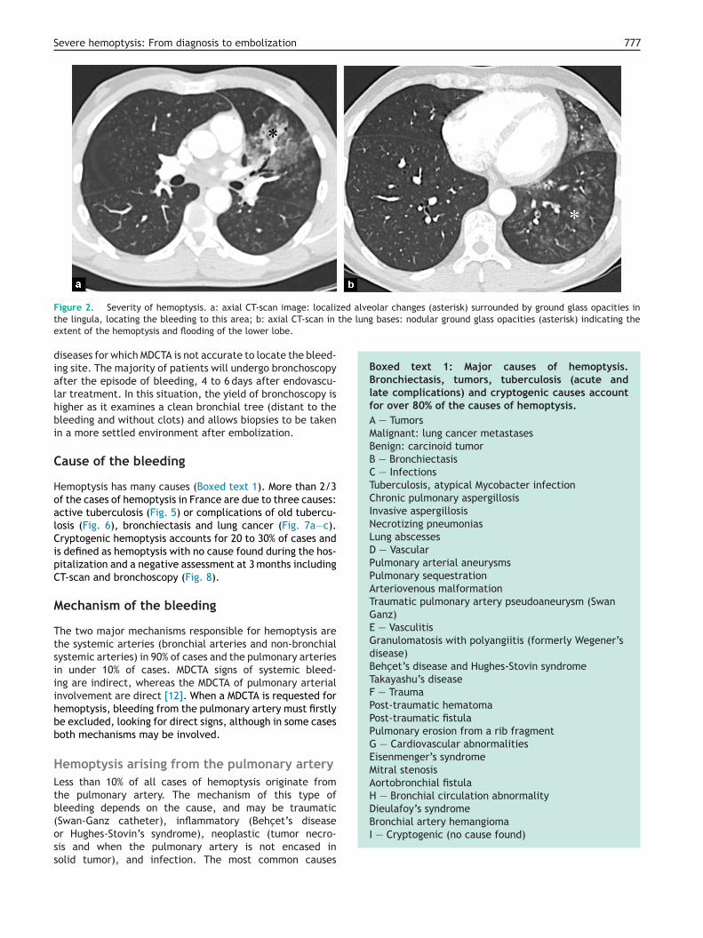
We have shown that the extent of parenchymal involve-ent on CT correlates with the magnitude of the bleed
nd with clinical severity. Involvement of more than 3 lobess usually associated with exteriorized bleeding of over00 mL/24—48 h and requires more interventionist treat-ent [10] even if the patient has not coughed up a large
olume of blood (Fig. 2).
ite of the bleed
ateralization (the bleeding side) and precise localization ofhe hemoptysis are essential for treatment. When hemopt-sis is causing asphyxia, simple selective protection of theespiratory tract can only be performed when the side of theleeding is known. Similarly, some embolization decisions inituations at high risk of complication can only be consid-red if the side of the hemoptysis is known with certainty.he decision to perform surgery to stop bleeds can also onlye made when there is certainty as to the lobe that is to bexcised.
The bleed is localized from the parenchymal windownvestigation that seeks to identify aground glass opacityr alveolar consolidation (Fig. 3a,b). This abnormality isf high localizing value [10—12]. The presence of severalreas of ground glass opacities and/or alveolar consolida-ion with a relatively unaffected subpleural area shoulduggest the possibility of intra-alveolar hemorrhage. If around glass opacity image is present in the bases togetherith an alveolar consolidation in the upper part of the
ung (Fig. 2), the site of the bleeding is the highestart and the other abnormalities are due to positionalooding [12].
Some signs reflect the cause (bronchiectasis, cavitationr pulmonary artery pseudoaneurysm) or consequence ofhe bleeding (endobronchial clot) and have lower localizingalue.
Lung consolidation with necrosis or cavitation associatedith the appearance of a pulmonary artery pseudoaneurysm
an uncommon situation) indicates a bleed originating fromhe pulmonary artery (Fig. 4).
The topographic diagnostic yield of MDCTA compared tolinical assessment at the patient’s bedside (including thelinical enquiry, clinical examination, chest radiography and
ronchoscopy) is similar and in the region of 80% [7]. As aesult, in our view, bronchoscopy can be delayed.Bronchoscopy is no longer the first line investigation toocate the bleeding and is reserved for diffuse or bilateral
Severe hemoptysis: From diagnosis to embolization 777
ed alveolar changes (asterisk) surrounded by ground glass opacities inthe lung bases: nodular ground glass opacities (asterisk) indicating the
Boxed text 1: Major causes of hemoptysis.Bronchiectasis, tumors, tuberculosis (acute andlate complications) and cryptogenic causes accountfor over 80% of the causes of hemoptysis.A — TumorsMalignant: lung cancer metastasesBenign: carcinoid tumorB — BronchiectasisC — InfectionsTuberculosis, atypical Mycobacter infectionChronic pulmonary aspergillosisInvasive aspergillosisNecrotizing pneumoniasLung abscessesD — VascularPulmonary arterial aneurysmsPulmonary sequestrationArteriovenous malformationTraumatic pulmonary artery pseudoaneurysm (SwanGanz)E — VasculitisGranulomatosis with polyangiitis (formerly Wegener’sdisease)Behcet’s disease and Hughes-Stovin syndromeTakayashu’s diseaseF — TraumaPost-traumatic hematomaPost-traumatic fistulaPulmonary erosion from a rib fragmentG — Cardiovascular abnormalitiesEisenmenger’s syndromeMitral stenosisAortobronchial fistulaH — Bronchial circulation abnormalityDieulafoy’s syndrome
Figure 2. Severity of hemoptysis. a: axial CT-scan image: localizthe lingula, locating the bleeding to this area; b: axial CT-scan in
extent of the hemoptysis and flooding of the lower lobe.
diseases for which MDCTA is not accurate to locate the bleed-ing site. The majority of patients will undergo bronchoscopyafter the episode of bleeding, 4 to 6 days after endovascu-lar treatment. In this situation, the yield of bronchoscopy ishigher as it examines a clean bronchial tree (distant to thebleeding and without clots) and allows biopsies to be takenin a more settled environment after embolization.
Cause of the bleeding
Hemoptysis has many causes (Boxed text 1). More than 2/3of the cases of hemoptysis in France are due to three causes:active tuberculosis (Fig. 5) or complications of old tubercu-losis (Fig. 6), bronchiectasis and lung cancer (Fig. 7a—c).Cryptogenic hemoptysis accounts for 20 to 30% of cases andis defined as hemoptysis with no cause found during the hos-pitalization and a negative assessment at 3 months includingCT-scan and bronchoscopy (Fig. 8).
Mechanism of the bleeding
The two major mechanisms responsible for hemoptysis arethe systemic arteries (bronchial arteries and non-bronchialsystemic arteries) in 90% of cases and the pulmonary arteriesin under 10% of cases. MDCTA signs of systemic bleed-ing are indirect, whereas the MDCTA of pulmonary arterialinvolvement are direct [12]. When a MDCTA is requested forhemoptysis, bleeding from the pulmonary artery must firstlybe excluded, looking for direct signs, although in some casesboth mechanisms may be involved.
Hemoptysis arising from the pulmonary arteryLess than 10% of all cases of hemoptysis originate fromthe pulmonary artery. The mechanism of this type ofbleeding depends on the cause, and may be traumatic
(Swan-Ganz catheter), inflammatory (Behcet’s diseaseor Hughes-Stovin’s syndrome), neoplastic (tumor necro-sis and when the pulmonary artery is not encased insolid tumor), and infection. The most common causesBronchial artery hemangiomaI — Cryptogenic (no cause found)

778 A. Khalil et al.
Figure 3. Hemoptysis associated with bronchiectasis. Forty-three-year-old female patient with no particular past history, transferredto intensive care for massive hemoptysis about 350 mL on one time. a: axial CT-scan image in the parenchymal window focused on theright middle lobe bronchus. Ground glass opacities (asterisk) with bronchiectasis fully filled with blood clots (black arrows); b: sagittalreconstruction image in the parenchymal window showing the bronchus fully filled with clots (black arrows) and distal consolidation int r lobi rom
a
oata
he right middle lobe. Note the increased density in the right lowenfracarenal region. Bronchial vessel with an atypical origin arising ffter embolization with particles and occlusion with coils (f).
f infections are predominantly tuberculosis (Rasmussenneurysm), necrotic infectious pneumonias and other infec-ions involving parenchymal necrosis such as invasivespergillosis.
dma
e; c, d: axial CT-scan images passing through the aortic arch andthe aortic arch (arrows); e, f: bronchial angiography before (e) and
The MDCTA signs of pulmonary arterial involvementepend on the cause [13]. In systemic disease with pul-onary artery aneurysms, the pulmonary artery aneurysm
t the origin of the bleeding is present within an area of

Severe hemoptysis: From diagnosis to embolization 779
Figure 4. Necrotic pneumonia with a pulmonary artery pseudo-aneurysm. a, b: six mm maximum intensity projection, axial (a) and of th
pseud
coronal (b) views showing lung consolidation of the ventral segmenta branch of the pulmonary artery representing a pulmonary arterylung consolidation or ground glass opacity (Fig. 4). This
aneurysm requires emergency treatment. The majority ofother pulmonary artery aneurysms respond well to appro-priate medical therapy.bcm
Figure 5. Hemoptysis and active tuberculosis. Nineteen-year-old femasis. Clinical enquiry assessed the hemoptysis volume as being over 250 mL
lung cavities in the left upper lobe with adjacent nodules, lung consolib: axial CT-scan passing through the apex confirming the CXR findings: cview passing through the main trunk of the pulmonary artery: bilateral hlymphadenopathy; d: axial CT-scan showing the tree-in-bud appearance
diagnosis was confirmed to be active bacillary pulmonary tuberculosis rarteries achieved immediate control without recurrence.
e culmen with necrosis (black arrow) and ectasia (white arrow) ofo-aneurysm.
In infectious disease, the main sign is necrosis which can
e identified as a hypodense area within lung parenchymalonsolidation which is enhanced by the iodinated contrastedia. Late phase CT images or enhanced images canle patient presenting to the emergency department with hemopty-during the last 24 hours. a: postero-anterior chest radiograph (CXR):dation in the lingula and right axillary micronodular appearances;avernous appearance with nodules in the culmen; c: axial CT-scanilar lymphadenopathy predominantly on the left and infracarenal
(branch nodules) indicating bronchiolar spread of the disease. Theesponsible for hemoptysis for which embolization of the bronchial

780 A. Khalil et al.
Figure 6. Hemoptysis and late complications of tuberculosis. Fifty-three-year-old man hospitalized for hemoptysis of 300 mL over theprevious 48 hours. a: postero-anterior chest radiograph: aerated atelectasis of the culmen with bronchiectasis(arrow) and raised left mainbronchus; b: CT-scan image passing through the apices: traction bronchiectasis combined with adjacent lung consolidation and a groundglass opacity indicating the site of the hemoptysis; c: CT-scan image passing through the bases: nodules of increased density with groundglass opacity in the left lower lobe indicating the extent of the hemoptysis and flooding from the culmen; d: coronal reconstruction: goodcorrelation with the chest radiograph. Raised left fissure with bronchiectasis in the culmen and increased ground glass opacity. A bronchiala as c
iitmmcb
HsBagar[tdi
ootod(oac7or5a(t
rteriography with embolization (in addition to antibiotic therapy) w
mprove visibility of the hypodense area. The second signs a pseudoaneurysm within the hypodense area. Investiga-ion for a pseudoaneurysm is performed on 5 to 8 mm thickaximal intensity projection (MIP) reconstructions in theediastinal window in all three spatial planes. A less spe-
ific sign is the presence of a pulmonary artery with irregularorders in the wall of the necrosis.
emoptysis of systemic origin and mapping ofystemic arteriesy excluding a pulmonary arterial mechanism, MDCTA allows
conclusion to be drawn that the hemoptysis has its ori-in in a systemic artery. MDCTA visualizes the bronchialrteries (BA) and the non-bronchial systemic arteries (NBSA)esponsible for the bleeding. In addition, Remy-Jardin et al.
8] have shown that MDCTA is more accurate than conven-ional angiography in identifying the bronchial arteries. Thisifference is explained by anatomical variants and catheter-zation difficulties associated with patient age becauset
r[
arried out with no recurrence after a follow-up period of 6 months.
f atheromatous plaques. We have compared two groupsf patients who underwent systemic arterial emboliza-ion or attempted embolization before and after the eraf MDCTA; we included 200 patients in each group andivided the cohorts into 3 groups according to patient ageunder 50 years old, 50 to 70 years old and over 70 yearsld) and compared the catheterization failure rate byge band in both cohorts (with and without MDCTA). Theatheterization failure rates in the group of patients over0 years old were 36.6% and 14.6% in the cohorts with-ut and with MDCTA respectively. By expressing the failureate as a ratio to the age band of patients between0 and 70 years old, we found that the failure rate waslmost stable when the procedure was preceded by MDCTA14.6 compared to 12%), whereas it increased by a fac-or of almost 3.5 if MDCTA was not used (36.6 compared
o 10.2%).The use of Volume rendering technic (VRT) is more accu-ate (Fig. 7c) than axial images to detect ectopic arteries9,14] and their path in the mediastinum.
Severe hemoptysis: From diagnosis to embolization 781
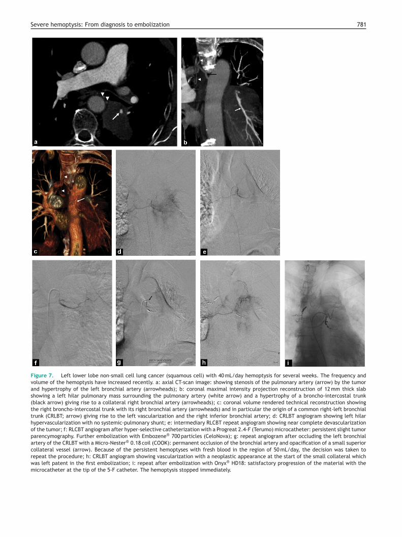
Figure 7. Left lower lobe non-small cell lung cancer (squamous cell) with 40 mL/day hemoptysis for several weeks. The frequency andvolume of the hemoptysis have increased recently. a: axial CT-scan image: showing stenosis of the pulmonary artery (arrow) by the tumorand hypertrophy of the left bronchial artery (arrowheads); b: coronal maximal intensity projection reconstruction of 12 mm thick slabshowing a left hilar pulmonary mass surrounding the pulmonary artery (white arrow) and a hypertrophy of a broncho-intercostal trunk(black arrow) giving rise to a collateral right bronchial artery (arrowheads); c: coronal volume rendered technical reconstruction showingthe right broncho-intercostal trunk with its right bronchial artery (arrowheads) and in particular the origin of a common right-left bronchialtrunk (CRLBT; arrow) giving rise to the left vascularization and the right inferior bronchial artery; d: CRLBT angiogram showing left hilarhypervascularization with no systemic-pulmonary shunt; e: intermediary RLCBT repeat angiogram showing near complete devascularizationof the tumor; f: RLCBT angiogram after hyper-selective catheterization with a Progreat 2.4-F (Terumo) microcatheter: persistent slight tumorparencymography. Further embolization with Embozene® 700 particles (CeloNova); g: repeat angiogram after occluding the left bronchialartery of the CRLBT with a Micro-Nester® 0.18 coil (COOK): permanent occlusion of the bronchial artery and opacification of a small superiorcollateral vessel (arrow). Because of the persistent hemoptyses with fresh blood in the region of 50 mL/day, the decision was taken torepeat the procedure; h: CRLBT angiogram showing vascularization with a neoplastic appearance at the start of the small collateral whichwas left patent in the first embolization; i: repeat after embolization with Onyx® HD18: satisfactory progression of the material with themicrocatheter at the tip of the 5-F catheter. The hemoptysis stopped immediately.

782 A. Khalil et al.
Figure 8. Cryptogenic hemoptysis. Forty-five-year-old man presenting to respiratory department with hemoptysis. Clinical enquiryassessed the hemoptysis as being 100 mL/24 h. a: axial CT-scan image in the parenchymal window showing an area of increased den-sity with focal ground glass opacity (asterisk) localizing the bleeding; b, c: axial CT-scan image in the mediastinal window: nodular orlinear enhancement in the mediastinum representing the bronchial vessels in their mediastinal path; d: frontal reconstruction image in themediastinal window showing the left bronchial trunk (black arrow) and bronchial artery (white arrowhead) of the right broncho-intercostaltrunk; e: coronal image with volume rendered technical reconstruction showing the left bronchial artery (black arrow), the right broncho-intercostal trunk (white arrow) and the right bronchial artery (white arrowheads) of this trunk; f: axial CT-scan image 1 month later showingdisappearance of the increased density of ground glass appearance. Bronchoscopy, bacteriology samples and the immunology assessmentwere negative. The diagnosis made was that of cryptogenic hemoptysis (without cause).

••
•
••
E
Severe hemoptysis: From diagnosis to embolization
Currently, the identification of the middle anterior spinalartery of the high thoracic spinal cord is only possible withconventional angiography (Fig. 9a—c).
The contribution to the bleeding from NBSA should beconsidered in chronic disease with a pleural symphysis. Yoonet al. showed that CT-scan offered excellent sensitivity andspecificity and had a high diagnostic value in detecting NBSA[15,16]. The NBSA may contribute to hypervascularizationand bleeding in chronic lung disease with pleural involve-ment.
Contribution from NBSA to the hypervascularization is
unequivocal if systemic vessels pass through pleura that areover 3 mm thick.The NBSA most often found are:• the intercostal arteries in posterior disease;
h
Ci
Figure 9. Median anterior spinal artery at the cervicothoracic junctioright superior bronchial artery; b, c: angiogram of the same artery macervicothoracic spine and showing a small narrow vessel leading towards
lateralized to the right because of slight rotation of the patient with respethe cervicothoracic region of the spinal cord; d: use of a microcatheter w2 mm in diameter and 7 cm long). Particles are formerly contra-indicated
as a result of reflux from the median anterior spinal artery.
783
branches of the subclavian arteries in the apices;the internal thoracic arteries in the anterosuperior seg-ments;the triangular ligament arteries in the bases and parame-diastinal region;the inferior diaphragmatic arteries in the bases;the coronary arteries in some diseases.
ndovascular treatment of severe
emoptysisurrently, this treatment can only be considered after annitial MDCTA assessment and in close collaboration with
n. a: angiogram of a right broncho-intercostal trunk showing thegnified (subtracted ‘‘b’’ and unsubtracted ‘‘c’’) centering on thethe middle with a ‘‘hairpin’’ shape. This structure, which is slightlyct to the X-ray beam, is the median anterior spinal artery supplyingith occlusion of the trunk using 3-F Micro-Nester® 0.18 coils (COOK;in this situation because of the large risk of accidental embolization

7
if
((raicrur
Tm
ENIrpoas
cr
clfli
bi
aahfptraTcc
aott
Fcbtu
84
ntensive care or respiratory physicians and surgical teamor overall management of the disease.
In our center, the indication for this treatmentFig. 10) is based on the volume of the hemoptysisover 200 mL/24—48 h), repercussions on oxygenation (acuteespiratory failure) and on any pulmonary artery dam-ge. Below 50 mL/24—48 h, endovascular management is notndicated and between 50 and 200 mL/24—48 h, the indi-ation depends on the patient’s underlying condition (poorespiratory reserve in patients with chronic respiratory fail-re) or causes such as aspergilloma with a high risk ofecurrence as massive hemoptysis.
echniques, approaches and embolizationaterials
mbolization of systemic arteries (BA andBSA)
n the majority of situations, the approach is via theight femoral artery after inserting a 5-F introducer. Werefer to use long 5-F introducers (45 cm) in patientsver 70 years old or in those with tortuous atheromatousrteries which makes navigation and catheterization more
traightforward.The two catheters used on first line are the JL4 (leftoronary catheter) and the spinal catheter. Others may beequired such as the left bronchial catheter, an AL1 or AL2
tlst
igure 10. Chart indicating the management of life-threatening hemhest-X-ray (CXR), and a multidetector CT-angiography (MDCTA) ± broncronchial arteriography with embolization (BAE) or pulmonary artery vareatment (BAE or PAVO) or medical treatment depends on the cause (anderlying diseases (chronic respiratory failure with a risk of major respi
A. Khalil et al.
atheter and internal thoracic artery catheter, etc. The twoeft bronchial and left coronary catheters can ‘‘scrape’’ theoor of the aortic arch and catheterize the bronchial arter-
es arising from it.By referring to MDCTA findings, arteries feeding the
leeding area are catheterized and an angiography seriess then performed.
A microcatheter is recommended for safer embolization,lthough the risk of spasm and dissection of the bronchialrteries during catheterization with a microcatheter isigher, which prevent optimal embolization. If gentle risk-ree embolization from the ostium of systemic arteries isossible (Fig. 7d—g), we recommend starting the emboliza-ion using the 5-F catheter. Early and repeated checks areequired in order to avoid reflux of the embolization materi-ls and to detect hazardous anastamoses as early as possible.hese anastamoses may be unmasked when the hypervas-ularization is reduced during ostial embolization by theatheter, or more distally with the microcatheter.
The first line embolization materials are particles thatre currently available in many sizes on the market, eachf which has slightly different physical features. In our cen-er, we use Embozene® 700 to 1300 microns, depending onhe hypervascularization, systemic to pulmonary shunts and
he lumen size of the catheter or micro-catheter. For aarge systemic-pulmonary shunt, the use of a large particleize is required. Coils can be used, however, the opera-or remembers that proximal occlusion (mediastinal andoptysis. The initial assessment includes a clinical assessment, ahoscopy. The first line of treatment is interventional radiology orso-occlusion (PAVO). * In this situation, the indication for invasivespergilloma with risk of recurrence by massive hemoptysis) or theratory impairment with a small amount of hemoptysis recurrence).

785
Table 1 Causes and results of endovascular treatmentfor massive hemoptysis managed in our center over10 years (unpublished data).
Causes Number ofpatients (n)
Success,n (%)
Tuberculosis (activeand latecomplications)
189 158 (84)
Bronchiectasis 159 126 (79)Cancer 149 91 (61)Cryptogenic 126 106 (84)Aspergilloma 48 21 (44)Chronic obstructive
pulmonary disease29 19 (66)
Other 67 48 (72)
(asebitfs(a
ttht[
sotrs0tolahbdatatpia
Severe hemoptysis: From diagnosis to embolization
proximal hilar) should be avoided as there is a highly devel-oped anastomotic network in the mediastinal and proximalregion of the hilum of the lung (Fig. 3e, f). If occluded proxi-mally, this anastomotic network can revascularize the vesseldistal to the occlusion, making embolization less effectiveor even hazardous by closing a recognized and straightfor-ward embolization approach. Coils should be reserved forpermanent very distal obstruction, or if catheterization isdifficult and/or hazardous (Fig. 9). The other embolizationmaterials should be considered in occasional cases and needto be understood in advance of their use, in the same wayas the liquids (Onyx®, Histoacryle®, etc.).
Pulmonary artery vascular occlusionIn most situations, the approach via the right femoral veinis sufficient, using a 7- or 8-French introducer. In specificsituations, such as a raised right hemidiaphragm and a pasthistory of right middle and lower lobe lobectomies, theinternal jugular approach is preferred for anatomical rea-sons. A guide catheter is highly recommended or may evenbe essential for at least three reasons: catheterization,changing catheters and optimal occlusion of the target pul-monary artery. The pulmonary arteries are wide and far fromthe puncture site with at least 2 curves before reaching thetarget, making direct catheterization difficult. The use ofa guide catheter helps to lead the catheter (coaxial) usingthe double curve (the curve of the guide catheter and thecurve of the coaxial catheter). Changing catheter is morestraightforward with a guide catheter and in addition whenocclusion is achieved using coils, the guide catheter offerstwo additional merits of compacting the coil and preventingpullback of the catheter, avoiding accidental occlusion ofoff-target arteries.
The occlusion should be as close as possible to thepulmonary artery lesion responsible for the bleeding. Ide-ally, for a pulmonary artery pseudoaneurysm, the occlusionshould be made proximal and distal to the aneurysm,although this carries an increased risk of iatrogenic rup-ture. Proximal occlusion is sufficient for a distal, acuterapid process or in cancer. Conversely, in inflammatory andchronic infectious processes, particularly tuberculosis, sand-wich occlusion is preferable in order to avoid the recurrenceof the bleeding through the systemic vessels. The aim is toproduce occlusion at the tip of the catheter without theneed for distal embolization. Attention should be paid tothe risk of rupturing a pseudoaneurysm during high-pressureinjection or when a coil or plug is deployed in an irregularartery surrounded by necrosis. In decreasing order of fre-quency, the occlusion materials most often used are coils,plugs (vascular plug or microvascular plug), graft stents andOnyx®. The graft stent should be used for localized lesionswhen the artery has to be kept patent.
Results and complications
Bronchial artery embolization provides immediate control(Table 1) of hemoptysis in over 80% of cases (65 to 92%)
depending on the underlying disease [6,17,18]. Control ismore effective with cryptogenic hemoptysis, bronchiecta-sis or active tuberculosis whereas this is less effective inaspergillosis or lung cancer. If an early recurrence occursheii
Total 767 569 (74)
10 to 20% of cases), the immediate response should be further review of the MDCTA looking for other bronchialystemic arterial conditions, either of atypical origin, orctopic, a NBSA supplying the territory responsible for theleed or alternatively a pulmonary source which was notnitially seen. The use of systemic vasoconstrictors medica-ion before bronchial artery embolization or of resorbableragments (Gelfoam) during embolization is also a possibleource of recurrence. In most cases a second angiographyFig. 7h, i) usually enables the bleeding to be controlledgain.
Long-term recurrence after bronchial arterial emboliza-ion occurs in 10 to 60% of cases and is due eithero recanalization of the embolized vessels or to furtherypervascularization when the cause of hypervasculariza-ion remains. This is common in aspergilloma and cancer17,19,20].
The major complications of embolization are rare buterious and predominantly involve accidental embolizationf the median anterior spinal artery which gives rise tohe right and left superior intercostal arteries and areesponsible for serious neurological events (Brown-Sequardyndrome and paraplegia) with an estimated incidence of.6 to 6.5% [18]. The other complications are necrosis ofhe esophageal or bronchial walls, myocardial infarctionr more systemic spread because of an unstable cathetereading to reflux of the embolization materials into theorta (ischemic stroke, gastrointestinal infarction, splenicematoma, etc.). The possibility of splenic infarction shoulde investigated if a patient develops abdominal pain pre-ominantly in the left hypochondrium together with shocknd a fall in hemoglobin [21]. There is a risk of acute dis-al ischemia at the arterial puncture site in patients withtherosclerosis as a result of thrombosis (partly predisposedo by the length of the procedure and multiple angiogra-hy sessions for recurrences with several punctures). Thencidence of these episodes is greatly influenced by oper-tor experience [18,22] and some can be avoided by usingyperselective microcatheters, which in expert hands allow
mbolization beyond the origin of an esophageal branch ornto a right bronchial artery well beyond the origin of thentercostal artery which may give rise to a median anterior
7 A. Khalil et al.
stc
C
Mppali
• Serious complications are rare (less than 5% intrained hands): cerebrovascular accident, spinalischemia or esophageal necrosis.
• Early recurrence (10 to 20%): review the mechanismof the hemoptysis on MDCTA, further embolization(unembolized collateral, incomplete embolization of
C
ToararmCRP were raised. A MDCTA was performed (Figs. 11—14).
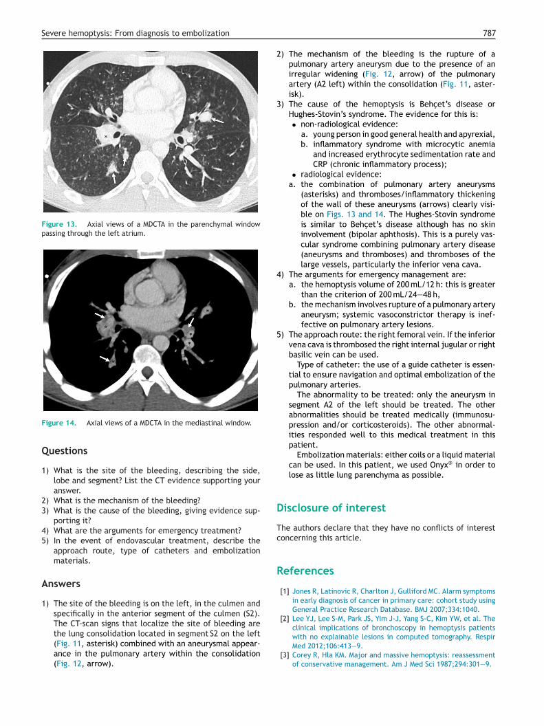
Figure 11. Axial views of a MDCTA in the parenchymal window.
86
pinal artery in patients with a right broncho-intercostalrunk or during unstable catheterization. The most commonomplications of embolization are transient chest pain.
onclusion
DCTA has become an unquestioned investigation in theretreatment assessment of massive hemoptysis. It enableserfect visualization of the bronchopulmonary vascular treend mechanisms responsible for severe hemoptysis. The firstine treatment in the acute phase and in inoperative patientss endovascular.
Take-home messagesGeneral concepts• Hemoptysis: blood arising from the infraglottic
region.• Hemoptysis, symptom (less than 50 mL/24—48 h):
alarm signal for investigation into the cause.• Hemoptysis, disease (serious) (volume over 200 mL/
24—48 h): life threatening requires emergencyspecific treatment.
The use of multidetector CT-angiography• Technique: optimal enhancement of all intra-
thoracic vascular structures.• Assesses the severity of the hemoptysis.• Site of the bleeding: ground glass opacity or localized
lung consolidation.• Identifies the pulmonary artery mechanism for the
bleeding (less than 10% of hemoptysis): necrosis,pseudoaneurysm, irregular pulmonary artery in thewall of a cavity or necrosis.
• Complete mapping of the bronchial and non-bronchial systemic arteries (over 90% of hemoptysis).
• Identifies the cause of the bleeding: tuberculosis,bronchiectasis, lung cancer (2/3 of causes). No causefound (cryptogenic hemoptysis) in 20 to 30% of cases.
Endovascular treatment• Management in a specialized unit: intensive care and
interventional radiology ± thoracic surgery.• Indication: at least one of the following criteria:
respiratory tolerance (acute respiratory failure),pulmonary arterial mechanism and hemoptysis ofover 200 mL/24—48 h.
• The approach (femoral artery or vein) depends onMDCTA findings.
• Embolization material: particularly microparticles(over 500 microns).
• The median anterior spinal artery (arising from theintercostal artery) must be looked for routinely andspecifically during opacification of the right broncho-intercostal trunk; if it is present or there is a doubt asto its presence the use of particles is formally contra-indicated and these should be replaced by occlusionwith micro-coils.
•
Embolization controls hemoptysis in 65 to 92% ofcases depending on cause.F
an artery, vasoconstrictor therapy, etc.).
linical case
his 26-year-old man was referred for hemoptysis of 200 mLn one occasion 12 hours ago. Clinical examination revealed
patient in good general health, apyrexial and withoutecent weight loss. He had no signs of respiratory distressnd his respiratory rate was 14/min with an oxygen satu-ation of 98%. Laboratory investigations showed moderateicrocytic anemia. His erythrocyte sedimentation rate and
igure 12. Axial views of a MDCTA in the mediastinal window.
Severe hemoptysis: From diagnosis to embolization
Figure 13. Axial views of a MDCTA in the parenchymal windowpassing through the left atrium.
2
3
4
5
D
Tc
R
with no explainable lesions in computed tomography. Respir
Figure 14. Axial views of a MDCTA in the mediastinal window.
Questions
1) What is the site of the bleeding, describing the side,lobe and segment? List the CT evidence supporting youranswer.
2) What is the mechanism of the bleeding?3) What is the cause of the bleeding, giving evidence sup-
porting it?4) What are the arguments for emergency treatment?5) In the event of endovascular treatment, describe the
approach route, type of catheters and embolizationmaterials.
Answers
1) The site of the bleeding is on the left, in the culmen andspecifically in the anterior segment of the culmen (S2).The CT-scan signs that localize the site of bleeding arethe lung consolidation located in segment S2 on the left
(Fig. 11, asterisk) combined with an aneurysmal appear-ance in the pulmonary artery within the consolidation(Fig. 12, arrow).787
) The mechanism of the bleeding is the rupture of apulmonary artery aneurysm due to the presence of anirregular widening (Fig. 12, arrow) of the pulmonaryartery (A2 left) within the consolidation (Fig. 11, aster-isk).
) The cause of the hemoptysis is Behcet’s disease orHughes-Stovin’s syndrome. The evidence for this is:• non-radiological evidence:
a. young person in good general health and apyrexial,b. inflammatory syndrome with microcytic anemia
and increased erythrocyte sedimentation rate andCRP (chronic inflammatory process);
• radiological evidence:a. the combination of pulmonary artery aneurysms
(asterisks) and thromboses/inflammatory thickeningof the wall of these aneurysms (arrows) clearly visi-ble on Figs. 13 and 14. The Hughes-Stovin syndromeis similar to Behcet’s disease although has no skininvolvement (bipolar aphthosis). This is a purely vas-cular syndrome combining pulmonary artery disease(aneurysms and thromboses) and thromboses of thelarge vessels, particularly the inferior vena cava.
) The arguments for emergency management are:a. the hemoptysis volume of 200 mL/12 h: this is greater
than the criterion of 200 mL/24—48 h,b. the mechanism involves rupture of a pulmonary artery
aneurysm; systemic vasoconstrictor therapy is inef-fective on pulmonary artery lesions.
) The approach route: the right femoral vein. If the inferiorvena cava is thrombosed the right internal jugular or rightbasilic vein can be used.
Type of catheter: the use of a guide catheter is essen-tial to ensure navigation and optimal embolization of thepulmonary arteries.
The abnormality to be treated: only the aneurysm insegment A2 of the left should be treated. The otherabnormalities should be treated medically (immunosu-pression and/or corticosteroids). The other abnormal-ities responded well to this medical treatment in thispatient.
Embolization materials: either coils or a liquid materialcan be used. In this patient, we used Onyx® in order tolose as little lung parenchyma as possible.
isclosure of interest
he authors declare that they have no conflicts of interestoncerning this article.
eferences
[1] Jones R, Latinovic R, Charlton J, Gulliford MC. Alarm symptomsin early diagnosis of cancer in primary care: cohort study usingGeneral Practice Research Database. BMJ 2007;334:1040.
[2] Lee YJ, Lee S-M, Park JS, Yim J-J, Yang S-C, Kim YW, et al. Theclinical implications of bronchoscopy in hemoptysis patients
Med 2012;106:413—9.[3] Corey R, Hla KM. Major and massive hemoptysis: reassessment
of conservative management. Am J Med Sci 1987;294:301—9.

7
[
[
[
[
[
[
[
[
[
[
[
[
88
[4] Patel U, Pattison CW, Raphael M. Management of massivehaemoptysis. Br J Hosp Med 1994;52:74—8.
[5] Fartoukh M, Khoshnood B, Parrot A, Khalil A, Carette M-F, Sto-clin A, et al. Early prediction of in-hospital mortality of patientswith hemoptysis: an approach to defining severe hemoptysis.Respir Int Rev Thorac Dis 2012;83:106—14.
[6] Fartoukh M, Khalil A, Louis L, Carette M-F, Bazelly B, CadranelJ, et al. An integrated approach to diagnosis and managementof severe haemoptysis in patients admitted to the intensivecare unit: a case series from a referral centre. Respir Res2007;8:11.
[7] Chalumeau-Lemoine L, Khalil A, Prigent H, Carette M-F, Far-toukh M, Parrot A. Impact of multidetector CT-angiography onthe emergency management of severe hemoptysis. Eur J Radiol2013;82:e742—7.
[8] Remy-Jardin M, Bouaziz N, Dumont P, Brillet P-Y, Bruzzi J,Remy J. Bronchial and non bronchial systemic arteries at multi-detector row CT angiography: comparison with conventionalangiography. Radiology 2004;233:741—9.
[9] Khalil A, Fartoukh M, Tassart M, Parrot A, Marsault C,Carette M-F. Role of MDCT in identification of the bleedingsite and the vessels causing hemoptysis. Am J Roentgenol2007;188:W117—25.
10] Khalil A, Soussan M, Mangiapan G, Fartoukh M, Parrot A, CaretteM-F. Utility of high-resolution chest CT scan in the emergencymanagement of haemoptysis in the intensive care unit: sever-ity, localization and aetiology. Br J Radiol 2007;80:21—5.
11] Revel MP, Fournier LS, Hennebicque AS, Cuenod CA, Meyer G,Reynaud P, et al. Can CT replace bronchoscopy in the detec-tion of the site and cause of bleeding in patients with large or
massive hemoptysis? Am J Roentgenol 2002;179:1217—24.12] Khalil A, Fartoukh M, Parrot A, Bazelly B, Marsault C, Carette M-F. Impact of MDCT angiography on the management of patientswith hemoptysis. Am J Roentgenol 2010;195:772—8.
[
A. Khalil et al.
13] Khalil A, Parrot A, Nedelcu C, Fartoukh M, Marsault C,Carette M-F. Severe hemoptysis of pulmonary arterial origin:signs and role of multidetector row CT angiography. Chest2008;133:212—9.
14] Hartmann IJC, Remy-Jardin M, Menchini L, Teisseire A, KhalilC, Remy J. Ectopic origin of bronchial arteries: assess-ment with multidetector helical CT angiography. Eur Radiol2007;17:1943—53.
15] Yoon YC, Lee KS, Jeong YJ, Shin SW, Chung MJ, Kwon OJ.Hemoptysis: bronchial and nonbronchial systemic arteries at16-detector row CT. Radiology 2005;234:292—8.
16] Yoon W, Kim YH, Kim JK, Kim YC, Park JG, Kang HK. Massivehemoptysis: prediction of nonbronchial systemic arterial sup-ply with chest CT. Radiology 2003;227:232—8.
17] Chun J-Y, Belli A-M. Immediate and long-term outcomesof bronchial and non-bronchial systemic artery embolizationfor the management of haemoptysis. Eur Radiol 2010;20:558—65.
18] Mal H, Rullon I, Mellot F, Brugière O, Sleiman C, MenuY, et al. Immediate and long-term results of bronchialartery embolization for life-threatening hemoptysis. Chest1999;115:996—1001.
19] Katoh O, Kishikawa T, Yamada H, Matsumoto S, Kudo S. Recur-rent bleeding after arterial embolization in patients withhemoptysis. Chest 1990;97:541—6.
20] Kim YG, Yoon H-K, Ko GY, Lim C-M, Kim WD, Koh Y. Long-termeffect of bronchial artery embolization in Korean patients withhaemoptysis. Respirol Carlton Vic 2006;11:776—81.
21] Labbe V, Roques S, Boughdène F, Razazi K, Khalil A,Parrot A, et al. Shock complicating successful bronchial
artery embolization for severe hemoptysis. Chest 2009;135:215—7.22] Wong ML, Szkup P, Hopley MJ. Percutaneous embolotherapy forlife-threatening hemoptysis. Chest 2002;121:95—102.