Embed Size (px)
Citation preview
Severe Headaches in Patients with shunts
Slit Ventricle Syndromes
Incidence Of Headaches In Shunted Hydrocephalus
• Essentially all adolescents and young adults with shunts will have headaches if asked
• Most of these headaches are mild, intermittent and lead to normal function
• Severe headaches in shunted patients with apparently working shunts is called the “Slit Ventricle Syndrome” (SVS)
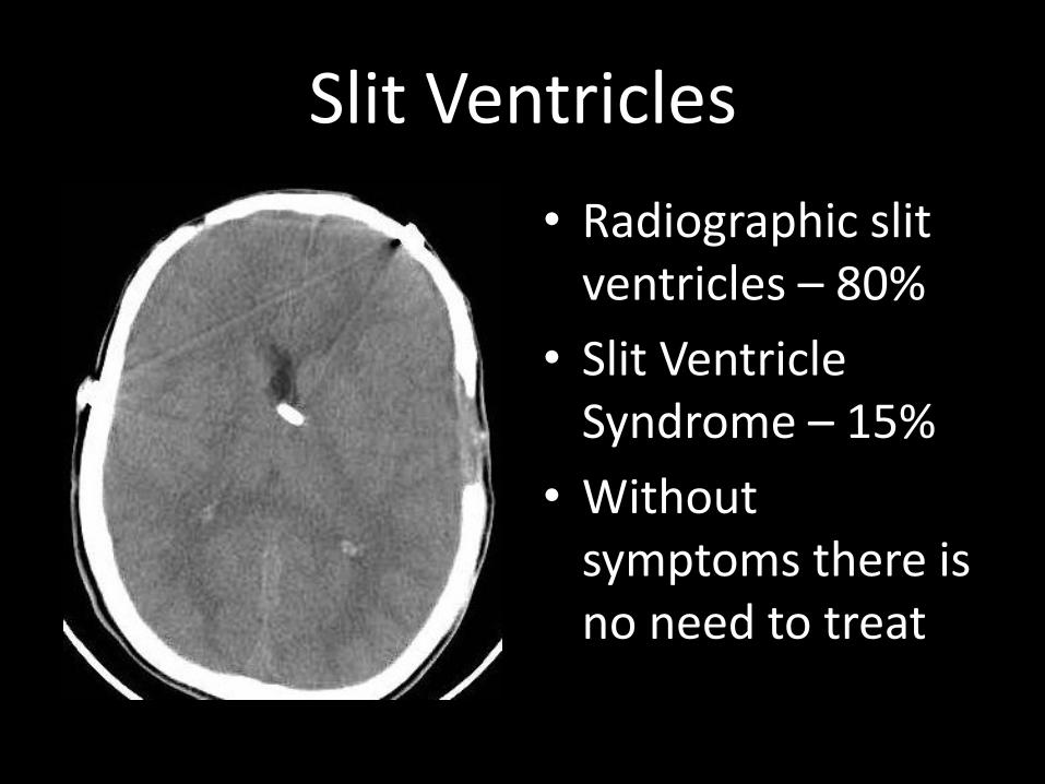
Slit Ventricles
• Radiographic slit ventricles – 80%
• Slit Ventricle Syndrome – 15%
• Without symptoms there is no need to treat
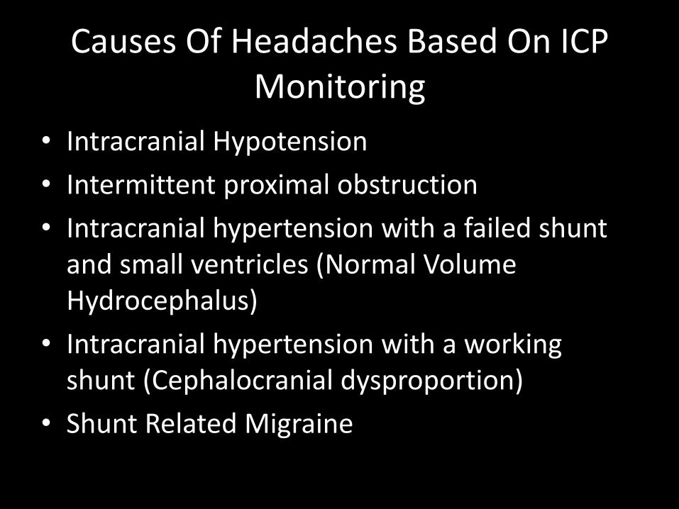
Causes Of Headaches Based On ICP Monitoring
• Intracranial Hypotension
• Intermittent proximal obstruction
• Intracranial hypertension with a failed shunt and small ventricles (Normal Volume Hydrocephalus)
• Intracranial hypertension with a working shunt (Cephalocranial dysproportion)
• Shunt Related Migraine
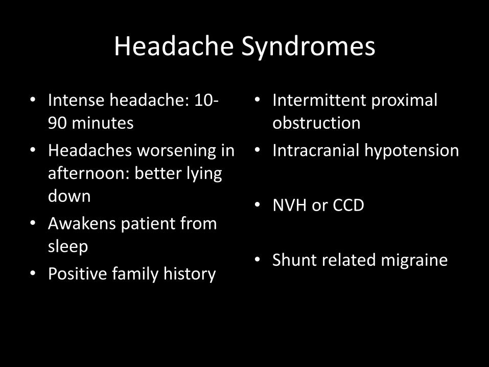
Headache Syndromes
• Intense headache: 10-90 minutes
• Headaches worsening in afternoon: better lying down
• Awakens patient from sleep
• Positive family history
• Intermittent proximal obstruction
• Intracranial hypotension
• NVH or CCD
• Shunt related migraine
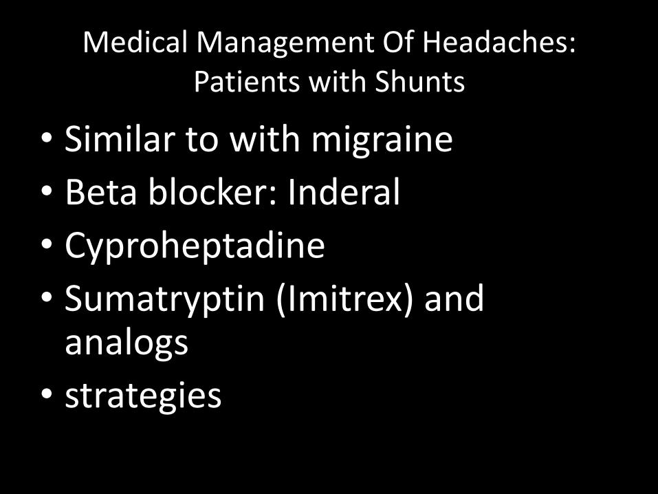
Medical Management Of Headaches: Patients with Shunts
• Similar to with migraine
• Beta blocker: Inderal
• Cyproheptadine
• Sumatryptin (Imitrex) and analogs
• strategies
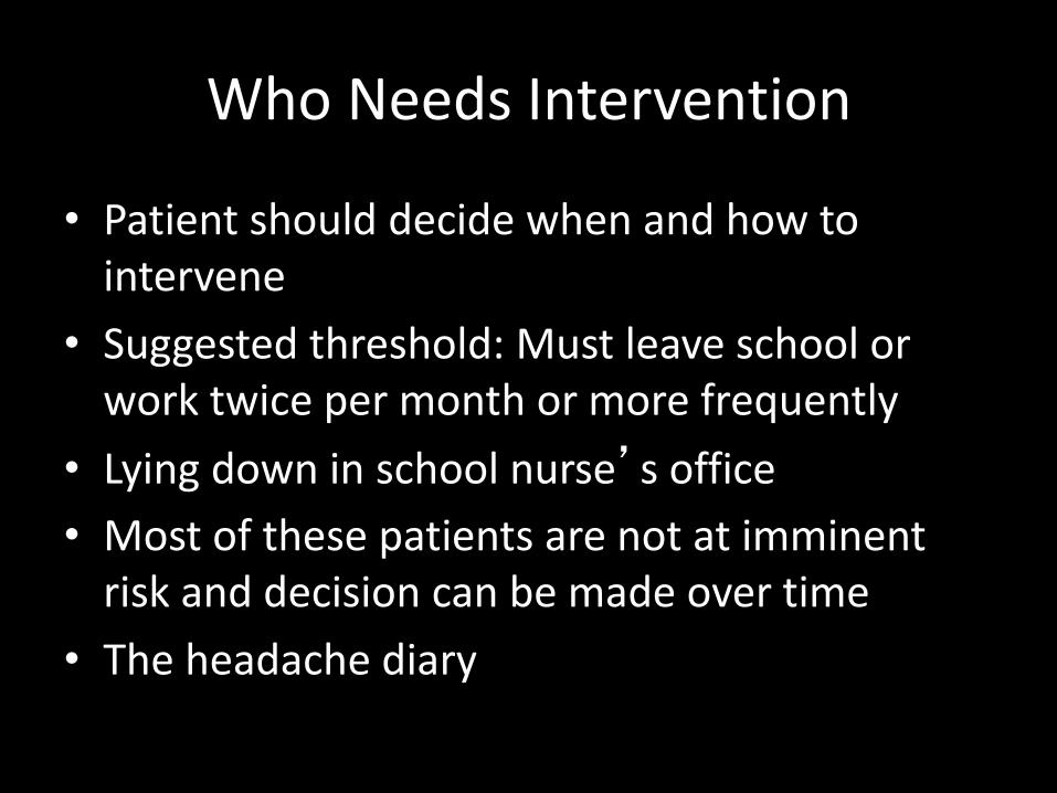
Who Needs Intervention
• Patient should decide when and how to intervene
• Suggested threshold: Must leave school or work twice per month or more frequently
• Lying down in school nurse’s office
• Most of these patients are not at imminent risk and decision can be made over time
• The headache diary
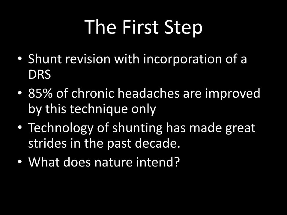
The First Step
• Shunt revision with incorporation of a DRS
• 85% of chronic headaches are improved by this technique only
• Technology of shunting has made great strides in the past decade.
• What does nature intend?
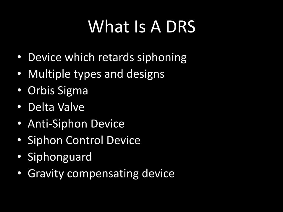
What Is A DRS
• Device which retards siphoning
• Multiple types and designs
• Orbis Sigma
• Delta Valve
• Anti-Siphon Device
• Siphon Control Device
• Siphonguard
• Gravity compensating device
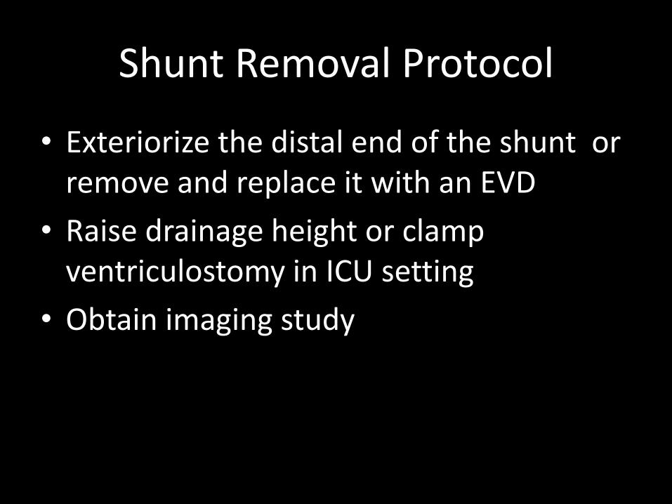
Shunt Removal Protocol
• Exteriorize the distal end of the shunt or remove and replace it with an EVD
• Raise drainage height or clamp ventriculostomy in ICU setting
• Obtain imaging study
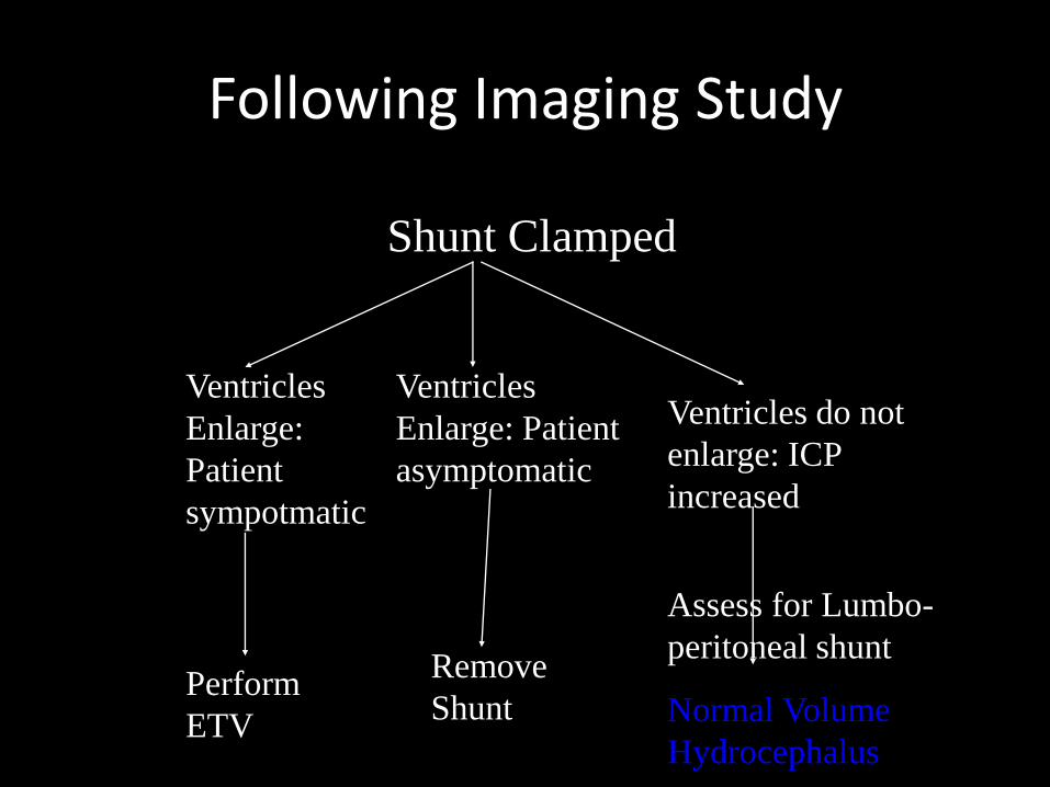
Following Imaging Study
Shunt Clamped
Ventricles
Enlarge:
Patient
sympotmatic
Perform
ETV
Ventricles
Enlarge: Patient
asymptomatic
Remove
Shunt
Ventricles do not
enlarge: ICP
increased
Assess for Lumbo-
peritoneal shunt
Normal Volume
Hydrocephalus
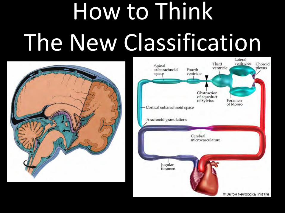
How to Think The New Classification
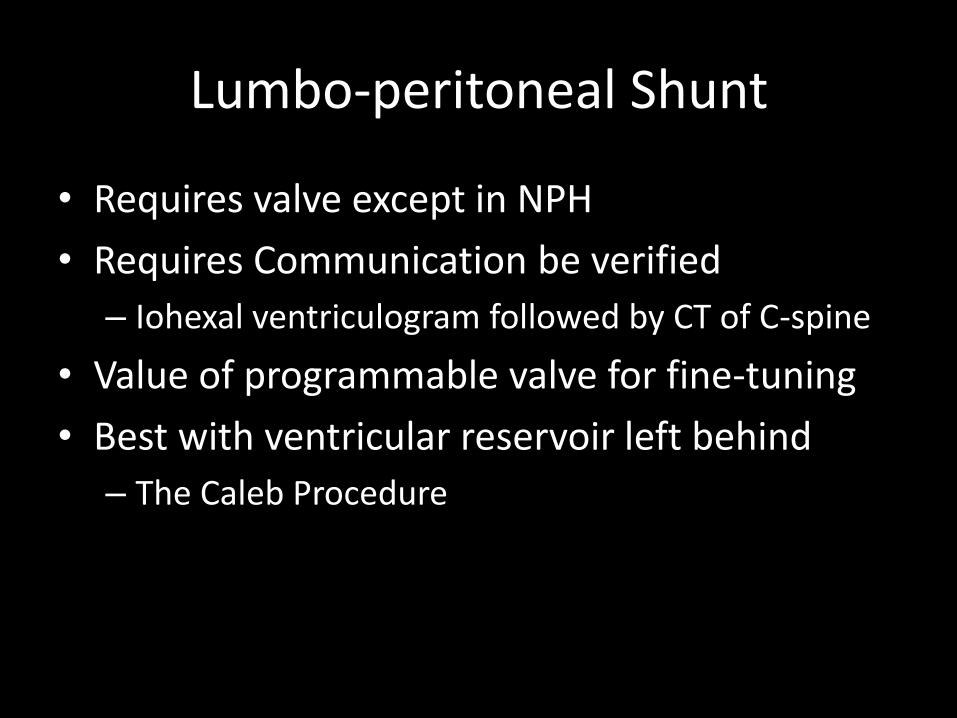
Lumbo-peritoneal Shunt
• Requires valve except in NPH
• Requires Communication be verified
– Iohexal ventriculogram followed by CT of C-spine
• Value of programmable valve for fine-tuning
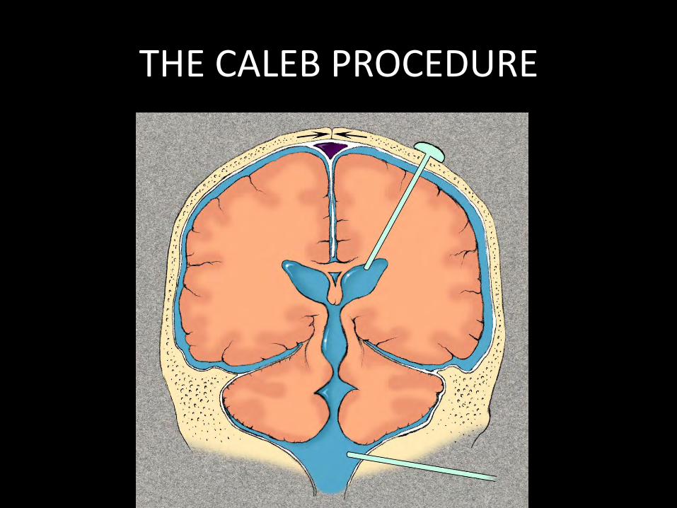
• Best with ventricular reservoir left behind
– The Caleb Procedure
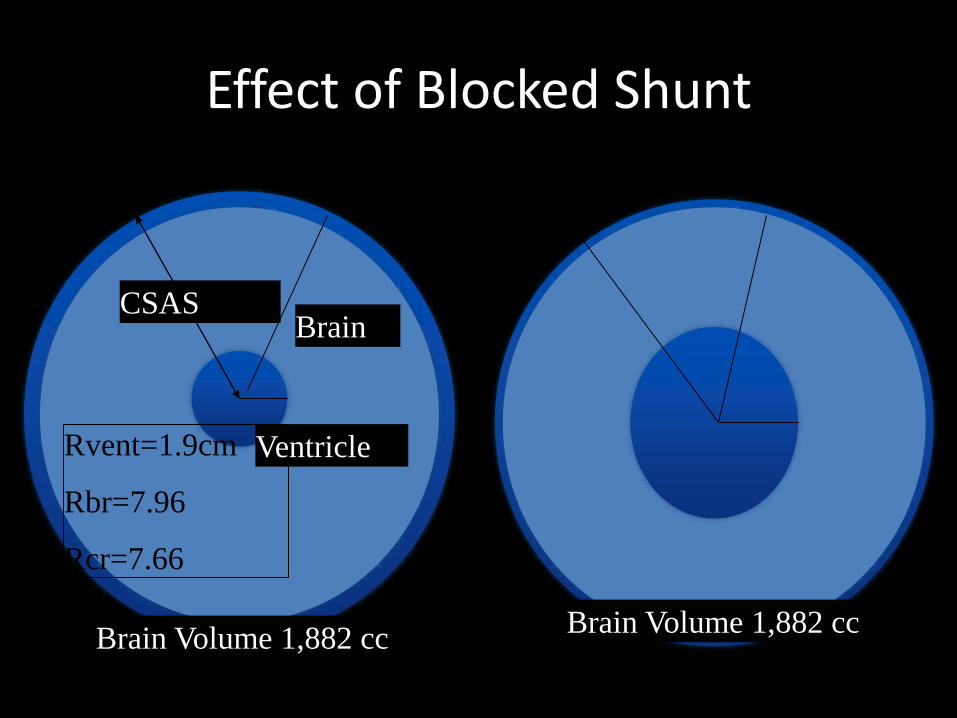
Effect of Blocked Shunt
Ventricle
Brain CSAS
Rvent=1.9cm
Rbr=7.96
Rcr=7.66
Brain Volume 1,882 cc Brain Volume 1,882 cc
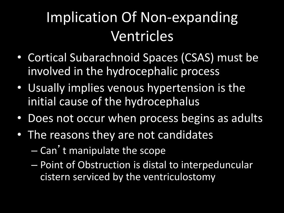
Implication Of Non-expanding Ventricles
• Cortical Subarachnoid Spaces (CSAS) must be involved in the hydrocephalic process
• Usually implies venous hypertension is the initial cause of the hydrocephalus
• Does not occur when process begins as adults
• The reasons they are not candidates – Can’t manipulate the scope
– Point of Obstruction is distal to interpeduncular cistern serviced by the ventriculostomy
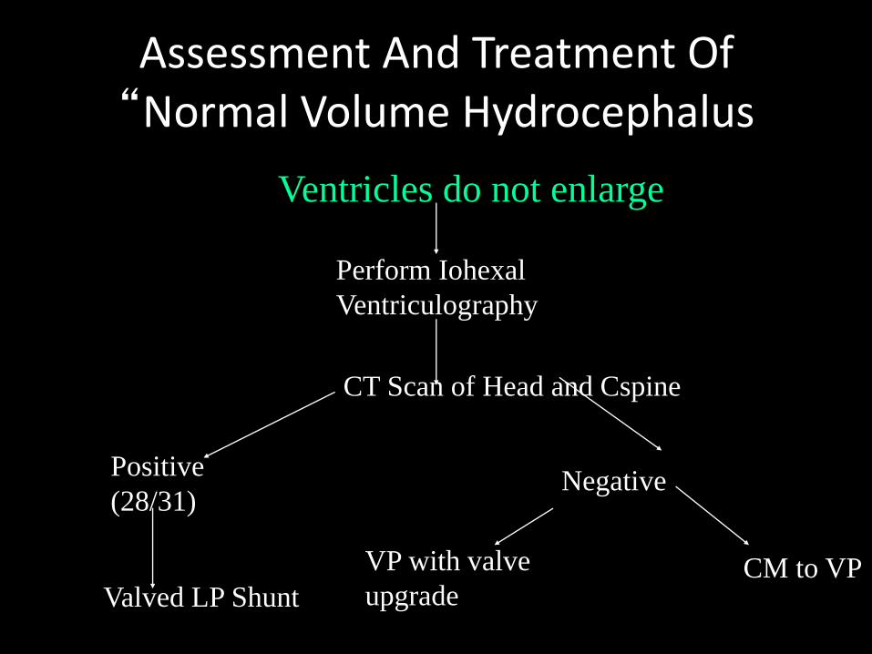
Assessment And Treatment Of “Normal Volume Hydrocephalus
Ventricles do not enlarge
Perform Iohexal
Ventriculography
CT Scan of Head and Cspine
Positive
(28/31) Negative
Valved LP Shunt
VP with valve
upgrade CM to VP
“Normal Volume Hydrocephalus”
• Must Involve the Cortical Subarachnoid Space
• One in 5 children will be found to have non-responding ventricles at the time of shunt failure
• All will have been sent away from ER while still sick
Strategies To Overcome Severe Slit Ventricle Syndrome (NVH)
• Opening pressure (>5mmHg)
• Lumbo-peritoneal shunt (Must have valve)
• Shunt to cisterna magna
• Cisterna magna to ventricle to peritoneal shunt with LP shunt tubing in cisterna magna
• pressure of valve must be higher than the sagittal sinus
WHAT WORKS • AVOID SHUNTING IF AT ALL POSSIBLE
• CORTICAL SUBARACHNOID SHUNT TO PERITONEUM
• VENTRICULAR SHUNTS WITH VERY HIGH RESISTANCE
• VALVES IN SERIES
• CISTERNA MAGNA SHUNT TO PERITONEUM
THE CALEB PROCEDURE
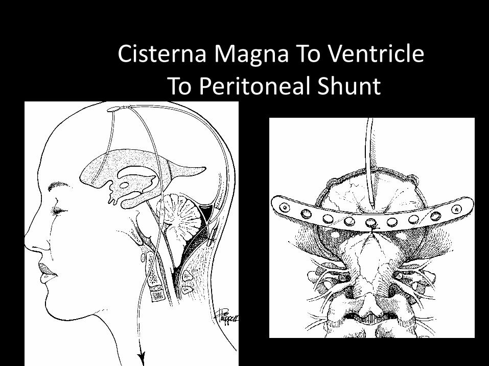
Cisterna Magna To Ventricle To Peritoneal Shunt
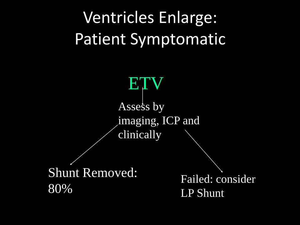
Ventricles Enlarge: Patient Symptomatic
ETV
Assess by
imaging, ICP and
clinically
Shunt Removed:
80% Failed: consider
LP Shunt
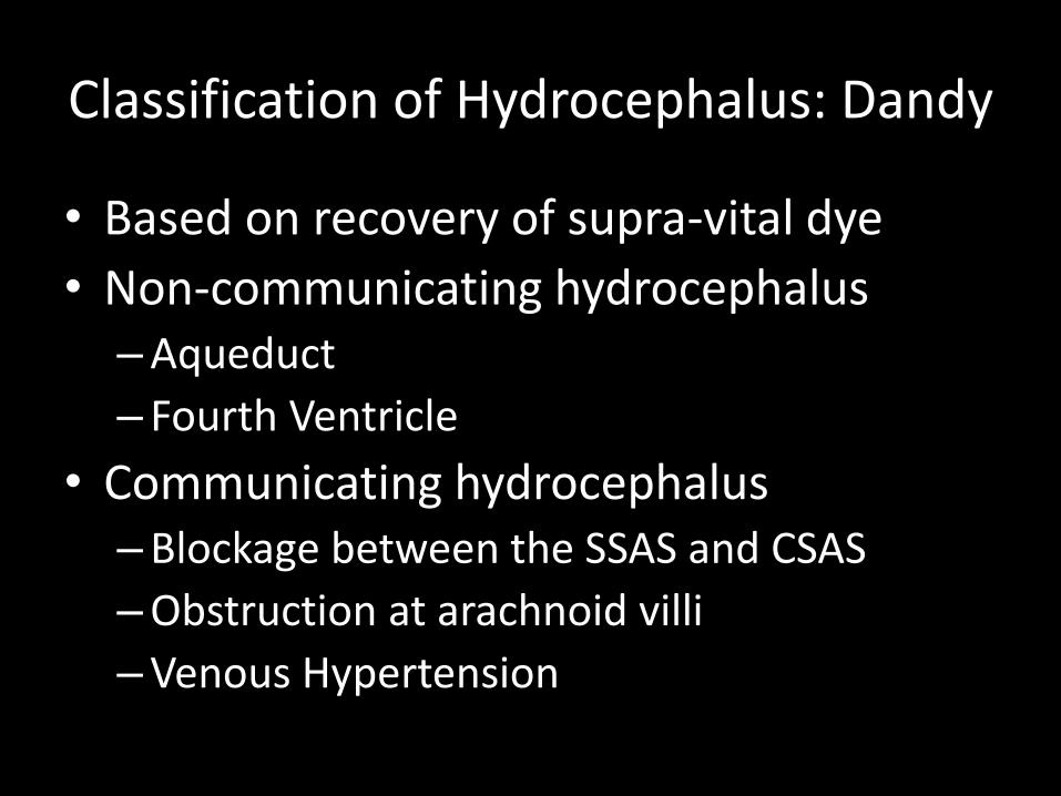
Classification of Hydrocephalus: Dandy
• Based on recovery of supra-vital dye
• Non-communicating hydrocephalus –Aqueduct
– Fourth Ventricle
• Communicating hydrocephalus –Blockage between the SSAS and CSAS
–Obstruction at arachnoid villi
–Venous Hypertension
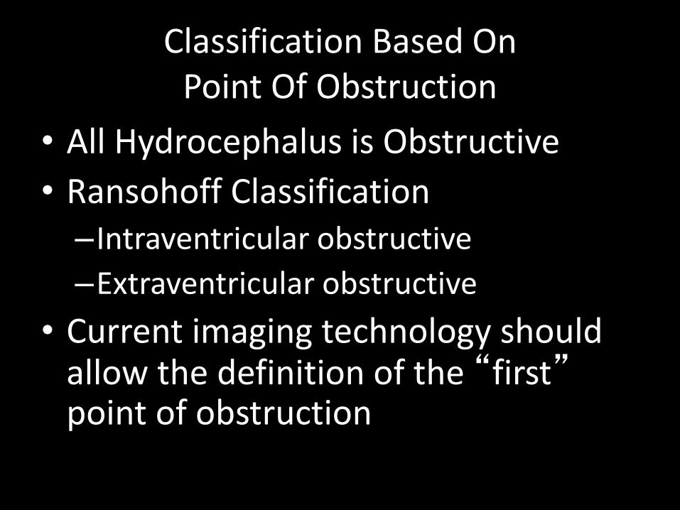
Classification Based On Point Of Obstruction
• All Hydrocephalus is Obstructive
• Ransohoff Classification –Intraventricular obstructive
–Extraventricular obstructive
• Current imaging technology should allow the definition of the “first” point of obstruction

Please Note • The point of obstruction in post-
hemorrhagic hydrocephalus
–Acutely it is in the area of the arachnoid villi
– Late, in all forms of PHH the blockage is between the SSAS and CSAS, an ideal case for ETV
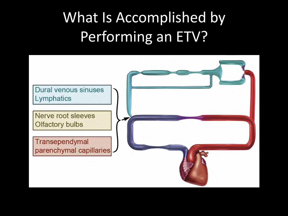
What Is Accomplished by Performing an ETV?
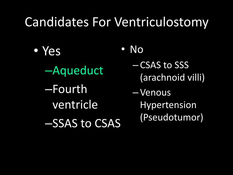
Candidates For Ventriculostomy
• Yes
–Aqueduct
–Fourth ventricle
–SSAS to CSAS
• No
–CSAS to SSS (arachnoid villi)
–Venous Hypertension (Pseudotumor)

Risk Assessment
• Shunt
–Infection: 8%
–Failure: 20-50%
–Death: 1%/yr
• ETV – Hormonal Difficulty
usually DI:
– Loss of recent memory
– Diplopia
– Hemiparesis
– All 3% acute/1% permanent
– Death
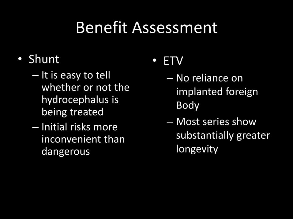
Benefit Assessment
• Shunt – It is easy to tell
whether or not the hydrocephalus is being treated
– Initial risks more inconvenient than dangerous
• ETV
– No reliance on implanted foreign Body
– Most series show substantially greater longevity

ETV Is Subtantially More Dangerous Than A Shunt
Procedure and Subtantially Less Dangerous Than A Lifetime Of
Shunt Dependency
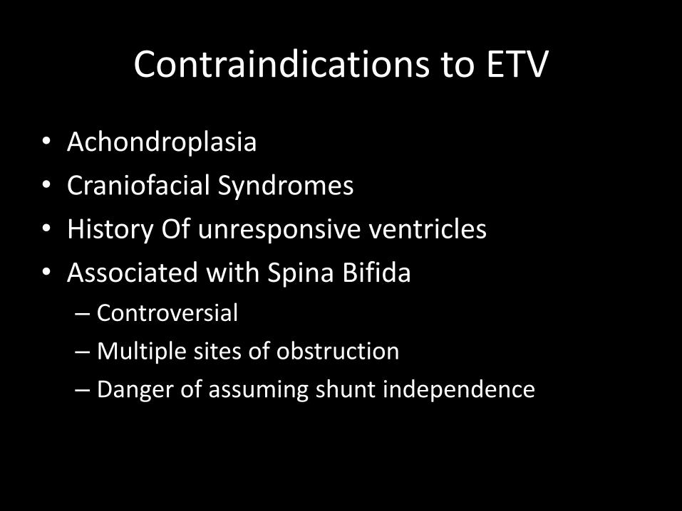
Contraindications to ETV
• Achondroplasia
• Craniofacial Syndromes
• History Of unresponsive ventricles
• Associated with Spina Bifida
– Controversial
– Multiple sites of obstruction
– Danger of assuming shunt independence
Late Outcome
• Personal Experience – After two weeks only
one patient has failed late
– That patient failed because of herniation of Basilar artery
– Late failures try second ETV
• Literature – Growing number of
late failures
– Drake et al see ETV failure as same as shunt failure
– Several Late Deaths
The New Rules
• Every shunt failure is a chance to test shunt dependency
• Shunts are evil
• Most patients with communicating hydrocephalus are candidates for ETV

Conclusions • At least 70% of shunted patients may be
candidates for shunt removal
• Communicating hydrocephalus is a misnomer
• Previously shunted patients are excellent candidates for ETV
• “If it aint broke don’t fix it”
–But • Make certain that you and your neurosurgeon
have a plan for the next step
Rekate’s Rules Of Problematic Shunt Management
• Make certain shunt is really needed
• Attempt shunt removal
• If remains shunt dependent make certain that all CSF compartments see the same pressure either internally or externally
• Make certain ICPs 5-15 mmHg recumbant and –5 to +5 mmHg standing