Embed Size (px)
Citation preview
1
INTRODUCTION
Proteases are a type of enzymes that are widely distributed in nature and are found ubiquitously in eukaryotes, prokary-otes, and viruses. They perform proteolytic reactions and pep-tide bond hydrolysis. Based on the important chemical groups in their active sites, proteases are typically categorized as 4 ma-jor classes (e.g., serine, metallo-, cysteine, and aspartic proteas-es). Of these classes, serine proteases are receiving increasing attention due to their diverse array of functions. They are in-volved in various aspects of physiological progression, such as digestion, apoptosis, signal transduction, blood coagulation, and wound healing through the proteolysis cascade action [1]. Aside from their roles in the physiology of organisms, they also play crucial roles in the pathogenesis of a number of dis-eases, such as cardiopulmonary disease and emphysema [2].
Parasitic helminths are one of the most important patho-
gens worldwide and are classified into nematodes (round-worms), trematodes (flatworms), and cestodes (tapeworms). Humans are constantly threatened by infections with these pathogens, which cause a wide variety of infectious diseases. During the process of infection, proteases derived from para-sites are thought to be significant factors for successfully estab-lishing infection. Experimental evidence has shown that serine proteases are involved in a wide variety of events in the life cy-cle of helminths.
The vast majority of serine proteases are digestive proteases involved in metabolic food processing or host tissue penetra-tion. Additional serine proteases that are involved in reproduc-tion, evasion of the host immune system, and development have also been characterized [3]. The essential roles of parasite serine proteases and their diverse activities make them attrac-tive targets for the development of novel immunotherapeutic, chemotherapeutic, and serodiagnostic agents for the next gen-eration of antiparasite interventions. The molecular and bio-chemical characterization of the serine proteases derived from these parasites is therefore central to the understanding of hel-minth-host interplay and the successful control of helminth infections. In this review, we summarized what is known of helminth serine proteases and their putative functions.
ISSN (Print) 0023-4001ISSN (Online) 1738-0006
Korean J Parasitol Vol. 53, No. 1: 1-11, February 2015 http://dx.doi.org/10.3347/kjp.2015.53.1.1▣MINI-REVIEW
•Received 27 March 2014, revised 15 September 2014, accepted 23 October 2014.*Corresponding author ([email protected]; [email protected]; [email protected])
© 2015, Korean Society for Parasitology and Tropical MedicineThis is an Open Access article distributed under the terms of the Creative Commons Attribution Non-Commercial License (http://creativecommons.org/licenses/by-nc/3.0) which permits unrestricted non-commercial use, distribution, and reproduction in any medium, provided the original work is properly cited.
Serine Proteases of Parasitic Helminths
Yong Yang1,2,*, Yun jun Wen1, Ya Nan Cai3, Isabelle Vallée2, Pascal Boireau2, Ming Yuan Liu4,5,*, Shi Peng Cheng1,*1State Key Laboratory for Molecular Biology of Special Economic Animals, Institute of Special Economic Animal and Plant Sciences, Chinese
Academy of Agricultural Sciences, Changchun, China; 2ANSES, ENVA, UPVM, PRES Paris Est, JRU BIPAR, Animal Health Laboratory, Maisons-Alfort, France; 3College of Animal Science and Technology, Jilin Agricultural University, Changchun, China; 4Key Laboratory of Zoonosis Research,
Ministry of Education, Institute of Zoonosis, Jilin University, Changchun, China; 5Jiangsu Co-innovation Center for Prevention and Control of Important Animal Infectious Diseases and Zoonoses, Yangzhou 225009, China
Abstract: Serine proteases form one of the most important families of enzymes and perform significant functions in a broad range of biological processes, such as intra- and extracellular protein metabolism, digestion, blood coagulation, regulation of development, and fertilization. A number of serine proteases have been identified in parasitic helminths that have putative roles in parasite development and nutrition, host tissues and cell invasion, anticoagulation, and immune evasion. In this review, we described the serine proteases that have been identified in parasitic helminths, including nem-atodes (Trichinella spiralis, T. pseudospiralis, Trichuris muris, Anisakis simplex, Ascaris suum, Onchocerca volvulus, O. lien-alis, Brugia malayi, Ancylostoma caninum, and Steinernema carpocapsae), cestodes (Spirometra mansoni, Echinococcus granulosus, and Schistocephalus solidus), and trematodes (Fasciola hepatica, F. gigantica, and Schistosoma mansoni). Moreover, the possible biological functions of these serine proteases in the endogenous biological phenomena of these parasites and in the host-parasite interaction were also discussed.
Key words: Serine protease, biological function, parasitic helminth, nematode, trematode, cestode
2 Korean J Parasitol Vol. 53, No. 1: 1-11, February 2015
SERINE PROTEASES AND ENZYME MECHANISMS
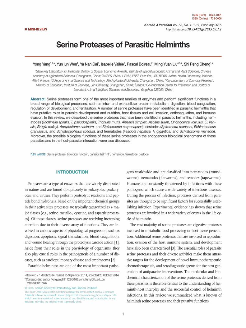
Serine proteases are named because of the presence of a nu-cleophilic serine residue at the active site. The serine residue plays important roles in mediating protein hydrolysis. Most members of the serine proteases contain 3 essential residues at their active sites: a serine (Ser), a histidine (His), and an aspar-tate (Asp). Although these 3 residues do not have continual distribution throughout the linear protein sequence, they are close to each other in the active 3-dimensional conformation. Chymotrypsin, which is a major serine protease, is found in helminths. Chymotrypsin can be divided into 3 main subfam-ilies based on its substrate specificity; trypsin-like, chymotryp-sin-like, and elastase-like. The proteases in these 3 subfamilies share a similar tertiary structure, but their substrate cleavage specificities differ; trypsin-like, in which a cleavage of amide substrates follows Arg or Lys at the P1 position (Fig. 1A); chy-
Fig. 1. The pattern and characterization of the binding pocket re-sponsible for specificity of serine proteases. (A) Trypsin specificity is due to a negatively charged aspartic acid (Asp) located in the base of the binding pocket. Thus, it specifically cleaves peptide bonds of positively charged residues, i.e., lysine (Lys) and arginine (Arg). (B) Chymotrypsin specificity is due to a deep hydrophobic pocket containing serine (Ser) and glycine (Gly). This contributes to specifically cleave peptide bonds of large hydrophobic resi-dues, i.e., phenylalanine (Phe), tryptophan (Trp), and tyrosine (Tyr). (C) Elastase has a much smaller binding pocket containing Arg and Lys than Trypsin or Chymotrypsin and prefers to cleave pep-tides of small, neutral residues, such as alanine (Ala), glycine (Gly), and valine (Val).
Trypsin Chymotrypsin Elastase
Asp189
Ser189
Val216
Thr226
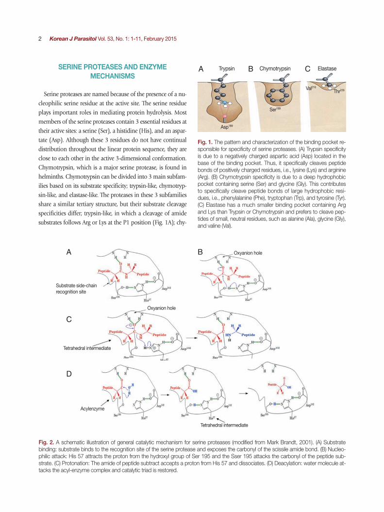
Fig. 2. A schematic illustration of general catalytic mechanism for serine proteases (modified from Mark Brandt, 2001). (A) Substrate binding: substrate binds to the recognition site of the serine protease and exposes the carbonyl of the scissile amide bond. (B) Nucleo-philic attack: His 57 attracts the proton from the hydroxyl group of Ser 195 and the Sser 195 attacks the carbonyl of the peptide sub-strate. (C) Protonation: The amide of peptide subtract accepts a proton from His 57 and dissociates. (D) Deacylation: water molecule at-tacks the acyl-enzyme complex and catalytic triad is restored.
Substrate side-chainrecognition site
Tetrahedral intermediate
Tetrahedral intermediate
Acylenzyme
Oxyanion hole
Oxyanion hole
A B C
A
C
D
B
Yang et al.: Serine proteases of helminths 3
motrypsin-like, in which a cleavage occurs following 1 of the hydrophobic amino acids at P1 (Fig. 1B); and elastase-like, in which a cleavage follows an Ala at P1 (Fig. 1C).
These enzymes are usually synthesized as inactive precursor zymogens, which are converted to the smaller activated en-zymes by cleavage processes involving a conformational change. Conformational change is necessary for hydrolytic ac-tivity. There are 4 steps involved in the chymotrypsin catalysis mechanism, which include substrate binding, nucleophilic at-tack, protonation, and deacylation. A variety of structural fea-tures are responsible for the catalytic effectiveness of these en-zymes [4] (Fig. 2A-D).
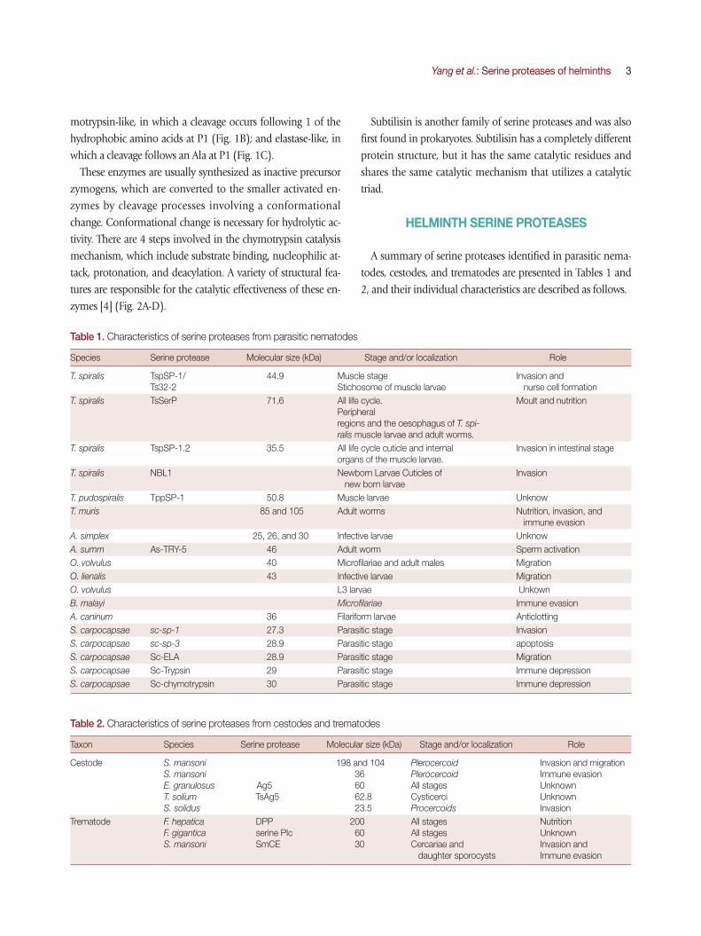
Table 1. Characteristics of serine proteases from parasitic nematodes
Species Serine protease Molecular size (kDa) Stage and/or localization Role
T. spiralis TspSP-1/Ts32-2
44.9 Muscle stage Stichosome of muscle larvae
Invasion and nurse cell formation
T. spiralis TsSerP 71.6 All life cycle. Peripheralregions and the oesophagus of T. spi-ralis muscle larvae and adult worms.
Moult and nutrition
T. spiralis TspSP-1.2 35.5 All life cycle cuticle and internal organs of the muscle larvae.
Invasion in intestinal stage
T. spiralis NBL1 Newborn Larvae Cuticles of new born larvae
Invasion
T. pudospiralis TppSP-1 50.8 Muscle larvae UnknowT. muris 85 and 105 Adult worms Nutrition, invasion, and
immune evasion A. simplex 25, 26, and 30 Infective larvae UnknowA. summ As-TRY-5 46 Adult worm Sperm activationO. volvulus 40 Microfilariae and adult males Migration O. lienalis 43 Infective larvae MigrationO. volvulus L3 larvae UnkownB. malayi Microfilariae Immune evasion A. caninum 36 Filariform larvae AnticlottingS. carpocapsae sc-sp-1 27.3 Parasitic stage Invasion S. carpocapsae sc-sp-3 28.9 Parasitic stage apoptosisS. carpocapsae Sc-ELA 28.9 Parasitic stage MigrationS. carpocapsae Sc-Trypsin 29 Parasitic stage Immune depressionS. carpocapsae Sc-chymotrypsin 30 Parasitic stage Immune depression
Table 2. Characteristics of serine proteases from cestodes and trematodes
Taxon Species Serine protease Molecular size (kDa) Stage and/or localization Role
Cestode S. mansoniS. mansoniE. granulosusT. soliumS. solidus
Ag5TsAg5
198 and 104 36 60 62.8 23.5
PlerocercoidPlerocercoidAll stagesCysticerciProcercoids
Invasion and migration Immune evasion UnknownUnknownInvasion
Trematode F. hepaticaF. giganticaS. mansoni
DPPserine PIcSmCE
200 60 30
All stagesAll stages Cercariae and daughter sporocysts
NutritionUnknownInvasion andImmune evasion
Subtilisin is another family of serine proteases and was also first found in prokaryotes. Subtilisin has a completely different protein structure, but it has the same catalytic residues and shares the same catalytic mechanism that utilizes a catalytic triad.
HELMINTH SERINE PROTEASES
A summary of serine proteases identified in parasitic nema-todes, cestodes, and trematodes are presented in Tables 1 and 2, and their individual characteristics are described as follows.
4 Korean J Parasitol Vol. 53, No. 1: 1-11, February 2015
PARASITIC NEMATODE SERINE PROTEASES
Serine proteases from Trichinella Trichinella is an intracellular nematode that infects a wide
variety of animals. The complete life cycle of the parasite is completed in a single host via the invasion of intestinal epi-thelial and skeletal muscle cells. A recent study of proteases throughout the life of Trichinella spiralis found that excretion-secretion (ES) and crude extracts of muscle stage larvae show substantial serine protease activity against structural proteins, whereas newborn larvae and adult worms principally degrade hematic proteins. This stage-specific proteolytic activity con-tributes to the breakdown of both mechanical and humoral barriers within the host during parasite infection. These serine proteases are targets of the antibody response, which can in-hibit the protease activity and possibly contribute to the im-pairment of the parasite in a sensitized host [5,6].
During the invasion of epithelial cells, the larvae released several glycoproteins that bear the highly antigenic sugar moi-ety, tyvelose (3, 6-dideoxy arabinohexose). Monoclonal anti-bodies against tyvelose protect against infection, which impli-cates that tyvelose-bearing glycoproteins play keys roles in in-testinal epithelium invasion and niche establishment. With the aim of investigating these glycoproteins at the molecular level, Romaris et al. [7] first isolated glycoproteins by affinity chromatography technique using monoclonal antibodies (mAbs). De novo peptide sequencing combined with cDNA library screening identified that these glycoproteins are serine proteases (TspSP-1). Western blot analysis and immunohisto-chemistry indicated that these glycoproteins are muscle larvae (ML) stage specific and are synthesized in α stichocytes. Fur-thermore, the inhibition of epithelial cell invasion and migra-tion by mAbs against TspSP-1 indicated that TspSP-1 could play an important role in degrading cytoplasmic or intercellu-lar proteins, thereby facilitating the movement of the larvae [7]. Subsequently, Nagano et al. [8] also isolated a serine protease, named Ts23-2, from a cDNA library of T. spiralis muscle larvae. The Ts23-2 gene is only transcribed after the completion of cyst formation. The protease activity of the recombinant cata-lytic domain was confirmed using synthetic peptide substrates, indicating that it is a plasmin-like protease [8].
Recently, another member of this subfamily, named Tsp-SP-1.2, was characterized. The anti-serum against TspSP-1.2 can partially prevent the larval invasion of intestinal epithelial cells. Furthermore, the recombinant TspSP-1.2 protein induced
a partial protective immunity in mice. These results indicated that TspSP-1.2 contributes to the larval invasion of host intes-tinal epithelial cells and could be a potential vaccine candidate against T. spiralis infection [9]. A similar protein (TppSP-1) from Trichinella pseudospiralis muscle larvae was identified by Cwiklinski et al. [10]. Analysis of the deduced amino acid se-quence found that the histidine residue of the catalytic triad in TsSP-1 was replaced with an arginine residue in the TppSP-1. This could lead to the loss of proteolytic activity, and the role in the T. pseudospiralis-host interaction needs further research [10]. Trap et al. [11] identified another putative serine protease by screening a library from T. spiralis adult-newborn larvae mixed stage with a radioisotope-labelled DNA probe. TsSerP contains 2 trypsin-like serine protease domains flanking a hy-drophilic domain. Northern blot analysis of the expression profile for TsSerP genes demonstrated that it was expressed in all life cycle stages of the parasite. Western blot analysis using soluble and E-S antigens found that it was not detected in ES products. Immunolocalization showed that TsSerP is expressed on the peripheral regions and the esophagus of T. spiralis mus-cle larvae and adult worms. Thus, TsSerP may be involved in the parasite’s moulting process and digestive function [11].
Liu et al. [12] identified a newborn larval stage-specific ser-ine protease gene (NBL1) via a subtractive cDNA library of T. spiralis newborn larvae. It includes 2 regions, a catalytic do-main and a C-terminal domain. Epitope mapping using trun-cated variants of rNBL1 indicated that the C-terminal part of NBL1 is the main immunodominant region. NBL1 showed encouraging potential in the early detection of Trichinella infec-tion and protective immunity against T. spiralis infection in pigs [13]. Based on the high immunogenicity of the C-termi-nal domain, we hypothesized that during the newborn larval invasion of the host, it may divert the immune response away from the functional regions of NBL1 to contribute to host in-vasion.
The multiple serine proteases identified at different stages of T. spiralis indicated the existence of a superfamily of serine pro-teases in T. spiralis. Each member of serine protease families may have different functions in parasite infection, which de-pends on stage-specific expression, location, and the presence of a regulation domain of the serine protease. Further studies are required to fully understand their functions in parasite-host interplay.
Yang et al.: Serine proteases of helminths 5
Serine proteases from Trichuris muris T. muris is a parasitic nematode of mice in which an infec-
tive larva invades host intestinal mucosa and develops into an adult worm. The anterior portion of an adult worm embeds within a syncytial tunnel derived from host cecal epithelium. There are 2 major serine peptidases with specific activity for collagen-like molecules in the ES antigens of T. muris adult worms. Interestingly, the activity of both serine peptidases was not observed in worm extract, which suggests that the enzymes are present in the adult worm as inactive precursors or inacti-vated by endogenous peptidase inhibitors. The ability for deg-radation of the basement membrane by live worms suggested that these peptidases could be involved in the invasive process. These peptidases also could play important roles in the pro-duction and subsequent maintenance of the parasites' syncy-tial habitat. Moreover, authors speculated that they could con-tribute to the pathology of trichuriasis by disrupting the integ-rity of epithelial cell membranes [14].
The intestinal mucus barrier plays a significant role in the expulsion of gastrointestinal nematodes. The mucin is pivotal during the formation of the mucus layer. A recent study found that serine proteases secreted by T. muris can degrade the ma-jor intestinal mucin Muc2 and depolymerize the mucin net-work. Thus, it was suggested that serine proteases secreted by T. muris could contribute to the modification of the parasitic niche to prevent clearance from the host or to facilitate effi-cient mating and egg laying [15]. Given its essential role in the intestinal stage, this serine protease could be an attractive drug target and should be characterized at the molecular level.
Serine proteases from Anisakis simplex Anisakiasis is a gastrointestinal tract disease, which is caused
by the consumption of raw or undercooked seafood that con-tains larvae of the nematode A. simplex. After ingestion, A. sim-plex larvae penetrate the mucosa, submucosa, and muscularis of the host stomach and intestine and may migrate to the omentum, liver, pancreas, or gall bladder. Sakanari and McK-errow [16] found that the secretion of infective larvae contains trypsin-like serine proteases that degrade connective tissue ex-tracellular matrix (ECM). Judy et al. [17] isolated 4 serine pro-tease genes of A. simplex using degenerate oligonucleotide probes based on the consensus regions of mammalian serine proteases. One of these genes is 67% identical to the rat tryp-sin II gene. Alignment of these 2 genes revealed that the in-tron-exon junctions are conserved between nematodes and
rats, confirming the structural and functional similarity of the 2 genes. Thus, serine proteases of infective larvae may be in-volved in the degradation or digestion of host tissue [17].
Meanwhile, Stephen et al. [18] purified 2 serine proteases from the infective larvae of A. simplex using different affinity chromatography approaches. The 26 kDa-protease is similar to the trypsin of the domestic pig, Sus scrofa. The second serine protease was similar to the extracellular serine protease of the pathogenic bacterium Dichelobacter nodosus, which can degrade elastin, keratin, and collagen. The mechanism involved in the tissue destruction caused by the 2 serine proteases is far from being decrypted and needs further research [18].
Serine proteases from Ascaris suum A. suum, also known as the large roundworm of pigs, is an
intestinal nematode. During the process of A. suum sperm acti-vation, sperm differentiates from immature spermatids into mature and motile spermatozoa. Recently, study of sperm acti-vation indicated that serine protease is responsible for A. suum sperm activation. This serine protease was purified and identi-fied by de novo sequencing. The purified protease showed strong activity in sperm activation, and it can be inhibited by the serine protease inhibitor PMSF. Finally, the full length cDNA named As-TRY-5 was cloned by RACE-PCR using the degenerative primers based on the peptide sequence. Sequence comparisons indicated that As-TRY-5 shares a high degree of homology with trypsin-like serine proteases of eukaryotes. A further study using a serine protease inhibitor (As-SRP-1) that was released by the activated sperm indicated that during the spermatogenesis process, the activity of As-TRY-5 was regulated by this serine protease inhibitor. This could be significant dur-ing postcopulatory sexual selection [19].
Serine proteases from filarial wormsOnchocerca volvulus is an important filarial nematode that
causes subcutaneous filariasis of humans and affects the eyes and skin. The infective larvae, male worms, and microfilariae migrate through the host tissue. A proteolytic activity study in-dicated that there is a 40 kDa neutral elastase in ES products of microfilariae, which can degrade components of the dermal extracellular matrix, collagen type IV, fibronectin, and laminin but cannot degrade intact immunoglobulins. Based on this proteolytic activity, authors suggested that the elastase of mi-crofilariae plays an important role in the degradation of elastic fibres of the host tissue. Moreover, the elastase proteolytic ac-
6 Korean J Parasitol Vol. 53, No. 1: 1-11, February 2015
tivity is also present in males, but absent in ES products of fe-males. This is correlated with the different behavior of adult worms; the adult males are able to migrate from 1 nodule to another, whereas adult females only reside in nodules [20]. In addition, a stage-specific 43-kDa serine elastase was also found in infective larvae of Onchocerca lienalis. The specific elastase of L3 larvae most likely contributes to L3 migration from the blackfly bite site to different tissues of the body where the adults will develop and form nodules [21]. Blisterase is a sub-tilisin-like serine protease and plays important roles in nema-tode biology including the cuticle production and mainte-nance, neural signalling, and nematode development. Thus, it is a potential drug target for controlling parasite infection. Catherine et al. [22] isolated blisterase from a cDNA library of the infective larvae (L3) of O. volvulus. A fragment of blisterase was cloned and expressed in insect cells with maximal activity at a neutral pH. However, the roles of the blisterase in the O. volvulus-host interaction remain unknown [22].
Complement plays multiple roles in both innate and adap-tive immunity, such as mediating the adherence of myeloid cells to the parasite and subsequently killing parasite and di-recting cellular recruitment. Rees-Roberts et al. [23] reported that secreted products of Brugia malayi microfilariae can cleave the C5a and completely abolish C5a-mediated chemotaxis of human peripheral blood granulocytes. The inhibition was blocked by a serine protease inhibitor, indicating 1 or more types of serine proteases are responsible for the cleavage of C5a. It has been speculated that serine proteases from B. ma-
layi may suppress the immune system and induce immune tolerance, hindering parasite elimination [23].
Serine proteases from Ancylostoma caninumHookworm disease results from infection by a hematopha-
gous nematode of the genus Ancylostoma that lives in the small intestine of the host. Now, more than 1 billion people are in-fected with this parasite worldwide. Hookworms cause anemia by extracting host blood from lacerated capillaries in the mu-cosa of the small intestine over an extended period of time. Peter et al. [24] found a 36 kDa elastolytic enzyme with anti-clotting properties in ES products of third-stage infective filari-form larvae of Ancylostoma caninum. This elastolytic enzyme interferes with fibrin clot formation and promotes fibrin clot dissolution. The protease can degrade fibrinogen into 5 small-er polypeptides with anticoagulant properties. In addition, this protease can convert plasminogen to a mini-plasminogen-like
molecule. This molecule is analogous to leukocyte elastase and could be related to the specific antihemostatic mechanism of the hookworm. According to their results, authors hypothe-sized that the parasite uses this enzyme to feed on the villous capillaries by preventing the blood from clotting. Thus, this protease is a potential target for chemotherapeutic or immu-nological intervention [24].
Serine proteases from Steinernema carpocapsaeS. carpocapsae is a parasitic nematode of insects and is used
as a biological control agent to kill several insect pests and vec-tors. The infective juvenile can enter the host by mouth or anus and invade the hemocoelium. Invasion has been de-scribed as a key factor in nematode virulence and is mediated by proteases. Recently, 2 novel serine protease cDNAs (sc-sp-1 and sc-sp-3) from the parasitic stage were isolated by a degen-erate RT-PCR based on conserved motifs near the catalytic his-tidine of serine protease. The sc-sp-1 expression time frame analysis showed that the sc-sp-1 stage-specifically expressed at parasitic stages. It is mainly expressed in the midgut of the in-sect (L3), where the nematodes will prepare to invade the in-sect hemocoelium. Further, analysis of the influence of insect tissue on sc-sp-1 expression showed that different tissues of the insect can induce the expression of sc-sp-1 at different times. This could contribute to the parasite’s ability to sense insect tissues at different time points. The peritrophic mem-brane of the gut wall and the basal lamina are major barriers of host tissue invasion. The study showed that the sc-sp-1 was highly efficient at destroying the peritrophic membranes and caused epithelium cell detachment from the basal lamina. Thus, the function of sc-sp-1 could be the invasion of hemo-coelium through the disruption of the midgut barrier [25]. The sc-sp-3 is a multifunctional chymotrypsin-like protease. It not only shares similar biochemical characteristics with sc-sp-1 but also induces caspase-dependent apoptosis in Sf9 insect cells [26]. Recently, a stage-specific elastase-like serine protease gene (Sc-ELA) was isolated by the suppression subtractive hy-bridization method during the parasitic stage. Sequence com-parison and evolutionary marker analysis revealed that Sc-ELA was a member of the elastase serine protease family with po-tential degradative, developmental, and fibrinolytic activities [27].
In addition to these serine proteases, Balasubranian et al. [28,29] purified 2 insect immune depression-related serine proteases from the ES products of infective-stage S. carpocapsae.

Yang et al.: Serine proteases of helminths 7
Melanotic encapsulation that is formed by the deposition of multiple layers of hemocytes and⁄or melanin is an important insect defence mechanism against parasites. The trypsin-like serine protease and chymotrypsin-like serine protease can pre-vent melanotic encapsulation by suppressing prophenoloxi-dase activity or by disrupting the insect hemocyte F-actin cyto-skeleton. Although this experimental evidence did not fully elucidate the exact biological roles of the serine proteases dur-ing host immune suppression, it contributes to the under-standing of the pathogenesis strategy used by S. carpocapsae. Further biochemical and molecular characterization of Sc-Trypsin and Sc-chymotrypsin is required for a complete delin-eation of their possible functions in helping parasites to infect and survive within the host [28,29].
CESTODE SERINE PROTEASES
Cestodes reside in the digestive tract of their host as adults. However, the larvae are involved in tissue invasion and can migrate into some visceral organs and the central nervous sys-tem, causing a range of serious diseases such as sparganosis, echinococcosis, and neurocysticercosis. Some serine proteases involved in host tissue invasion and immune evasion have been characterized in cestodes.
Sparganosis caused by the plerocercoid larvae of Spirometra mansoni usually results from ingesting contaminated food or water. The parasite can migrate to any part of the body, but it usually resides in the skin where it develops into a nodule. Kong et al. [30] purified 3 neutral serine proteases from the ex-tracts of the plerocercoids. Analysis of proteolytic activities showed that 2 trypsin-like proteases of 198 and 104 kDa have collagenolytic activities; however, the 36 kDa chymotrypsin-like serine protease prefers to cleave human recombinant in-terferon-g and bovine myelin basic protein. In addition, all purified proteins elicited strong antibody responses in infected patients, suggesting that they could be potential antigens in se-rologic diagnosis of human sparganosis [30].
Cystic echinococcosis (CE), caused by Echinococcus granulosus has a public health importance not only in areas of endemicity but also in countries or regions where the migration of infect-ed people and exchanges of livestock occurs. Antigen 5 (Ag5) is a major secreted component of the larvae of E. granulosus. It has been used as a diagnostic antigen for detection of echino-coccosis in humans for many years. To characterize the biologi-cal function, Lorenzo et al. [31] isolated the Ag5 gene by RT-
PCR on the basis of the amino acid sequences of tryptic frag-ments. Analysis of the nucleotide sequence indicated that Ag5 is synthesized as a single polypeptide chain, which afterwards is processed into 2 subunits. The 22-kDa subunit contains a highly conserved glycosaminoglycan-binding motif. This motif may help confine Ag5 to the host tissue surrounding the para-site. The amino acid sequence of the 38 kDa subunit shows high similarity to serine proteases of the trypsin family, specifi-cally to the neutral proteases of mast cells. However, the cata-lytic serine residue is replaced by threonine. The biochemical characterization of Ag5 showed that neither proteolytic activity nor binding to protease inhibitors could be found in native purified Ag5. This intriguing feature of Ag5 suggests that it could possess a highly specific substrate or a specific activation step to carry out new biological function [31].
Furthermore, immunolabelling with specific antibodies against rAg5 showed that Ag5 is strongly expressed in the tegu-ment of the protoscolex and the embryonic membrane of the egg as well as on the surface of the oncosphere. Meanwhile, it is also weakly expressed in the tegument of the adult. Never-theless, the roles of Ag5 remain unknown, but the expression in all stages of the life cycle confirms that Ag5 is a potential antigen for use in diagnosis and vaccine development in both intermediate and definitive hosts [32].
In a recent study, a trypsin-like serine protease of Taenia soli-um cysticercus termed TsAg5 was identified. It is the first serine protease gene characterized from T. solium so far, and it is highly homologous to E. granulosus antigen Ag5. Western blot analysis showed that TsAg5 can be detected in the cyst fluid and ES antigens of the cysticercus. The recombinant trypsin-like domain of TsAg5 showed trypsin-like activity and can be inhibited with chymostatin. Furthermore, evaluation of the di-agnostic potential of this domain in detecting human cysticer-cosis by immunoblot assay showed that the trypsin-like do-main was moderately sensitive and specific for neurocysticer-cosis [33].
Schistocephalus solidus is a tapeworm that infects fish. A 23.5-kDa chymotrypsin-like serine protease with collagenolytic ac-tivity was identified in the extracts of procercoids. However, it was absent in plerocercoids and adults. The specific expression of the chymotrypsin-like serine protease in procercoids may be necessary for procercoid invasion via the penetration of the host's intestinal wall [34].
Although increasingly fruitful reports on serine proteases suggest their importance in cestode infections, they still re-
8 Korean J Parasitol Vol. 53, No. 1: 1-11, February 2015
main to be extensively characterized and assessed for their therapeutic values.
TREMATODE SERINE PROTEASES
Serine proteases from the liver flukeFasciola hepatica and F. gigantica are the parasites that cause
liver fluke disease (fascioliasis). It is not only an important hu-man disease but it also affects cattle and sheep. Infection causes worldwide economic losses of approximately 2 billion dollars per year. Carmona et al. [35] purified a secreted dipep-tidylpeptidase (DPP) from F. hepatica by gel-filtration and ion-exchange chromatography. It was found to be a serine protease that is expressed in newly excysted juvenile, immature, and mature flukes. Authors suggested that the parasite DPP may function in the digestion of host macromolecules into pep-tides. These peptides could be absorbed as nutrients by the parasite’s intestine, which could profit the parasite [35]. In a recent study, a 60-kDa neutral serine protease designated as serine PIc was separated from F. gigantica. A study of biological characteristics found that the activity and stability of the serine protease depended on divalent cations and temperature. En-zyme activity assays indicated that proteolytic activity increased followed by the development of F. gigantica, which suggests that it has a very important physiological role, but the precise function remains unknown [36].
Serine proteases from schistosomes Schistosomiasis is a serious human disease in the tropics,
which affects millions of people. Infection of humans by Schis-
tosoma mansoni begins following the invasion of intact skin by the cercariae. The penetration of the skin is facilitated by secre-tions from the acetabular and head glands. Disruption of these potential mechanisms by specific drugs/vaccines may provide therapeutic benefits. Thus far, a number of studies have con-firmed that cercarial elastase (SmCE), which has a chymotryp-sin-like activity, is a major histolytic protease involved in skin invasion. Northern blot analysis indicated that it is stage-spe-cific and is only expressed in cercariae. Further anatomical lo-cation of SmCE mRNA in tissue sections of developing larvae showed that it is only synthesized in acetabular gland cells of developing cercariae. This further indicated that SmCE is regu-lated within a limited developmental frame in a specialized cell [37]. A subsequence protease activities assay indicated that a serine protease with “trypsin-like” activities from secretions
of cercariae could also be involved in host invasion [38]. To evaluate the relative roles of these 2 serine proteases in larvae invasion, both of 2 serine proteases were analyzed by southern blot, genomic PCR, and immunolocalization. These results demonstrated that only single SmCE with activities against macromolecular substrates is responsible for human skin in-vasion, and serine protease with trypsin-like activities is a con-taminant from the intermediate host snail [39].
To date, 8 isoforms of S. mansoni stage-specific elastases have been identified based on amino acid and promoter sequence homology. In addition, investigation of SmCE ortholog genes in the related species Schistosoma haematobium and S. douthitti found that multiple CE isoforms exist in both species [40]. By contrast, in another schistosome species, Schistosoma japoni-cum, no SmCE ortholog was identified. This result indicates that the expansion of the cercarial elastase genes is limited to the human-specific schistosomes [41,42]. To further explore the roles of SmCE gene expansion in S. mansoni, James et al. [43] investigated the profile of transcript and protein expres-sion patterns and substrate preferences of the expanded SmCE gene family. The results revealed that these SmcE isoforms are similarly expressed throughout the S. mansoni life cycle. They have largely similar substrate specificities. According to these results, authors suggested that the majority of protease iso-forms share a conserved function for a common pool of sub-strates [43]. Thus, the expansion of the SmCE gene family is functionally redundant and is a direct increase in the amount of protease. In addition, activity-based profiling showed that the activity of SmCE also presented in 6-week daughter sporo-cysts, suggesting that 1 or more of the SmCE isoforms could have a novel role in its intermediate host [43].
Proteases have long been hypothesized as aiding parasites in evading the immune response of the host by degrading im-mune effectors or modulating the cellular immune response. Studies have shown a direct correlation between levels of spe-cific IgE and protective immunity against schistosomes in hu-mans [44,45]. Analysis of the immune evasion ability of para-sites showed that extracts from cercarial and schistosomular stages of S. mansoni can cleave human, mouse, and rat IgE, but not human IgA1, IgA2, or IgG1. This cleavage can be inhibited by serine protease inhibitors. This indicates that during the es-tablishment of mature infections, an elastase-like serine prote-ase helps the parasite to evade IgE-mediated protective reac-tions [46]. A recent study using a highly purified SmCE found that these SmCE cleave IgE at solvent-exposed interdomain re-
Yang et al.: Serine proteases of helminths 9
gions of the IgE-Fc. This sequence of cleavage is also present in numerous key molecules involved in regulating immunity, in-cluding FcγRI, IL-2, IL-10R, IL-12R, and TLR3. Thus, additional studies are required to determine more genuine substrates for SmCE, which will help us to completely understand the roles of SmCE in immune evasion [47].
To confirm such a role in vivo would require analysis of the immune clearance of parasites in a living host following chemical or genetic knockout of the protease. Because the deg-radation of immune effectors is vital for parasite immune eva-sion, the inhibition of this degradation pathway offers a valid approach for developing novel chemotherapeutic agents. Thus, further investigation to determine serine protease biological function and the evaluation of its potential as a drug target are needed.
CONCLUSIONS AND PERSPECTIVES
Worm disease remains a major neglected disease of human-ity in many regions, especially in developing countries. Be-cause there is an emerging drug resistance, and there is an in-ability of current drugs to prevent reinfection, the identifica-tion of novel chemotherapeutic agents and vaccines for protec-tion from helminth pathogens is a public health priority. The challenge of developing new therapies involves several steps, the first of which is to identify and characterize potential tar-gets of drug or vaccine treatments. This review presents evi-dence that serine proteases do not only have important func-tions in the regulation of endogenous physiological processes of parasitic helminths, but are also actively involved in host-parasite interactions. These findings will undoubtedly make serine proteases to be an exciting field of helminth research. The newly available genome sequences of some helminths combined with large EST libraries should facilitate future work in this area enormously; for example, a bioinformatic analysis of the genome sequence dataset along with proteomic and microarray data will accelerate the identification of more ser-ine proteases. Useful tools to characterize helminth serine pro-tease function such as development regulation, fertilization, invasion of host tissues, and immune evasion include crystal-lography for the determination of the 3D structure, RNA inter-ference for the silencing of gene expression, and monoclonal antibodies for the inhibition of protease activity as well as co-localization studies to validate an association between a par-ticular serine protease and a putative substrate.
This comprehensive analysis not only expands the growing knowledge base regarding helminth serine proteases but also provides a platform for the exploration of their biological functions and potential as targets of effective chemotherapeu-tic or immunological treatments. Anthelmintic drugs found via high throughput screening for small molecule inhibitors of some of the critical serine proteases involved in the host-para-site interplay will become a practical reality in the near future. Moreover, anthelmintic drug discovery needs to take into ac-counts not only the target enzymes within the parasite but also similar enzymes in the host because inhibition of host enzymes can result in toxicity to the host. Thus, the increase in information that is becoming available on the serine proteases of humans is also beneficial to parasitologists. In addition, substrate identification will also undoubtedly yield insight into many different areas of helminth biology. The fine speci-ficity of the relationships between serine proteases and their substrate proteins could provide a new molecular paradigm for understanding host-parasite co-evolution.
ACKNOWLEDGMENTS
We thank the Med-Vet-Net EU contract (WP 27 TrichiMED), Chinese Academy of Agricultural Sciences Engineering Innova-tion fund (20140204066NY), ANR TrichiVac contract, and China scholarship council for financial support.
CONFLICT OF INTEREST
We have no conflict of interest related to this work.
REFERENCES
1. Neurath H. The versatility of proteolytic enzymes. J Cell Bio-chem 1986; 32: 35-49.
2. Tetley TD. New perspectives on basic mechanisms in lung dis-ease. 6. Proteinase imbalance: its role in lung disease. Thorax 1993; 48: 560-565.
3. Dzik JM. Molecules released by helminth parasites involved in host colonization. Acta Biochim Pol 2006; 53: 33-64.
4. Hedstrom L. Serine protease mechanism and specificity. Chem Rev 2002; 102: 4501-4524.
5. Ros-Moreno RM, Vázquez-López C, Giménez-Pardo C, de Ar-mas-Serra C, Rodríguez-Caabeiro F. A study of proteases through-out the life cycle of Trichinella spiralis. Folia Parasitol (Praha) 2000; 47: 49-54.
6. Todorova VK, Stoyanov DI. Partial characterization of serine pro-
10 Korean J Parasitol Vol. 53, No. 1: 1-11, February 2015
teinases secreted by adult Trichinella spiralis. Parasitol Res 2000; 86: 684-687.
7. Romaris F, North SJ, Gagliardo LF, Butcher BA, Ghosh K, Beiting DP, Panico M, Arasu P, Dell A, Morris HR, Appleton JA. A putative serine protease among the excretory-secretory glycoproteins of L1 Trichinella spiralis. Mol Biochem Parasitol 2002; 122: 149-160.
8. Nagano I, Wu Z, Nakada T, Boonmars T, Takahashi Y. Molecular cloning and characterization of a serine proteinase gene of Trich-inella spiralis. J Parasitol 2003; 89: 92-98.
9. Wang B, Wang Z Q, Jin J, Ren HJ, Liu L N, Cui J. Cloning, ex-pression and characterization of a Trichinella spiralis serine prote-ase gene encoding a 35.5 kDa protein. Exp Parasitol 2013; 134: 148-154.
10. Cwiklinski K, Meskill D, Robinson MW, Pozio E, Appleton JA, Connolly B. Cloning and analysis of a Trichinella pseudospiralis muscle larva secreted serine protease gene. Vet Parasitol 2009; 159: 268-271.
11. Trap C, Fu B, Le Guerhier F, Liu M, Le Rhun D, Romand T, Perret C, Blaga R, Boireau P. Cloning and analysis of a cDNA encoding a putative serine protease comprising two trypsin-like domains of Trichinella spiralis. Parasitol Res 2006; 98: 288-294.
12. Liu MY, Wang XL, Fu BQ, Li CY, Wu XP, Le Rhun D, Chen QJ, Boireau P. Identification of stage-specifically expressed genes of Trichinella spiralis by suppression subtractive hybridization. Para-sitology 2007; 134: 1443-1455.
13. Boireau P, Liu M, Fu B, Le Rhun D, Bahuon C, Vallee I, Le Guer-hier F, Bello RH, Wu X. Polpeptides recognized by anti-Trichinella antibodies, and uses thereof. 2007 (WO Patent 2,007,090,960).
14. Drake LJ, Bianco AE, Bundy DA, Ashall F. Characterization of pep-tidases of adult Trichuris muris. Parasitology 1994; 109: 623-630.
15. Hasnain SZ, McGuckin MA, Grencis RK, Thornton DJ. Serine protease (s) secreted by the nematode Trichuris muris degrade the mucus barrier. PLoS Negl Trop Dis 2012; 6: e1856.
16. Sakanari JA, McKerrow JH. Identification of the secreted neutral proteases from Anisakis simplex. J Parasitol 1990; 76: 625-630.
17. Sakanari JA, Staunton CE, Eakin AE, Craik CS , McKerrow JH. Serine proteases from nematode and protozoan parasites: isola-tion of sequence homologs using generic molecular probes. Proc Natl Acad Sci USA 1989; 86: 4863-4867.
18. Morris SR, Sakanari JA. Characterization of the serine protease and serine protease inhibitor from the tissue-penetrating nema-tode Anisakis simplex. J Biol Chem 1994; 269: 27650-27656.
19. Zhao Y, Sun W, Zhang P, Chi H, Zhang MJ, Song CQ , Ma X, Shang Y, Wang B, Hu Y, Hao Z, Hühmer AF, Meng F, L’Hernault SW, He SM, Dong MQ, Miao L. Nematode sperm maturation triggered by protease involves sperm-secreted serine protease in-hibitor (Serpin). Proc Natl Acad Sci USA 2012; 109: 1542-1547.
20. Haffner A, Guilavogui AZ, Tischendorf FW, Brattig NW. Oncho-cerca volvulus: microfilariae secrete elastinolytic and males non-elastinolytic matrix-degrading serine and metalloproteases. Exp Parasitol 1998; 90: 26-33.
21. Lackey A, James ER, Sakanari JA, Resnick SD, Brown M, Bianco AE, McKerrow JH. Extracellular proteases of Onchocerca. Exp Par-
asitol 1989; 68: 176-185.22. Poole CB, Jin J, McReynolds LA. Cloning and biochemical char-
acterization of blisterase, a subtilisin-like convertase from the fi-larial parasite, Onchocerca volvulus. J Biol Chem 2003; 278: 36183-36190.
23. Rees-Roberts D, Mullen LM, Gounaris K, Selkirk ME. Inactiva-tion of the complement anaphylatoxin C5a by secreted products of parasitic nematodes. Int J Parasitol 2010; 40: 527-532.
24. Hotez PZ, Cerami A. Secretion of a proteolytic anticoagulant by Ancylostoma hookworms. J Exp Med 1983; 157: 1594-1603.
25. Toubarro D, Lucena-Robles M, Nascimento G, Santos R, Montiel R, Veríssimo P, Pires E, Faro C, Coelho AV, Simões N. Serine pro-tease-mediated host invasion by the parasitic nematode Steiner-nema carpocapsae. J Biol Chem 2010; 285: 30666-30675.
26. Toubarro D, Lucena-Robles M, Nascimento G, Costa G, Montiel R, Coelho AV, Simões N. An apoptosis-inducing serine protease secreted by the entomopathogenic nematode Steinernema carpo-capsae. Int J Parasitol 2009; 39: 1319-1330.
27. Hao YJ, Montiel R, Nascimento G, Toubarro D, Simões N. Iden-tification and expression analysis of the Steinernema carpocapsae elastase-like serine protease gene during the parasitic stage. Exp Parasitol 2009; 122: 51-60.
28. Balasubramanian N, Hao YJ, Toubarro D, Nascimento G, Simões N. Purification, biochemical and molecular analysis of a chymotrypsin protease with prophenoloxidase suppression ac-tivity from the entomopathogenic nematode Steinernema carpo-capsae. Int J Parasitol 2009; 39: 975-984.
29. Balasubramanian N, Toubarro D, Simões N. Biochemical study and in vitro insect immune suppression by a trypsin-like secret-ed protease from the nematode Steinernema carpocapsae. Parasite Immunol 2010; 32: 165-175.
30. Kong Y, Chung YB, Cho SY, Choi SH, Kang SY. Characterization of three neutral proteases of Spirometra mansoni plerocercoid. Parasitology 1994; 108: 359-368.
31. Lorenzo C, Salinas G, Brugnini A, Wernstedt C, Hellman U, González-Sapienza, G. Echinococcus granulosus antigen 5 is close-ly related to proteases of the trypsin family. Biochem J 2003; 369: 191-198.
32. Li Y, Xu H, Chen J, Gan W, Wu W, Hu X. Gene cloning, expres-sion, and localization of antigen 5 in the life cycle of Echinococ-cus granulosus. Parasitol Res 2012; 110: 2315-2323.
33. Rueda A, Sifuentes C, Gilman RH, Gutiérrez AH, Piña R, Chile N, Carrasco S, Larson S, Mayta H, Verástegui M, Rodriguez S. TsAg5, a Taenia solium cysticercus protein with a marginal trypsin-like activity in the diagnosis of human neurocysticercosis. Mol Bio-chem Parasitol 2011; 180: 115-119.
34. Polzer M, Conradt U. Identification and partial characterization of the proteases from different developmental stages of Schisto-cephalus solidus (cestoda: Pseudophyllidae). Int J Parasitol 1994; 24: 967-973.
35. Carmona C, McGonigle S, Dowd AJ, Smith AM, Coughlan S, McGowran E, Dalton JP. A dipeptidylpeptidase secreted by Fas-ciola hepatica. Parasitology 1994; 109: 113-118.
Yang et al.: Serine proteases of helminths 11
36. Mohamed SA, Fahmy AS, Mohamed TM, Hamdy SM. Proteases in egg, miracidium and adult of Fasciola gigantica. Characteriza-tion of serine and cysteine proteases from adult. Comp Biochem Physiol B Biochem Mol Biol 2005; 142: 192-200.
37. Newport GR, McKerrow JH, Hedstrom R, Petitt M, McGarrigle L, Barr PJ, Agabian N. Cloning of the proteinase that facilitates in-fection by schistosome parasites. J Biol Chem 1988; 263: 13179-13184.
38. Dalton JP, Clough KA, Jones MK, Brindley PJ. The cysteine pro-teinases of Schistosoma mansoni cercariae. Parasitology 1997; 114: 105-112.
39. Salter JP, Choe Y, Albrecht H, Franklin C, Lim KC, Craik CS, McKerrow JH. Cercarial elastase is encoded by a functionally conserved gene family across multiple species of schistosomes. J Biol Chem 2002; 277: 24618-24624.
40. Young ND, Jex AR, Li B, Liu S, Yang L, Xiong Z, Li Y, Cantacessi C, Hall RS, Xu X, Chen F, Wu X, Zerlotini A, Oliveira G, Hofmann A, Zhang G, Fang X, Kang Y, Campbell BE, Loukas A, Rangana-than S, Rollinson D, Rinaldi G, Brindley PJ, Yang H, Wang J, Wang J, Gasser RB. Whole-genome sequence of Schistosoma hae-matobium. Nature Genetics 2012; 44: 221-225.
41. Dvořák J, Mashiyama ST, Braschi S, Sajid M, Knudsen GM, Han-sell E, Lim KC, Hsieh I, Bahgat M, Mackenzie B. Medzihradszky KF, Babbitt PC, Caffrey CR, McKerrow JH. Differential use of protease families for invasion by schistosome cercariae. Biochi-mie 2008; 90: 345-358.
42. Zhou Y, Zheng H, Chen Y, Zhang L, Wang K, Guo J, Huang Z, Zhang B, Huang W, Jin K. Dou T, Hasegawa M, Wang L, Zhang Y, Zhou J, Tao L, Cao Z, Li Y, Vinar T, Brejova B, Brown D, Li M,
Miller DJ, Blair D, Zhong Y, Chen Z, Liu F, Hu W, Wang ZQ, Zhang QH, Song HD, Chen S, Xu X, Xu B, Ju C, Huang Y, Brind-ley PJ, McManus DP, Feng Z, Han ZG, Lu G, Ren S, Wang Y, Gu W, Kang H, Chen J, Chen X, Chen S, Wang L, Yan J, Wang B, Lv X, Jin L, Wang B, Pu S, Zhang X, Zhang W, Hu Q, Zhu G, Wang J, Yu J, Wang J, Yang H, Ning Z, Beriman M, Wei CL, Ruan Y, Zhao G, Wang S, Liu F, Zhou Y, Wang ZQ, Lu G, Zheng H, Brindley PJ, McManus DP, Blair D, Zhang QH, Zhong Y, Wang S, Han ZG, Chen Z, Wang S, Han ZG, Chen Z. The Schistosoma japonicum ge-nome reveals features of host-parasite interplay. Nature 2009; 460: 345-351.
43. Ingram JR, Rafi SB, Eroy-Reveles AA, Ray M, Lambeth L, Hsieh I, Ruelas D, Lim KC, Sakanari J, Craik CS, Jacobson MP, McKerrow JH. Investigation of the proteolytic functions of an expanded cercarial elastase gene family in Schistosoma mansoni. PLoS Negl Trop Dis 2012; 6: e1589.
44. Hagan P, Blumenthal UJ, Dunn D, Simpson AJ, Wilkins HA. Human IgE, IgG4 and resistance to reinfection with Schistosoma haematobium. Nature 1991; 349: 243-245.
45. Zhang P, Mutapi F. IgE: a key antibody in Schistosoma infection. Electronic J Biol 2006; 2: 11-14.
46. Pleass RJ, Kusel JR, Woof JM. Cleavage of human IgE mediated by Schistosoma mansoni. Int Arch Allergy Immunol 2000; 121: 194-204.
47. Aslam A, Quinn P, McIntosh RS, Shi J, Ghumra A, McKerrow JH, Bunting KA, Dunne DW, Doenhoff MJ, Morrison SL, Zhang K, Pleass RJ. Proteases from Schistosoma mansoni cercariae cleave IgE at solvent exposed interdomain regions. Mol Immunol 2008; 45: 567-574.