Embed Size (px)
Citation preview
270
http://journals.tubitak.gov.tr/botany/
Turkish Journal of Botany Turk J Bot(2013) 37: 270-275© TÜBİTAKdoi:10.3906/bot-1111-6
Seed coat ultrastructure of hard-seeded and soft-seededvarieties of Vicia sativa
Hatice Nurhan BÜYÜKKARTAL1,*, Hatice ÇÖLGEÇEN2, Nur Münevver PINAR1, Neslihan ERDOĞAN3
1 Department of Biology, Faculty of Science, Ankara University, 06100 Tandoğan, Ankara, Turkey2 Department of Biology, Faculty of Arts and Science, Bülent Ecevit University, 67100 İncivez, Zonguldak, Turkey
3 Department of Biology, Faculty of Arts and Science, Mehmet Akif Ersoy University, 15030 Burdur, Turkey
* Correspondence: [email protected]
1. IntroductionVicia L. (Fabaceae) is a widespread, moderately large ge-nus of ±150 species with 64 species in Turkey (Davis & Plitmann, 1970; Maxted, 1989). It is a forage plant of eco-nomic importance and is cultivated in Turkey for forage or grain production, as a pasture plant, or as a green fertiliser plant. Its green, dry hay is of high quality and nutritious. The seeds are high in protein and are used as animal feed. The species is naturally widespread in the Mediterranean region and Asia Minor.
Soft-seeded summer and hard-seeded winter lines of Vicia sativa were developed by Elçi (1975) via selection, reformation, and breeding from a single plant. While stand establishment is easy in the summer line, it is dif-ficult in the winter line. The winter line has a waxy sub-stance on the surface of the seed coat. The objective of this study was to examine the ultrastructure of the seed coat in these 2 lines using light microscopy (LM) and transmis-sion electron microscopy (TEM).
2. Materials and methodsSeed of both lines were obtained from the Ankara Univer-sity Faculty of Agriculture, Department of Crop Science.
From each line, 10 seeds (20 seeds total) were measured for length and width under a binocular lens to the nearest 0.1 mm. In addition, 10 seeds from each line were pooled and weighed. For TEM studies, mature seeds were cut into small segments (1–2 mm3) and fixed in 3% glutaraldehyde buffered with 0.1 M phosphate (pH 7.2) for 3 h at room temperature. The tissue was washed 3 times in 0.1 M phos-phate buffer and postfixed in 1% osmium tetroxide in 0.1 M phosphate buffer (pH 7.2) for 3 h at room temperature. Samples were then dehydrated in an ethanol series, trans-ferred to 100% propylene oxide, and embedded in epoxy resin. Tissue for TEM was sectioned on a Reichert ultra-microtome, stained with uranyl acetate and lead citrate, and examined with a JEOL CX-100 transmission electron microscope at 80 kW. Epoxy-embedded material for LM was cut at 1.5 or 2 µm and stained with 1% methylene blue.
For scanning electron microscopy (SEM) studies, dry seeds were mounted on stubs and coated with gold for 4 min before viewing with a JEOL 3000 scanning electron microscope at 30 kW. Terminology followed that of Brochmann (1992). SPSS-PC and the Simpson and Roe graphical test (Van der Pluyma & Hideux, 1997) were used for statistical analysis.
Abstract: The mature seed coat structure in hard-seeded and soft-seeded varieties of Vicia sativa L. was examined by scanning and transmission electron microscopy to investigate differentiation in cytological and morphological features between these varieties. The outer layer of the seed coats was composed of macrosclereid (Malpighian) cells in the hard-seeded and soft-seeded varieties of Vicia sativa. The walls of the macrosclereid cells were thickened, and the cell vacuoles were filled with tannin. The cytoplasm of the macrosclereids contained small- and large-sized vacuoles and ribosomes, mitochondria, and endoplasmic reticulum. Under this layer there was a hypodermal layer composed of osteosclereids separated by large intercellular spaces. The bottom layer consisted of parenchyma cells. Thin-walled parenchyma cells were filled with amyloplasts that contained large starch grains, protein bodies, and a few organelles. Seed size and colour are important characteristics for distinguishing between hard-seeded and soft-seeded varieties. The hard seeds of Vicia sativa are smaller than the soft seeds.
Key words: Seed coat, ultrastructure, Vicia sativa
Received: 07.11.2011 Accepted: 23.07.2012 Published Online: 15.03.2013 Printed: 15.04.2013
Research Article
BÜYÜKKARTAL et al. / Turk J Bot
271
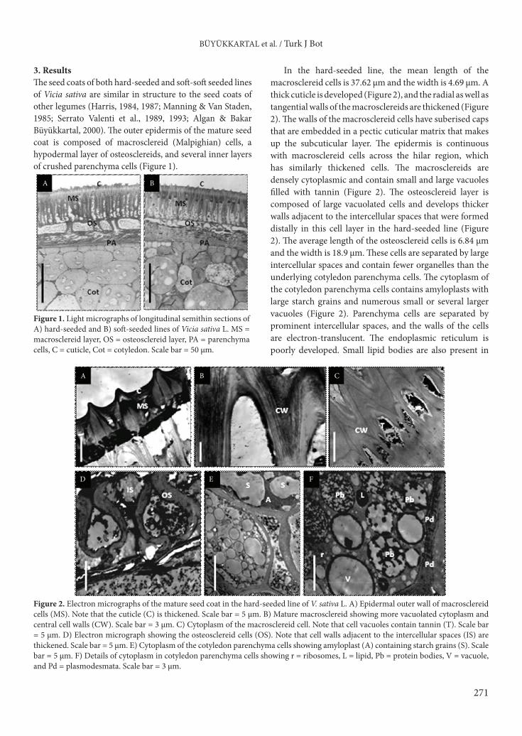
3. ResultsThe seed coats of both hard-seeded and soft-soft seeded lines of Vicia sativa are similar in structure to the seed coats of other legumes (Harris, 1984, 1987; Manning & Van Staden, 1985; Serrato Valenti et al., 1989, 1993; Algan & Bakar Büyükkartal, 2000). The outer epidermis of the mature seed coat is composed of macrosclereid (Malpighian) cells, a hypodermal layer of osteosclereids, and several inner layers of crushed parenchyma cells (Figure 1).
In the hard-seeded line, the mean length of the macrosclereid cells is 37.62 µm and the width is 4.69 µm. A thick cuticle is developed (Figure 2), and the radial as well as tangential walls of the macrosclereids are thickened (Figure 2). The walls of the macrosclereid cells have suberised caps that are embedded in a pectic cuticular matrix that makes up the subcuticular layer. The epidermis is continuous with macrosclereid cells across the hilar region, which has similarly thickened cells. The macrosclereids are densely cytoplasmic and contain small and large vacuoles filled with tannin (Figure 2). The osteosclereid layer is composed of large vacuolated cells and develops thicker walls adjacent to the intercellular spaces that were formed distally in this cell layer in the hard-seeded line (Figure 2). The average length of the osteosclereid cells is 6.84 µm and the width is 18.9 µm. These cells are separated by large intercellular spaces and contain fewer organelles than the underlying cotyledon parenchyma cells. The cytoplasm of the cotyledon parenchyma cells contains amyloplasts with large starch grains and numerous small or several larger vacuoles (Figure 2). Parenchyma cells are separated by prominent intercellular spaces, and the walls of the cells are electron-translucent. The endoplasmic reticulum is poorly developed. Small lipid bodies are also present in
A B
Figure 1. Light micrographs of longitudinal semithin sections of A) hard-seeded and B) soft-seeded lines of Vicia sativa L. MS = macrosclereid layer, OS = osteosclereid layer, PA = parenchyma cells, C = cuticle, Cot = cotyledon. Scale bar = 50 µm.
A
D E F
CB
Figure 2. Electron micrographs of the mature seed coat in the hard-seeded line of V. sativa L. A) Epidermal outer wall of macrosclereid cells (MS). Note that the cuticle (C) is thickened. Scale bar = 5 µm. B) Mature macrosclereid showing more vacuolated cytoplasm and central cell walls (CW). Scale bar = 3 µm. C) Cytoplasm of the macrosclereid cell. Note that cell vacuoles contain tannin (T). Scale bar = 5 µm. D) Electron micrograph showing the osteosclereid cells (OS). Note that cell walls adjacent to the intercellular spaces (IS) are thickened. Scale bar = 5 µm. E) Cytoplasm of the cotyledon parenchyma cells showing amyloplast (A) containing starch grains (S). Scale bar = 5 µm. F) Details of cytoplasm in cotyledon parenchyma cells showing r = ribosomes, L = lipid, Pb = protein bodies, V = vacuole, and Pd = plasmodesmata. Scale bar = 3 µm.
BÜYÜKKARTAL et al. / Turk J Bot
272
the cytoplasm. Large vacuole-like vesicles become visible and seem to be entirely filled with protein (Figure 2). Plasmodesmata occur in the common walls between the parenchyma cells.
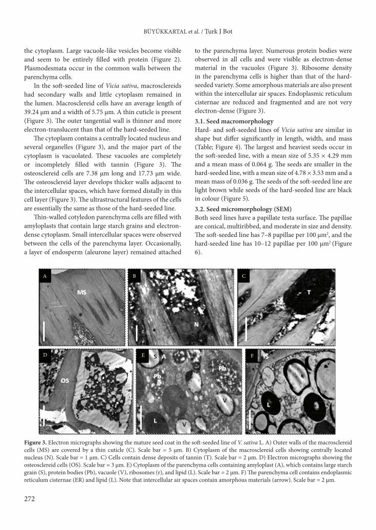
In the soft-seeded line of Vicia sativa, macrosclereids had secondary walls and little cytoplasm remained in the lumen. Macrosclereid cells have an average length of 39.24 µm and a width of 5.75 µm. A thin cuticle is present (Figure 3). The outer tangential wall is thinner and more electron-translucent than that of the hard-seeded line.
The cytoplasm contains a centrally located nucleus and several organelles (Figure 3), and the major part of the cytoplasm is vacuolated. These vacuoles are completely or incompletely filled with tannin (Figure 3). The osteosclereid cells are 7.38 µm long and 17.73 µm wide. The osteosclereid layer develops thicker walls adjacent to the intercellular spaces, which have formed distally in this cell layer (Figure 3). The ultrastructural features of the cells are essentially the same as those of the hard-seeded line.
Thin-walled cotyledon parenchyma cells are filled with amyloplasts that contain large starch grains and electron-dense cytoplasm. Small intercellular spaces were observed between the cells of the parenchyma layer. Occasionally, a layer of endosperm (aleurone layer) remained attached
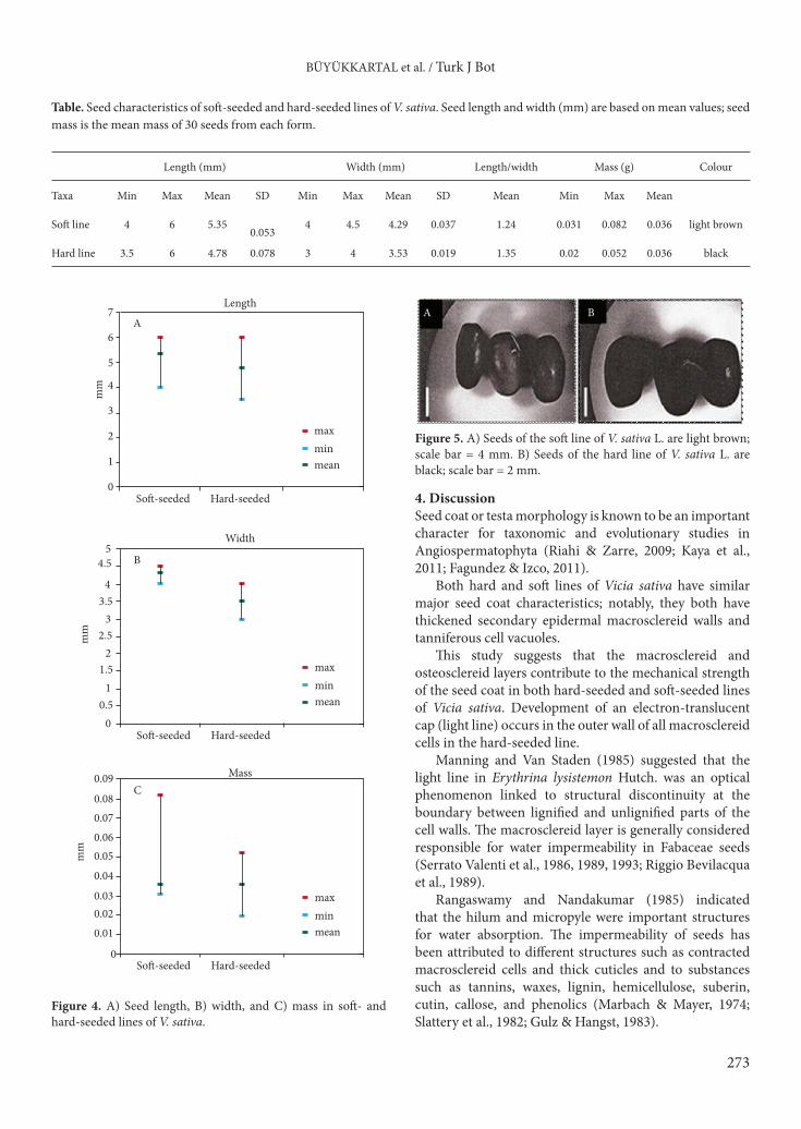
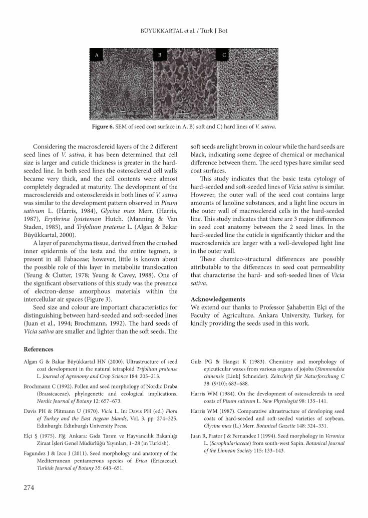
to the parenchyma layer. Numerous protein bodies were observed in all cells and were visible as electron-dense material in the vacuoles (Figure 3). Ribosome density in the parenchyma cells is higher than that of the hard-seeded variety. Some amorphous materials are also present within the intercellular air spaces. Endoplasmic reticulum cisternae are reduced and fragmented and are not very electron-dense (Figure 3).3.1. Seed macromorphologyHard- and soft-seeded lines of Vicia sativa are similar in shape but differ significantly in length, width, and mass (Table; Figure 4). The largest and heaviest seeds occur in the soft-seeded line, with a mean size of 5.35 × 4.29 mm and a mean mass of 0.064 g. The seeds are smaller in the hard-seeded line, with a mean size of 4.78 × 3.53 mm and a mean mass of 0.036 g. The seeds of the soft-seeded line are light brown while seeds of the hard-seeded line are black in colour (Figure 5).3.2. Seed micromorphology (SEM) Both seed lines have a papillate testa surface. The papillae are conical, multiribbed, and moderate in size and density. The soft-seeded line has 7–8 papillae per 100 µm2, and the hard-seeded line has 10–12 papillae per 100 µm2 (Figure 6).
A
D E F
CB
Figure 3. Electron micrographs showing the mature seed coat in the soft-seeded line of V. sativa L. A) Outer walls of the macrosclereid cells (MS) are covered by a thin cuticle (C). Scale bar = 5 µm. B) Cytoplasm of the macrosclereid cells showing centrally located nucleus (N). Scale bar = 1 µm. C) Cells contain dense deposits of tannin (T). Scale bar = 2 µm. D) Electron micrographs showing the osteosclereid cells (OS). Scale bar = 3 µm. E) Cytoplasm of the parenchyma cells containing amyloplast (A), which contains large starch grain (S), protein bodies (Pb), vacuole (V), ribosomes (r), and lipid (L). Scale bar = 2 µm. F) The parenchyma cell contains endoplasmic reticulum cisternae (ER) and lipid (L). Note that intercellular air spaces contain amorphous materials (arrow). Scale bar = 2 µm.
BÜYÜKKARTAL et al. / Turk J Bot
273
4. DiscussionSeed coat or testa morphology is known to be an important character for taxonomic and evolutionary studies in Angiospermatophyta (Riahi & Zarre, 2009; Kaya et al., 2011; Fagundez & Izco, 2011).
Both hard and soft lines of Vicia sativa have similar major seed coat characteristics; notably, they both have thickened secondary epidermal macrosclereid walls and tanniferous cell vacuoles.
This study suggests that the macrosclereid and osteosclereid layers contribute to the mechanical strength of the seed coat in both hard-seeded and soft-seeded lines of Vicia sativa. Development of an electron-translucent cap (light line) occurs in the outer wall of all macrosclereid cells in the hard-seeded line.
Manning and Van Staden (1985) suggested that the light line in Erythrina lysistemon Hutch. was an optical phenomenon linked to structural discontinuity at the boundary between lignified and unlignified parts of the cell walls. The macrosclereid layer is generally considered responsible for water impermeability in Fabaceae seeds (Serrato Valenti et al., 1986, 1989, 1993; Riggio Bevilacqua et al., 1989).
Rangaswamy and Nandakumar (1985) indicated that the hilum and micropyle were important structures for water absorption. The impermeability of seeds has been attributed to different structures such as contracted macrosclereid cells and thick cuticles and to substances such as tannins, waxes, lignin, hemicellulose, suberin, cutin, callose, and phenolics (Marbach & Mayer, 1974; Slattery et al., 1982; Gulz & Hangst, 1983).
Table. Seed characteristics of soft-seeded and hard-seeded lines of V. sativa. Seed length and width (mm) are based on mean values; seed mass is the mean mass of 30 seeds from each form.
Length (mm) Width (mm) Length/width Mass (g) Colour
Taxa Min Max Mean SD Min Max Mean SD Mean Min Max Mean
Soft line 4 6 5.350.053
4 4.5 4.29 0.037 1.24 0.031 0.082 0.036 light brown
Hard line 3.5 6 4.78 0.078 3 4 3.53 0.019 1.35 0.02 0.052 0.036 black
Figure 4. A) Seed length, B) width, and C) mass in soft- and hard-seeded lines of V. sativa.
A B
Figure 5. A) Seeds of the soft line of V. sativa L. are light brown; scale bar = 4 mm. B) Seeds of the hard line of V. sativa L. are black; scale bar = 2 mm.
7Length
A
maxminmean
So�-seeded Hard-seeded
mm
6
5
4
3
2
1
0
5Width
B
maxminmean
So�-seeded Hard-seeded
mm
4.5
4
3
21.5
0.5
2.5
3.5
1
0
0.09 MassC
maxminmean
So�-seeded Hard-seeded
mm
0.080.07
0.05
0.030.02
0.04
0.06
0.01
0
BÜYÜKKARTAL et al. / Turk J Bot
274
Considering the macrosclereid layers of the 2 different seed lines of V. sativa, it has been determined that cell size is larger and cuticle thickness is greater in the hard-seeded line. In both seed lines the osteosclereid cell walls became very thick, and the cell contents were almost completely degraded at maturity. The development of the macrosclereids and osteosclereids in both lines of V. sativa was similar to the development pattern observed in Pisum sativum L. (Harris, 1984), Glycine max Merr. (Harris, 1987), Erythrina lysistemon Hutch. (Manning & Van Staden, 1985), and Trifolium pratense L. (Algan & Bakar Büyükkartal, 2000).
A layer of parenchyma tissue, derived from the crushed inner epidermis of the testa and the entire tegmen, is present in all Fabaceae; however, little is known about the possible role of this layer in metabolite translocation (Yeung & Clutter, 1978; Yeung & Cavey, 1988). One of the significant observations of this study was the presence of electron-dense amorphous materials within the intercellular air spaces (Figure 3).
Seed size and colour are important characteristics for distinguishing between hard-seeded and soft-seeded lines (Juan et al., 1994; Brochmann, 1992). The hard seeds of Vicia sativa are smaller and lighter than the soft seeds. The
soft seeds are light brown in colour while the hard seeds are black, indicating some degree of chemical or mechanical difference between them. The seed types have similar seed coat surfaces.
This study indicates that the basic testa cytology of hard-seeded and soft-seeded lines of Vicia sativa is similar. However, the outer wall of the seed coat contains large amounts of lanoline substances, and a light line occurs in the outer wall of macrosclereid cells in the hard-seeded line. This study indicates that there are 3 major differences in seed coat anatomy between the 2 seed lines. In the hard-seeded line the cuticle is significantly thicker and the macrosclereids are larger with a well-developed light line in the outer wall.
These chemico-structural differences are possibly attributable to the differences in seed coat permeability that characterise the hard- and soft-seeded lines of Vicia sativa.
AcknowledgementsWe extend our thanks to Professor Şahabettin Elçi of the Faculty of Agriculture, Ankara University, Turkey, for kindly providing the seeds used in this work.
A B C
Figure 6. SEM of seed coat surface in A, B) soft and C) hard lines of V. sativa.
References
Algan G & Bakar Büyükkartal HN (2000). Ultrastructure of seed coat development in the natural tetraploid Trifolium pratense L. Journal of Agronomy and Crop Science 184: 205–213.
Brochmann C (1992). Pollen and seed morphology of Nordic Draba (Brassicaceae), phylogenetic and ecological implications. Nordic Journal of Botany 12: 657–673.
Davis PH & Plitmann U (1970). Vicia L. In: Davis PH (ed.) Flora of Turkey and the East Aegean Islands, Vol. 3, pp. 274–325. Edinburgh: Edinburgh University Press.
Elçi Ş (1975). Fiğ. Ankara: Gıda Tarım ve Hayvancılık Bakanlığı Ziraat İşleri Genel Müdürlüğü Yayınları, 1–28 (in Turkish).
Fagundez J & Izco J (2011). Seed morphology and anatomy of the Mediterranean pentamerous species of Erica (Ericaceae). Turkish Journal of Botany 35: 643–651.
Gulz PG & Hangst K (1983). Chemistry and morphology of epicuticular waxes from various organs of jojoba (Simmondsia chinensis [Link] Schneider). Zeitschrift für Naturforschung C 38: (9/10): 683–688.
Harris WM (1984). On the development of osteosclereids in seed coats of Pisum sativum L. New Phytologist 98: 135–141.
Harris WM (1987). Comparative ultrastructure of developing seed coats of hard-seeded and soft-seeded varieties of soybean, Glycine max (L.) Merr. Botanical Gazette 148: 324–331.
Juan R, Pastor J & Fernandez I (1994). Seed morphology in Veronica L. (Scrophulariaceae) from south-west Sapin. Botanical Journal of the Linnean Society 115: 133–143.
BÜYÜKKARTAL et al. / Turk J Bot
275
Kaya A, Ünal M, Özgökçe F, Doğan B & Martin E (2011). Fruit and seed morphology of six species previously placed in Malcolmia (Brassicaceae) in Turkey and their taxonomic value. Turkish Journal of Botany 35: 653–662.
Manning JP & Van Staden J (1985). The development and ultrastructure of the testa and tracheid bar in Erythrina lysistemon Hutch. (Leguminosae: Papilionoideae). Protoplasma 129: 157–167.
Marbach I & Mayer AM (974). Permeability of seed coats to water as related to drying conditions and metabolism of phenolics. Plant Physiology 54: 817–820.
Maxted N (1989). A new Vicia from South-west Turkey. Notes from the Royal Botanic Garden Edinburgh 453: 453–456.
Rangaswamy NS & Nandakumar L (1985). Correlative studies on seed coat structure. Chemical composition and impermeability in the legume Rhynchosia minima. Botanical Gazette 146: 501–509.
Riahi M & Zarre S (2009). Seed development in Astragalus cemerinus and A. ruscifolius (Fabaceae), and its systematic implications. Acta Biologica Cracoviensia Series Botanica 51: 111–117.
Riggio Bevilacqua L, Roti-Mihelozzı G & Modenesi P (1989). The watertight dormancy of Melilotus alba seeds: further observations on the palisade cell wall. Canadian Journal of Botany 67: 3453–3456.
Serrato Valenti G, Cornara L, Ferrando M & Modenesi P (1993). Structural and histochemical features of Stylosanthes scabra (Leguminosae: Papilionoideae) seed coat as related to water entry. Canadian Journal of Botany 71: 834–840.
Serrato Valenti G, Melone L, Ferrando M & Bozzini A. (1989). Comparative studies on testa structure of hard-seeded and soft-seeded varieties of Lipinus angustifolius L. (Leguminosae) and mechanisms of water entry. Seed Science and Tecnology 17: 563–581.
Serrato Valenti G, Modenesi P, Roti-Mihelozzi G & Bevilacqua L (1986). Structural and histochemical characters of the Prosopis tamarugo Phil. seed coat in relation to its hardness. Acta Botanica Neerlandica 35: 475–487.
Slattery HD, Atwell BJ & Kuo J (1982). Relationship between colour, phenolic content and impermeability in the seed coat of various Trifolium subterraneum L. genotypes. Annals of Botany 50: 373–378.
Van der Pluyma A. & Hideux M (1997). Applications d’une methodologie quantitative a’la palynologie d’Eryngium maritimum (Umbelliferae). Plant Systems and Evolution 127: 55–85.
Yeung EC & Cavey MJ (1988). Cellular endosperm formation in Phaseolus vulgaris I. Light and scanning electron microscopy. Canadian Journal of Botany 66: 1209–1216.
Yeung EC & Clutter ME (1978). Embryogeny of Phaseolus coccineus: growth and microanatomy. Protoplasma 94: 19–40.


![Practice For May: Cell Ultrastructure [114 marks]blogs.4j.lane.edu/.../2018/02/Cell-Ultrastructure-Test-1.pdfPractice For May: Cell Ultrastructure [114 marks]1. Which structure found](https://img.dokumen.tips/doc/110x75/5eda4db5b3745412b5711d9c/practice-for-may-cell-ultrastructure-114-marksblogs4jlaneedu201802cell-ultrastructure-test-1pdf.jpg)