Embed Size (px)
Citation preview

Seasonal 4-year investigation into the role of the alpine
marmot (Marmota marmota) as a carrier of zoophilic
dermatophytes
M.G. GALLO*, P. LANFRANCHI$, G. POGLAYEN%, S. CALDEROLA§, A. MENZANO*, E. FERROGLIO* &
A. PEANO*
*Dipartimento di Produzioni Animali, Epidemiologia ed Ecologia, Sezione Parassitologia e Malattie Parassitarie, Grugliasco,Turin, $Dipartimento di Patologia Animale, Igiene e Sanita Pubblica, Sezione di Patologia Generale e Parassitologia, Milan and%Dipartimento di Sanita Pubblica Veterinaria, Sezione di Parassitologia e Malattie Parassitarie, Messina, Italy, and §ResearchInstitute for Wildlife Ecology, Veterinary Faculty, Vienna, Austria
Two hundred and six samples of alpine marmot (Mamota marmota) hair (148 from
adults and 58 from young subjects), 102 soil samples from the entrances to the
burrows of the above individuals and 20 control specimens (obtained from
adjoining areas away from the burrow systems where the rodents are not usually
present) were examined from May 1994 to September 1997. Seventy-five isolates
belonging to six species of dermatophytes were found in 69 of the 206 hair samples
examined (33.5%). Two species were zoophilic, Microsporum canis (7.8%) and
Trichophyton mentagrophytes (11.2.%), and four geophilic, Microsporum cookei
(2%), M. gypseum (5.8%), Trichophyton ajelloi (3.9%) and T. terrestre (5.8%). The
prevalence of each species in the hair samples did not change significantly
according to year, season (chi-squared test [limit significance: P B/0.05] gives no
significant values [P�/0.05] both in year and in season comparison) or age/sex
(adult versus juvenile: P�/ 0.1; male versus female: P�/0.8) of the marmot.
Twenty-three of the 102 soil samples (22.5%) were positive for dermatophytes
found in the hair of marmots from the same burrow systems.
Five of the 20 control soil samples (25%) were positive for dermatophytes. One
isolate of M. gypseum , three of T. terrestre and one of T. mentagrophytes were
obtained.
Compared with other free-ranging rodent hosts studied in Europe, this
mycoflora is characterized by the presence and relatively high prevalence of M.
canis, frequently reported in symptomatic and asymptomatic cats, dogs and fur
animals. M. canis has not been isolated in other rodents in the wild. However, it
has recently been reported in asymptomatic foxes (Vulpes vulpes) from northern
Italy. The close link between V. vulpes and M. marmota , with the former
representing the most important mammal predator of the latter in the Alps (only
a fraction of the predator’s attacks result in the death of the rodent) may have
favoured the adaptation of M. canis to this rodent host. The stable character of the
The procedures followed during this study conform to the current
laws regulating such research in Switzerland.
Correspondence: Maria Grazia Gallo, Dipartimento di Produzioni
Animali, Epidemiologia ed Ecologia, Sezione Parassitologia e
Malattie Parassitarie, Via Leonardo da Vinci 44, I-10095
Grugliasco, Torino, Italy. Tel: �/39 0116 709001; Fax �/39 0116
709000; E-mail: [email protected]
Received 15 April 2004; Accepted 3 August 2004
– 2005 ISHAM DOI: 10.1080/13693780400008282
Medical Mycology June 2005, 43, 373�/379
Med
Myc
ol D
ownl
oade
d fr
om in
form
ahea
lthca
re.c
om b
y R
adbo
ud U
nive
rsite
it N
ijmeg
en o
n 12
/17/
14Fo
r pe
rson
al u
se o
nly.

M. canis/M. marmota relationship (no seasonally or annually related difference in
the prevalence of this dermatophyte has been found) suggests the inclusion of the
alpine marmot in the reservoir of this zoophilic pathogenic agent. In this situation,
hibernation in labyrinthine burrow systems, where temperature and moisture
ranges are quite uniform the whole year round, may favour the viability of M. canis
arthroconidia, whose survival in mountain habitat might otherwise be compro-
mised. This seems to be confirmed by the fact that the fungus has never been found
in the control samples collected at a distance of 300 m from the outer edge of the
sampled burrow systems.
Keywords alpine marmot, dermatophytes, burrow soil, Microsporum canis,
zoonotic agents
Introduction
Surveys on keratinophilic fungi have been carried out
examining many wild species in several European
countries [1�/7]. Most of them deal with rodents or
foxes, observed both as affected animals or carriers of
these fungi [1�/3,6,8,9,11,12]. Some authors also point
out the zoonotic aspect revealed by their studies [6,11�/
17]. Until now only two surveys have referred to the
alpine marmot as a carrier of keratinophilic fungi
[17,18]. However, neither of these studies, both carried
out in the Italian Alps, investigates marmot hair, but
only the soil outside the entrance to the animals’
burrows. Marmots are a very common inhabitant of
the Alps and they share their environment both with
wild animals (especially foxes, small mammals and
ungulates) and domestic ones (such as cattle, sheep and
sheepdogs).
Moreover, the marmot’s home range is often found
in tourist resorts where the marmots have, to some
extent, adapted fairly well to man [19], making
indirect contact between marmots and humans possi-
ble. The alpine marmot is a very social species [20]
which spends 6 months of the year in hibernation in
complex, labyrinthine burrow systems, creating a soil
substrate enriched in keratinic particles, which Man-
tovani has called ‘animalized soil’ [21]. Here the
temperature and moisture ranges are quite uniform
all the year round [22] and most probably suitable for
the survival and circulation of fungi such as derma-
tophytes.
The aim of the present survey, therefore, is to
investigate the presence and the epidemiological role
played by alpine marmot colonies in the diffusion of
dermatophyte fungi, with particular reference to zoo-
philic dermatophyte fungi.
Materials and methods
Habitat and field of research
This survey was carried out in Bivio, a winter tourist
resort in the Engandine region of the Canton of
Graubunden in the Rhaetian Alps of eastern Switzer-
land (46828?N/9841?E).
The study field was a rectangular, south-facing area
of a roughly 5 ha pasture from 1950 to 2000 m above
sea level. The climate and vegetation are typical of the
high alpine habitat. From November until the end of
April, the area is mostly covered by snow. In summer,
the area is populated by livestock, especially cattle and
sheep, with man present to tend the livestock. Roe deer
live in this territory the whole year round, while red
deer, ibex and chamois are found only in spring. Fox
and small mammals are also present.
The socio-economic implications of mountain live-
stock rearing in the area studied provided the oppor-
tunity for the present investigation. The damage caused
by marmot activity to high-value pastures led the
Game and Fishing Inspectorate of the Canton of
Graubunden to initiate a culling programme as part
of its Regional Pest Control Project. No animal
selection was made, as culling applied to all marmots
found within range of fire. According to the data listed
on individual cards, all animals were of normal weight
and healthy. None was found to be affected by
dermatophytosis. A multidisciplinary project was car-
ried out on the marmots culled.
Hair samples
From 1994 to 1997, 206 hair samples were collected in
spring (May), summer (July) and autumn (September)
from the marmots culled.
– 2005 ISHAM, Medical Mycology, 43, 373�/379
374 Gallo et al.
Med
Myc
ol D
ownl
oade
d fr
om in
form
ahea
lthca
re.c
om b
y R
adbo
ud U
nive
rsite
it N
ijmeg
en o
n 12
/17/
14Fo
r pe
rson
al u
se o
nly.

The age of the marmots was calculated by dental
eruption and body dimensions following Ratti [23] anddivided into two age classes: juvenile (B/12 months old)
and adult (]/12 months old). In total, 206 hair samples
were collected, 148 samples from adults (78 male, 70
female) and 58 from young subjects.
The marmots were brushed before any other body
examination was carried out, to avoid contamination.
The procedure was essentially the same as that
described by Mackenzie [24]. Each animal was brushedall over using a sterilized plastic hair brush measuring 8
cm in diameter. None of the marmots showed clinical
signs of dermatophyte infection. The brushes were sent
to the mycological laboratory of the Department of
Animal Production of the University of Turin, Italy,
where each of them was pressed into a Petri dish
containing Sabouraud’s dextrose agar supplemented
with chloramphenicol (0.05 g/l) and cycloheximide (0.5g/l). The hairs left on the brush were inoculated on a
second agar plate using sterile forceps as described by
Okafor and Gugnani [25]. All the plates were incubated
at 288C and examined daily for 45 days.
Soil samples
In 102 cases, the marmots were culled at the entrance totheir burrows and a soil sample was collected from in
front of the burrow entrances. Twenty control speci-
mens were obtained by sampling soil from adjoining
areas 300 m away from the outer edge of the burrow
systems where the rodents are not usually present. (The
Alpine marmot does not usually move more than
200�/250 m from its burrow system.) Each soil speci-
men, weighing 100 g, was taken by scooping the top 2cm of surface soil likely to be rich in organic matter and
put directly into a paper bag to keep the soil dry and to
avoid dermatophyte growth before laboratory culture.
These specimens were each divided into five sub-
samples, each weighing 20 g, which were placed in a
sterile 10-cm diameter Petri dish and processed by
Vanbreuseghem’s method [26] using short pieces
(7�/12 mm) of horse hair as keratinic bait. Thisprocedure was modified by the addition of horse hair
powder obtained from ground sterile hair; 0.5 g was
uniformly mixed with the contents of each Petri dish
while a further 0.5 g was sprinkled on the surface of the
samples to increase the amount of keratinous material
available for the keratinophilic fungi.
The samples were moistened with sterile distilled
water, incubated at 288C and kept moistened as needed.The inoculated dishes were examined three times a
week for 2 months. When the hair fragments showed
good fungal growth, they were microscopically exam-
ined for preliminary morphological identification. Sub-
sequently, the pieces of positive hair were transferred toSabouraud’s dextrose agar medium supplemented with
chloramphenicol (0.05 g/l) and cycloheximide (0.5 g/l),
and kept at 288C for 45 days to obtain pure cultures for
identification.
Fungus identification
Fungus identification was carried out both on hair and
soil samples by observing the macroscopic and micro-
scopic features of the tallus, the colony characteristics
and the asexual reproductive structures (macro- and
microconidia). When needed, slide culture technique
was employed to facilitate detailed observations ofmicroscopic morphology. Each isolate was identified
following the analytical keys given by Rebell and Taplin
[27] and de Hoog et al . [28].
Statistical analyses
Differences between categorical data were compared by
the chi-squared test (limit significance: P�/0.05), when
necessary by kappa statistics and general agreement
(McNemar test) with a 95% confidence interval.
Results
In total, 75 isolates of dermatophytes were cultured
from 69 (33.5%) of the 206 marmot hair samples
examined. Of the six species identified, two were
zoophilic: Microsporum canis (7.8%) and Trichophyton
mentagrophytes (11.2%), and four geophilic:
Microsporum gypseum (5.8%), M. cookei (1.9%),
Trichophyton terrestre (5.8%) and T. ajelloi (3.9%)(Table 1).
Infection was monospecific in 63 positive samples
(91.3%) while the following associations were observed
in the remaining six samples: T. mentagrophytes�/
M. gypseum (two samples each); and T.
mentagrophytes�/M. canis ; M. gypseum�/T. ajelloi ;
Table 1 Presence of dermatophyte fungi and their prevalence in 206
marmot hair samples collected at Bivio, Canton of Graubunden,
Switzerland, during a 4-year investigation (1994�/97)
Species No. isolates Prevalence (%)
Microsporum canis 16 7.8
Microsporum cookei 4 1.9
Microsporum gypseum 12 5.8
Trichophyton ajelloi 8 3.9
Trichophyton mentagrophytes 23 11.2
Trichophyton terrestre 12 5.8
– 2005 ISHAM, Medical Mycology, 43, 373�/379
Alpine marmot as a carrier of zoophilic dermatophytes 375
Med
Myc
ol D
ownl
oade
d fr
om in
form
ahea
lthca
re.c
om b
y R
adbo
ud U
nive
rsite
it N
ijmeg
en o
n 12
/17/
14Fo
r pe
rson
al u
se o
nly.
M. cookei�/T. terrestre ; M. canis�/T. ajelloi (one
sample each). In addition, four strains of Chrysospor-
ium spp. were obtained.
The prevalence of positive hair samples did not
change according to year or season (Table 2; chi-
squared limit of significance: PB/0.05), host age
(36.5% of positive adults versus 25.9% of positive
juveniles: P�/ 0.1) or sex (35.0% of positive males
versus 37.1% of positive females: P�/ 0.8).
Dermatophytes were isolated from 23 of the 102 soilsamples (22.5%) collected from the entrances to the
burrows. The same six species identified in the hair
samples were found: M. canis, T. mentagrophytes and
T. terrestre each with six samples (5.9%), M. cookei
with three (2.9%) and T. ajelloi with one (0.9%). A
single dermatophyte species was present in 22 positive
samples (95.6%), while the association T. menta-
grophytes�/T. terrestre was observed in one sample.Eight isolates of Chrysosporium spp. were also ob-
tained.
The prevalence of dermatophytes in contaminated
soil samples decreased in summer for all the species
found, except for M. canis which remained unchanged
(Table 3). Application of the chi-squared test shows
that the summer decrease of dermatophyte fungi in the
soil is due to the decrease of geophilic fungi (zoophilicversus geophilic fungi: P�/0.005), whereas the presence
of a dermatophyte species both in hair and in the soil
from the corresponding burrow was significantly
and positively correlated for M. canis (P B/0.01),
M. gypseum (P B/0.01) and T. terrestre (P�/0.01). No
correlation was found for T. mentagrophytes and
Chrysosporium spp. Data regarding M. cookei and T.
ajelloi were deemed insufficient for a statistical check.Moreover, the correlation between all dermatophytes
harboured in the marmot hair and those found in the
soil collected from the entrance to the corresponding
burrows was calculated using kappa statistics and
general agreement. Whereas the first test indicates
borderline intermediate values (kappa: 0.38, CI�/0.18
and 0.59) [29], the second gives significant values(McNemar test: P�/0.0011).
Five of the 20 control soil samples (25%) were
positive for dermatophytes. One isolate of M. gypseum ,
three of T. terrestre and one of T. mentagrophytes were
obtained.
Discussion
Dermatophytes, both geophilic (M. cookei , M. gyp-
seum , T. terrestre, T. ajelloi ) and zoophilic (M. canis
and T. mentagrophytes ) were found on the fur of
Marmota marmota . M. cookei and T. ajelloi were
occasional findings, whereas M. canis, M. gypseum,
T. mentagrophytes and T. terrestre were repeatedlyfound in fur cohorts from different seasons and years.
T. mentagrophytes, T. terrestre and M. gypseum were
found in the marmot hair, in the burrow soil and in the
samples taken from the control areas. Some remarks
are worth making about these findings.
The presence of zoophilic T. mentagrophytes, whose
recognized carriers are rodents, was to be expected.
Besides marmot, other sympatric rodent hosts maycontribute to the persistence of this fungus in the
habitat studied, including the control areas, for exam-
ple Apodemus sylvaticus and Clethrionomys glareolus.
These two hosts have frequently been implicated in the
spread of T. mentagrophytes elsewhere [6].
Similarly, it is likely that these rodents participate in
the spread of T. terrestre [6,11], whose possibilities of
isolation, it would seem, are influenced little by soilcomposition [30,31], unlike some other geophilic der-
matophytes, for example M. gypseum [32]. This may
explain why T. terrestre is found to be as prevalent in
the control soil as in the marmot fur and environment.
The presence of domestic and wild ungulates may
play a fairly important role, as they enrich the soil with
organic matter. Such a habitat is particularly favourable
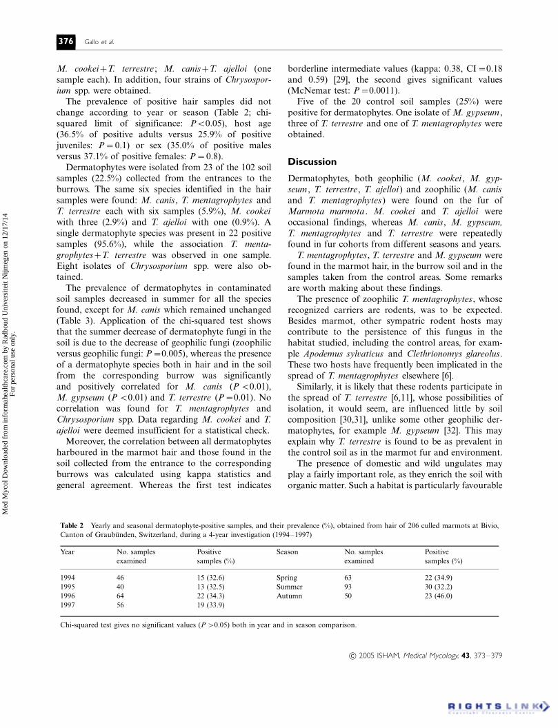
Table 2 Yearly and seasonal dermatophyte-positive samples, and their prevalence (%), obtained from hair of 206 culled marmots at Bivio,
Canton of Graubunden, Switzerland, during a 4-year investigation (1994�/1997)
Year No. samples
examined
Positive
samples (%)
Season No. samples
examined
Positive
samples (%)
1994 46 15 (32.6) Spring 63 22 (34.9)
1995 40 13 (32.5) Summer 93 30 (32.2)
1996 64 22 (34.3) Autumn 50 23 (46.0)
1997 56 19 (33.9)
Chi-squared test gives no significant values (P �/0.05) both in year and in season comparison.
– 2005 ISHAM, Medical Mycology, 43, 373�/379
376 Gallo et al.
Med
Myc
ol D
ownl
oade
d fr
om in
form
ahea
lthca
re.c
om b
y R
adbo
ud U
nive
rsite
it N
ijmeg
en o
n 12
/17/
14Fo
r pe
rson
al u
se o
nly.

for M. gypseum [32] and justifies its presence also in the
samples taken from the control areas.Compared with other studies of European free-
ranging rodents, the mycotic flora obtained in this
survey is characterized by the presence and relatively
high prevalence of M. canis. This species is usually
reported world-wide in symptomatic and asymptomatic
cats, dogs and fur animals [33�/36] as well as in
synanthropic species (Rattus rattus, Rattus norvegicus
and Mus musculus) within heavily anthropogenic areas[36]. M. canis has not been isolated in other free-
ranging small mammals [3,4,6,10,12] nor in wild boar
in Europe [8], but it has been reported in asymptomatic
wild foxes (Vulpes vulpes ) in northern Italy [8,9]. The
close ecological link between V. vulpes and M. mar-
mota , with the former representing the most important
mammal predator of the latter in those alpine areas
where the two share the same territory, [37,38] mayhave favoured the apparent adaptation of M. canis to
this rodent host. As only a fraction of the predator’s
attacks result in the death of the rodent, M. canis may
be transferred to the marmot hair. In addition, a fox
explores the entrances to marmot burrows and may
shed M. canis arthroconidia during its search.
Previously, M. canis had been isolated in soil samples
collected from four out of 15 M. marmota burrowsystems surveyed in the western Italian Alps [18], while
a similar investigation carried out in the central Italian
Alps had yielded negative results [17].
The steady character of the M. canis/M. marmota
association in this 4-year survey suggests the inclusion
of the alpine marmot among the reservoirs of this
pathogenic dermatophyte. Indeed, should a mere spill-
over of M. canis from other reservoirs have occurred,an age-related (adults versus juveniles) and/or season-
related prevalence (spring [just after emergence from a
6-month hibernation] versus summer/autumn) would
probably have arisen. Further evidence of the adapta-
tion of M. canis to this new rodent host is afforded by
its unchanged seasonal presence in soil samples col-
lected in the burrow systems as opposed to its absence
in the soil obtained from outside the marmot colonies,where other hosts, including man, might spread the
dermatophyte. [Unchanged seasonal presence of course
refers to the waking moments of the marmot’s life
cycle, excluding winter when all rodent activity stops as
the animal hibernates in burrows under a deep layer of
snow.]
Obviously, man and his pets may be suspected as the
original source for the past introduction of M. canis
into the marmot environment. For a long time, Bivio
has been a tourist resort and the anthropic impact is
quite noticeable, also in the neighbourhood of the areaTa
ble
3S
easo
na
lis
ola
tes
an
dth
eir
pre
va
len
ce(%
)o
fd
erm
ato
phy
tes
fro
mso
ilsa
mp
les
coll
ecte
do
uts
ide
the
entr
an
ces
tom
arm
ot
bu
rro
ws,
atB
ivio
,C
an
ton
of
Gra
ub
un
den
,S
wit
zerl
an
d,
du
rin
ga
4-y
ear
inves
tig
atio
n(1
99
4�/9
7)
Sea
son
Zo
op
hil
icd
erm
ato
phy
tes
Geo
ph
ilic
der
mato
phy
tes
No
.sa
mp
les
exa
min
ed
Mic
rosp
oru
mca
nis
Tri
cho
phy
ton
men
tag
rop
hyte
sM
.co
ok
eiM
.gy
pse
um
T.
aje
llo
iT
.te
rres
tre
Sp
rin
g1
(4.3
)2
(8.7
)0
1(4
.3)
1(4
.3)
2(8
.7)
23
Su
mm
er3
(6.5
)0
00
01
(2.2
)4
6
Au
tum
n2
(6.0
)3
(9.0
)3
(2.9
)2
(6.0
)0
2(6
.0)
33
To
tal
6(5
.9)
5(4
.9)
3(2
.9)
3(2
.9)
1(1
.0)
5(4
.9)
10
2
Ch
i-sq
uare
dte
stsh
ow
ssi
gn
ific
an
tvalu
esin
sum
mer
com
pari
son
of
zoo
ph
ilic
ver
sus
geo
ph
ilic
der
mato
phyte
s(P
�/0
.00
5).
– 2005 ISHAM, Medical Mycology, 43, 373�/379
Alpine marmot as a carrier of zoophilic dermatophytes 377
Med
Myc
ol D
ownl
oade
d fr
om in
form
ahea
lthca
re.c
om b
y R
adbo
ud U
nive
rsite
it N
ijmeg
en o
n 12
/17/
14Fo
r pe
rson
al u
se o
nly.
studied, due to the presence of many footpaths, much
used by hikers, and a busy road. Nevertheless, it isimportant to emphasize that M. canis was found only
in marmot settlements, never in the control areas away
from the rodents’ environment, where, on the contrary,
man, dog and cat are present. Therefore, in our
opinion, M. canis nowadays seems fully adapted to
the wild environment of the marmot.
Burrow systems probably play a key role in the
preservation of M. canis arthroconidia. First, theburrow microclimate varies little during the course of
the year, particularly in winter when the internal
temperature never drops below 08C [22], thereby
avoiding the damage caused by the sudden, extreme
temperature and moisture variations [39] typical of an
alpine environment. Furthermore, as marmot spends
around 80% of its life in the burrows [40], in particular
during the hibernation period, a keratin-enrichedmedium is created which is important for the preserva-
tion of the fungus [41].
The presence of two zoophilic dermatophytes (M.
canis and T. mentagrophytes ) and of M. gypseum
(geophilic with parasitic aptitude) both on the marmot
hair and in the burrow soil suggests that gamekeepers,
veterinarians and anyone who may come into contact
with the rodent should take all precautions necessary toavoid infection from any arthroconidia present.
Acknowledgements
We thank a sponsor who wishes to remain anonymous,
for financing part of this project.The authors thank the management and game-
keepers of the Game and Fishing Inspectorate, Canton
of Graubunden, Switzerland, for the use of their
facilities and for their collaboration during field work.
We also thank the former Director, P. Ratti, the leading
spirit of the project, U. Bruns, F. Frey-Roos and M.
Giacometti for their co-operation in collecting samples.
References
1 English MP. Ringworm in wild mammals. J Zool 1967; 153: 556�/
561.
2 Alteras I, Nesterov V, Ciolofan I. The occurrence of dermato-
phytes in wild animals from Rumania. Sabouraudia 1966; 4: 215�/
218.
3 Otcenasek M, Hubalec Z, Sixl W. Survey of dermatophytes on
hair of small mammals from Austria. Folia Parasitologica
(Praha) 1980; 27: 83�/87.
4 Poglayen G, Tamperi MP, Mantovani AL, Bacchi-Reggiani G.
Ricerca di dermatofiti in ruminanti selvatici in Italia. Arch Sclavo
1981; 123: 225�/226.
5 Mantovani A, Morganti L, Battelli G, et al . The role of wild
animals in the ecology of dermatophytes and relates fungi. Folia
Parasitologica (Praha) 1982; 20: 279�/283.
6 Chabasse D, Guiguen G, Couatamanac’h A, et al . Contribution a
la connaissance de la flore keratinophile isolee des petites
mammiferes sauvages et du lapin de Garenne en France. Discus-
sion sur les especes fongiques rencontrees. Ann Parasitol Hum
Comp 1987; 62: 357�/368.
7 Mancianti F, Mignone W, Papini R. Keratinophylic fungi from
coats of wild boars in Italy. J Wildlife Dis 1997; 33: 167�/169.
8 Gallo MG, Gagliardo D, Cabeli P. Flora micotica del mantello
della volpe (Vulpes vulpes ) in Piemonte. Parassitologia 1990;
32(Suppl.): 114�/115.
9 Mancianti F, Papini R, Poli A. Mycological survey from coats of
red foxes in Italy. J Mycol Med 1993; 3: 109�/111.
10 Otcenasek M, Dvorak J. The isolation of Trichophyton terrestre
and other keratinophylic fungi from small mammals in South
Western Moravia. Sabouraudia 1962; 2: 111�/113.
11 Mariat F, Chatelain J, Rouffaud MA. Study of the contamination
by dermatophytes of population of small mammals in Alsace.
Mycopathologia 1976; 58: 71�/78.
12 Banaszkiewicz H. A study on fungi in the fur of small rodents
from the environs of Warsaw. Mycosen 1985; 28: 520�/523.
13 LaTouche CJ. The importance of the animal reservoir of infection
in the epidemiology of animal-type ringworm in man. Vet Rec
1955; 67: 666�/669.
14 Kaplan W, George LK, Ajello L. Recent developments in animals
ringworm and their public health implications. Ann NY Acad Sci
1958; 70: 636�/648.
15 English MP, Bayley JA. Dermatophytes in a population of bank-
voles and woodmice. Mycopathologia 1978; 66: 67�/71.
16 Arrese JE, Martalo O, Pierard-Franchimont C, Pierard GE.
Urban and rural mycozoonoses. Rev Med Liege 2000; 55: 998�/
1002.
17 Battelli G, Bianchedi M, Frigo W, et al . Survey of keratinophilic
fungi in alpine marmot (Marmota marmota ) burrow soil and
adjoining soils. Sabouraudia 1978; 16: 83�/86.
18 Gallo MG, Bassano B, Milan F. Dermatophytes in Alpine
marmots (Marmota marmota ) burrows soil from Western Alps.
In: Bassano B, Durio P, Gallo Orsi U, Macchi E, eds. Proceedings
of the First International Symposium on Alpine Marmot ( Mar-
mota marmota) and on genus Marmota, 28�/30 October 1991, S.
Vincent, Aosta, Italy. Turin: Desk Top Pre-Press Color Type
Setting, 1992: 199�/202.
19 Neuhaus P, Mainini B, Ingold P. Human impact on marmot
behaviour. In: Bassano B, Durio P, Gallo Orsi U, Macchi E, eds.
Proceedings of the First International Symposium on Alpine
Marmot ( Marmota marmota) and on genus Marmota, 28�/30
October 1991, S. Vincent, Aosta, Italy. Turin: Desk Top Pre-Press
Color Type Setting, 1992: 165�/169.
20 Armitage KB. Social organisation and fitness strategies of
marmots. In: Bassano B, Durio P, Gallo Orsi U, Macchi E, eds.
Proceedings of the First International Symposium on Alpine
Marmot ( Marmota marmota) and on genus Marmota, 28�/30
October 1991, S. Vincent, Aosta, Italy. Turin: Desk Top Pre-Press
Color Type Setting, 1992: 89�/94.
21 Mantovani A. The role of animals in the epidemiology of the
mycoses. Mycopathologia 1979; 65: 61�/66.
22 Lenti D. Distribuzione degli insediamenti e stima della densita di
marmotta alpina (Marmota marmota L.): i metodi d’indagine per
zone campione. In: Spagnesi M, Toso S, eds. Atti del I Convegno
Nazionale dei Biologi della Selvaggina (Suppl Ricerche Biologia
Selvaggina) , 28�/30 January 1998, Bologna, Italy. Savignano al
Panaro-Modena: Tipolitografia F.G 1988; 14: 353�/364.
23 Ratti P. Beitrag zur Kenntnis des Alpenmurmeltieres. Schweiz
Arch Tierheilk 1970; 112: 283�/295.
– 2005 ISHAM, Medical Mycology, 43, 373�/379
378 Gallo et al.
Med
Myc
ol D
ownl
oade
d fr
om in
form
ahea
lthca
re.c
om b
y R
adbo
ud U
nive
rsite
it N
ijmeg
en o
n 12
/17/
14Fo
r pe
rson
al u
se o
nly.
24 Mackenzie DWR. ‘Hairbrush diagnosis’ in detection and eradica-
tion of non fluorescen scalp-ringworm. Br Med J 1963; 2: 363�/
365.
25 Okafor JI, Gugnani H. Dermatophytes and other keratinophilic
fungi associated with hairs of Rodents in Nigeria. Mycosen 1981;
24: 616�/620.
26 Vanbreuseghem R. Technique biologique pour l’isolement des
dermatophytes du soil. Ann Soc Belge Med Trop 1952; 32: 173�/
178.
27 Rebell G, Taplin D. Dermatophytes �/ Their Recognition and
Identification , 2nd edn. Coral Gables, Florida: University of
Miami Press, 1974.
28 De Hoog GS, Guarro J, Gene J, Figueras MJ. Atlas of Clinical
Fungi , 2nd edn. Utrecht, The Netherlands/Reus, Spain: Central-
bureau voor Schimmelcultures/Universitat Rovira i Virgili, 2000.
29 Landis JR, Kock GG. The measurement of observer agreement
for categorial data. Biometrics 1977; 33: 159�/174.
30 Otcenasek M. Ecology of the dematophytes. Mycopathologia
1978; 65: 67�/72.
31 Alteras I, Evolceanu R. A ten year survey of Romanian soil
screening for keratinophylic fungi (1958�/1967). Mycopath Mycol
Appl 1969; 38: 151�/159.
32 Sommerville D, Marples MJ. The effects of soil enrichment on the
isolation of keratinophylic fungi from soil samples. Sabouraudia
1967; 6: 70�/76.
33 Moriello KA, DeBoer DJ. Fungal flora of the haircoat of cats
with and without dermatophytosis. J Med Vet Mycol 1991; 29:
285�/292.
34 Kwon-Chung KJ, Bennett JKE. Medical Mycology, 1st edn.
Philadelphia/London: Lea & Febiger, 1992.
35 Foil CS. Dermatophytosis. In: Griffin CE, Kwochka KW,
MacDonald JM, (eds.) Current Veterinary Dermatology: The
Science and Art of Therapy. St Louis, MO: Mosby Year Book,
1993: 22�/33.
36 Mantovani A, Morganti L. Dermatozoonoses in Italy. Vet Sci
Comm 1977; 1�/2: 171�/177.
37 Chiesura Corona M. Observation on distribution and abundance
of Alpine marmot (Marmota marmota L) in the territory of
Belluno (south-eastern Alps). In: Bassano B, Durio P, Gallo
Orsi U, Macchi E, eds. Proceedings of the First International
Symposium on Alpine Marmot ( Marmota marmota) and on
genus Marmota, 28�/30 October 1991, S. Vincent, Aosta,
Italy. Turin: Desk Top Pre-Press Color Type Setting, 1992: 117�/
121.
38 Calderola S. Indagine sui parassiti intestinali della marmotta
alpina (Marmota marmota ). Tesi di laurea. Veterinary Faculty,
University of Bologna, Bologna, Italy, 1994.
39 Alteras MD. On the long-term survival of keratinophilic fungi in
non-sterile soil. Mycopathologia 1971; 44: 177�/181.
40 Svendsen GE. Behavioural and environmental factors in the
spatial distribution and population dynamics of a yellow-bellied
Marmot population. Ecology 1974; 55: 760�/771.
41 Ajello L. Present day concept of the dermatophytes. Mycopathol
Mycol Appl 1962; 17: 315�/324.
– 2005 ISHAM, Medical Mycology, 43, 373�/379
Alpine marmot as a carrier of zoophilic dermatophytes 379
Med
Myc
ol D
ownl
oade
d fr
om in
form
ahea
lthca
re.c
om b
y R
adbo
ud U
nive
rsite
it N
ijmeg
en o
n 12
/17/
14Fo
r pe
rson
al u
se o
nly.