Embed Size (px)
Citation preview
SDS PAGE
M.SRI DEVI2nd Yr, M.Tech BIOTECH
SDS PAGE Sodium dodecyl sulphate polyacrylamide gel electrophoresis is a
type of denaturing electrophoresis, used to separate proteins.
SDS- Sodium dodecyl sulphate
Anionic detergent
SDS disrupts the secondary, tertiary and quaternary
structure of the protein to produce a linear polypeptide
chain coated with negatively charged SDS molecule.
Amount of SDS bound to protein is always proportional to
molecular weight of polypeptide.
The detergent binds to hydrophobic regions in a constant
ratio of about 1.4 g of SDS per gram of protein.
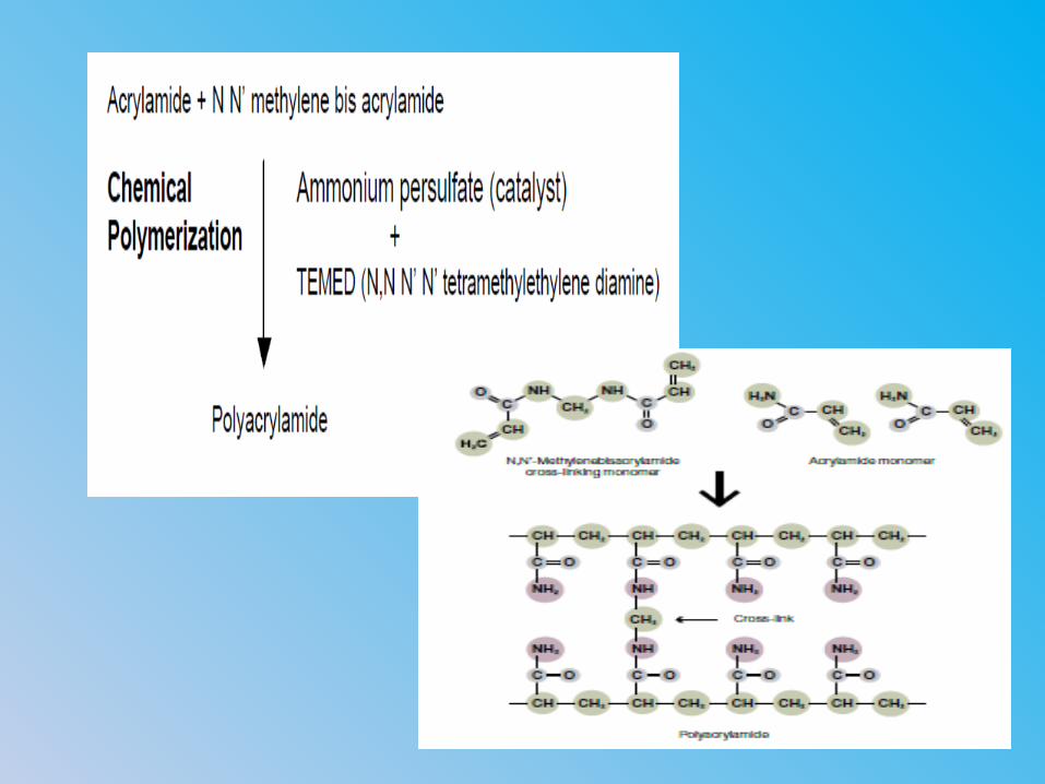
PAGE POLYACRYLAMIDE GEL ELECTROPHORESIS
Electrophoresis- study of movement of charged molecules
in electric field.
Support medium- polyacrylamide.
Polyacrylamide- synthetic polymer of acrylamide
monomers.
SDS- polypeptide complexes migrate through the
polyacrylamide gel in accordance with the size of
polypeptide.
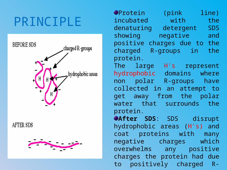
PRINCIPLEProtein (pink line) incubated
with the denaturing detergent SDS showing negative and positive charges due to the charged R-groups in the protein. The large H's represent hydrophobic domains where non polar R-groups have collected in an attempt to get away from the polar water that surrounds the protein.
After SDS: SDS disrupt hydrophobic areas (H's) and coat proteins with many negative charges which overwhelms any positive charges the protein had due to positively charged R-groups.
The resulting protein has been denatured by SDS (reduced to its primary structure-amino acid sequence) and as a result has been linearized.
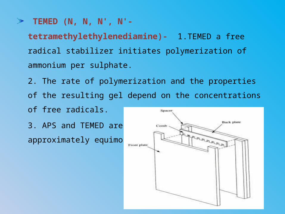
PROCEDURE
Preparation of plates.
Spacers are kept and sealed with agarose
Separating gel.
Stacking gel.
Sample preparation.
Protein samples made to run in electric field.
Addition of staining solution to gel.
De staining solution.
IN DETAIL……..
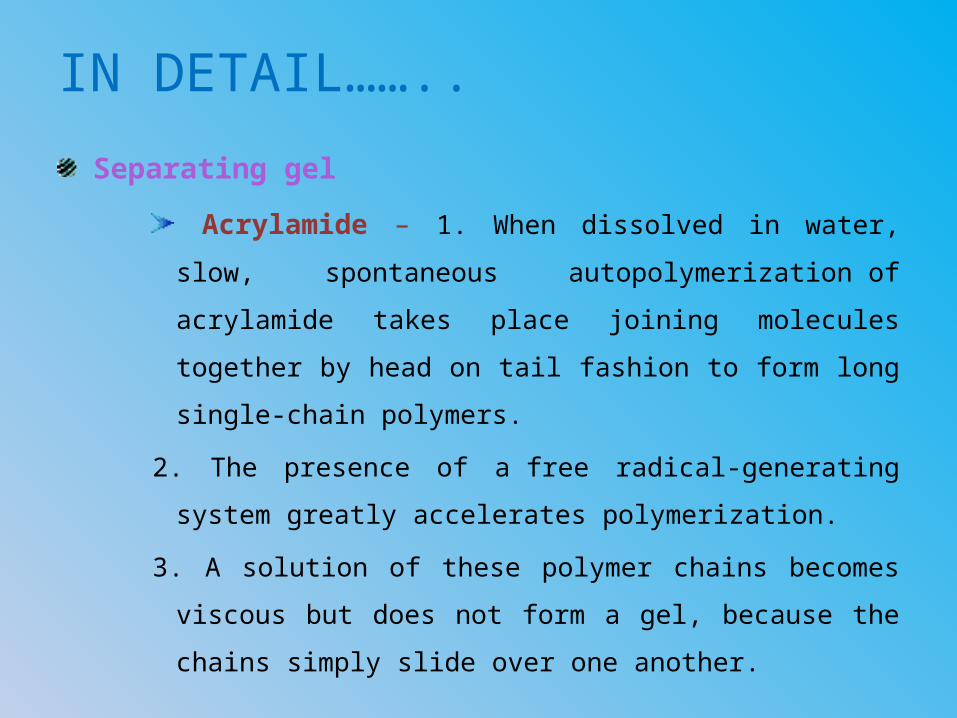
Separating gel
Acrylamide – 1. When dissolved in water, slow,
spontaneous autopolymerization of acrylamide takes
place joining molecules together by head on tail
fashion to form long single-chain polymers.
2. The presence of a free radical-generating system
greatly accelerates polymerization.
3. A solution of these polymer chains becomes viscous
but does not form a gel, because the chains simply
slide over one another.
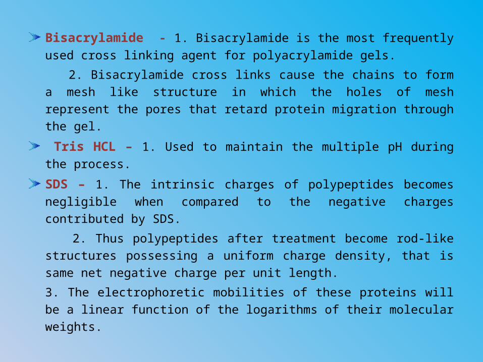
Bisacrylamide - 1. Bisacrylamide is the most frequently used cross linking agent for polyacrylamide gels.
2. Bisacrylamide cross links cause the chains to form a mesh like structure in which the holes of mesh represent the pores that retard protein migration through the gel.
Tris HCL – 1. Used to maintain the multiple pH during the process.
SDS – 1. The intrinsic charges of polypeptides becomes negligible when compared to the negative charges contributed by SDS.
2. Thus polypeptides after treatment become rod-like structures possessing a uniform charge density, that is same net negative charge per unit length.
3. The electrophoretic mobilities of these proteins will be a linear function of the logarithms of their molecular weights.
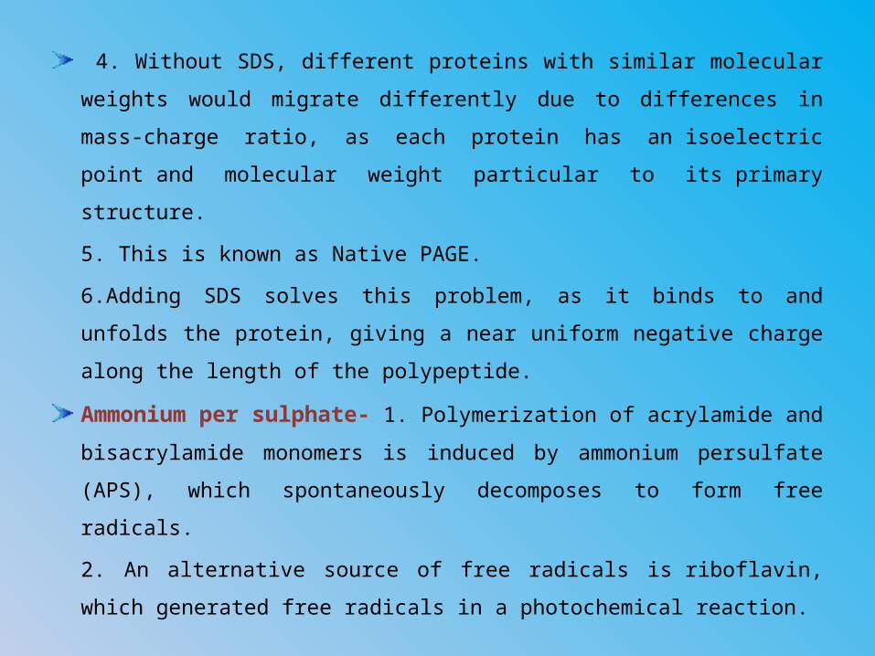
4. Without SDS, different proteins with similar molecular
weights would migrate differently due to differences in mass-
charge ratio, as each protein has an isoelectric point and
molecular weight particular to its primary structure.
5. This is known as Native PAGE.
6.Adding SDS solves this problem, as it binds to and unfolds the
protein, giving a near uniform negative charge along the length
of the polypeptide.
Ammonium per sulphate- 1. Polymerization of acrylamide
and bisacrylamide monomers is induced by ammonium
persulfate (APS), which spontaneously decomposes to form free
radicals.
2. An alternative source of free radicals is riboflavin, which
generated free radicals in a photochemical reaction.
TEMED (N, N, N', N'-tetramethylethylenediamine)-
1.TEMED a free radical stabilizer initiates polymerization of
ammonium per sulphate.
2. The rate of polymerization and the properties of the
resulting gel depend on the concentrations of free radicals.
3. APS and TEMED are typically used at approximately
equimolar concentrations .

Stacking gel
Acrylamide
Bisacrylamide
Tris HCl
SDS
APS
TEMED
1. Pore size of
the gels.
2. pH.
DIFFERENCE???
??
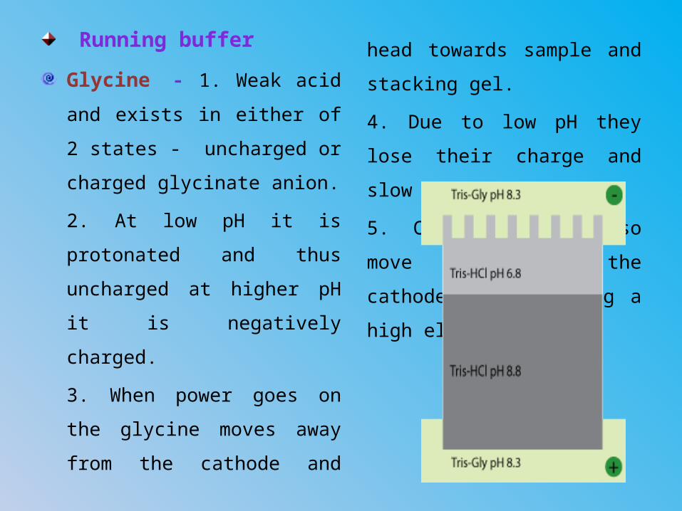
Running buffer
Glycine - 1. Weak acid and
exists in either of 2 states -
uncharged or charged
glycinate anion.
2. At low pH it is protonated
and thus uncharged at
higher pH it is negatively
charged.
3. When power goes on the
glycine moves away from
the cathode and head
towards sample and
stacking gel.
4. Due to low pH they lose
their charge and slow down.
5. Chloride ions also move
away from the cathode, thus
leaving a high electric field.
6. This helps the highly negative charged protein to move
towards the anode.
7. The effect of moving in high voltage is all proteins reach
the separating gel at the same time, so migration is truly a
function of molecular size.
8. When they reach the separating gel everything changes.
9. The pH goes up and glycine becomes deprotonated
making them to move fast.
10. The protein thus moves in a slow and relaxed manner.
Gel loading buffer
Glycerol – 1. It is used to obtain higher viscosity of the
gel, to make the handling with it easier.
2. Makes the sample more dense than the sample
buffer, so the sample will remain in the bottom of a well
rather than float out.
Tris HCl - 1. Cl used to
facilitate glycine.
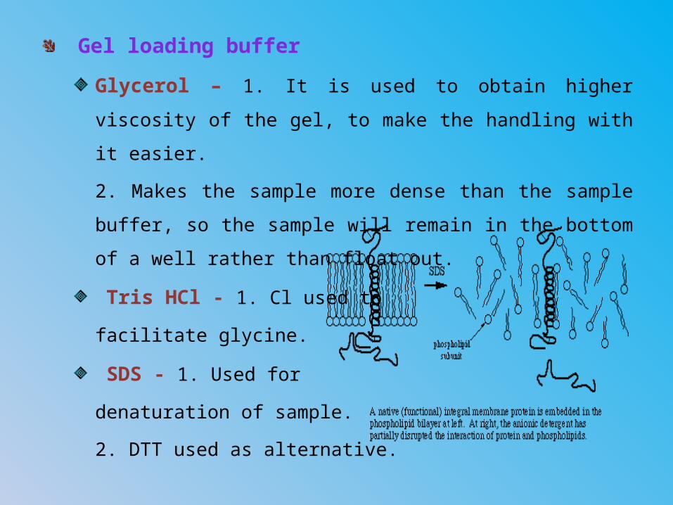
SDS - 1. Used for
denaturation of sample.
2. DTT used as alternative.
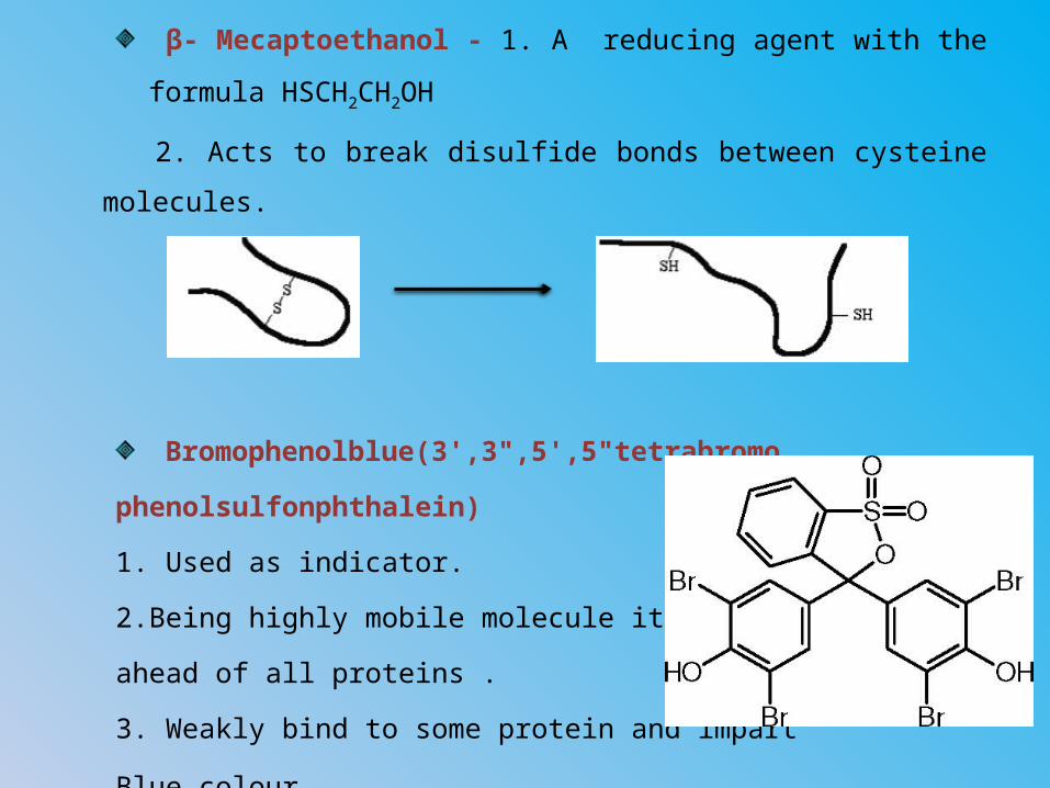
β- Mecaptoethanol - 1. A reducing agent with the
formula HSCH2CH2OH
2. Acts to break disulfide bonds between cysteine
molecules.
Bromophenolblue(3',3",5',5"tetrabromo
phenolsulfonphthalein)
1. Used as indicator.
2.Being highly mobile molecule it moves
ahead of all proteins .
3. Weakly bind to some protein and impart
Blue colour.
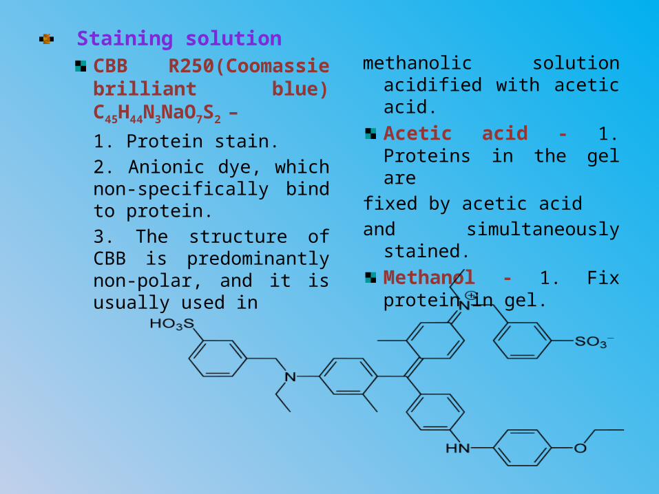
Staining solutionCBB R250(Coomassie brilliant blue) C45H44N3NaO7S2 –
1. Protein stain.2. Anionic dye, which non-specifically bind to protein.3. The structure of CBB is predominantly non-polar, and it is usually used in
methanolic solution acidified with acetic acid.
Acetic acid - 1. Proteins in the gel are
fixed by acetic acid and simultaneously
stained.
Methanol - 1. Fix protein in gel.
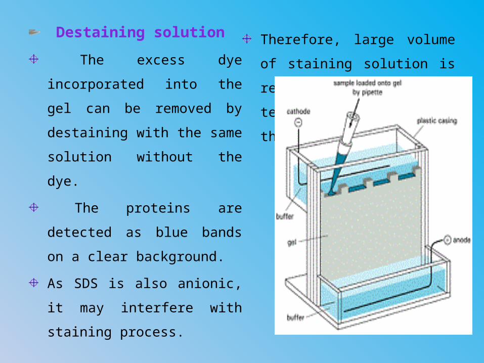
Destaining solution
The excess dye
incorporated into the gel
can be removed by
destaining with the same
solution without the dye.
The proteins are detected
as blue bands on a clear
background.
As SDS is also anionic, it
may interfere with staining
process.
Therefore, large volume of
staining solution is
recommended, at least ten
times the volume of the gel.
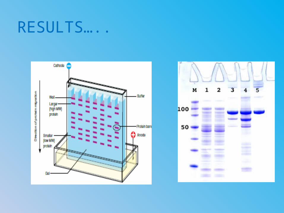
RESULTS…..

QUERIES????