Embed Size (px)
Citation preview
Sclera-related gene polymorphisms in high myopia
Hui-Ju Lin,1,2,3 Lei Wan,2,3,4 Yuhsin Tsai,3 Su-Ching Liu,2 Wen-Chi Chen,2,5 Shih-Wei Tsai,6 Fuu-Jen Tsai2,3
1Department of Ophthalmology, China Medical University Hospital, Taichung, Taiwan; 2Department of Medical Genetics, ChinaMedical University Hospital, Taichung, Taiwan; 3School of Chinese Medicine, College of Chinese Medicine, China MedicalUniversity, Taichung, Taiwan; 4Department of Biotechnology, Asia University, Taichung, Taiwan; 5Graduate Institute of IntegratedMedicine, College of Chinese Medicine, China Medical University, Taichung, Taiwan; 6Institute of Environmental Health, Collegeof Public Health, National Taiwan University, Taipei, Taiwan
Purpose: Transforming growth factor-β2 (TGF-β2), basic fibroblast growth factor (bFGF), and fibromodulin (FMOD)are important extracellular matrix components of the sclera and have been shown to be associated with the developmentof high myopia. Our aim was to examine the association between myopia and the polymorphisms within TGF-β2,bFGF, and FMOD.Methods: The study group comprised of patients (n=195; age range: 17−24 years) with a spherical equivalent of −6.5diopters (D) or a more negative refractive error. The control group comprised of individuals (n=94; age range: 17−25years) with a spherical equivalent ranging from −0.5 D to +1.0 D. The subjects with astigmatism over –0.75 D wereexcluded from the study. High resolution melting (HRM) genotyping and restriction fragment length polymorphism(RFLP) genotyping were used to detect single nucleotide polymorphisms (SNPs). The polymorphisms detected were TGF-β2 (rs7550232 and rs991967), bFGF (rs308395 and rs41348645), and FMOD (rs7543418). Moreover, a stepwise logisticregression procedure was used to detect which of the significant SNPs contributed to the main effects of myopiadevelopment.Results: There were significant differences in the frequency of the A allele and A/A genotype in TGF-β2 (rs7550232;p=0.0178 and 0.03, respectively). Moreover, the haplotype distribution of haplotype 2 (Ht2; A/A) of TGF-β2 differedsignificantly between the two groups (p=0.014). The results of the stepwise logistic regression procedure revealed thatTGF-β2 (rs7550232) contributed significantly to the development of high myopia.Conclusions: TGF-β2 is an important structure of sclera and might contribute to the formation of myopia. TGF-β2(rs7550232) polymorphisms, A allele and A/A genotype, had a protective role against the development of high myopia.
The prevalence of myopia ranges from 20% to 30% inNorth American, European, and Australian populations [1]and is as high as 90% in Asian populations [2,3]. Low tomoderate degrees of myopia present a relatively minorinconvenience as the symptoms are easily corrected usingspectacles or contact lenses. High degrees of myopia, whilealso correctable using these optical approaches, are of majorconcern because of the increased risk of myopia-relatedpathology such as chorioretinal degeneration and retinaldetachment [4]. The ocular pathology associated with highmyopia is among the leading causes of registered blindness inthe developed world [5].
Many studies have suggested that myopia is a complexdisease with multiple causes, including the interaction ofmultiple genes with environmental stimuli [6]. Therefore, itis necessary to take both genes and environment into accountto understand myopia [6]. As for pathogenesis of high myopia,studies of high myopia in animal models have demonstratedthat increasing eye size facilitated by remodeling of the sclera
Correspondence to: Lei Wan, Ph.D., Department of MedicalGenetics, China Medical University Hospital, No. 2 Yuh Der Road,Taichung 404, Taiwan; Phone: 886-4-22052121 ext. 2041; FAX:886-4-22033295; email: [email protected]
was one of the most important etiologies in the progression ofmyopia [7]. The sclera consists of fibrous connective tissuecomprised largely of heterologous collagen fibrils, which arein turn composed mainly of type I collagen with smallamounts of other fibrillar and fibril-associated collagens [8].An important candidate in the search for factors involved inscleral remodeling in myopia is transforming growth factor-β(TGF-β). Transforming growth factor-β is known to initiatemyofibroblast formation and fibrosis [9]. Although fivemembers of the TGF-β family have currently been identified,only TGF-β isoforms 1, 2, and 3 have been detected in eyesand TGF-β2 is the predominant form [9,10].
In addition to TGF-β, basic fibroblast growth factors(bFGFs) are also involved in the remodeling of sclera. bFGFswith their receptors (FGFRs) and signaling cascades areinvolved in a diverse range of cellular processes includingproliferation, apoptosis, cell survival, chemotaxis, celladhesion, motility, and differentiation. bFGF-2 is a potentmitogen for fibroblasts and myofibroblasts [11]. Moreover,the major extracellular matrix components of the fibrousmammalian sclera are collagen types I and III and the smallleucine rich proteoglycans (SLRPs), which include decorin,biglycan, lumican, and fibromodulin (FMOD) [12]. There isconsiderable evidence that FMOD is involved in regulating
Molecular Vision 2009; 15:1655-1663 <http://www.molvis.org/molvis/v15/a177>Received 15 February 2009 | Accepted 16 August 2009 | Published 20 August 2009
© 2009 Molecular Vision
1655

the formation of the network of collagen fibrils that makes upthe extracellular matrix [13]. Among the polymorphisms,rs7550232 (TGF-β2) and rs308395 (bFGF) are found in thepromoter region and rs991967 (TGF-β2) and rs41348645(bFGF) are found in the 3′-UTR region. Polymorphisms in 5′-UTR and 3′-UTR might have effects on the translation andstability of the genes. We selected these polymorphisms ascandidates in this study.
METHODSPatients: From February to November 2004, we measured therefractive error in 3,000 volunteers. All of the participantswere medical students, unrelated and Taiwan-born HanChinese (Table 1). The included subjects had a visual acuitywith distance correction of 0.2 logMAR (logarithm of theminimal angle of resolution; 20/32) or better. The includedsubjects did not have any systemic diseases. They also did notreceive ocular surgery before. Refractive error was measuredin diopters (D) and determined by the mean sphericalequivalent (SE) of the two eyes of each individual afteradministering one drop of cycloplegic drug (1% mydricyl;Alcon, Berlin, Germany). Individuals with −6.5 D or a morenegative refractive error (both eyes) were included in the studygroup, and those with myopia between −0.5 D and +1.0 D(both eyes) were included in the control group. Patients withastigmatism with a refractive error more negative than -0.75D were excluded from the study. Other volunteers did not meetthe criteria to be included in either the study or control groups,and they were excluded from our study. Our study wasreviewed by the ethics committee, and informed consent wasobtained from all patients and control subjects. The study wasperformed according to the tenets of the Declaration ofHelsinki for research involving human subjects.Comprehensive ophthalmic examinations and bloodcollection were performed. As with all data collectionprocedures, auto-refraction (Auto-refractor/auto-keratometer; ARK 700A; Nikon, Tokyo, Japan) was taken onboth eyes by experienced optometrists who were trained andcertified on study protocols. Refractive data, sphere(s),negative cylinder©, and axis measurements were analyzed bycalculating refractive error with SE.
Genotype determinations: The single nucleotidepolymorphisms (SNPs) were selected based on three criteria.(1) The selected SNP had a frequency in the CHB (HanChinese in Beijing, China) or JPN(Japanese) population asdocumented in the HapMap project, and the heterozygosityshould be more than 10%. (2) The selected SNP should be inthe region of the promoter, 5′-UTR, exons, and 3′-UTR of thegene. We excluded any intron SNP because it is hard to predictthe outcome of the polymorphism. (3) In exons, only non-synonymous SNPs were selected. Based on these criteria, onlya few SNPs could be selected. For FMOD, the selected SNPwas based on sequencing experiments. The exons of FMODwere sequenced among 50 randomly selected individuals toreveal the SNPs in FMOD. Only one SNP was detected. Asfor selecting haplotype, HapMap genotypes were analysedwithin Haploview and tag SNPs were selected using tagger.Four tag SNPs were selected for each gene with r2≥0.80 tocapture 80% of genotype information in the region. Theaverage tag SNP was with r2=0.901.
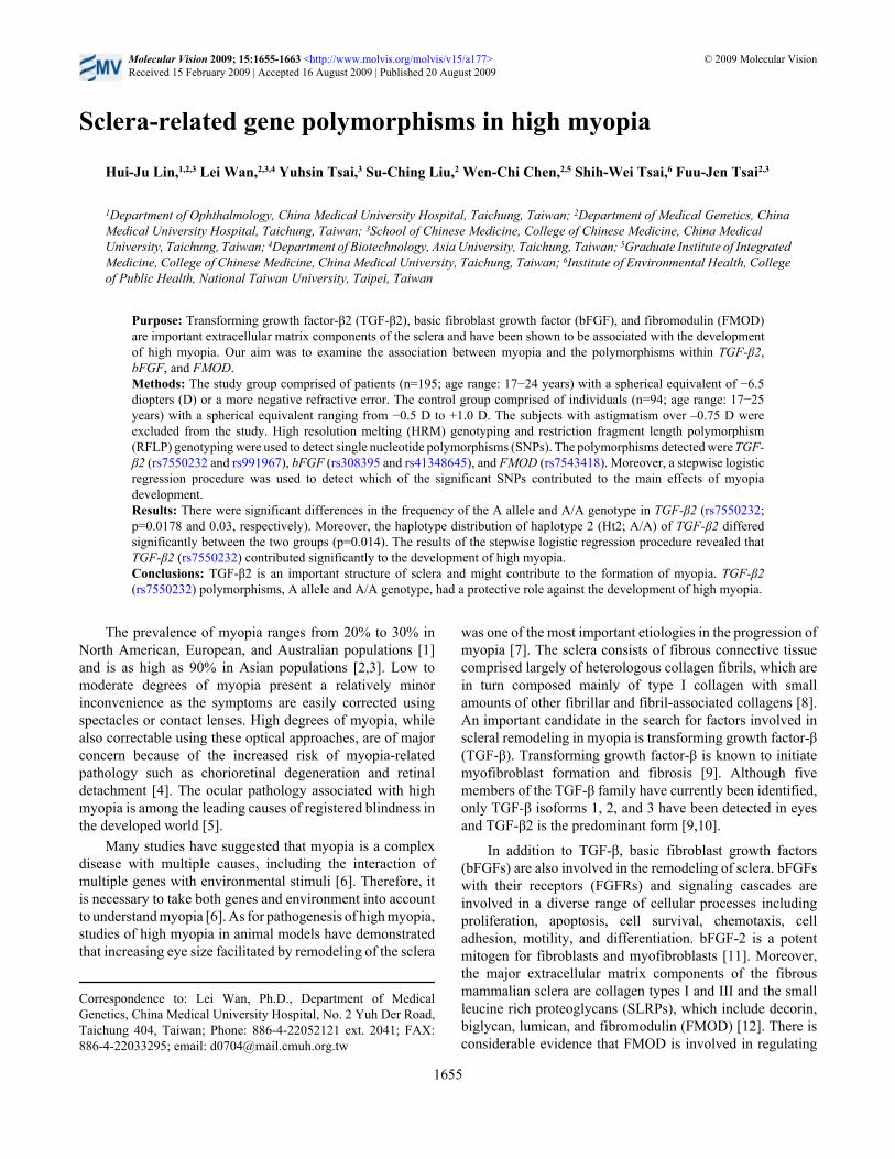
In this study, we used high resolution melting (HRM)genotyping and restriction fragment length polymorphism(RFLP) genotyping to detect the SNPs. Genomic DNA wasextracted from whole blood samples after a standard protocolof digestion by proteinase K and purification with phenol-chloroform. RFLP was used to detect genotypes of TGF-b2(rs991967), bFGF (rs308395), and FMOD (rs7543148). Theamplification protocol and restriction enzymes used todetermine the genotype are listed in Table 2. Preventivecontamination measures were taken by including apolymerase chain reaction (PCR) reaction mixture withoutDNA (negative control) in each run of amplification. TheDNA fragments were separated by horizontal electrophoresison 3% agarose gels, stained with ethidium bromide, andphotographed under ultraviolet lights. Data were analyzed byABI prism GeneMapper Version 3.0 software (AppliedBiosystems Co, Foster City, CA). The PCR product of TGF-β2 (rs991967) was digested with HpyCH4III (New EnglandBiolabs, Mississauga, Ontario, Canada). The A allele size was239 bp, and the C allele size was 100 bp+139 bp as shown inFigure 1. The PCR product of bFGF (rs308395)polymorphism was digested with BsrI (New England
TABLE 1. CHARACTERISTICS OF THE STUDY SUBJECTS.
Characteristics Control (n=94) Cases (n=195) All subjects (n=289)Age, mean (SD), yr 18 (2.9) 18 (3.4) 18 (3.2)Female, Number (%) 33 (35.1%) 70 (35.9%) 103 (35.6%)SE, mean (SD), D 0.02 (0.32) −8.83 (2.5) −4.5 (4.8)AXL, mean (SD), mm 23.56 (0.78) 26.8 (1.8) 24.8 (2.5)CD, mean (SD), D 43.5 (0.9) 44.2 (1.9) 43.8 (1.5)ACD, mean (SD), mm 3.56 (0.25) 3.88 (0.33) 3.62 (0.3)LT, mean (SD), mm 3.8 (0.65) 4.0 (0.58) 3.9 (0.6)
SE: spherical equivalent, AXL: axial length, CD: cornea diopter, ACD: anterior chamber depth, LT: lens thickness.
Molecular Vision 2009; 15:1655-1663 <http://www.molvis.org/molvis/v15/a177> © 2009 Molecular Vision
1656
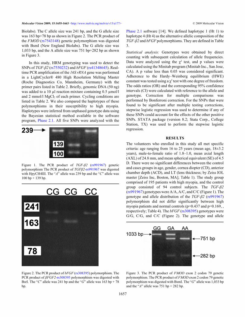
Biolabs). The C allele size was 241 bp, and the G allele sizewas 163 bp+78 bp as shown in Figure 2. The PCR product ofthe FMOD (rs7543148) genetic polymorphism was digestedwith BsmI (New England Biolabs). The G allele size was1,033 bp, and the A allele size was 751 bp+282 bp as shownin Figure 3.
In this study, HRM genotyping was used to detect theSNPs of TGF-β2 (rs7550232) and bFGF (rs41348645). Real-time PCR amplification of the 16S rRNA gene was performedin a LightCycler® 480 High Resolution Melting Master(Roche Diagnostics Co, Mannheim, Germany) with theprimer pairs listed in Table 2. Briefly, genomic DNA (50 ng)was added to a 10 µl reaction mixture containing 0.5 µmol/land 2 mmol/l MgCl2 of each primer. Cycling conditions arelisted in Table 2. We also compared the haplotypes of thesepolymorphisms in their susceptibility to high myopia.Haplotypes were inferred from unphased genotype data usingthe Bayesian statistical method available in the softwareprogram, Phase 2.1. All five SNPs were analyzed with the
Figure 1. The PCR product of TGF-β2 (rs991967) geneticpolymorphism The PCR product of TGFβ2-rs991967 was digestedwith HpyCH4III. The “A” allele was 239 bp and the “C” allele was100 bp + 139 bp.
Figure 2. The PCR product of bFGF (rs308395) polymorphism. ThePCR product of βFGF2-rs308395 polymorphism was digested withBsrI. The “C” allele was 241 bp and the “G” allele was 163 bp + 78bp.
Phase 2.1 software [14]. We defined haplotype 1 (Ht 1) tohaplotype 4 (Ht 4) as the alternative allelic composition of theTGF-β2 and bFGF polymorphisms. They are defined in Table3.Statistical analysis: Genotypes were obtained by directcounting with subsequent calculation of allele frequencies.Data were analyzed using the χ2 test, and p values werecalculated using the Minitab program (Minitab Inc., San Jose,CA). A p value less than 0.05 was considered significant.Adherence to the Hardy–Weinberg equilibrium (HWE)constant was tested using a χ2 test with one degree of freedom.The odds ratios (OR) and the corresponding 95% confidenceintervals (CI) were calculated with reference to the allele andgenotype. Correction for multiple comparisons wasperformed by Bonferroni correction. For the SNPs that werefound to be significant after multiple testing corrections,stepwise logistic regression was used to determine if any ofthese SNPs could account for the effects of the other positiveSNPs. STATA package (version 8.2; Stata Corp., CollegeStation, TX) was used to perform the stepwise logisticregression.
RESULTSThe volunteers who enrolled in this study all met specificcriteria: age ranging from 16 to 25 years (mean age, 18±3.2years), male-to-female ratio of 1.8–1.0, mean axial length(AXL) of 24.8 mm, and mean spherical equivalent (SE) of 4.5D. There were no significant differences between the controland cases groups in age, gender, cornea diopter (CD), anteriorchamber depth (ACD), and LT (lens thickness; by Zeiss IOLmaster [Zeiss Inc, Boston, MA]; Table 1). The study groupcomprised of 195 patients with high myopia, and the controlgroup consisted of 94 control subjects. The TGF-β2(rs991967) genotypes were A/A, A/C, and C/C (Figure 1). Thegenotype and allele distribution of the TGF-β2 (rs991967)polymorphism did not differ significantly between highmyopia patients and normal controls (p=0.437 and p=0.169, ,respectively; Table 4). The bFGF (rs308395) genotypes wereG/G, C/G, and C/C (Figure 2). The genotype and allele
Figure 3. The PCR product of FMOD exon 2 codon 79 geneticpolymorphism. The PCR product of FMOD exon 2 codon 79 geneticpolymorphism was digested with BsmI. The “G” allele was 1,033 bpand the “A” allele was 751 bp + 282 bp.
Molecular Vision 2009; 15:1655-1663 <http://www.molvis.org/molvis/v15/a177> © 2009 Molecular Vision
1657
TAB
LE 2
. CO
ND
ITIO
NS O
F PC
R A
ND
HR
M. T
HE P
RIM
ER PA
IRS,
CO
ND
ITIO
N, P
RO
DU
CT S
IZE,
AN
D R
ESTR
ICTI
ON
ENZY
ME O
F TG
F-Β2
(RS9
9196
7), B
FGF
(RS3
0839
5) IN
THE P
CR
-RFL
P A
ND
TG
F-Β2
(RS7
5502
32) A
ND
BFG
F (R
S413
4864
5) IN
TH
E R
EAC
TIO
N O
F HR
M R
EAC
TIO
N.
Sequ
ence
Poly
mor
phis
m (r
snu
mbe
r)
Prim
erPC
R P
rodu
ct si
ze(b
p)PC
R c
ondi
tions
(ann
ealin
gte
mpe
ratu
re)
Res
tric
tion
enzy
me
site
Alle
les
DN
AFr
agm
ent
size
(bp)
TGF-
β2 rs
9919
67F:
5′-T
GA
CC
GA
GA
AA
GTC
TGC
ATT
-3′
239
55 °C
Hpy
CH
4III
A23
9
R: 5
′-AA
GG
TCTG
AA
GTT
TGA
CC
AG
TAC
A-3
′
C10
0+13
9bF
GF
rs30
8395
F: 5
′-GC
ATG
GC
CTT
TTG
AA
AC
CTA
-3′
241
55 °C
Bsr
IC
241
R
: 5′-C
AG
CG
TCTC
AC
AC
AC
TGA
GG
-3′
G
163+
78FM
OD
rs75
4314
8F:
5′-G
CTG
G C
TTG
C T
CTG
T TC
TCT-
3′10
3355
°CB
smI
G10
33
R: 5
′-GC
CA
A G
GTC
T C
AC
CA
TTG
AT-
3′
A75
1+28
2Se
quen
cePo
lym
orph
ism
(rs
num
ber)
Prim
erD
NA
conc
entr
atio
nM
g co
ncen
trat
ion
PCR
cond
ition
s(a
nnea
ling
tem
pera
ture
)
Alle
les
TGF-
β2 rs
7550
232
F: 5
′-AA
CG
GG
AG
AC
TTG
ATT
GTC
CT-
3′7.
8 ng
2 m
Mto
uchd
own
60/5
3 °C
A
R
: 5′-C
GA
AC
CG
TTG
AG
GG
AG
TGT-
3′
CbF
GF
rs41
3486
45F:
5′-A
CC
ATA
GA
CTG
TCTT
AC
CC
A-3
′7.
8 ng
2 m
Mto
uchd
own
60/5
3 °C
A
R
: 5′-C
AA
TTG
TAA
GG
GA
AG
TCA
GC
-3
G
Molecular Vision 2009; 15:1655-1663 <http://www.molvis.org/molvis/v15/a177> © 2009 Molecular Vision
1658
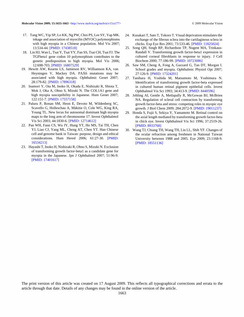
distributions of the bFGF (rs308395) polymorphism did notdiffer significantly between high myopia patients and normalcontrols (p=0.215 and p=1.0, respectively; Table 4). In theFMOD (rs7543418) genetic polymorphism, the genotype andallele distributions of the FMOD (rs7543148) polymorphismalso did not differ significantly between high myopia patientsand normal controls (p=0.362 and p=0.129, respectively;Table 4).
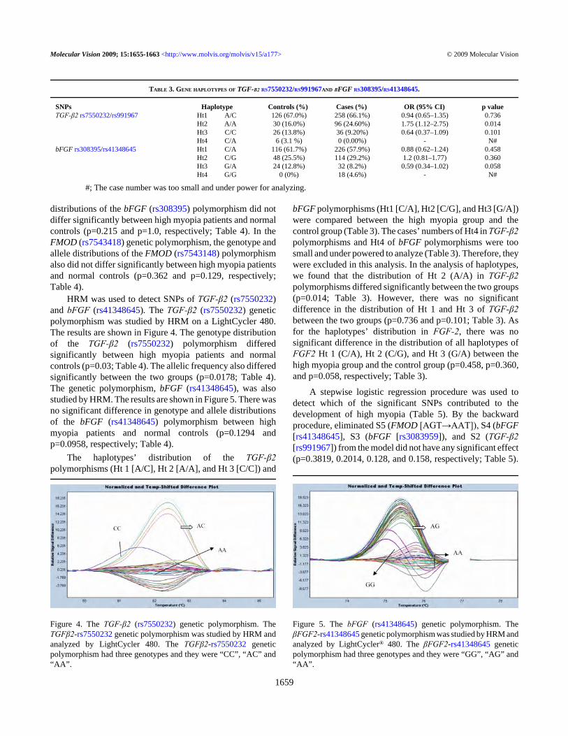
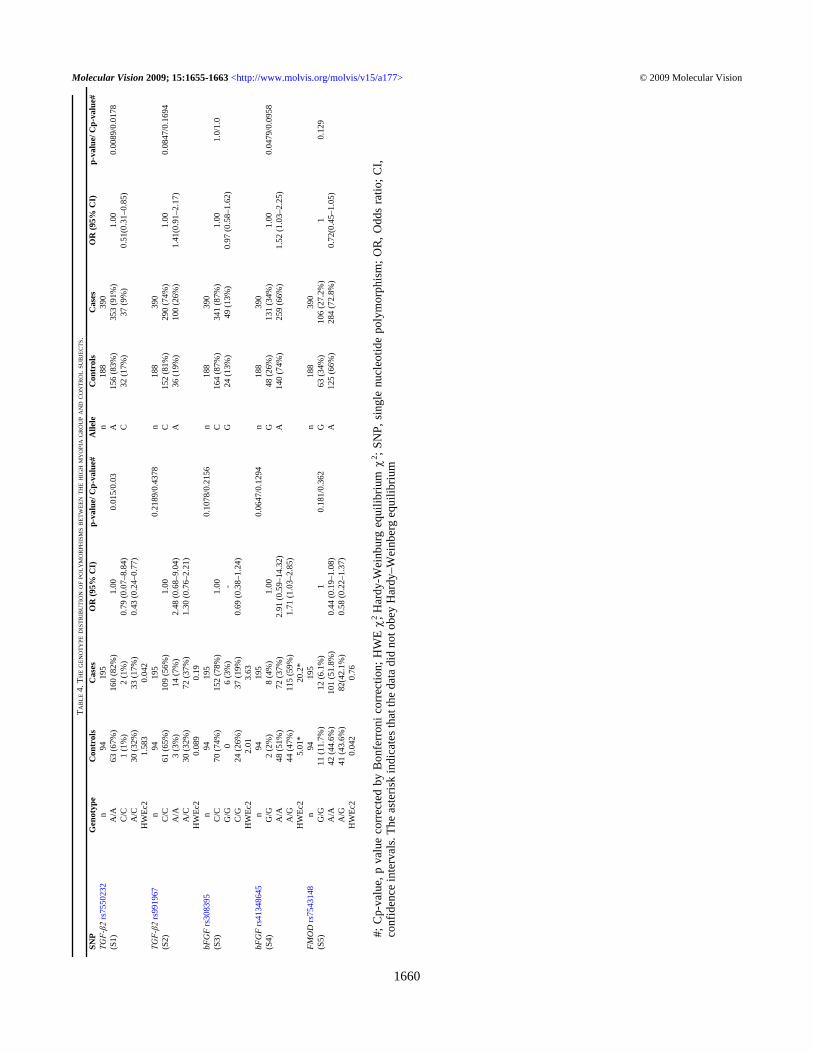
HRM was used to detect SNPs of TGF-β2 (rs7550232)and bFGF (rs41348645). The TGF-β2 (rs7550232) geneticpolymorphism was studied by HRM on a LightCycler 480.The results are shown in Figure 4. The genotype distributionof the TGF-β2 (rs7550232) polymorphism differedsignificantly between high myopia patients and normalcontrols (p=0.03; Table 4). The allelic frequency also differedsignificantly between the two groups (p=0.0178; Table 4).The genetic polymorphism, bFGF (rs41348645), was alsostudied by HRM. The results are shown in Figure 5. There wasno significant difference in genotype and allele distributionsof the bFGF (rs41348645) polymorphism between highmyopia patients and normal controls (p=0.1294 andp=0.0958, respectively; Table 4).
The haplotypes’ distribution of the TGF-β2polymorphisms (Ht 1 [A/C], Ht 2 [A/A], and Ht 3 [C/C]) and
Figure 4. The TGF-β2 (rs7550232) genetic polymorphism. TheTGFβ2-rs7550232 genetic polymorphism was studied by HRM andanalyzed by LightCycler 480. The TGFβ2-rs7550232 geneticpolymorphism had three genotypes and they were “CC”, “AC” and“AA”.
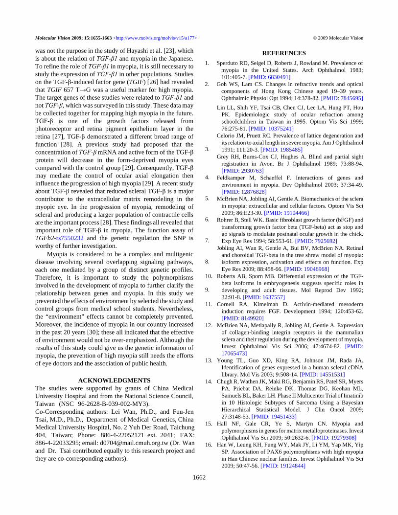
bFGF polymorphisms (Ht1 [C/A], Ht2 [C/G], and Ht3 [G/A])were compared between the high myopia group and thecontrol group (Table 3). The cases’ numbers of Ht4 in TGF-β2polymorphisms and Ht4 of bFGF polymorphisms were toosmall and under powered to analyze (Table 3). Therefore, theywere excluded in this analysis. In the analysis of haplotypes,we found that the distribution of Ht 2 (A/A) in TGF-β2polymorphisms differed significantly between the two groups(p=0.014; Table 3). However, there was no significantdifference in the distribution of Ht 1 and Ht 3 of TGF-β2between the two groups (p=0.736 and p=0.101; Table 3). Asfor the haplotypes’ distribution in FGF-2, there was nosignificant difference in the distribution of all haplotypes ofFGF2 Ht 1 (C/A), Ht 2 (C/G), and Ht 3 (G/A) between thehigh myopia group and the control group (p=0.458, p=0.360,and p=0.058, respectively; Table 3).
A stepwise logistic regression procedure was used todetect which of the significant SNPs contributed to thedevelopment of high myopia (Table 5). By the backwardprocedure, eliminated S5 (FMOD [AGT→AAT]), S4 (bFGF[rs41348645], S3 (bFGF [rs3083959]), and S2 (TGF-β2[rs991967]) from the model did not have any significant effect(p=0.3819, 0.2014, 0.128, and 0.158, respectively; Table 5).
Figure 5. The bFGF (rs41348645) genetic polymorphism. TheβFGF2-rs41348645 genetic polymorphism was studied by HRM andanalyzed by LightCycler® 480. The βFGF2-rs41348645 geneticpolymorphism had three genotypes and they were “GG”, “AG” and“AA”.
TABLE 3. GENE HAPLOTYPES OF TGF-Β2 RS7550232/RS991967AND ΒFGF RS308395/RS41348645.
SNPs Haplotype Controls (%) Cases (%) OR (95% CI) p valueTGF-β2 rs7550232/rs991967 Ht1 A/C 126 (67.0%) 258 (66.1%) 0.94 (0.65–1.35) 0.736 Ht2 A/A 30 (16.0%) 96 (24.60%) 1.75 (1.12–2.75) 0.014 Ht3 C/C 26 (13.8%) 36 (9.20%) 0.64 (0.37–1.09) 0.101 Ht4 C/A 6 (3.1 %) 0 (0.00%) - N#bFGF rs308395/rs41348645 Ht1 C/A 116 (61.7%) 226 (57.9%) 0.88 (0.62–1.24) 0.458 Ht2 C/G 48 (25.5%) 114 (29.2%) 1.2 (0.81–1.77) 0.360 Ht3 G/A 24 (12.8%) 32 (8.2%) 0.59 (0.34–1.02) 0.058 Ht4 G/G 0 (0%) 18 (4.6%) - N#
#; The case number was too small and under power for analyzing.
Molecular Vision 2009; 15:1655-1663 <http://www.molvis.org/molvis/v15/a177> © 2009 Molecular Vision
1659
TAB
LE 4
. TH
E G
ENO
TYPE
DIS
TRIB
UTI
ON
OF P
OLY
MO
RPH
ISM
S BET
WEE
N T
HE
HIG
H M
YO
PIA
GR
OU
P AN
D C
ON
TRO
L SU
BJE
CTS
.SN
PG
enot
ype
Con
trol
sC
ases
OR
(95%
CI)
p-va
lue/
Cp-
valu
e#A
llele
Con
trol
sC
ases
OR
(95%
CI)
p-va
lue/
Cp-
valu
e#TGF-β2
rs75
5023
2n
9419
5n
188
390
(S1)
A/A
63 (6
7%)
160
(82%
)1.
000.
015/
0.03
A15
6 (8
3%)
353
(91%
)1.
000.
0089
/0.0
178
C
/C1
(1%
)2
(1%
)0.
79 (0
.07–
8.84
)C
32 (1
7%)
37 (9
%)
0.51
(0.3
1–0.
85)
A
/C30
(32%
)33
(17%
)0.
43 (0
.24–
0.77
)
HW
Ec2
1.58
30.
042
TGF-β2
rs99
1967
n94
195
0.21
89/0
.437
8n
188
390
(S2)
C/C
61 (6
5%)
109
(56%
)1.
00C
152
(81%
)29
0 (7
4%)
1.00
0.08
47/0
.169
4
A/A
3 (3
%)
14 (7
%)
2.48
(0.6
8–9.
04)
A36
(19%
)10
0 (2
6%)
1.41
(0.9
1–2.
17)
A
/C30
(32%
)72
(37%
)1.
30 (0
.76–
2.21
)
HW
Ec2
0.08
90.
19bF
GF
rs30
8395
n94
195
0.10
78/0
.215
6n
188
390
(S3)
C/C
70 (7
4%)
152
(78%
)1.
00C
164
(87%
)34
1 (8
7%)
1.00
1.0/
1.0
G
/G0
6 (3
%)
-G
24 (1
3%)
49 (1
3%)
0.97
(0.5
8–1.
62)
C
/G24
(26%
)37
(19%
)0.
69 (0
.38–
1.24
)
HW
Ec2
2.01
3.63
bFG
F rs
4134
8645
n94
195
0.06
47/0
.129
4n
188
390
(S4)
G/G
2 (2
%)
8 (4
%)
1.00
G48
(26%
)13
1 (3
4%)
1.00
0.04
79/0
.095
8
A/A
48 (5
1%)
72 (3
7%)
2.91
(0.5
9–14
.32)
A14
0 (7
4%)
259
(66%
)1.
52 (1
.03–
2.25
)
A/G
44 (4
7%)
115
(59%
)1.
71 (1
.03–
2.85
)
HW
Ec2
5.01
*20
.2*
FMO
D rs
7543
148
n94
195
n18
839
0(S
5)G
/G11
(11.
7%)
12 (6
.1%
)1
0.18
1/0.
362
G63
(34%
)10
6 (2
7.2%
)1
0.12
9
A/A
42 (4
4.6%
)10
1 (5
1.8%
)0.
44 (0
.19–
1.08
)A
125
(66%
)28
4 (7
2.8%
)0.
72(0
.45–
1.05
)
A/G
41 (4
3.6%
)82
(42.
1%)
0.58
(0.2
2–1.
37)
H
WEc
20.
042
0.76
#; C
p-va
lue,
p v
alue
cor
rect
ed b
y B
onfe
rron
i cor
rect
ion;
HW
E χ2 , H
ardy
-Wei
nbur
g eq
uilib
rium
χ2 ; S
NP,
sin
gle
nucl
eotid
e po
lym
orph
ism
; OR
, Odd
s ra
tio; C
I,co
nfid
ence
inte
rval
s. Th
e as
teris
k in
dica
tes t
hat t
he d
ata
did
not o
bey
Har
dy–W
einb
erg
equi
libriu
m
Molecular Vision 2009; 15:1655-1663 <http://www.molvis.org/molvis/v15/a177> © 2009 Molecular Vision
1660

In conclusion, the stepwise regressionprocedure demonstrated that S1 (TGF-β2 [rs7550232])significantly contributed the development of high myopia(p=0.004) (Table 5).
DISCUSSIONSome genes have been found to be closely related to highmyopia such as matrix metalloproteinases [15], paired box 6[16], myocilin [17], TGF-β1 [18], Paired box gene 6(PAX-6) [19], and Collagen, type I, alpha 1 (COL1A1) [20].These genes all indicate an evident predisposition for thedevelopment of high myopia. However, none of these geneshave been found to be solely responsible for the developmentof myopia in different ethnic groups or to cover all myopiapatients [15-21]. The other difficulty in myopic study is theuncertainty of what are the surrounding environmentalinfluences that promote the progression of myopia.Individuals with a higher education have a higher prevalenceof myopia than people in the general population, which is whystudents in a medical school were chosen as candidates in thisstudy. The medical school’s students in this study decreasedthe bias of environmental influence.
In this study, we found that the distribution of the TGF-β2(rs7550232) A/A genotype and A allele differed significantlybetween the myopia and control groups. The A/A genotypeand A allele of TGF-β2 (rs7550232) had a protective effectagainst myopia (Table 4). In the analysis of haplotypes, wefound that Ht 2 (A/A) of TGF-β2 was significantly differentin the incidence of high myopia. The frequency of the Ht 2(A/A) of TGF-β2 was higher in the control group then in themyopia group (p=0.014), indicating that Ht 2 of TGF-β2polymorphisms had a protective effect against myopia. InTGF-β2, combining the analyses of allelic and haplotypestudy, we could indicate that the A allele of TGF-β2(rs7550232) had a protective effect against high myopia, andits effect would increase when the subjects had the A allele inTGF-β2 (rs991967; Ht2 [A/A] of TGF-β2; Table 3). As forbFGF, the frequencies of the haplotypes were all withoutsignificant differences between the two groups (Table 3).According to our study, the genetic polymorphisms in bFGFdid not play any important role in the suffering of high myopia.Besides, the results of stepwise regressionprocedure demonstrated that S1 (TGF-β2 [rs7550232])
contributed significantly to the development of myopia(p=0.04) (Table 5). There were no significant effects when S5(FMOD [rs7543148]), S4 (bFGF [rs41348645]), S3 (bFGF[rs3083959]), and S2 (TGF-β2 [rs991967]) were eliminatedfrom the model (Table 5). This also reinforced the importanceof TGF-β2 (rs7550232) in the development of high myopia.In the check of the Hardy–Weinberg equilibrium (HWE), allSNPs in this study obeyed HWE except bFGF (rs41348645;Table 4). To validate our findings, we repeated the genotypinganalysis several times and consistent results were obtained.The errors in genotyping were kept to a minimum. We triedto trace the ancestry background of the controls and patientsto check the probability of population stratification.According to the paper published by Pan et al. [22], theymentioned that on the SNP profiles in the majorhistocompatibility complex (MHC) region (6p21.3), thecomposition of our population is as follows: 85% are Minnandescendants, 5% are Hakka descendants, and the remaining10% are mixed population of Minnan, Hakka, and Cantondescendants. There was no significant difference in the MHCregion among these groups, this indicated that the populationin this study was homogeneous and population stratificationshould not affect this study. Moreover, the deviations fromHWE might be a sign of mutation. This disequilibrium seemsto also support the association of the polymorphisms in TGF-β2 with high myopia.
The expression of TGF-β1 and TGF-β2 were different inmyopia. A study by Hayashi et al. [23] revealed that there wasno significant relationship between high myopia and TGF-β1among the Japanese [23]. Furthermore, Kusakari et al. [24]reported that TGF-β1 expression was reduced in the sclera,choroid, and retina during myopia development in an isoform-and time-specific manner. However, it has been shown thatthe TGF-β2 content increased in the retina, choroid, and scleraduring myopia development [25]. In this study, we found thatexpression of TGF-β2 was different from the result of TGF-β1among the Japanese. The different expressions of TGF-β2 andTGF-β1 in myopia could be understood by the differentbehavior of the two isoforms in sclera during the progressionof myopia. Moreover, our other study has found that the CCgenotype in the TGF-β1 codon 10 polymorphism(rs1982073) contributes to the genetic predisposition of highmyopia [18]. The SNP we detected (TGF-β1 [rs1982073])
TABLE 5. SNPS ANALYZED BY STEPWISE LOGISTIC REGRESSION PROCEDURE
Null model Alternative model p valueS1+S2+S3+S4+S5 S1+S2+S3+S4 0.3819S1+S2+S3+S4+S5 S1+ S2+S3+S5 0.2014S1+S2+S3+S4+S5 S1+S2+S4+S5 0.128S1+S2+S3+S4+S5 S1+S3+S4+S5 0.158S1+S2+S3+S4+S5 S2+S3+S4+S5 0.004
S1: TGF-β2 -rs7550232 , S2: TGF-β2 -rs991967, S3: βFGF -rs308395, S4: βFGF -rs41348645, S5: FMOD -rs7543148 .
Molecular Vision 2009; 15:1655-1663 <http://www.molvis.org/molvis/v15/a177> © 2009 Molecular Vision
1661
was not the purpose in the study of Hayashi et al. [23], whichis about the relation of TGF-β1 and myopia in the Japanese.To refine the role of TGF-β1 in myopia, it is still necessary tostudy the expression of TGF-β1 in other populations. Studieson the TGF-β-induced factor gene (TGIF) [26] had revealedthat TGIF 657 T→G was a useful marker for high myopia.The target genes of these studies were related to TGF-β1 andnot TGF-β, which was surveyed in this study. These data maybe collected together for mapping high myopia in the future.TGF-β is one of the growth factors released fromphotoreceptor and retina pigment epithelium layer in theretina [27], TGF-β demonstrated a different broad range offunction [28]. A previous study had proposed that theconcentration of TGF-β mRNA and active form of the TGF-βprotein will decrease in the form-deprived myopia eyescompared with the control group [29]. Consequently, TGF-βmay mediate the control of ocular axial elongation theninfluence the progression of high myopia [29]. A recent studyabout TGF-β revealed that reduced scleral TGF-β is a majorcontributor to the extracellular matrix remodeling in themyopic eye. In the progression of myopia, remodeling ofscleral and producing a larger population of contractile cellsare the important process [28]. These findings all revealed thatimportant role of TGF-β in myopia. The function assay ofTGFb2-rs7550232 and the genetic regulation the SNP isworthy of further investigation.
Myopia is considered to be a complex and multigenicdisease involving several overlapping signaling pathways,each one mediated by a group of distinct genetic profiles.Therefore, it is important to study the polymorphismsinvolved in the development of myopia to further clarify therelationship between genes and myopia. In this study weprevented the effects of environment by selected the study andcontrol groups from medical school students. Nevertheless,the “environment” effects cannot be completely prevented.Moreover, the incidence of myopia in our country increasedin the past 20 years [30]; these all indicated that the effectiveof environment would not be over-emphasized. Although theresults of this study could give us the genetic information ofmyopia, the prevention of high myopia still needs the effortsof eye doctors and the association of public health.
ACKNOWLEDGMENTSThe studies were supported by grants of China MedicalUniversity Hospital and from the National Science Council,Taiwan (NSC 96-2628-B-039-002-MY3).Co-Corresponding authors: Lei Wan, Ph.D., and Fuu-JenTsai, M.D., Ph.D., Department of Medical Genetics, ChinaMedical University Hospital, No. 2 Yuh Der Road, Taichung404, Taiwan; Phone: 886-4-22052121 ext. 2041; FAX:886-4-22033295; email: [email protected] (Dr. Wanand Dr. Tsai contributed equally to this research project andthey are co-corresponding authors).
REFERENCES1. Sperduto RD, Seigel D, Roberts J, Rowland M. Prevalence of
myopia in the United States. Arch Ophthalmol 1983;101:405-7. [PMID: 6830491]
2. Goh WS, Lam CS. Changes in refractive trends and opticalcomponents of Hong Kong Chinese aged 19–39 years.Ophthalmic Physiol Opt 1994; 14:378-82. [PMID: 7845695]
3.
Lin LL, Shih YF, Tsai CB, Chen CJ, Lee LA, Hung PT, HouPK. Epidemiologic study of ocular refraction amongschoolchildren in Taiwan in 1995. Optom Vis Sci 1999;76:275-81. [PMID: 10375241]
4.
Celorio JM, Pruett RC. Prevalence of lattice degeneration andits relation to axial length in severe myopia. Am J Ophthalmol1991; 111:20-3. [PMID: 1985485]
5.
Grey RH, Burns-Cox CJ, Hughes A. Blind and partial sightregistration in Avon. Br J Ophthalmol 1989; 73:88-94.[PMID: 2930763]
6.
Feldkamper M, Schaeffel F. Interactions of genes andenvironment in myopia. Dev Ophthalmol 2003; 37:34-49.[PMID: 12876828]
7.
McBrien NA, Jobling AI, Gentle A. Biomechanics of the sclerain myopia: extracellular and cellular factors. Optom Vis Sci2009; 86:E23-30. [PMID: 19104466]
8.
Rohrer B, Stell WK. Basic fibroblast growth factor (bFGF) andtransforming growth factor beta (TGF-beta) act as stop andgo signals to modulate postnatal ocular growth in the chick.Exp Eye Res 1994; 58:553-61. [PMID: 7925692]
9.
Jobling AI, Wan R, Gentle A, Bui BV, McBrien NA. Retinaland choroidal TGF-beta in the tree shrew model of myopia:isoform expression, activation and effects on function. ExpEye Res 2009; 88:458-66. [PMID: 19046968]
10. Roberts AB, Sporn MB. Differential expression of the TGF-beta isoforms in embryogenesis suggests specific roles indeveloping and adult tissues. Mol Reprod Dev 1992;32:91-8. [PMID: 1637557]
11. Cornell RA, Kimelman D. Activin-mediated mesoderminduction requires FGF. Development 1994; 120:453-62.[PMID: 8149920]
12. McBrien NA, Metlapally R, Jobling AI, Gentle A. Expressionof collagen-binding integrin receptors in the mammaliansclera and their regulation during the development of myopia.Invest Ophthalmol Vis Sci 2006; 47:4674-82. [PMID:17065473]
13. Young TL, Guo XD, King RA, Johnson JM, Rada JA.Identification of genes expressed in a human scleral cDNAlibrary. Mol Vis 2003; 9:508-14. [PMID: 14551531]
14. Chugh R, Wathen JK, Maki RG, Benjamin RS, Patel SR, MyersPA, Priebat DA, Reinke DK, Thomas DG, Keohan ML,Samuels BL, Baker LH. Phase II Multicenter Trial of Imatinibin 10 Histologic Subtypes of Sarcoma Using a BayesianHierarchical Statistical Model. J Clin Oncol 2009;27:3148-53. [PMID: 19451433]
15. Hall NF, Gale CR, Ye S, Martyn CN. Myopia andpolymorphisms in genes for matrix metalloproteinases. InvestOphthalmol Vis Sci 2009; 50:2632-6. [PMID: 19279308]
16. Han W, Leung KH, Fung WY, Mak JY, Li YM, Yap MK, YipSP. Association of PAX6 polymorphisms with high myopiain Han Chinese nuclear families. Invest Ophthalmol Vis Sci2009; 50:47-56. [PMID: 19124844]
Molecular Vision 2009; 15:1655-1663 <http://www.molvis.org/molvis/v15/a177> © 2009 Molecular Vision
1662
19. Hewitt AW, Kearns LS, Jamieson RV, Williamson KA, vanHeyningen V, Mackey DA. PAX6 mutations may beassociated with high myopia. Ophthalmic Genet 2007;28:179-82. [PMID: 17896318]
20. Inamori Y, Ota M, Inoko H, Okada E, Nishizaki R, Shiota T,Mok J, Oka A, Ohno S, Mizuki N. The COL1A1 gene andhigh myopia susceptibility in Japanese. Hum Genet 2007;122:151-7. [PMID: 17557158]
21. Paluru P, Ronan SM, Heon E, Devoto M, Wildenberg SC,Scavello G, Holleschau A, Mäkitie O, Cole WG, King RA,Young TL. New locus for autosomal dominant high myopiamaps to the long arm of chromosome 17. Invest OphthalmolVis Sci 2003; 44:1830-6. [PMID: 12714612]
22. Pan WH, Fann CS, Wu JY, Hung YT, Ho MS, Tai TH, ChenYJ, Liao CJ, Yang ML, Cheng AT, Chen YT. Han Chinesecell and genome bank in Taiwan: purpose, design and ethicalconsiderations. Hum Hered 2006; 61:27-30. [PMID:16534213]
23. Hayashi T, Inoko H, Nishizaki R, Ohno S, Mizuki N. Exclusionof transforming growth factor-beta1 as a candidate gene formyopia in the Japanese. Jpn J Ophthalmol 2007; 51:96-9.[PMID: 17401617]
24. Kusakari T, Sato T, Tokoro T. Visual deprivation stimulates theexchange of the fibrous sclera into the cartilaginous sclera inchicks. Exp Eye Res 2001; 73:533-46. [PMID: 11825024]
25. Song QH, Singh RP, Richardson TP, Nugent MA, Trinkaus-Randall V. Transforming growth factor-beta1 expression incultured corneal fibroblasts in response to injury. J CellBiochem 2000; 77:186-99. [PMID: 10723086]
26. Saw SM, Cheng A, Fong A, Gazzard G, Tan DT, Morgan I.School grades and myopia. Ophthalmic Physiol Opt 2007;27:126-9. [PMID: 17324201]
27. Tanihara H, Yoshida M, Matsumoto M, Yoshimura N.Identification of transforming growth factor-beta expressedin cultured human retinal pigment epithelial cells. InvestOphthalmol Vis Sci 1993; 34:413-9. [PMID: 8440596]
28. Jobling AI, Gentle A, Metlapally R, McGowan BJ, McBrienNA. Regulation of scleral cell contraction by transforminggrowth factor-beta and stress: competing roles in myopic eyegrowth. J Biol Chem 2009; 284:2072-9. [PMID: 19011237]
29. Honda S, Fujii S, Sekiya Y, Yamamoto M. Retinal control onthe axial length mediated by transforming growth factor-betain chick eye. Invest Ophthalmol Vis Sci 1996; 37:2519-26.[PMID: 8933768]
30. Wang TJ, Chiang TH, Wang TH, Lin LL, Shih YF. Changes ofthe ocular refraction among freshmen in National TaiwanUniversity between 1988 and 2005. Eye 2009; 23:1168-9.[PMID: 18551136]
Molecular Vision 2009; 15:1655-1663 <http://www.molvis.org/molvis/v15/a177> © 2009 Molecular Vision
The print version of this article was created on 17 August 2009. This reflects all typographical corrections and errata to thearticle through that date. Details of any changes may be found in the online version of the article.
1663
17. Tang WC, Yip SP, Lo KK, Ng PW, Choi PS, Lee SY, Yap MK.inkage and association of myocilin (MYOC) polymorphismswith high myopia in a Chinese population. Mol Vis 2007;13:534-44. [PMID: 17438518]
18. Lin HJ, Wan L, Tsai Y, Tsai YY, Fan SS, Tsai CH, Tsai FJ. TheTGFbeta1 gene codon 10 polymorphism contributes to thegenetic predisposition to high myopia. Mol Vis 2006;12:698-703. [PMID: 16807529]