Embed Size (px)
Citation preview

GSJ: Volume 7, Issue 10, October 2019, Online: ISSN 2320-9186
www.globalscientificjournal.com
Schistosomiasis Presenting as Acute Appendicitis
with Mesenteric Nodule Filled with Bilharzial
Ova: Case study
2
, DoaaM.Hasan1Amir Mounir
1 Head of General surgery department at Imbaba general Hospital, El-Giza, Egypt
2 General Surgery resident at Imbaba general Hospital, El-Giza, Egypt
Abstract: developing countries. The incommonly humans, infestation inSchistosomiasis is a parasitic
on the location of the basedinfection manifests itself as a variety of different pathologies,
manifestation is that of a common surgical presentation, unusual Anparasite and its eggs.
for ctomyeappendicyoung male who underwent acute appendicitis. We present a case of a
upon pathological established, infestation Schistosomiasisacute appendicitis caused by a
examination of the resected appendix.
Introduction:
chronic granulomatous disease that can affect any prevalentSchistosomiasis is a tropical
vary according to schistosoma species, such Schistosomiasisorgan. Clinical manifestations of
, all of japonicum Schistosoma and ,Schistosoma haematobium ,Schistosoma mansoni as
. symptomsgastrointestinal cause S. japonicum and S. mansoni lifecycles.which have similar
typically survives in the human body for up to 5 years, but Schistosoma Without treatment,
due illness life threatening results in usuallyChronic infection up to 40 years. continuemay
foundto eggs reactioninflammatory bodyand fibrosis caused by damagetissue persistentto
S. infects the intestine and liver, while S. mansoni ,Ordinarilyin the affected organs.
of exhibitions uncommonTwo infects the bladder, kidney and ureters. haematobium
discomfortepigastric periodicare appendicitis and chronic Schistosomiasisgastrointestinal
patient presented with here a case where document mesenteric thrombosis. We caused by
pathological histon upo hedestablisinfection, Schistosomiasisdue to acute appendicitis
appendix. excisedof the investigation
GSJ: Volume 7, Issue 10, October 2019 ISSN 2320-9186
772
GSJ© 2019 www.globalscientificjournal.com

Clinical presentation:
Our case is a 28-year-old male, who was until that time healthy. He came to the emergency
department complaining of acute right iliac fossa pain that he had been living through for
quite a few days. The pain was not associated with nausea or vomiting, and he had no
noteworthy symptoms as headache, hematuria, dysuria, myalgia, arthralgia, cough, diarrhea
or rash.
Proceeding with the clinical examination, the patient had tenderness localized to the right
lower quadrant of the abdomen and rebound tenderness. Lab work revealed normal
hemoglobin (14.2 g/L) and total leukocyte count (22 × 109/L) , no eosinophilia; platelets were
440 × 109/L.
The preliminary diagnosis of his case was acute appendicitis. Pelvi-abdominal
ultrasonography displayed nonspecific intraperitoneal inflammatory change in the area of the
terminal ileum and ileoceacal junction, the patient was managed surgically and open
appendicectomy was done.
Upon surgical excision of the appendix, solitary nodule was found on the mesentery of the
ilium about 8 inches from the ileoceacal junction (Figure 1), and excision of the nodule was
done.
Histopathological analysis of the resected appendix and the nodule exposed acute
inflammatory process, marked with copious ova found within the nodule. Further evaluation
of the specimen by microbiology and parasitology lab established that these were
Schistosoma ova which were designated as ovoid to spherical in shape, and with lateral spines
visualized on the eggs (Figure 2). These criteria were indicative of S. mansoni.
The patient was referred to the tropical disease hospital for additional assessment and
treatment. Later the patient told us that he was born and had lived most of his earliest life in
the rural areas of the Delta in Egypt. He was given 60 mg/kg/d of praziquantel divided into 3
doses to which he was well tolerant.
GSJ: Volume 7, Issue 10, October 2019 ISSN 2320-9186
773
GSJ© 2019 www.globalscientificjournal.com
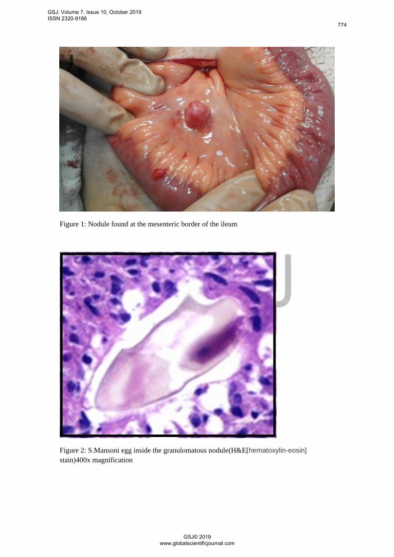
Figure 1: Nodule found at the mesenteric border of the ileum
eosin]-hematoxylin[(H&EFigure 2: S.Mansoni egg inside the granulomatous nodule
stain)400x magnification
GSJ: Volume 7, Issue 10, October 2019 ISSN 2320-9186
774
GSJ© 2019 www.globalscientificjournal.com

Conclusion:
Schistosomiasis is a devastating tropical disease due to its high dominance in various
life threatening manyChronic Schistosomiasis can lead to developing countries.
the stopthe parasite and eliminatecan managementa simple nevertheless, complications
causes of common infrequent of awareness of worththe attractsThis . complication cascade
. like Egypt rural areas in endemic areas mostlyns surgical presentatio
Conflicts of interest:
There are no conflicts of interest.
References:
1. Cox N, Yates P. Schistosomiasis: a rare cause of acute appendicitis. J Surg Case
Reports;2010(4):4–4.
2. Mosli MH, Chan WW, Morava-Protzner I, Kuhn SM. Schistosomiasis Presenting as a Case
of Acute Appendicitis with Chronic Mesenteric Thrombosis. Can J Infect Dis Med Microbiol
:2016:1–3.
3. U Olveda D. Bilharzia: Pathology, Diagnosis, Management and Control. Trop Med Surg .
2013;01(04).
4. Aldossary M, Almabyouq F, Mashhour M, Hassan K. Schistosomal appendicitis presenting
as acute peritonitis: A case report and literature review. J Heal Spec [Internet]. 2017;5(4):225.
5-Woldegerima E, Bayih AG, Tegegne Y, Aemero M, Jejaw Zeleke A. Prevalence
and Reinfection Rates of Schistosoma mansoni and Praziquantel Efficacy against the
Parasite among Primary School Children in Sanja Town, Northwest Ethiopia. J
Parasitol Res .2019, 24;1–8.
6-Le Govic Y, Kincaid-Smith J, Allienne J-F, Rey O, de Gentile L, Boissier J.
Schistosoma haematobium – Schistosoma mansoni Hybrid Parasite in Migrant Boy,
France, 2017. Emerg Infect Dis . 2019;25(2):365–7.
GSJ: Volume 7, Issue 10, October 2019 ISSN 2320-9186
775
GSJ© 2019 www.globalscientificjournal.com


![Acute Appendicitis[1]](https://img.dokumen.tips/doc/110x75/577cd3341a28ab9e7896e8e0/acute-appendicitis1.jpg)


