Embed Size (px)
Citation preview
, . t I~
PRESENCE OF E. coli AND Salmonella spp IN Polymesoda expansa (CLAM) SAMPLED IN SUNGAI SARAWAK
Dyg. Syafiznur Aisyah Binti Abg. Ya'kub 23402
QL 431.6 D996 2012 Bachelor of Science with Honours
(Resource Biotechnology) 2012
• • , I ,.
PUSlt Khidmlt Mlklumat Akademik UNlVERSm MALAYSIA SARAWAK
P.KHIDMAT MAKLUMAT AKADIMIK
111111111rnn 111111111 1000235673
PRESENCE OF E. coli AND Salmonella spp IN Polymesoda expansa (CLAM)
SAMPLED IN SUNGAI SARAWAK
DYG. SY AFIZNUR AISY AH BINTI ABG. Y A'KUB (23402)
This project is submitted in partial fulfiUment of the requirement for the degree of
Bachelor of Science with Honours
(Resource Biotechnology)
I

·,
ACKNOWLEDGEMENT
First of a'll, I would like to thank my supervisor, Professor Dr. Kasing Apun, who had given
me numerous advice, guidance and supervision in the development of my Final Year Project. I
would also like to thank my co-supervisor, Dr. Samuel Lihan, for his help.
This small token of appreciation I would also dedicate to my bench mate, Arfazieda
Binti Anuar for the sharing of information and the amount of time we had spent together in the
making of this project. Apart from that, I would also thank my family for their undivided
support and financial resources contributed for me.
I
I
Pusat Khidmat Maklumat Akademik· . UN1VEltSm MALAYSIA SAKAWA){
T ABLE OF CONTENT
Acknowledgement. .............................. . ... .. ......... . .............. .. '" .............. ... .. i
List of Abbreviation......................................... . ....................... . .............. .. .. iv
List of Tables and Figures ... . ... .. ... .. ... .. ....... ... ............ . ......... . ........ .. ..... . ... . ... . v
Abstract. ................................................................................................. 1
1. Introduction.......... . ............... . .. . ......... ..... . . ....... .. ........... .. ......... . .......... 2
1.1. Rationale . .... . . ... .. .. ... .... . ...... .. ...... . ..... . ... ........ . .................... . .. '" 3
1.2. Research Hypothesis ............. .. .. .... ..... .. .... ... . ... . .. .... . ................. " 3
1.3. Research Question / Problem ..................... .. .. ... ... . ........ .. ........... . ... 4
1.4. Aims and Objectives ................................................................... 5
2. Literature Review ... '" .. . ........ . ....... .... ... . .... ..... ... ... ............. . .... . ...............6
2.1. Indicator Microorganism ... .. .... .. .. . ....... '" ....... . ..... .. ... .. .. .. .............. 6
2.1.1. Escherichia coli (E. coli) .. . ............. ....... . ...... . ........... ... ... ... 6
2.1.2. Vibrio parahaemolYlicus ..... ... .. .. '" ...... .. . ..... ... .... ... '" .. . '" ... ... ... 7
2.1.3. Klebsiella pneumoniae ...... .. . '" ... '" ... '" ... ...... .......... ... .. '" .. . ...... 7
2.1.4. Salmonella spp......................................................... '" 7
2.2. Health Effect of Microorganisms Contamination . .... .. ....... . ..... . ........ .. .. 8
2.3. Indicator Microorganisms Standard ... ... ... ... ...... .. ... .......... .. . .......... .. . 9
2.3 .1. Water Quality Standard (Interim National Water Quality Standard,
INWQS) ........ . ........ .. .... . ... ... .. . .... . .... . ... ... ...... . ..... .. ..... 9
2.3.2. Seafood Microorganisms Standard (International Commission
on Microbiological Specification of Food, ICMSF) .................... 10
ii
• • , I:.
2.4. Method to Determine Level of Bacterial Contamination ... '" ... .. .......... .. .. 10
2.4.1. Most Probable Number Test. ............................................. 10
2.4.2. Membrane Filter Method ................................... .. ............. . 11
2.4.3. Presence Absence Test. ... .... .. ........ .............................. .. .... 11
2.4.4. Standard Plate Count. .. ..... ....... ............. ... .......... . ....... .. ... .. 11
2.5 . Sungai Sarawak .......................................................................... 11
3. Material and Methods ....... .. ... .. .......... ............. ..... . ............... .. ........... .. ... 13
3.1. Sample collection ... .. ....... ... ....... ... ... .. ............ ... ............ . .... ... ....... 13
3.1.1. Bivalve Collection .......................................................... 13 J 3.1.2. Water Sample Collection ........ ....... '" ................................ 14
3.2. Bivalve Dissection .............................. '" .................................... 15
3.3. Microbiological Examination.... .... .................................... . ... ... ...... 15
3.3 .1. Preliminary Morphological Identification ......... .. ....... .. ... ... .. ... 16
3.3.2. Biochemical Test. ...... ... ...... .. ............................... .. ..... .. .. 16
3.4. Microbiological Quality of Water................................................... 17
4. Result. ....... .. ... ....... .. , .... " ................. , . .... " .. ..... ,. . . . ... ... . .. . . . ... .. . . . . .. . . .. . 18
4.1. Colony Count. ... ... .... .. . ...... .... ........ .. .. .. ... ... ... ...... ... .. ............ ... . ... 23
5. Discussion .. .. ........ .. ... .. .......... .. .. . .... . ......... ... ................ . .... . .. '" ............ 26
6. Conclusion............ .. ...... ..... .. . .... . .... .. .. . ... . ... ... ... .. .... . .. ... ... .......... .... .. ... 28
7. Appendix......... '" .. .. .. ' " ... '" ........ . .. .... ... ... ..... .. .... . .... ... .. .. . ' " ................ 29
References ...... .... . ..... .. ..... . .......... .. .. .. .... .. ... . .... ...... .... .. ... .. ....... .. .... .. .......... 32
iii
I
I __...-.----.
t' ' ..
List of Abbreviation
CFU - Colony Forming Unit
EMB Agar - Eosin Methylene Blue
ICMSF - International Commision on Microbiological Specification of Food
INWQS - Interim National Water Quality Standards for Malaysia
MF - Membrane Filter
MPN - Most Probable Number
MRVP - Methyl Red and Voges Proskauer
PBS - Phosphate Buffered Saline
SPC - Standard Plate Count
XLD - Xylose Lysine Deoxycholate Agar
iv
, _., r
List of Tables and Figures
Table 1: Water Quality Standard as shown by Interim National Water Quality Standard
Table 2: Seafood Microorganisms Standard by International Commision on Microbiological
Specification of Food
Table 3: Plate count of E. coli in clams sampled from three different sites during each
sampling.
Table 4: Plate count of bacteria in water sampled from three different area during each
sampling
Table 5: Biochemical test for clams sampled from three different areas for Sampling 1
Table 6: Biochemical test for clams sampled from three different areas for Sampling 2
Table 7: Biochemical test for clams sampled from three different areas for Sampling 3
Table 8: Biochemical test for clams sampled from three different areas for Sampling 4
Figure }: Map of collection sites in Sungai Sarawak
Figure 2: Clam (Polymesoda Expansa)
Figure 3: Plate count of E. coli in clams sampled from three different sites during each
sampling.
Figure 4: Plate counts of bacteria in water sampled from three different area during each
sampling
v

• • I ,"
Presence of E. coli and Salmonella spp. in Bivalve Sampled from Sungai Sarawak
Dyg. SyaflZnur Aisyab Binti Abg Ya'kub
(23402)
Programme: Resource Biotechnology Department: Molecular Biology Department Faculty of Resource Science and Technology
University Malaysia Sarawak
ABSTRACT
Recently, there are increasing interests on studies of microbial contamination in seafood. Indicator microorganisms are a group of organisms that are used to suggest the presence of pathogens. Clams are used as the species of seafood in this study. Presence of the indicator organisms (Escherichia coli and Salmonella) were determined in clams sampled from Sungai Sarawak at three sites, Sungai Sabang, Sungai Santubong and Sungai Tabo. The presence of the microorganisms were determined by morphological characteristic and biochemical test, such as the use of selective agar, MRVP test, Indole test, Citrate Test and Motility test. In addition, the quality of seafood was also determined by comparing the standard plate count of the seafood with the standard from ICMSF. River water of Sungai Sarawak was determined by comparing the water taken from Sungai Sarawak with the standard from INWQS. In this study, Sungai Sabang and Sungai Santubong has been determined as in Normal Raw Water Quality as having average count of 1.47 x 10² and 1.88 x 10² respectively while Sungai Tabo showed as Excellent Water Quality as having average count of 0.64 x 10²
• Food count showed that all samples from the three sites are of 104 104Good Quality Product, with average count of 2.56 x (Sungai Sabang), 4.19 x (Sungai
Santubong) and 1.83 x 10⁴ (Sungai Tabo). The level of indicator microorganisms in the water and seafood are shown to be within the standard limit (3x10² for INWQS and 5xl05 for ICMSF).
Key words: Indicator microorganisms, bivalve, water quality
ABSTRAK
Minat terhadap kajian pencemaran mikrob dalam makanan laut meningkat sejak akhir-akhir ini. Mikroorganisme penunjuk adalah sekumpulan organisme yang digunakan untuk menunjukkan kehadiran patogen. Lokan digunakan sebagai spesies makanan laut dalam kajian ini. Kehadiran organisme penunjuk (Escherichia coli dan Salmonella) telah ditunjukkan dalam sampel lokan yang diambil dari tiga tapak di Sungai Sarawak iaitu Sungai Sabang, Sungai Santubong dan Sungai Tabo. Kehadiran mikroorganisme tersebut telah ditentukan oleh beberapa ujian termasuk ujian morfologi dan ujian biokimia, seperti penggunaan agar selektif, ujian MRVP, ujian indol, ujian 'Citrate' dan ujian motiliti. Selain itu, kualiti makanan laut juga ditentukan dengan membandingkan jumlah koloni dari 'standard plate count' dengan piawaian dari ICMSF, manakala air dari sungai Sarawak ditentukan dengan membandingkannya dengan piawaian dari INWQS. Dalam kajian ini, Sungai Sabang dan Sungai Santubong telah ditentukan sebagai Kualiti Air Mentah Normal dan mempunyai kiraan purata sebanyak 1,47 x 10² dan 1,88 x J(j masingmasing manakala Sungai Tabo mempunyai Kualiti Air Cemerlang dengan kiraan purata sebanyak 0.64 x 1~. Sampel makanan menunjukkan bahawa kesemua sampel dari tiga lokasi dibawah kategori Kualiti Produk Baik, dengan kiraan purata 2.56 x 10⁻¹ (Sungai Sabang), 4.19 x }O-I
(Sungai Santubong) dan 1.83 x 10⁴ (Sungai Tabo) . Tahap pencemaran mikroorganisme penunjuk di dalam sampel air dan sampel makanan juga dibawah tahap piawai (3x10² untuk INWQS dan 5x1rY untuk ICMSF) .
Kata kunci: Mikoorganisme penunjuk, bivalvia, kuali/i air
1
· .
1. INTRODUCTION
Recently, there had been increasing amount of interest in studying the presence and
distribution of various microbial pollution indicators in seafood. This is because it is widely
known that seafood is prone to contamination of microorganisms (Wilson & Moore, 1996).
The studies also need to be done as one step to prevent microbial food poisoning.
Indicator microorganisms are microorganisms that are used to suggest the presence of
pathogens. There are three groups of indicators, which are process indicator, fecal indicator
and index and model organism (Ashbolt et ai, 2001). Process indicator is a group of
microorganisms that shows effectiveness of a process in question. Faecal indicator is a group
of microorganisms that indicates the presence of faecal contamination. An index and model
organism is a group of microorganisms that indicate the presence of pathogen and behavior.
As indicator microorganisms are used to detect presence of pathogens, the pathogens
that are always present in aquatic environment must be known. The pathogens that are always
present in aquatic environment are Clostridium perjringens, Staphylococcus, Erysipeiotric,
Edwardsiella, Salmonella spp., Shigella, Franciscella and Vibrio species (Miget, 1991).
As river is one of the water sources in the world, it is essential to examine the quality
and level of pollution of the water. There are three types of analysis of water, which are
physical analysis, chemical analysis and biological analysis of water (Diersing, 2009). Besides
that, the source of the sea food needs to be studied as well, as contaminated seafood frequently
came from polluted rivers (Kay et al., 2008). Contaminated rivers can be caused by improper
disposal of human waste, sewage, agricultural lands and also by animal wastes.
2
. ' "
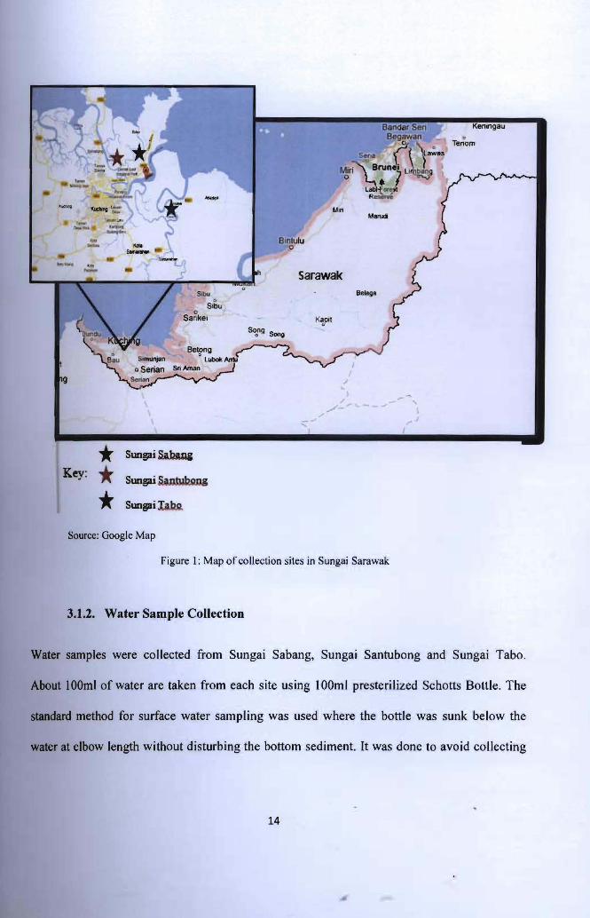
In this research, the river studied was Sungai Sarawak. Sungai Sarawak is one of the
water sources in Sarawak. It is used as drinking water, reservoir and for everyday needs. It
consists on an area of 2459 km2, with length of 120 km and has tributaries such as Sungai
Maong, Sungai Sekama, Sungai Bintangor, Sungai Padungan, Sungai Gersik, Sungai Bedil,
Sungai Kudei, Sungai Sarawak Kiri, Sungai Sarawak Kanan, Sungai Samarahan, Sungai
Sabang, Sungai Bako (Tabo) and Sungai Santubong (Hydrological Year Book, 2004).
Bivalves were sampled from Sungai Sabang which is a village area, Sungai Tabo which is a
recreation area and Sungai Santubong which is a tourism area. ,1.1. Rationale
There is no research on microbial contamination on food sources in Sungai Sarawak.
Therefore, the research on microbial contamination on food sources in Sungai Sarawak is
essential as Sungai Sarawak is one of the main aquatic food sources at the area, and to
compare the indicator microorganisms' content in the seafood with the lCMSF (International
Commission on Microbiological Specifications for Food).
1.2. Research Hypothesis
As the level of pollution will be determined either in water quality or seafood quality, it is
predicted that the amount of indicator organisms in bivalves in village area is higher than
those in recreational park and the amount of indicator organisms in the water in recreation area
is higher than those in tourism area. It is because village area is prone to contamination than
the other area. Apart from that, when the level of water contamination is high, then the level
of seafood quality to be consumed is low.
3
· .
1.3. Research Question / Problem
There may be a relationship between the level of water contamination with public safety and
health associated with their lack of water and seafood quality awareness for consumption. It is
widely known that contaminated food and water can cause severe illness and death. Lack of
awareness of the relationship between contaminated water and seafood quality can cause fatal
epidemic disease, for example diarrheal disease, which is caused by contaminated food and
water, and is one of the most common causes of death (Sherbinin, 1998). Hence, assessment
of current status and also predicting and inspecting of changes in water quality for reference,
supervisory and other controlling purposes can help identifying threats arising from Sungai
Sarawak (Lau & Murtedza, 2000).
~ I
4
Pusat Khidmat Maklumat Akademik • . I' • UNlVERSm MALAYSIA SARAWAK
1.4. Aims and Objectives
The aims of this research are to determine the level of water contamination of Sungai Sarawak
and to raise awareness of better water and seafood quality for public safety and health. The
objectives of the study were to:
I) detect the presence of indicator microorganisms (E. coli and Salmonella spp.) In
bivalves in Sungai Sarawak,
2) relate the water quality of Sungai Sarawak with the seafood quality of bivalves ,taken
from Sungai Sarawak, and
3) determine the degree of contamination in clam and water samples in Sungai Sarawak. ~ I
I I
5
t , _- ~ _
, '
2. LITERATURE REVIEW
2.1. Indicator Microorganisms
The use of indicator microorganisms is to avoid routine examination of environmental
samples, which is often tedious, difficult and time-consuming (Gerba, 2009). From this study,
the criteria for ideal indicator microorganisms are stated. The criteria are the organisms should
be useful for any types of environment, must be present each and every time the enteric
pathogens are present, should have longer survival time than the toughest enteric pathogen,
must not grow in water, testing technique must be easy to be done, their amount must have
direct relationship to level of fecal pollution and should be a member of intestinal micro flora
of warm-blooded animals. The indicator microorganisms that are usually associated with
bivalves are E. coli, Vibrio parahaemolyticus, Klebsiella pneumonae and Salmonella spp.
Other types of indicator microorganisms include other species of microorganism from genus
enterococcus (Ash bolt, 2004).
2.1.1. Escherichia coli (E. coil)
E. coli is one of the well-known indicator microorganisms. According to Desmarais et al.
(2002), E. coli complies the characteristic of good indicator organisms. Scott et al. (2002)
states that it does not typically pathogenic to human and that it existed at a concentration
which is much higher than the pathogen it predicts. It is gram negative, rod-shaped bacterium
and is one of the enteric bacteria. Most of E. coli strains are harmless, but some of its
serotypes cause food poisoning in humans (Vogt & Dippold, 2005).
6
· .
2.1.2. Vibrio parahaemolyticus
Vibrio parahaemolyticus, like other indicator organism, is a gram negative bacterium. It is
usually found in shellfishes, including bivalves. Apart from being an indicator microorganism,
V. parahaemolyticus is also a human pathogen by causing gastrointestinal disease (Bilung et
al., 2005; Nelapati et aI., 2011). It is a member of genus Vibrio and is motile with a single
flagellum (Ryan, 2004). It was first discovered as an agent of foodborne gastrointestinal
disease in Japan in 1951 (Nelapati et al., 2011).
2.1.3. Klebsiella pneumonia
Klebsiella pneumonia is a member of Klebsiella genus of Enterobacteria, which is bacterium
found in the normal flora of human. K. pneumonia is closely related to K. oxytoca, but is
differentiated from the latter by being indole negative. As a normal flora bacterium, K.
pneumonia is considered harmless, but now, Klebsiella spp is regarded as agent of nosocomial
infection (Podschun & Ullman, 1998).
2.1.4. Salmonella spp
Salmonella is also one of the enteric bacteria of cold and warm-blooded animals (Todar,
2011). There are two recognized species of Salmonella, which are S. enterica and S. bongori,
and there are six main subspecies, which are enterica (I), salamaer (II), arizonae (III),
diarizonae (IV), houtenae(V) and indica (VI) (Janda & Abbott, 2006). Even in the subspecies,
the member f each subspecies have a distinct variation with one another, hence, classified as
serovar. Salmonella typhi and Salmonella enteritidis are the serovar of S. enterica enterica
(Gianella, 1996).
7
t I., ,
2.2. Health Effect of Indicator Microorganisms Contamination
Salmonella spp and Campylobacler spp are known as zoonotic bacteria, which are bacteria
that cause diseases that can be transmitted from animals to humans or from humans to animals
(also known as reverse zoonotics) (Taylor el al., 2001).
Salmonella spp can cause salmonellosis and gastroenteritis (caused by Salmonella
enlerica serotype) and had caused 811 of 8640 cases of food poisoning in year 1994 in
Malaysia (Thong et ai, 2002). Salmonella typhi caused typhoid fever (McClelland, 2004). In
Malaysia, report of this fever has dropped since 1992 to 1999 (Malik, A & Malik R, 200 I).
Campylobacler jejuni caused enteritis with symptoms such as abdominal pam,
diarrhea, fever and malaise and can persists between 24 hours to a week, but maybe longer
(Fujimoto & Amako, 1990). Campylobacter jejuni can also causes Campylobacteriosis that is
characterized by inflammatory, cramps, fever, pain, and sometimes bloody, diarrhea or
dysentery syndrome (Javid, 2011).
Escherichia coli are mostly harmless, but some serotypes, such as E. coli 01 04:H4,
and E. coli 0157:H7 can cause serious food poisoning in humans (Vogt & Dippold, 2005).
Virulent E. coli strain can cause gastroenteritis, urinary tract infections and neonatal
meningitis (Todar, 2007).
Vibrio parahaemolylicus causes acute gastroenteritis which is characterized by
diarrhea, vomiting and abdominal cramps. It can also cause traveller's diarrhea (Nelapati et
al., 20 II), ear infection and secondary septicemia (Pavia el al., 1989). In 1951, V.
8
• ., r
parahaemolyticus caused a large outbreak of 272 cases and 20 death in Japan (Nelapati et al.,
20 II).
Klebsiella pneumonia is a common hospital-acquired pathogen, which can cause
urinary tract infection, nosocomial infection and intra-abdominal infection. In Taiwan, more
than 900 reported cases of Klebsiella spp infection in the last 10 years (Ko et al., 2002). In a
study conducted by Ko et al. (2002), 44.4% of 455 cases of Klebsiella pneumoniae infection is
community-acquired, which proved that Klebsiella pneumoniae is not only hospital-acquired,
but also community-acquired pathogen.
Clam, through contamination of bacteria, also cause food poisoning. In a three decade
observation by Potasman et al. (2002) from year 1969 until 2001, there were a total of 42
outbreaks with nearly 300,000 cases in all over the world. In all over the world too, clams had
been contaminated by E. coli and caused nearly 300,000 cases in China until 2010 (Love et
01., 2010) while for Salmonella, 4-7% of outbreaks from 1988-1992 of Salmonellosis were
caused by consumption of contaminated bivalve (Kumar et al., 2009). Asian country had
reported 99% of the cases, in which the reports are mainly from China. Malaysia had also
reported 120 cases of Vibrio cholera from bivalve consumption (Potasman et al., 2002) .
2.3. Indicator Microorganisms Standard
2.3.1. Water Quality Standard (Interim National Water Quality Standards for
Malaysia, INWQS)
To ensure the river water is not harmful for human everyday needs, Water Quality Standard
must be used to compare the level of the contamination of the river with the safe level of
9
· .
contamination for raw water. In Malaysia, the water quality standard used is Interim National
Water Quality Standard (INWQS). Table 1 shows the water quality standard as shown by
INWQS. There are different standard used by different countries, but the international water
quality standard is ISO Water Quality (International Organization for Standardization, 2004).
2.3.2. Seafood Microorganisms Standard (ICMSF)
To ensure the seafood is not harmful for human consumption, Seafood Microorganisms
Standard must be used to compare the level of the contamination of the seafood with the safe
level of contamination of seafood. In Malaysia, the seafood microorganisms' standard used is
International Commission on Microbiological Specification of Food (ICMSF). If the value of
the standard plate out of the seafood is higher than the standard, it showed that the seafood
was not safe to be consumed.
2.4. Metbod to Determine Level of Bacterial Contamination
2.4.1. Most Probable Number Test
Most probable number (MPN) test is described by Gerba (2009), when he stated that it shows
the 'presence of coliforms in a sample and estimation of their numbers' and it consists of three
steps, which are the presumptive test, confirmed test and a completed test (Gerba, 2009).
There were some disadvantages where MPN test is proved to be time consuming. According
to Schiemann (1972), there are two assumptions when working with MPN and one of them is
assumed that the distributions of individual cells are "random in the suspension with complete
independence" (Schiemann, 1972). The second assumption is that the growth will follow the
introduction of the cells into a tube of medium.
10
• • I
2.4.2. Membrane Filter Method
According to Gerba (2009), MF is easier to be done than MPN, because it needs fewer test
tubes, and less labor, and also require less time than MPN. The appropriate volume of
samples are to pass through a membrane filter with 0.451lm porosity, which is the most
appropriate porosity for a membrane filter so that the microorganisms would not pass through
(Dufour et al., 1981).
2.4.3. Presence Absence Test
The third way to test water quality is by presence absence test. As Rice et al. (1989) reported,
the presence absence (PA) test is a qualitative method rather than a quantitative method. In
Pipes et al. (1986), they believed that the presumptive coliform test is considered positive
when there is a production of acid and gas. They also stated that there are more positive
samples by PA test than MF method.
2.4.4. Standard Plate Count
Standard plate count (SPC) is considered to be better than coliform index (LeChevallier et al.,
1980) and used collection of samples streaked, and then counted with the help of dissecting
microscope at 15X magnification. LeChevallier et al. (1980) stated that the standard plate
count at 35°C is the best indicator for bacteria.
2.5. Sungai Sarawak
Sungai Sarawak has a few tributaries, which are Sungai Maong, Sungai Sekama, Sungai
Bintangor, Sungai Padungan, Sungai Gersik, Sungai Bedil, Sungai Kudei, Sungai Sarawak
Kir, Sungai Sarawak Kanan, Sungai Samarahan, Sungai Sabang, Sungai Bako and Sungai
11
i •
Santubong. (Bernama - Sungai Sarawak To Be Cleaned in 10th Malaysian Plan, 2010).
Sungai Sarawak flows from the Sungai Sarawak Kiri and Sungai Sarawak Kanan towards
Muara Tebas where it flow off to the Laut Cina Selatan (Hydrological Year Book, 2004).
According to a report by Jabatan Alam Sekitar (2004), Sungai Sarawak through all its
tributaries, is considered semi-contaminated either chemically, or biologically.
Sungai Sarawak has two main tributaries, which are Sungai Sarawak Kiri and Sungai
Sarawak Kanan. Both tributaried meet at Batu Kitang, which is about 34 km from Kuching.
The water from Sungai Sarawak then converged with Sungai Kuap and further downstream
before discharging to the Laut Cina Selatan at Muara Tebas (Jabatan Alam Sekitar, 2004).
Sungai Sarawak provides the residents in Kuching what any other river provided.
Sungai Sarawak is the major water source in Kuching, used by the residents as drinking water,
reservoir and as everyday needs. There are also recreational park, villages, tourism area and
industrial park along Sungai Sarawak.
12
, .,
3. MATERIALS and METHODS
3.1. Sample Collection
Sampling were done once in a fortnight for two months on the 29 January, 12 February, 26
February, and 11 March, 2012. Sampling was done on both clam samples and water samples.
3.1.1. Bivalve Collection
Clams (Polymesoda expansa) were bought from fishermen who took the clams from three
different sites, which are recreational area (Sungai Tabo), tourism area (Sungai Santubong)
and village area (Sungai Sabang). The clams were selected and the clams were gapping or
cannot be used are immediately discarded (Kronkvist, 2006). Only five clams from each site
were used in the experiment. The three areas were selected because of the possible difference
on level of contamination in village area, recreational area and Tourism Park. The clams were
chosen is because of it is the most popular species of bivalve for consumption among Sarawak
residents than the other species of bivalve. The clams were placed in a plastic bag and
positioned inside an ice box and transported to the laboratory within an hour.
13
- -----_ ..
. .
....,. Min
1_' MaNdl
,-iIIq .......- Blnlulu 0
-...,.. Sarawak
Sibu _gft
S,bu 0
Sar. ke, K~pit
* Sung;si~ Key: Sung;si S~Qog* Sung;si Ia.b,g,*
Source: Google Map
Figure 1: Map of collection sites in Sungai Sarawak
3.1.2. Water Sample Collection
Water samples were collected from Sungai Sabang, Sungai Santubong and Sungai Tabo.
About IOOml of water are taken from each site using 100m I presterilized Schotts Bottle. The
standard method for surface water sampling was used where the bottle was sunk below the
water at elbow length without disturbing the bottom sediment. It was done to avoid collecting
14
rain water, which IS usually found at the uppermost surface of water (SESD Operating
Procedure, 2007).
3.2. Bivalve Dissection
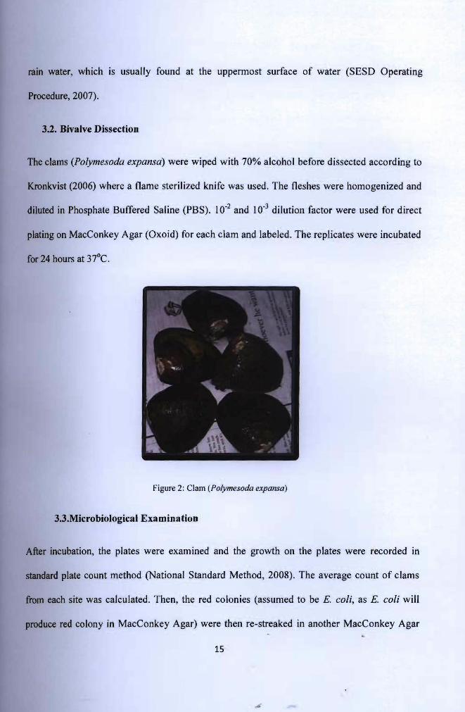
The clams (Polymesoda expansa) were wiped with 70% alcohol before dissected according to
Kronkvist (2006) where a flame sterilized knife was used. The fleshes were homogenized and
diluted in Phosphate Buffered Saline (PBS). 10.2 and 10.3 dilution factor were used for direct
plating on MacConkey Agar (Oxoid) for each clam and labeled. The replicates were incubated
for 24 hours at 37°C.
Figure 2: Clam (Polymesoda expansa)
3.3.Microbiological Examination
After incubation, the plates were examined and the growth on the plates were recorded in
standard plate count method (National Standard Method, 2008). The average count of clams
from each site was calculated. Then, the red colonies (assumed to be E. coli, as E. coli will
produce red colony in MacConkey Agar) were then re-streaked in another MacConkey Agar
15
and the same goes for the yellow colony (tentatively identified as Salmonella, as Salmonella
will produce yellow colony on MacConkey Agar) and then incubated. After one day
incubation, the plates of the red colony were re-streaked into Eosin Methylene Blue Agar
(EMBA) (Merck) and the plates of the yellow colony were re-streaked into Xylose Lysine
Deoxycolate (XLD) (Oxoid) agar to ensure pureness. If pure colony were still not achieved,
the colonies were re-streaked again in EMBA and XLD until pure colony is achieved.
3.3.1. Preliminary Morphological Identification
After obtaining pure colony, a single colony from the plate was selected and allowed to grow
for 24 hours before checking for its Gram stain. After Gram stain, if only one type of cell were
observed, the colony were placed into Nutrient Broth and incubated for stock. The other
colonies were used for biochemical test.
3.3.2. Biochemical Test
Single colonies from the plate were inoculated and tested for its biochemical properties with
IMViC test (MRVP, Indole, Citrate and Mobility test) (National Standard Method, 2008).
Colonies undergo Biochemical test were incubated for two days inside the agar used for
biochemical tests (Indole test and Mobility test were using SIM (Sulfite Indole Motility) agar,
Citrate test was using Simmon Citrate agar and MRVP (Methyl Red and Voges Proskauer) test
was using MRVP broth) before tested. After incubation, few drops of Kovacs Reagent were
dropped into SIM agar, and Barritt's Reagent A and B were dropped into MRVP broth.
For Indole test, a clear red colour on the surface of the SIM agar indicates positive
reaction, while negative reaction is indicated by yellow or creamy colour on the surface of the
agar. For Mobility test, positive reaction is indicated by turbid area extending away from
16
inoculation site while negative reaction has no turbid area. For Citrate test, positive reaction is
shown by the green colour of the media turned blue and negative reaction shows no changes of
colour. For MRVP test, the MR and VP test are conducted separately. For MR test, a few
drops of Barritt's reagent A were added into the broth and red or pink colour indicates positive
reaction while yellow colour indicates negative reaction (Gilles, 2008). Lastly for VP test, a
few drops of Barritt's reagent B were added to the broth. Positive reaction is presented by
pink-burgundy colour, while negative reaction is presented by yellow or creamy colour
(National Standard Method, 2008)
3.4. Microbiological Quality of Water
The water samples were monitored for its level of contamination by membrane filter
techniques. A 0.45J.l1ll diameter pore filter membrane (Pall) was placed on top of m-FC agar
(Merck) and were incubated for 24 hours at 37°C (Merck, 2002). After incubation, the number
of colonies was counted. The values were compared with Interim National Water Quality
Standard (INWQS) to detennine water quality.
17