Embed Size (px)
Citation preview

SAFETY AND MORBIDITY OF INTRA-ORAL ZYGOMATIC BONE GRAFT HARVESTINGDevelopment of a novel bone harvesting technique
VESAKAINULAINEN
Faculty of Medicine,Institute of Dentistry,
University of Oulu;Department of Oral and
Maxillofacial Surgery,Oulu University Hospital
OULU 2004

VESA KAINULAINEN
SAFETY AND MORBIDITY OF INTRA-ORAL ZYGOMATIC BONE GRAFT HARVESTINGDevelopment of a novel bone harvesting technique
Academic Dissertation to be presented with the assent ofthe Faculty of Medicine, University of Oulu, for publicdiscussion in Auditorium 1 of the Institute of Dentistry,on October 29th, 2004, at 12 noon.
OULUN YLIOPISTO, OULU 2004
Copyright © 2004University of Oulu, 2004
Supervised byProfessor Kyösti OikarinenDocent George K. B. Sàndor
Reviewed byProfessor Anders HolmlundDocent Tom C. Lindholm
ISBN 951-42-7473-3 (nid.)ISBN 951-42-7474-1 (PDF) http://herkules.oulu.fi/isbn9514274741/
ISSN 0355-3221 http://herkules.oulu.fi/issn03553221/
OULU UNIVERSITY PRESSOULU 2004

Kainulainen, Vesa, Safety and morbidity of intra-oral zygomatic bone graftharvesting. Development of a novel bone harvesting techniqueFaculty of Medicine, University of Oulu, P.O.Box 5000, FIN-90014 University of Oulu, Finland,Institute of Dentistry, Department of Oral and Maxillofacial Surgery, University of Oulu, P.O.Box5281, FIN-90014 University of Oulu, Finland; Oulu University Hospital, P.O.Box 10, FIN-90029OYS, Finland 2004Oulu, Finland
AbstractThis study focuses on the development of a bone collecting device for intra-oral bone harvesting andon the introduction of a new bone graft donor site, zygomatic bone.
A bone collector was constructed and tested in vitro. This bone collector is suitable and efficientin dental implant related bone grafting surgery. It was also found to be more efficient and with a largercapacity in bone harvesting when compared to the two commercially available bone collectors.
A zygomatic bone harvesting technique is introduced in this study. The safety and morbidity ofthe method was assessed in a cadaver and a prospective clinical study. In the cadaver study, 40procedures were performed. The complications during the cadaver harvesting included 15perforations into the maxillary sinus and 7 perforations into the infratemporal fossa. The only intra-operative complication in 32 clinical operations was perforation of the maxillary sinus in 33% of thezygomatic sites. None of these patients experienced any post-operative problems related to theperforation. Patients needed pain medication for a mean time of four days and they did notdemonstrate any paresthesias or altered sensations in the donor area.
The yield of the bone graft from zygomatic bone was quantified in cadaver and clinical studies. Inthe cadaver study, the average yield of the graft was 0.59 ml. In the clinical study the average graftvolume was 0.90 ml. The required reconstructions were accomplished in all clinical cases.
In the prospective clinical study, the bone grafts from the zygomatic bone were usedsimultaneously with one-stage dental implants placement. Bone grafting was employed at 72 of the82 implant sites. Two of the bone grafted implants failed, yielding a survival rate of 97.2% for bonegrafted implants and 97.6% for the whole study group. Grafted sites healed remarkably well, and noobvious signs of graft resorption were noted during the 26.9 months follow-up period.
The bone collector developed in this study is an effective instrument in intra-oral bone harvesting.The zygomatic bone can be regarded as a safe bone harvesting donor site and the yield of bone graftfrom this area is sufficient for moderate defects in resorbed alveolar ridges.
Keywords: autogenous bone grafts, bone graft volume, donor site morbidity, zygomaticbone

Acknowledgements
The research was carried out during the years 1996-2004 at the Department of Oral and Maxillofacial Surgery, Institute of Dentistry, University of Oulu, Finland and at the Department of Oral and Maxillofacial Surgery, Faculty of Dentistry, University of Toronto, Canada.
I wish to express my deepest gratitude to my supervisors, Professor Kyösti Oikarinen, D.D.S., Ph.D., the Head of the Department of Oral and Maxillofacial Surgery at the University of Oulu and Docent George Sàndor, D.D.S., M.D., Ph.D., the Director of Post Graduate Program at the Department of the Oral and Maxillofacial Surgery at the University of Toronto. Professor Oikarinen introduced me to oral and maxillofacial surgery and has been a great inspiration to my career. Dr. Sàndor gave me the opportunity to work at the University of Toronto during the years 2000-2001. Besides his substantial scientific knowledge, his vast clinical experience, his encouraging attitude and true friendship, I highly appreciate all those conversations with him which gave me strength to keep on working with this project until its completion.
I would like to express my gratitude to Professor Cameron Clokie D.D.S., Ph.D., the Head of the Department of Oral and Maxillofacial Surgery at the University of Toronto, for providing support and research facilities. Grateful thanks are also due to my co-worker Robert Carmichael D.M.D., M.Sc.. I wish to thank Robert Barron D.D.S. and Hani Abd-Ul-Salam D.D.S., Ph.D. who helped me during those very many late hours doing the experimental surgery for this study.
To Professor Anders Holmlund and Docent Tom Lindholm I would like to express my thanks for their valuable comments on the manuscript of this thesis.
I want to express my warmest thanks to the staff of the Department of Oral and Maxillofacial Surgery of the University of Oulu for their always constructive attitude and friendly atmosphere. I also like to thank my colleagues and friends in Toronto.
Mrs. Liisa Kärki and Mrs. Seija Leskelä deserve my thanks for their friendly collaboration during these years. My special thanks are also due to Mr. Eino Kemppainen for helping to manufacture the bone collecting instruments.
There are no words great enough to describe my gratitude to my parents, Liisa and Juhani. Special thanks are due to Timo for being my Big Brother. Warm thanks are also
due to Mrs. Sirkka Haapalainen. I would like to also thank all my friends for the great moments during the years.
Finally I want to express my thanks to my beloved wife Tiina, for the support, help and love during the years. I dedicate this work to our children Vilma and Sampo, the sunshines of our life.
This work was financially supported by the Finnish Dental Society Apollonia, the Northern Finland Cultural Foundation, the Research Foundation of the University of Oulu, the Foundation of Emil Aaltonen, and the Finnish Medical Society Duodecim.
Oulu, October 2004
Vesa Kainulainen

Abbreviations
BMP bone morphogenetic protein CT computed tomography scanning DNA deoxyribonucleic acid DO distraction osteogenesis GBR guided bone regeneration GM-CSF granulocyte macrophage colony stimulating factor e-PTFE expanded polytetrafluoroethylene IGF insulin-like growth factor IL interleukin N2O/O2 nitrous-oxide oxygen sedation OCT® Osseous coagulum trap® OP-1 osteogenic protein -1 PRP platelet-rich-plasma SLA® sand-blasted, large grit, acid-etched® TGF-β transforming growth factor –beta TMJ temporomandibular joint TNF tumor necrosis factor


Glossary of terms
Allograft: A graft derived from tissue taken from another individual of the same species. Alloplastic graft: Synthetic graft material. Autograft: A graft derived from tissue of the same individual. Alveolar bone: That portion of bone in either the maxilla or the mandible
which surrounds and supports the teeth. Bone graft: Bone tissue to repair or replace diseased or missing anatomical
structures. Bone transplanted from a donor site to a recipient site.
Distraction osteogenesis: Bone lengthening after an osteotomy by gradual mechanical distraction.
Le Fort osteotomy: An osteotomy often done to correct a maxillary skeletal deformity. Classified as Le Fort osteotomy I, II, or III, depending upon the location.
Mandibular corpus: The body of the mandible between the ramus and symphysis. Mandibular ramus: The posterior ascending part of the mandible. Mandibular symphysis: The most anterior part of the body of mandible between the
canine teeth. Mandibular torus: An exostosis protruding from the lingual aspect of the
mandible, usually opposite the premolar teeth. Maxillary tuberosity: The bulging lower extremity of the posterior surface of the
body of the maxilla, behind the root of the last molar tooth. Osseointegration: A direct structural and functional connection between living
bone and the surface of an implant. Osteoconduction: Bone formation by the ingrowth from the bone graft recipient
bed into the graft by capillaries, perivascular tissue and osteoprogenitor cells.
Osteogenesis: The formation of new bone from osteocompetent cells. Osteoinduction: New bone is produced in an area where there was no bone
before, where one tissue or its derivative causes another undifferentiated tissue to differentiate into bone.


List of original papers
The thesis is based on the following original articles, which are referred to in the text by numerals I to V: I Oikarinen K, Kainulainen V & Kainulainen T (1997) A method of harvesting
corticocancellous bone chips for reconstructive maxillofacial surgery. International Journal of Oral and Maxillofacial Surgery 26: 103-105.
II Kainulainen V & Oikarinen K (1998) Comparison of four bone collectors designed for oral and maxillofacial surgery - an in vitro study. Clinical Oral Implants Research 9: 327-332.
III Kainulainen VT, Sàndor GKB, Clokie CML, Keller A & Oikarinen KS (2004) The zygomatic bone as a potential donor site for alveolar reconstruction - A quantitative anatomic cadaver study. International Journal of Oral and Maxillofacial Surgery. In press.
IV Kainulainen VT, Sàndor GKB, Oikarinen KS & Clokie CML (2002) Zygomatic Bone – An additional donor site for alveolar bone reconstruction – A Technical note. International Journal of Oral and Maxillofacial Implants 17: 723-728.
V Kainulainen VT, Sàndor GKB, Carmichael RP & Oikarinen KS (2004) Safety of zygomatic bone harvesting - A prospective study of 32 consecutive patients with simultaneous zygomatic bone grafting and one stage implant placement. International Journal of Oral and Maxillofacial Implants. Accepted for publication.
Reprints were made with the permission of the journals.


Contents
Abstract Acknowledgements Abbreviations Glossary of terms List of original papers Contents 1 Introduction ...................................................................................................................15 2 Review of the literature .................................................................................................17
2.1 Bone biology and bone graft healing......................................................................17 2.1.1 Terminology of bone graft healing ..................................................................19
2.2 The histological origin of bone autografts ..............................................................19 2.3 Differences between membranous and endochondral bone grafts in maxillo-facial reconstruction..................................................................................20 2.4 Autogenous bone grafting in the maxillo-mandibular region .................................20
2.4.1 Procedures to augment alveolar bone for dental implants ...............................21 2.4.1.1 Maxillary sinus floor augmentation..........................................................21 2.4.1.2 Onlay grafting of the alveolar bone ..........................................................22 2.4.1.3 Interpositional bone grafting of alveolar bone..........................................24 2.4.1.4 Guided bone regeneration.........................................................................24 2.4.1.5 Techniques to augment alveolar bone without a bone graft......................25
2.5 Intra-oral bone harvesting donor sites ....................................................................26 2.5.1 Mandibular symphysis.....................................................................................27 2.5.2 Mandibular corpus and ramus .........................................................................28 2.5.3 Coronoid process of the mandible ...................................................................29 2.5.4 Maxilla.............................................................................................................29 2.5.5 Zygomatic bone ...............................................................................................30 2.5.6 Mandibular tori ................................................................................................30
2.6 Intra-oral bone harvesting with bone collectors......................................................30 2.7 Morbidity and complications associated with intra-oral bone harvesting ..............31 2.8 The yield of bone graft from various intra-oral donor sites ....................................33
2.8.1 Mandibular symhysis.......................................................................................33
2.8.2 Mandibular corpus and ramus .........................................................................34 2.8.3 The coronoid process of the mandible .............................................................34
2.9 Anatomy of the zygomatic bone .............................................................................35 3 Aims of the study...........................................................................................................36 4 Materials and methods...................................................................................................37
4.1 Development and comparison of bone collectors and bone harvesting drills.........37 4.2 Intra-oral zygomatic bone harvesting .....................................................................40
4.2.1 Zygomatic bone harvesting from the cadavers ................................................40 4.2.2 Surgical procedure of intra-oral zygomatic bone harvesting in patients..........41
4.3 Safety and morbidity of intra-oral zygomatic bone harvesting procedure..............44 4.3.1 Cadaver operations ..........................................................................................44
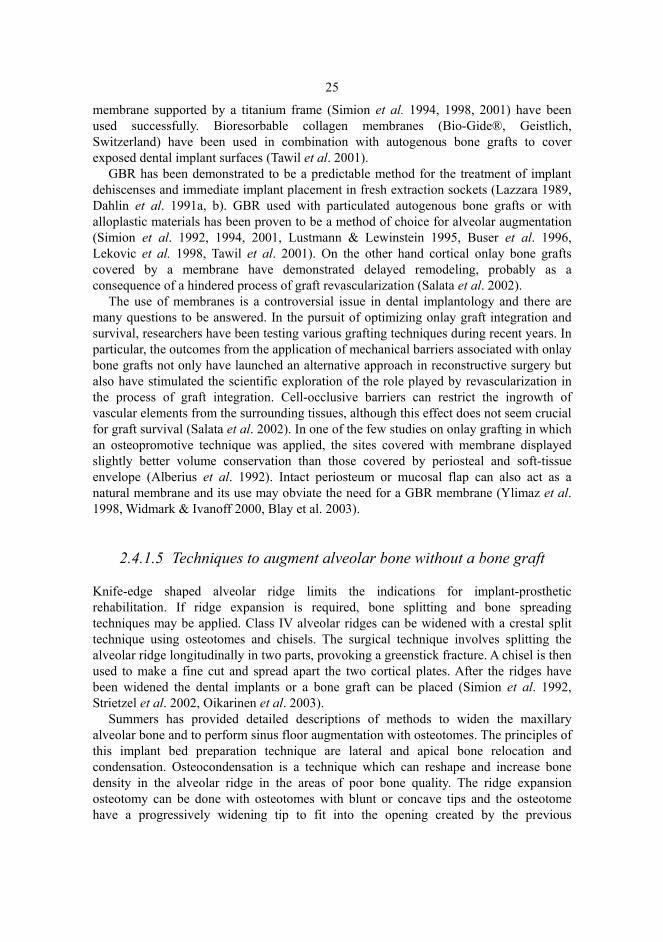
4.3.1.1 Pre-operative CT analysis .........................................................................45 4.3.1.2 Observations and measurements made during cadaver bone harvesting ..46 4.3.1.3 Post-operative CT analysis .......................................................................46
4.3.2 Clinical operations...........................................................................................46 4.4 The volume of bone graft harvested from the zygomatic bone ..............................49
4.4.1 Bone graft volume measurements in cadaver specimens.................................49 4.4.2 Bone graft volume measurement in clinical operations...................................50
4.5 Simultaneous one-stage implant placement and bone grafting with particulated bone ....................................................................................................51
4.5.1 Prospective clinical study ................................................................................51 4.6 Statistical analysis...................................................................................................52
5 Results ...........................................................................................................................54 5.1 Development and comparison of bone collectors and bone harvesting drills.........54 5.2 Safety and morbidity of intra-oral zygomatic bone harvesting procedure..............56
5.2.1 Cadaver study ..................................................................................................56 5.2.1.1 Pre-operative CT scan and intra-operative measurements........................56 5.2.1.2 Post-operative clinical observations and CT scan measurements .............56
5.2.2 Clinical study...................................................................................................57 5.3 The volume of bone graft harvested from the zygomatic bone ..............................58
5.3.1 Cadaver study ..................................................................................................58 5.3.2 Clinical study...................................................................................................58
5.4 Survival of dental implants with simultaneously placed particulated bone grafts ..58 6 Discussion .....................................................................................................................60
6.1 Development and comparison of bone collectors and bone harvesting drills.........60 6.2 Intra-oral zygomatic bone graft harvesting.............................................................63 6.3 Safety and morbidity of zygomatic bone harvesting ..............................................63 6.4 The volume of bone harvested from the zygomatic bone.......................................65 6.5 Survival of dental implants with simultaneously placed particulated bone grafts ..66 6.6 Clinical implications and future prospects..............................................................67
7 Summary and conclusions .............................................................................................69 References

1 Introduction
Bone grafting of the resorbed dental alveolus is often necessary prior to dental implantation. Many allografts and alloplastic materials have been used as bone graft substitutes, but autogenous corticocancellous bone grafts have still remained the gold standard for the reconstruction of alveolar bone. Research in the field of oral and maxillofacial surgery has produced new surgical techniques and bone harvesting donor sites for bone augmentation in deficient sites. The goal of these studies is the same - to reduce complications and post-operative morbidity, and to minimize the economic costs of the treatment. The use of the extra-oral bone harvesting donor sites, such as the anterior and posterior iliac crest, is still the standard when large reconstructions are performed in the maxillo-mandibular region for example after tumor surgery or in dental implant treatment to totally edentulous jaws. However, the current trend when implant surgery is done to partially edentulous resorbed dentoalveolar ridges is to harvest bone from an intra-oral donor site (Jensen & Sindet-Pedersen 1991, Misch 1997, Cordaro et al. 2002, Cordaro 2003).
The use of the dental implants for the reconstruction of edentulous jaws has been a progressively growing treatment modality since the late 1970´s. Brånemark and co-workers published their first follow-up report of osseointegrated implants in the treatment of the edentulous jaw in 1977 (Brånemark et al. 1977). Bone grafting of the resorbed alveolus for dental implants was employed later and Breine and Brånemark (1980) Kahnberg (1989), Sailer (1989) and Adell (1990) reported results on prosthetic reconstruction of the resorbed edentulous jaws with autologous bone grafts and dental implants (Kahnberg et al. 1989, Sailer 1989, Adell et al. 1990). Boyne and James were the first to report experiences with inlay bone grafting of the maxillary sinus for dental implants (Boyne & James 1980). After these studies dozens of articles were published concerning alveolar bone augmentation in edentulous or partially edentulous alveolar ridges prior to or in conjunction with dental implant placement.
The first reports of intra-oral bone harvesting and bone grafting for dental implants were published at the beginning of the 1990´s (Jensen & Sindet-Pedersen 1991, Misch et al. 1992). Most of these reports highlighted the intra-oral harvesting sites as having convenient surgical access. The ischemic time of the bone graft has reported to be short. Furthermore, since both the donor and recipient sites are intra-oral, there was no

16
morbidity from a second surgical site. The morbidity associated with intra-oral donor sites was also found to be lower compared to extra-oral donor sites and the use of a trans-oral approach does not cause visible scarring. One major disadvantage of intra-oral bone harvesting was also found - the limited amount of available bone (Sindet-Pedersen & Enemark 1988, 1990, Jensen & Sindet-Pedersen 1991, ten Bruggenkate et al. 1992, Misch et al. 1992, Jensen et al. 1994).
The most commonly utilized intra-oral bone harvesting donor sites in dental implant related surgery are the mandibular symphysis, (Jensen & Sindet-Pedersen 1991, Misch et al. 1992) and ramus (Misch 1996). Smaller amounts of particulated bone graft may be harvested from the maxillary tuberosity, extraosseous tori or with residual alveolar ridge osteoplasty (ten Bruggenkate et al. 1992, Misch & Misch 1999).
The present study focused on developing better bone harvesting instruments and to find an alternative for the already known intra-oral bone harvesting donor sites with minimal complications and patient discomfort. Better bone harvesting instruments and techniques should make it possible to reduce patient morbidity and the economic costs of the treatment.

2 Review of the literature
2.1 Bone biology and bone graft healing
The transplantation of tissues and organs represents one of the most fascinating strategies to repair or replace diseased or missing anatomical structures. Bone, by its character, differs substantially from solid organs and immediately revascularized tissues with respect to transplantation. Bone regenerates, and does so with autogenous resources including cells, cytokines and blood vessels, regardless of the source of graft material. Bone also shares, with other transplantable organs and tissues, the ability to induce a variety of immunological responses reflecting its nature (Friedlaender 1987).
The process of bone regeneration is common to skeletal homeostasis, the repair of fractures and the incorporation of bone grafts. The cascading sequence of biologic events common to this wide spectrum of regenerative activity is often described as the remodelling cycle. Cell populations are activated and become committed to resorption of pre-existing bone matrix (osteoclasts) followed by the accretion of new mineralised tissue (osteoblasts) (Heiple et al. 1963, Burchardt 1983, Friedlaender 1987). These events require a blood supply as well as a system of humoral factors (cytokines) that integrate and regulate these events. The circular sequence, or continuum of cellular and molecular events, is in large measure regulated by soluble factors, cytokines that facilitate cell-cell interactions and modulate their activities in an autocrine or, more frequently, paracrine fashion (Goldring & Goldring 1996, Mundy 1996).
Cytokine families include interleukins (IL-1, IL-6), tumour necrosis factor (TNF), insuline-like growth factors (IGF) and particularly members of the transforming growth factor –beta (TGF-β) super family, such as bone morphogenetic proteins (BMPs), and granulocyte macrophage colony stimulating factor (GM-CSF). Many of these factors have multiple and overlapping activities. They have been found to be produced by and influential in more than one biological system (Goldring & Goldring 1996). Several members of the TGF-β super family have been shown to cause the recruitment of mesenchymal stem cells and their differentiation into chondrogenic and osteogenic populations (Mundy 1996). Osteogenic protein-1 (OP-1 or BMP-7) and BMP-2 have been particularly well-characterised and produced by recombinant DNA techniques

18
(Cook & Rueger 1996, Riley et al. 1996). Some of these factors (BMP-2, BMP-7) are already commercially available for the promotion of osteo-inductive activity, including bone graft enhancement or substitution.
Bone metabolism is associated with cycles of active bone resorption and new bone formation. If bone grafts are transplanted to hard tissue defects, they undergo cellular regeneration followed by remodelling. Such bone regeneration is divided into two phases. The first phase is cellular proliferation and production of osteoid in a random fashion. This bone lacks the haversian systems and lamellae of more mature bone. Bone will undergo an obligatory resorption and is then replaced by organised bone (Phase II). The physiology is common to all bone healing. The identical physiology is observed in the formation-replacement-remodelling-formation cycle of both internal and external calluses and in normal skeletal remodelling (Yim 2003).
Non-vascularized free autogenous bone grafts are either cortical or cancellous. Cancellous bone grafts can revascularize more rapidly and completely than cortical grafts. Creeping substitution of a cancellous graft initially involves an appositional bone formation phase, followed by a resorptive phase, whereas cortical grafts undergo a reverse creeping substitution process. Cancellous grafts are usually repaired completely but cortical grafts remain as a mixture of necrotic and viable bone. Non-vascularized bone grafts act mostly as scaffolds and are thus more osteoconductive than osteoinductive. However, osteogenic activity may have remained in the cancellous part of the bone graft (Burchardt 1983, Bonutti et al. 1998, Keller et al. 1998, Vinzenz et al. 1998).
In particulated bone graft transplantation, endosteal osteoblasts primarily, and mesenchymal fibroblasts secondarily, are responsible for bone formation. This initial phase of bone regeneration is directly proportional to the cellular density of the transplanted bone and will result in the maximum amount of bone achievable by the graft system. These, particulated bones and cancellous marrow grafts that transplant a greater quantity of osteoprogenitor cells have been found to produce superior bone ossicles in mandibular continuity defects over block-type grafts containing fewer osteoprogenitor cells (Marx 1994).
The second phase of bone is not derived from transplanted cells as is phase I bone. It is instead derived from host tissue cells that eventually replace phase I bone with mature, organised bone and establish an endosteum and periosteum. This is the transitional sequence between phase I and II bone. As the osteoclast resorbs phase I bone, it is thought to secrete coupling factors or release osteogenin from the mineral matrix of the resorbed bone. This process occurs in normal everyday physiologic bone resorption as well. Such osteogenin release or coupling factor couples bone resorption and new bone apposition through the induction and mitogenesis of host connective tissue cells into functioning osteoblasts. Second-phase bone develops into a trabecular bone ossicle with more well-defined lamellae and greater mineral density. The second-phase bone will only resorb and replace phase I bone in a 1:1 ratio at best. Such phase II resorption-remodelling occurs throughout the life of the particulate bone graft as it does in all other bone (Yim 2003).

19
2.1.1 Terminology of bone graft healing
Osteogenesis is the formation of new bone from surviving cells within a bone graft – namely the cells from the inner cambium layer of periosteum that survive autogenous transplantation. Osteogenesis does not occur with allograft transplantation (Nather 2003).
Osteoinduction is the mechanism in which new bone is formed by the active recruitment of host pluripotential cells that differentiate into chondroblasts and osteoblasts. It is accomplished by diffusion of osteogenic bone matrix referred to as BMPs from bone matrix (Nather 2003). Specifically, new bone is produced in an area where there was no bone before, where one tissue or its derivative causes another undifferentiated tissue to differentiate into bone. The phenomenon of osteoinduction was first described in the classic works of Urist (Urist & McLean 1952, Urist 1965, Urist et al. 1977).
Osteoconductive bone formation refers to the ingrowth from a bone graft recipient bed into the graft by capillaries, perivascular tissue and osteoprogenitor cells. The graft acts as an inert scaffold for the ingrowth of this host tissue (Buchardt 1983, Nather 2003).
2.2 The histological origin of bone autografts
The bones of the human skeleton are formed by intramembranous or endochodral ossification. Depending on the mechanism of formation, bones are labelled membranous or endochondral (Manson 1994).
Endochondral bone originates from a hyaline cartilage matrix which is replaced by bone. Endochondral bones include the skeletal long bones, the ribs, the vertebrae, and the base of the skull (Mulliken et al. 1984, Manson 1994).
Endochodral ossification involves the initial generation of a cartilaginous model, which is then replaced by new bone. The matrix of the cartilage is calcified following vascular invasion. The processes of vascular invasion bring undifferentiated mesenchymal cells into the area and are then caused to differentiate into osteoblasts. Osteoid is produced that is then mineralized, forming spicules of bone trabeculae. The bone trabeculae are organized into woven bone and then compact bone (Albrektsson & Albrektsson 1978, Albrektsson 1980a, b, c, Frost & Jee 1994, Manson 1994).
In intramembranous ossification bone replaces the connective tissue proper and no cartilage intermediate is formed. Bones that develop by intramembranous bone formation are the nasal bones, maxilla, zygoma, mandibular body and ramus, the squamous and tympanic portion of the temporal bone, portions of the greater wing and pterygoid plates of the sphenoid bone, and the upper squamous portion of the occipital bone, clavicle and scapula (Mulliken et al. 1984, Manson 1994).
The formation of membranous bone does not require a cartilage precursor. Mesenchymal cells from the membrane differentiate directly into osteoblasts, which then form osteoid, which is subsequently converted into mineralized bone (Manson 1994, Frost & Jee 1994).

20
2.3 Differences between membranous and endochondral bone grafts in maxillo-facial reconstruction
There is experimental and clinical evidence which suggests that membranous bone grafts in the maxillo-mandibular area undergo less rapid resorption than grafts of endochodral origin (Freihofer & Kuijpers-Jagtman 1989, Borstlap et al. 1990, Sindet-Pedersen & Enemark 1990). Membranous bone grafts such as cranial bone and chin bone used in experimental studies, showed a tendency to maintain their volume, and also revascularised more rapidly when used as on onlay graft, compared to endochondral bone (Smith & Abramson 1974, Zins & Whitaker 1979, Kusiak et al. 1985). It has been proposed that membranous bone grafts revascularize more rapidly, which enhances early healing and allows for a more predictable maintenance of bone volume. This phenomenon may be explained by the similar embryonic origin of both the donor and recipient site bone (Zins & Whitaker 1983, Kusiak et al. 1985). In the studies by Koole, the superiority of either mandibular or iliac crest grafts in comparison of the architectural, histomorphometric findings in sheep could not be determined (Koole et al. 1989, 1991).
In the studies by Wong and Rabie (1999) and Lu and Rabie (2003) the animal model was used to compare integration and bone formation of membranous and endochondral autogeneous bone grafts in membranous bony defects. The results show that membranous autogenous bone produced more bone than the endochondral bone when grafted in the skull. Membranous bone grafts integrate better than endochondral bone grafts in three-dimensions when they are grafted into membranous bony defects. Clinically, they recommended that membranous bone is used to replace lost membranous bone in the oral cavity, as well as in skull defects, whenever possible (Wong & Rabie 1999, Lu & Rabie 2003).
The healing time of membranous and endochodral bone grafts in the maxillo-mandibular region may vary. It has been shown that a 4-month healing period is sufficient for membranous mandibular bone grafts (Misch et al. 1992, Williamson 1996), whereas a 6- to 9-month healing period is needed for bone grafts of endochondral origin (Liström & Symington 1988, Lundgren et al. 1997). A much shorter healing time for cancellous bone marrow grafts from iliac crest have been used, when bone healing is supported with platelet-rich-plasma (PRP). Marx recommended a 3-4 month healing time in mandibular reconstruction with a tent pole procedure (Marx et al. 2002). Different healing times for mandibular and maxillary recipient sites have been suggested when intra-oral bone grafts are employed. Intra-oral block grafts are allowed to heal for a minimum of 4 months for maxillary recipient sites and 5 to 6 months for mandibular sites (Misch & Misch 1999).
2.4 Autogenous bone grafting in the maxillo-mandibular region
Most bone grafts in the maxillo-mandibular region are performed to reconstruct alveolar bone prior placement of dental implants. Other indications where bone grafting is needed include congenital defects (e.g. cleft lip and palate), orthognathic surgery, reconstruction of traumatic defects, tumor surgery and TMJ replacements. Reconstruction of osseous

21
defects in the oral and maxillofacial region can be accomplished by the use of a variety of materials and techniques. While many bone substitutes are available, autogenous bone is considered the gold standard for bone grafting in maxillo-mandibularr area, as its successful use is well documented. Autogenous bone is osteogeneic, osteoconductive and immunologically safe (Boyne 1992, Marx 1993, 1994, Jensen et al. 1994, Block et al. 1998).
Several donor sites are available for the surgeon for autogenous bone harvesting. The most commonly used sites in maxillo-mandibular reconstruction are the ilium, rib, calvarium, tibia, mandible and maxilla (Ellis & Sinn 1993, Misch & Misch 1999, Kainulainen et al. 2003a, b). Extra-oral harvest sites usually require hospitalization of the patient and general anesthesia. Intra-oral bone harvesting, on the contrary, can usually be accomplished under local anesthesia combined with nitrous oxide-oxygen sedation (N2O/O2), intravenous or per os sedation, if necessary. The most significant advantages of the intra-oral harvesting sites are that local donor sites have convenient surgical access and the ischemic time of the bone graft is short. Furthermore since both the donor and recipient sites are intra-oral, there is no morbidity from a second surgical site (Marx & Morales 1988). The morbidity associated with intra-oral donor sites is usually low (Sindet-Pedersen & Enemark 1988, 1990, Jensen & Sindet- Pedersen 1991). The disadvantage of intra-oral harvesting is the limited amount of available bone (Misch 1997, Montazem et al. 2000). In addition, the harvested bone is almost completely cortical. Possible complications of intra-oral harvesting include endodontic problems, neurosensory disturbances, infections and wound dehiscence (Misch 1997, Raghoebar et al. 2001a, Nkenke et al. 2001, 2002, Clavero & Lundgren 2003).
2.4.1 Procedures to augment alveolar bone for dental implants
Several procedures have been described in the literature to augment resorbed alveolar ridges prior to dental implant placement. These procedures include: sinus floor augmentation, inlay and onlay grafting, interpositional grafting, guided bone regeneration, alveolar ridge splitting and condensation, distraction osteogenesis and orthodontic augmentation.
2.4.1.1 Maxillary sinus floor augmentation
Insertion of endosseous implants into atrophic maxilla is often complicated because of lack of supporting bone. Maxillary sinus floor elevation with autogenous bone grafts has been proven to be a reliable pre-implantology method to enable insertion of endosseous implants in a resorbed edentulous posterior maxilla (Lundgren et al. 1996, Block et al. 1998, Jensen et al. 1998, Raghoebar et al. 2001b). Various authors reference H Tatum Jr as the first to be performing sinus augmentations in the mid to late 1970´s (Misch 1987, Smiler et al. 1992). Boyne and James were the first to report their 4-year experience with autogenous bone placed into the sinus, which was allowed to heal for 6 months, followed by the placement of blade implants. In this technique, the maxillary sinus floor membrane

22
is elevated through a lateral window. The lateral wall of the sinus creates a roof for the space which is grafted with a bone graft material (Boyne & James 1980).
Many grafting materials have been used for sinus floor augmentation, and good results with both, alloplastic graft materials (Wheeler 1997) and autogenous bone grafts (Block & Kent 1997) have been achieved. However, autogenous bone has been shown to have a better bone regenerative potential in sinus augmentation when compared with bone substitutes (Moy et al. 1993).
Autologous bone grafts harvested from extra-oral sites have been successfully used in sinus floor augmentation (Raghoebar et al. 1993, 1999, 2001b). Mandibular bone has been used in sinus floor augmentation procedures in blocks (Hirsch & Ericsson 1991, Khoury 1999) or particulated (Lundgren et al. 1996, Cordaro 2003). Mandibular ramus has also been used in sinus augmentation surgery with good results (Clavero & Lundgren 2003). The yield of bone graft from intra-oral donor sites is usually enough to perform a simple augmentation of the maxillary sinus in partially edentulous patients (Lundgren et al. 1996, Cordaro 2003). It has even been stated that the extra-oral bone harvesting may be considered an over-treatment in such cases (Cordaro 2003).
In a study by Raghoebar the long-term (12-124 months) clinical and radiographic outcome of the sinus floor elevation, with regard to the grafts, the implants and the satisfaction of the patients with their implant-supported overdenture, was studied in 99 patients. The bone grafts were derived from the iliac crest, the mandibular symphysis, or the maxillary tuberosity. Perforation of the sinus membrane occurred in 47 cases, which did not predispose to the development of sinusitis. Loss of bone particles and sequestration were observed in one diabetic patient only, in whom a dehiscence of the oral mucosa occurred. Symptoms of transient sinusitis were observed in 3 patients and these symptoms were successfully treated with decongestants and antibiotics. Two other patients developed a purulent sinusitis which resolved after a nasal antrostomy. In all cases, the bone volume was sufficient for implant insertion. Thirty-two of 392 inserted implants (8.2%) were lost during the follow-up. Overall, the patients were very satisfied with the prosthetic construction (Raghoebar et al. 2001b). Similar, succesful long term results on sinus augmentation with autologous bone have been shown by Block (Block & Kent 1997, Block et al. 1998).
2.4.1.2 Onlay grafting of the alveolar bone
Loss of alveolar bone in the mandible and maxilla may preclude implant placement or compromise positioning and thus diminish the final esthetic result of the restoration. A sufficient volume of healthy bone should be present in order to allow a predictable long-term prognosis for dental implants in alveolar bone. Alveolar ridge defects and deformities can be the result of congenital maldevelopment, trauma, periodontal disease or iatrogenic causes (Buser et al. 1996). Resorption after tooth-loss occurs in a predictable pattern, where the labial aspect of the alveolar crest is the principal site of resorption, which first reduces in width and later in height (Atwood 1971, Tallgren 1972, Cawood & Howell 1988).

23
Several onlay reconstruction procedures have been introduced to increase alveolar volume both vertically and laterally to prepare the site for the correct placement of dental implants (Breine & Brånemark 1980, Kahnberg et al. 1989, Adell et al. 1990). The use of autogenous onlay block grafts has been reported as effective both in edentulous and partially edentulous patients. The grafts were harvested from both intra-oral end extra-oral donor sites (Keller et al. 1987, Misch et al. 1992, ten Bruggenkate et al. 1992, McGrath et al. 1996, Raghoebar et al. 1996, Triplett & Schow 1996, Misch 1997, Lekholm et al. 1999, Cordaro et al. 2002).
Bone graft resorption is a well-known phenomenon, especially when endochodral iliac crest bone is used as an onlay in maxillo-mandibular reconstruction (Weingart et al. 1993, Verhoeven et al. 2000). A substantial reduction of the grafted bone, especially of onlay grafts, was reported in a follow-up study of 43 patients. The resorption occurred mostly during the bone graft healing period (Widmark et al. 2001). Maxillary onlay grafting with mandibular bone grafts have been shown to be a very predictable procedure with minimal graft resorption (Misch 1997, Cordaro et al. 2002). The excessive resorption of the intra-orally harvested bone grafts has also been reported. In these studies the graft was harvested from the maxillary tuberosity. These grafts were more cancellous than cortical in nature and they were not fixed to the recipient site (ten Bruggenkate et al. 1992, Krekeler et al. 1993). To reduce the amount of onlay graft bone resorption, it is advisable to fix the graft securely in place with osteosynthesis screws. Rigid fixation is the method of choice in all circumstances where onlay bone grafts may be exposed to motion, shear, and torsional forces (Lin et al. 1990, Phillips & Rhan 1990, Triplett & Schow 1996, Cordaro et al. 2002). It has also been recommended to use membranous bone to reduce the graft resorption in intra-oral recipient sites (Phillips & Rhan 1990, Cordaro et al. 2002, Orsini et al. 2003).
Various results concerning the dental implant survival in onlay grafted alveolar bone has been reported. In the study of Widmark, 25 % of the implants in grafted bone were lost after two-year follow-up. The failure rate was higher in smokers than in non-smokers (Widmark et al. 2001). A retrospective, multicenter, Scandinavian study of bone grafting of alveolar processes of severely atrophic jaws in combination with implant insertion was conducted with 150 patients. Five different grafting techniques were assessed: local or full onlay, inlay, combination of onlay/inlay grafts, and Le Fort I osteotomies. A total of 781 dental implants were inserted, of which 624 were placed in bone grafts and alveolar bone. Within the remaining patients, 77% of the inserted implants were still in function at the end of the follow-up period. Onlays, inlays and Le Fort I osteotomies showed almost the same success rates (76-84%), whereas the onlay/inlay technique gave less favourable results (60%). Most of the observed losses (n = 131) took place during healing and the first year of loading. More implants were lost when they were inserted simultaneously with the bone grafts (23%) than if the implants were placed in a second stage (10%) (Lekholm et al. 1999).
Cordaro reported a 100 % implant survival rate after a mean follow-up time of 12-months. The partially edentulous alveolar process was grafted with bone graft harvested from the mandible. Forty dental implants were placed to the grafted bone after 6-month healing period (Cordaro et al. 2002). Favourable long term results with mandibular block grafts have also been shown (Misch 1997, Sethi & Kaus 2001).

24
2.4.1.3 Interpositional bone grafting of alveolar bone
Exposure of an underlying residual anterior maxillary edentulous ridge frequently reveals bone which is adequate in height but too narrow to accommodate dental implants. Studies have shown that the patterns of resorption of the residual alveolar ridges are predictable (Cawood & Howell 1988). In the anterior maxilla, the direction of bone loss is horizontal, with progressive loss of bone on the labial aspect of the ridge. This converts the broad Class III ridge, with adequate height and width of bone, into a knife edge Class IV ridge (Cawood classification I-IV) (Cawood & Howell 1991, Richardson & Cawood 1991). A horseshoe type osteotomy extending from the ridge crest into the floor of nose has been developed which allows advancement of the outer cortex to restore lost facial form and placement of an interpositional bone graft and endosseous implants to restore lost function (Richardson & Cawood 1991). Modifications to this technique have been presented with successful application to clinical situations (Simion et al. 1992, Lustmann & Lewinstein 1995).
A method to treat the extremely resorbed maxilla was described by Sailer (1989). He proposed a Le Fort I osteotomy in which the maxilla is repositioned forward and downward and the inter-maxillary relationship and vertical dimensions are corrected at the same time. Corticocancellous bone grafts are placed in the floor of the maxillary sinus and nasal floor. Dental implants are placed simultaneously and a vestibuloplasty performed during one surgical procedure (Sailer 1989). The modifications to this technique have been presented with good results in two studies. A two-stage technique was proposed, where the patients were treated with an inter-positional bone graft and Le Fort I osteotomy and the endosteal implants were placed six months after the osteotomy (Cawood et al. 1994, Nystrom et al. 1997).
2.4.1.4 Guided bone regeneration
Guided bone regeneration (GBR) is an alternative technique to onlay grafting for localized alveolar ridge augmentation. Barrier membranes were first tested in the late 1950s and early 1960s for the healing of bone defects in orthopaedic applications (Hurley et al. 1959, Bassett et al. 1961). The clinical potential of membrane techniques for bone regeneration was recognized by Nyman and co-workers (Nyman et al. 1989). They demonstrated that membranes act as a physical barrier when applied over bone defects, preventing the ingrowth of competing, non-osteogenic cells into the membrane-protected space (Dahlin et al. 1988, 1990, Seibert & Nyman 1990).
GBR has been used for minor augmentation procedures in the cranio-maxillofacial skeleton and prior to dental implant placement (Buser et al. 1990, 1996, Simion et al. 2001). Different types of membranes have been tested over the years. The first membranes used were of H-cellulose acetate laboratory filters (Hurley et al. 1959, Bassett et al. 1961). Gore Tex® membranes (W.L. Gore and Associates, Flagstaff, Arizona, USA) are made of e-PTFE with small pores in the membrane. This is probably the most used non-resorbable membrane material (Buser et al. 1990, 1993, 1994, 1995, 1996, Becker et al. 1999). Titanium membranes (von Arx et al. 1996) or an e-PTFE
25
membrane supported by a titanium frame (Simion et al. 1994, 1998, 2001) have been used successfully. Bioresorbable collagen membranes (Bio-Gide®, Geistlich, Switzerland) have been used in combination with autogenous bone grafts to cover exposed dental implant surfaces (Tawil et al. 2001).
GBR has been demonstrated to be a predictable method for the treatment of implant dehiscenses and immediate implant placement in fresh extraction sockets (Lazzara 1989, Dahlin et al. 1991a, b). GBR used with particulated autogenous bone grafts or with alloplastic materials has been proven to be a method of choice for alveolar augmentation (Simion et al. 1992, 1994, 2001, Lustmann & Lewinstein 1995, Buser et al. 1996, Lekovic et al. 1998, Tawil et al. 2001). On the other hand cortical onlay bone grafts covered by a membrane have demonstrated delayed remodeling, probably as a consequence of a hindered process of graft revascularization (Salata et al. 2002).
The use of membranes is a controversial issue in dental implantology and there are many questions to be answered. In the pursuit of optimizing onlay graft integration and survival, researchers have been testing various grafting techniques during recent years. In particular, the outcomes from the application of mechanical barriers associated with onlay bone grafts not only have launched an alternative approach in reconstructive surgery but also have stimulated the scientific exploration of the role played by revascularization in the process of graft integration. Cell-occlusive barriers can restrict the ingrowth of vascular elements from the surrounding tissues, although this effect does not seem crucial for graft survival (Salata et al. 2002). In one of the few studies on onlay grafting in which an osteopromotive technique was applied, the sites covered with membrane displayed slightly better volume conservation than those covered by periosteal and soft-tissue envelope (Alberius et al. 1992). Intact periosteum or mucosal flap can also act as a natural membrane and its use may obviate the need for a GBR membrane (Ylimaz et al. 1998, Widmark & Ivanoff 2000, Blay et al. 2003).
2.4.1.5 Techniques to augment alveolar bone without a bone graft
Knife-edge shaped alveolar ridge limits the indications for implant-prosthetic rehabilitation. If ridge expansion is required, bone splitting and bone spreading techniques may be applied. Class IV alveolar ridges can be widened with a crestal split technique using osteotomes and chisels. The surgical technique involves splitting the alveolar ridge longitudinally in two parts, provoking a greenstick fracture. A chisel is then used to make a fine cut and spread apart the two cortical plates. After the ridges have been widened the dental implants or a bone graft can be placed (Simion et al. 1992, Strietzel et al. 2002, Oikarinen et al. 2003).
Summers has provided detailed descriptions of methods to widen the maxillary alveolar bone and to perform sinus floor augmentation with osteotomes. The principles of this implant bed preparation technique are lateral and apical bone relocation and condensation. Osteocondensation is a technique which can reshape and increase bone density in the alveolar ridge in the areas of poor bone quality. The ridge expansion osteotomy can be done with osteotomes with blunt or concave tips and the osteotome have a progressively widening tip to fit into the opening created by the previous

26
osteotome. Osteotomes can offer several significant advantages over the implant drilling techniques. Compression creates a denser area for implant placement and the osteotome technique does not generate heat (Summers 1994a, b, c, 1995, Hahn 1999, Rosen et al. 1999).
Distraction osteogenesis (DO) has been used to correct a variety of maxillofacial skeletal deformities, as in Le Fort III advancement, mandibular ramus lengthening, segmental alveolar reconstruction and vertical augmentation of alveolar bone (McCarthy et al. 1992, Chin & Toth 1996, Chin 1999). The DO of the long bones has been used for decades to gradually lengthen bones without a bone graft. The resulting distraction gap is initially filled with callus, which later matures into bone (Ilizarov 1989). In alveolar bone DO the residual ridge is osteotomized and distracted with special devices designed for that purpose. In some systems the instrument is used to augment the site, and then is removed at the time of dental implant placement (Chin & Toth 1996, Chin 1999, Watzek et al. 2000). Implant borne DO devices have also been designed (Gaggi et al. 1999, 2000, Klein et al. 2001). The daily advancement of the DO instrument can be 0.25-0.5 mm and is started from two days to one week after the operation. The DO is continued a period of time when the desired amount of alveolar bone has been achieved. The consolidation phase of the distracted bone is from several weeks to months (Chin 1999, Gaggi et al. 2000).
Orthodontic extrusion can be used to increase the vertical bone height and volume of the alveolar bone and to establish a more favourable soft-tissue profile prior to implant placement. In addition, the increase in the vertical osseous dimension at interproximal sites may assist in the preservation of the interdental papillae and can further enhance gingival aesthetics. Orthodontic extrusion of teeth with poor prognosis has been used for implant site development prior to immediate implant placement (Salama & Salama 1993, Mantzikos & Shamus 1998, Danesh-Meyer & Brice 2000, Zuccati & Bocchieri 2003).
2.5 Intra-oral bone harvesting donor sites
Intra-oral bone grafts have been used for alveolar augmentation to allow implant placement with good results (Jensen & Sindet-Pedersen 1991, ten Bruggenkate et al. 1992, Misch et al. 1992, Collins & Nunn 1994, Jensen et al. 1994, Misch & Misch 1995, Buser et al. 1996, Williamson 1996, Cordaro et al. 2002, Cordaro 2003). Intra-oral bone grafts have also been used in other reconstructive procedures in maxillo-facial area, for example in tumor, trauma and orthognathic surgery (Wolford & Cooper 1985, Bagatin 1987, Waite et al. 1996, Muto & Kanazawa 1997, Choung & Kim 2001). Block type bone grafts may be harvested from the mandibular symphysis, corpus or ramus area. Smaller amounts of bone and particulate autograft may be harvested from the maxillary tuberosity, zygoma, extraosseous tori, residual ridge osteoplasty, extraction sites and implant osteotomy with bone collection devices (Misch & Misch 1999, Kainulainen et al. 2003a).

27
2.5.1 Mandibular symphysis
In the initial reports, the symphyseal bone was used for preprosthetic reconstruction of the atrophic alveolar process in edentulous and cleft lip and palate patients (Hofer & Mehnert 1964, Neuner 1965). Later the mandibular symphysis was used for reconstruction of bone in various sites in the maxillofacial skeleton. Symphyseal bone has been used for early secondary and secondary alveolar cleft bone grafting (Sindet-Pedersen & Enemark 1988, 1990, Freihofer & Kuijpers-Jagtman 1989, Koole et al. 1989, Borstlap et al. 1990). Several articles have described the use of the mandibular symhysis in alveolar bone augmentation prior to dental implant placement. In maxillary sinus floor elevation it has been used as a block or particulated graft (Hirsch & Ericsson 1991, Jensen & Sindet-Pedersen 1991, Lundgren et al. 1996, Khoury 1999, Clavero & Lundgren 2003, Cordaro 2003). Bone blocks from the symphysis have been used as onlay grafts for the alveolar process augmentation (Jensen & Sindet-Pedersen 1991, Misch et al. 1992, Triplett & Schow 1996, Misch 1997).
The mandibular symphysis has also been used as a bone graft for the reconstruction of the orbital floor after blow-out fracture (Bagatin 1987), for stabilizing the Le Fort I osteotomy (Waite et al. 1996) and in facial plastic and reconstructive surgery (Li & Schwartz 1996).
The mandibular symphysis is the intra-oral donor site which produces the largest amount of bone. Particulating the symphysis graft with rongeurs or a bone mill will usually produce sufficient quantities of bone for a unilateral sinus floor augmentation procedure (Lundgren et al. 1996). Cordaro has described a surgical technique that permits the achievement of bilateral simultaneous augmentation of the maxillary sinus floor with the use of autologous bone harvested from the mandibular symphysis alone. He used 9 or 11 mm trephine drills for bone harvesting from symphysis. After the blocks were harvested, bone was particulated with a bone mill. Such milling doubled the bone volume when compared with the original blocks (Cordaro 2003). Some cancellous bone can also be harvested from symphysis but the quantity is highly variable and at times only small amounts can be collected. If cancellous bone is preferred for a reconstruction then an alternative donor site should be considered (Montazem et al. 2000).
Bone graft harvesting from the mandibular symphysis can be performed under local anesthesia. The incision is initiated through vestibular mucosa and is continued through symphyseal muscles and periosteum in two stages. Exposure of the symphyseal bone is undertaken using periosteal elevators and the mental nerve is identified. The roots of the incisors and canines should be localized and bone cuts should be made at least 5 mm inferior to the root apices. The roots of canines can impede the operation and limit the size of the graft. Similarly, the surgeon should stay at least 5 mm away from the inferior border of symphysis and the mental foramen. A bone strip should also be maintained in the midline to prevent the collapse of the soft tissues into the donor defect. Bone cuts can be made with a bur or reciprocating saw under copious saline irrigation. When the bone cuts have been completed, thin straight or curved osteotomes are used to deliver the graft. Hemostasis can be achieved using resorbable hemostatic agents or fibrin glue. Large amounts of bone wax or other similar hemostats are not recommended because they delay bone healing (Mattson et al. 1990.). Long lasting local anesthetic such as bupivacaine can

28
be applied to area to achieve longer analgesia. The wound closure is done in two layers. Flexible skin tape can be used over the chin to prevent swelling and wound dehiscence (Clavero & Lundgren 2003, Cordaro 2003, Kainulainen et al. 2003a).
2.5.2 Mandibular corpus and ramus
The mandibular ascending ramus and corpus are common sites for cortical bone harvesting. Bone can be harvested from the buccal side of the mandible in the molar area and distal to the molars (Misch 1996, 1997, Bedrossian et al. 2000, Clavero & Lundgren 2003, Kainulainen et al. 2003a). Mandibular ramus and retromolar areas are associated with fewer complications than the symphyseal site (Misch 1997, Nkenke et al. 2001, 2002, Clavero & Lundgren 2003).
The mandibular ascending ramus and corpus area have been used in many studies with good results for reconstructing the resorbed alveolar ridge prior to dental implant placement (Misch 1996, 1997, Cordaro et al. 2002, Proussaefs et al. 2002, Capelli 2003, Clavero & Lundgren 2003). The mandibular lingual plate has been used as a source of cortical bone for reconstructing orbital floor defects (Girdler & Hosseini 1992). Mandibular reconstruction after resection, using the anterior part of ascending ramus, has been presented (Muto & Kanazawa 1997). Fracture of the zygomaticomaxillary complex with an associated defect of the infraorbital rim and orbital floor has been reconstructed with the lateral plate of the mandibular ramus (Laskin & Edwards 1977).
Before mandibular ramus bone graft harvesting, mandibular block anesthesia is performed and local infiltration anesthesia is given in the retromolar region. The concavity formed by the border between the ascending ramus and the external oblique ridge is identified and used as a starting point for the mucosal incision. The incision can be placed in the gingival margin or in the buccal vestibule and the retromolar area and anterior ramus are exposed. The margins of the bone block to be harvested are outlined by holes drilled through the cortex with a small round bur. The inferior alveolar nerve is near the buccal cortex and sawing or drilling should be performed with extra caution so as not to damage the neurovascular bundle. Just the tip of the saw or drill is used to avoid damaging these underlying anatomic structures. The osteotomy can be started anterior to the coronoid process at a point where adequate bone thickness is available. The osteotomy then continues along the anterior border of the ramus medial to the external oblique ridge. The anterior cut is placed in the mandibular body, in the molar region. The posterior vertical cut is made in the lateral aspect of ramus perpendicular to the external oblique osteotomy. The inferior osteotomy connecting the posterior and anterior vertical cuts is made with a round bur in a straight handpiece (shallow cut to create a line of fracture). The bone block is released using a thin curved or spatula osteotome. The bone block is carefully lifted to ensure that the inferior alveolar nerve is not trapped within the graft. Sharp bony edges are smoothed with burs or bone files. Trephine burs for implant explantation can also be used here to harvest plugs of bone. The retromolar area is also suitable for harvesting bone chips with a suction trap. Bleeding is controlled and the wound is closed with a running suture for a sulcus incision or with interdental interrupted

29
sutures if there was a gingival sulcular incision (Misch 1996, Gungormus & Yavuz 2002, Clavero & Lundgren 2003, Kainulainen et al. 2003a)
2.5.3 Coronoid process of the mandible
The coronoid process is membranous bone in origin. It was introduced as a bone graft in 1969 for the repair of small discontinuity defects of the mandible (Youmans & Russell 1969). However, it has not been used widely because of its small dimensions. The coronoid process of the mandible can be harvested by an intra-oral approach during orthognathic surgery, especially during mandibular ramus surgery (Choung & Kim 2001).
Wood and Moore have used the bone graft from the coronoid process for sinus floor augmentation procedure (Wood & Moore 1988). The coronoid process has also been used for orbital floor reconstruction after a blow out fracture (Honig 1996, Mintz et al. 1998), for nasal augmentation (Berry et al. 1994) and for paranasal augmentation in conjuction with orthognathic surgery (Choung & Kim 2001). Removal of coronoid process can be a difficult operation and usually it requires general anesthesia. Coronoid bone is very thin and contains only cortical bone. It is not optimal for onlay grafting in implant surgery but it can be used as a particulated graft for sinus floor augmentation (Wood & Moore 1988).
2.5.4 Maxilla
Bone harvested from the maxillary tuberosity can be used to fill small local alveolar defects before dental implantation (ten Bruggenkate et al. 1992). It has also been used in sinus floor augmentation prior to dental implant placement (Raghoebar et al. 2001).
The maxillary tuberosity is exposed by means of buccodistal mucoperiosteal flap. A wedge-shaped cortico-cancellous bone graft can be harvested with a mallet and chisel. Cancellous bone and marrow can be scooped from the area with curettes (ten Bruggenkate et al. 1992). The volume of bone in the posterior maxilla is rather limited and the bone is mostly cancellous. The thicker soft tissue in the tuberosity region can mislead the assessment of this donor site (Tatum 1986, Misch & Dietsch 1993). The anatomic limitations of this area include the maxillary sinus, pterygoid plates, and the greater palatine canal. Bone harvesting from the tuberosity is useful if additional bone is required to extend bone volumes in conjunction with other intra-oral grafts. For example this may occur with maxillary sinus floor augmentation where it is quite simple to extend the incision and harvest more bone from the tuberosity area with drills and a suction trap (Misch & Misch 1999, Kainulainen et al. 2003a).
Use of bone from the maxillary antrum to repair defects in the orbit and zygomatic area has been described. The method is appealing because the source is adjacent with the recipient site. Copeland and Meisner (1991) assessed the potential advantages of local over extra-oral bone grafts. They evaluated maxillary antral bone grafts obtained through buccal sulcus incisions in 14 patients for restoration following fractures of the orbit. The success of the procedure in their experience, coupled with the safety of bone harvesting

30
from this source, and the avoidance of an external scar make maxillary antral bone well suited to reconstruction of some areas of the orbit (Copeland & Meisner 1991).
Palatal bone can be used to stabilize the segmental Le Fort I osteotomies. The palate is easily accessible after the maxilla is down fractured, and the bone is usually sufficiently thick to serve as a bone graft donor area. The bone is usually harvested in the area of the canine and bicuspids (Wolford & Cooper 1985).
2.5.5 Zygomatic bone
A bone graft from the zygomatic eminence and arch area has been used to graft the maxillary step osteotomy and interdental osteotomy gaps during segmental Le Fort I –osteotomies. The subperiosteal dissection through a circumvestibular maxillary incision is carried further superiorly onto the zygomatic eminence and anterior aspect of the arch. The donor area is outlined with a bur, and an osteotome is used to mobilize the bone graft. It is possible to obtain a 1 cm by 1.5 cm graft from this area without untoward esthetic or functional problems (Wolford & Cooper 1985).
2.5.6 Mandibular tori
Mandibular tori can be used for alveolar augmentation with cortical bone. Considerable amounts of bone can be harvested from tori with a suction trap (Kainulainen et al. 2003a). If a torus is removed as a block it can be particulated with a bone mill or used as a block graft. Mandibular tori are composed of very dense cortical bone and are not ideal for use as block onlay or inlay grafts. The tori are difficult to shape, mortise and fixate to the host bone. Bilateral tori have been used as the donor site in conjunction with dental implant placement (Ganz 1997). As revascularization and remodelling of the cortical grafts occurs through the existing haversian system, incorporation of the graft is delayed and sequestration of the tori has been seen (Misch & Misch 1999).
2.6 Intra-oral bone harvesting with bone collectors
Small amounts of particulate bone grafts may be collected from the implant drills and screw taps during implant osteotomies. A bone collection device is attached to the suction tubing during osteotomies to harvest bone debris. The resulting bone paste can then be used to fill small defects or may be mixed with other graft materials. A filter which can be used for the intra-oral collection of osseous coagulum was first described by Robinson (1970). Dayoub used a stainless steel filter in a plastic housing to collect bone depris (Dayoub 1981). A bone collection suction device for use during dental implant surgery was introduced in 1994. In this report the bone collector consisted of a standard irrigation syringe, the plastic packing cartridge of a 3 ml syringe and ordinary filter paper from a tea bag or a coffee filter (Berarducci & Joseph 1994). Commercially available bone

31
collectors have been tested and found useful in dental implant surgery for augmenting deficient alveolar ridges (Haessler et al. 1995, Widmark & Ivanoff 2000, Zide 2000, Young et al. 2002b, Blay et al. 2003).
When using an inline bone collector to harvest implant osteotomy sites, 0.195 ml of bone can be obtained from an implant size of approximately 4 × 13 mm. Clinically, this translates into 1 ml of dried bone per 5 implant sites. When multiple implant sites are planned, and only a few need augmentation, sufficient bone can often be obtained using the inline bone collector alone. Surgical expense and time can also be saved when this technique is used, which is beneficial to both the patient and the surgeon. When combining alloplastic material with autogenous bone, decreased amounts of alloplastic material will have to be used to increase the bulk of the graft. This also reduces the expense of alloplastic materials and provides a more osteogenic graft than alloplastic materials alone (Savant et al. 2001). Corticocancellous bone chips from a bone collector have been shown to be viable after harvesting procedure (Blay et al. 2003) and cortical bone chips obtained with a bone collector from the zygomatic bone has been used for culturing of cells in vitro (Lindholm et al. 2003).
Autogenous bone chips from a bone collector have been used to augment exposed implant threads with and without GBR –membranes (Haessler et al. 1995, Widmark & Ivanoff 2000, Blay et al. 2003). In a study by Widmark and Ivanoff twenty-one consecutive patients treated with screw-shaped implants with exposed threads due to buccal fenestration or marginal defects were augmented with bone chips from a bone collector without a barrier membrane. Complete bone coverage of the exposed implant threads was seen in 12 of the 21 implant sites. The mean bone gain was 81% in patients with a marginal defect and 82% in patients with a fenestration defect (Widmark & Ivanoff 2000). Similar results were achieved in another study where the augmentation was also done without GBR –membrane (Blay et al. 2003). In cases of insufficient periosteal coverage over the implant and the bone graft, membranes have been used successfully to enhance the implant osseointegration and graft survival (Haessler et al. 1995).
Collection of bone chips during the preparation of an implant bed is done using copious irrigation but contamination from oral bacteria is possible. Several oral microbes have been found in the collected bone coagulum. Therefore, it is suggested to use two surgical aspirators. One of them only for saliva and another directly applied to the drilling site, collecting only osteotomized bone and saline solution, thus reducing the risk of excessive bacterial contamination (Young et al. 2001, 2002a, Blay et al. 2003). Pre-operative chlorhexidine mouthrinse have been shown to be effective to reduce the bacterial contamination of collected bone debris (Young et al. 2002a).
2.7 Morbidity and complications associated with intra-oral bone harvesting
Donor site morbidity is one of several important factors that must be considered when harvesting bone. Other factors to take into account are the amount of bone required, the type (cortical or cancellous) of bone needed, the recipient site, and the expected biologic

32
behavior (neovascularization and resorption). Complications and morbidity associated with intra-oral donor sites include: altered sensation and numbness of teeth, paresthesia of the sensory nerves, meteorotropism (weather-dependent discomfort), altered sensations in mucosa and skin, limited mouth opening, bleeding, bruising, swelling, pain, contour changes in donor areas and post-operative infections (Misch 1997, Nkenke et al. 2001, 2002, Raghoebar et al. 2001a, Clavero & Lundgren 2003).
The mandibular symphysis is the largest intra-oral donor site, but the major disadvantage with its use is the potential for post-operative altered sensation of the teeth and chin area. In a recent study 9 of 21 patients (43 %) reported a decreased sensibility in the symphyseal donor site. In 7 patients the paresthesia persisted, and 4 of these 7 patients also reported meteorotropism (Raghoebar et al. 2001a). Nkenke reported a prospective study in which 8 of 20 patients had sensory disturbances of the chin area after one month following chin bone harvesting. Sensory disturbances resolved in most cases during 12 months of follow-up, but in one case the paresthesia, and in two cases hypoaesthesia and hypalgesia, remained until the twelfth month (Nkenke et al. 2001).
In a case series reported by Cordaro the patients did not complain about significant side-effects at the symphyseal donor site. In all cases, a post-operative ecchymosis occurred in the chin area, and all patients reported numbness in the region of the lower front teeth during chewing for a period varying from 2 to 4 months. All patients reported the use of anti-inflammatory non-steroidal drugs for at least 3 days. Pain was reported to be well controlled with the prescribed medication (Cordaro 2003). When a large amount of bone is harvested from the symphysis to perform a bilateral sinus lift procedure, swelling and pain may be present in the days following surgery. However, no hospitalization is needed after surgery and the operation can be performed under local anaesthesia (Cordaro et al. 2002, Cordaro 2003).
In a comparative study of complications associated with mandibular symphysis and ramus donor sites the difference was clear. Immediately after the operation (0-2 weeks), the intensity and duration of pain seemed to be more pronounced in patients receiving grafts from the symphysis area. Their need for analgesics was also higher. Functional limitations in speaking, eating, and drinking were experienced equally by both groups, but mouth opening and chewing were reported to be more difficult for patients whose grafts were harvested from the ramus. Swelling was a complication that most patients experienced. Altered sensation was reported much more frequently (22 of 29 patients) in patients receiving grafts from the chin area than in patients who received a ramus graft (5 of 24). At 1 month after the operation, none of the 53 patients reported persistent pain. Twenty-two patients in the symphysis group experienced altered sensation in the mental and lower lip area. Five patients in the ramus group experienced altered sensation localized in the region of the buccal nerve terminal branch. Anatomic variations of the buccal nerve have been reported and might explain the impaired function of the buccal nerve after surgery (Hendy & Smith 1994.). At 18 months after the operation, altered sensation was considered permanent in 15 of the patients in the symphysis group and in 1 of the ramus group patients. In addition 35% of the symphysis group patients reported changes in the chin contour, however, this was not verified by clinical examination. There were no complaints of contour changes from the patients whose bone had been harvested from the ramus. When harvesting bone from the symphysis, the amount of bone obtained is directly proportional to the occurrence and persistence of morbidity and complications,

33
whereas the volume of bone harvested from the mandibular ramus does not seem to be related to the morbidity or complications experienced (Clavero & Lundgren 2003).
Functional limitations have not been reported after removing the coronoid process of the mandible (Youmans & Russell 1969, Honig 1996, Mintz et al. 1998). Choung and Kim performed the objective assessment of function (mandibular movement, including maximum mouth opening) by using Visitrainer (electrognathography; BioPak®, BioResearch Inc, Milwaukee, Wisconsin, USA), visual pain scale, and electromyography before and after harvesting the coronoid process. Though patients showed a wide variation of pre-operative symptoms in the temporomandibular joint, they could not find any differences between pre-operative and post-operative symptoms. Patients showed normal masticatory function after surgery. None of the patients showed limited mouth opening or disfigurement of the donor site. They did not find any complications in the temporomandibular joint associated with the coronoid graft harvesting (Choung & Kim 2001).
2.8 The yield of bone graft from various intra-oral donor sites
Although intra-oral bone grafting has been used extensively in oral and maxillofacial surgery, only few studies have been done to quantify the available bone from different donor sites. The yield of the bone grafts have been measured from mandibular symphysis, ramus and coronoid process.
2.8.1 Mandibular symhysis
Montazem and co-workers measured the quantity of bone in the mandibular symphysis in dentate adult cadavers. The average volume of monocortical bone harvest from the symphysis was 4.84 ml when a 5 mm safety margin from the donor site to the tooth apices, the mental foramen and the inferior border of the mandible was maintained. The maximum average block size was approximately 21 x 10 x 7 mm and they were able to harvest two blocks of this size from each symphysis (Montazem et al. 2000). In another study, 26 mandibles with mixed dentition before the eruption of the canines were examined on CT scans to assess the volume of the donor site in the mandibular symphysis. The quantification gave an average bone volume of 1 ml, if the buccal and lingual cortices were included (Bähr & Coulon 1996).
In a clinical study by Misch the average volume of symphysis graft was 1.74 ml (Misch 1997). Particulating the symphysis graft with rongeurs or a bone mill will usually allow for a unilateral sinus lift procedure. Some cancellous bone can also be harvested from symphysis but the quantity is highly variable and at times only small amounts can be collected. If cancellous bone is preferred for a reconstruction an alternative donor site should be considered (Lundgren et al. 1996, Montazem et al. 2000). Cordaro has performed bilateral augmentation of the maxillary sinus floor with the bone harvested from the mandibular symphysis alone. He harvested the bone graft with trephine drills and particulated it with a bone mill. The bone volume was not measured in this study but

34
the yield of the bone graft was enough for bilateral sinus augmentations in all eight cases (Cordaro 2003).
2.8.2 Mandibular corpus and ramus
Volumetric and dimensional measurements of the ramus harvests have been made in a couple of studies. In a clinical study by Misch, the average volume of the mandibular retromolar harvest was 0.9 ml (Misch 1997). In a cadaver study by Gungormous and Yavuz the average anterior side length of the anterior ascending ramus graft was 37.6 mm, and the average posterior side length was 33.2 mm. The average upper horizontal side length of the graft material was 22.5 mm, and the lower horizontal side length was 9.2 mm. The thickest area of the graft was 12.2 mm and the thinnest area was 2.5 mm. The average bone volume obtained from the anterior ascending ramus was 2.4 ml. The average surface area of the block of graft material was 495.13 mm2 (Gungormous & Yavuz 2002).
The block size which can be harvested from the mandibular ramus and retromolar areas depends upon the individual anatomy and if larger blocks are harvested the complications become more severe and common (Misch 1997). Although in another study it was found that the volume of bone harvested from the mandibular ramus does not seem to be related to the morbidity or complications experienced (Clavero & Lundgren 2003).
In a comparative study between symphysis and ascending ramus it was stated that, although the accessibility of the mandibular symphysis area seems to be better than that of the mandibular ramus, a greater amount of bone with higher density and more cortical content can be harvested with less morbidity and fewer complications from the ramus. Clavero and Lundgren modified their harvesting technique, mainly by increasing the access of the inferior mandibular body area, by using a long-shafted round bur to create a groove instead of an inferior border osteotomy. In this way, the accessible bone for harvest was increased to a volume much more than that possible from the mandibular symphysis (Clavero & Lundgren 2003).
2.8.3 The coronoid process of the mandible
The dimensions of the coronoid process of the mandible have been measured in a cadaver study, where 15 dry skulls were used. The average horizontal and vertical lengths of grafts were 18.5 mm and 17.5 mm, respectively. Comparing the size of the graft with the size of the paranasal recipient site in this study, the coronoid process was found suitable for grafting in the paranasal area. The thickness of the coronoid process was 5.4 mm on the right and 5.8 mm on the left side (Choung & Kim 2001).

35
2.9 Anatomy of the zygomatic bone
The zygomatic bone is formed by ossification in connective tissue (intramembranous ossification). The zygoma consists of frontal and temporal processes and an orbital surface. It is surrounded by the maxilla, frontal, sphenoid and temporal bones. The frontal and zygomatic bones are joined by the frontozygomatic suture. The zygomaticomaxillary suture lies between the zygomatic bone and the maxilla, and the temporozygomatic suture is found between the zygomatic and temporal bones. From the squamous part of the temporal bone the zygomatic process extends anteriorly, and with the temporal process of the zygomatic bone it forms the zygomatic arch. The zygomatic bone together with the maxilla forms a further part of the boundary of the orbital opening (Kahle et al. 1986).
In the middle section of zygomatic bone, about 5 mm caudal to the inferolateral corner of the orbital rim, there are one or two small foramina in the zygomatic bone. These are called the zygomatico-facial foramina. The zygomaticofacial nerve passes through the foramina. The infraorbital foramen and nerve are on the medial side of the zygomatic bone and below the infraorbital margin (Fig. 1). On the lateral wall of the orbital cavity the zygomatic nerve passes through the zygomatico-orbital foramen. The zygomaticotemporal nerve is located superior to the zygomaticofacial nerve and is on a posterior surface of the temporal process of the zygomatic bone. On the dorsal side of the zygoma is the infratemporal fossa where the maxillary artery (which divides into the infraorbital artery and superior alveolar arteries), maxillary veins, pterygopalatine nerve ganglion, maxillary nerve, buccal nerve, mandibular nerve, zygomatic nerve and temporalis muscle are located. Mimetic muscles, the zygomaticus major and minor arise from the zygomatic bone. The masseter muscle arises from the zygomatic arch and is inserted into the masseteric tuberosity on the angle of the mandible (Kahle et al. 1986).
Fig. 1. Anterior view of the skull and the zygomatic bone. 1. infraorbital foramen, 2. zygomaticofacial foramina.

3 Aims of the study
The purpose of this study was to develop a new intra-oral bone harvesting technique which is safe, with low morbidity and where the bone graft harvested is sufficient for alveolar bone augmentation. Bone harvesting instruments, bone collectors, were tested and further developed for clinical use. The developed bone collecting instrument was used in the clinical part of this study. The specific aims of this study were:
1. To develop and test different kind of bone collecting instruments, which would be suitable for dental implant-related surgery.
2. To introduce a novel intra-oral bone graft harvesting donor site, the zygomatic bone, and a new method for harvesting bone for alveolar bone reconstruction.
3. To evaluate the safety and morbidity of the intra-oral zygomatic bone harvesting procedure.
4. To quantify the amount of bone harvested from the zygomatic bone. 5. To study the outcome of particulated cortico-cancellous bone grafts harvested from
the zygomatic bone and used simultaneously with one stage dental implants to reconstruct edentulous resorbed alveolar ridges.

4 Materials and methods
This work was divided into two parts, which together combined five studies. The first part of the project comprised studies I, II and III. Studies I and II were in vitro studies where pig mandibles were used for experiments to study, develop and compare bone collecting instruments designed for oral and maxillofacial surgery. In study III 20 cadavers from the Department of Anatomy of the University of Toronto, Canada were used to study the safety and to quantify the amount of bone graft harvested from the zygomatic bone. The second part of the project was clinical and comprised studies IV and V in which a total of 32 subjects in whom bone graft material was harvested from the zygomatic bone for alveolar bone augmentation. Studies I and II were accomplished at the Department of Oral and Maxillofacial Surgery, Faculty of Dentistry, University of Oulu, Finland. Studies III, IV and V were done at the Department of Oral and Maxillofacial Surgery, University of Toronto, Canada and at the Department of Oral and Maxillofacial Surgery, Faculty of Dentistry, University of Oulu, Finland.
The study protocols were within the guidelines of the research ethics committees at the University of Toronto, and by the Department of Anatomy, University of Toronto, Toronto, Canada.
4.1 Development and comparison of bone collectors and bone harvesting drills
Two in vitro studies were done to develop and test different bone collecting instruments. In study I a novel bone collector was tested with different stainless steel sieves and bone harvesting drills. In study II four bone collectors connected to a surgical suction device were compared. Two commercially available bone collectors and two custom made models were tested.
In study I a bone collector with a plastic chamber and a stainless steel sieve was constructed. The bone collector was connected to a surgical suction device and the bone shavings were collected during bone drilling and harvesting on to the sieve.

38
Three sieves (0.355, 0.50 and 0.83 mm pore diameter, Tammet Oy, Finland), were tested in this study. The thicknesses of the wires in these sieves were 0.22, 0.20 and 0.50 mm, respectively. Four dental technician’s drills (Meisinger, Germany) connected to a high-speed handpiece were used for bone harvesting. The drills were named by the manufacturer as HM 77SX 060, HM 77GX 060, HM 77 G 070 and 77 080 (Meisinger, Germany) (Fig. 2).
Four dissected, dry, pig mandibles were used in study I. The lateral cortex and spongiosa in the posterior part of the mandible were drilled. An area of approximately three square centimetres was drilled using saline irrigation for 60 seconds or until the holes in the collecting sieve became clogged. The test was repeated three times with each sieve and drill. Bone chips and powder on the nets were dried at room temperature over a filter paper for 5 minutes, and weighed.
After weighing the bone samples were frozen at -20 oC. Semiliquid carboxymethyl cellulose gel was used as a mounting medium and frozen sections (15 µm thick) were prepared and collected on a plastic tape, fixed in 96% methanol and stored at -20oC until used. Sections were stained using haematoxylin and eosin, and viewed using a Leitz Ortholux (Leitz, Germany) microscope. The bone samples were photographed using an automatic photo camera (Wild Leitz MP551, Germany) and the size of the bone chips of each drill were measured.
In study II four bone collectors, which were connected to a surgical suction device, were compared. Two commercially manufactured bone collectors and two custom made models were used. The Osseous Coagulum Trap® (OCT) (Quality Aspirators, Duncanville, Texas, USA) consists of a two-part, aluminum (type 6061) housing that contains an inner mesh cylinder of nylon (mesh pore size 0.17 x 0.17 mm). The Frios® (Friatec, Mannheim, Germany) bone collector comprises titanium (grade-2), in a two-part housing with an internal disposable, single-use titanium collecting sieve. The sieve has 246 slots with a width of 0.3 mm and a range of lengths, including: 0.5, 3.6 and 5.3 mm. The bone collectors designed by the authors were fabricated of plastic. Model I is the same collector which was used in study I. Sieves with 0.355 and 0.50 mm pore diameter were used. In Model II a cone shaped standard plastic sieve was used (Siemens, Germany), in which the pore size was 0.80 mm (Fig. 3).
All four bone collectors were connected to the suction tubing and the same standard suction tip was used with all collectors. A hard metal dental technician drill HM 77SX 060 (Meisinger, Germany), which was found suitable for this purpose in study I, was connected to a dental handpiece.
In study II five pig mandibles were used. Soft tissue and periosteum were dissected and cortical bone in the posterior part of the mandible was drilled using saline irrigation. Bone chips and saline was carefully suctioned during 30 seconds of drilling or until the collecting sieve became clogged. Bone chips on the sieves were air dried for 10 minutes, and weighed. The test was repeated five times and in every round of tests only one mandible was used, because bone quality in the mandibles may vary.

39
Fig. 2. The drills tested in this study (study I). They are named from left to right by the manufacturer as HM 77SX 060, HM 77GX 060, HM 77 G 070 and 77 080 (Meisinger, Germany). The HM 77SX 060 was also used in study II.
Fig. 3. Bone collectors tested in study II. The collector chambers are opened and show the collecting sieves. A. OCT®, B. Frios®, C. Model I, D. Model II.

40
4.2 Intra-oral zygomatic bone harvesting
4.2.1 Zygomatic bone harvesting from the cadavers
In study III twenty freshly preserved human cadaver skulls were used to develop a bone harvesting method from the zygomatic bone. The mean age of this study sample was 75 years (40-94 years). Ten were females and ten were males. Nine of the cadavers were edentulous. This study was done at the facilities of the University of Toronto, Canada. The cadavers were bequeathed for medical research to the Department of Anatomy at the University of Toronto, Canada.
The zygomatico-maxillary area was exposed through a vestibular incision similar to the incision used in a Le Fort I osteotomy. The overlying soft tissue was lifted from the bone, extending around the base of the piriform rim, up to the inferior half of the zygomatic bone and the maxillary sinus wall, including the infraorbital nerve. This permitted an adequate view of the surgical field. The lateral border of the maxillary sinus was identified and marked. A round bur was used to initially grind on the lateral portion of the anterior maxillary wall until the border of the maxillary sinus was identified. Once the sinus border was identified, the bone harvesting was continued laterally to the maxillary sinus. An arbitrary safety margin of at least 5 mm caudal to the infraorbital rim and 3 mm cranial to the inferior border of the zygomatic bone was used in an attempt to avoid perforation into the orbit or the infratemporal fossa. A 4.6 / 3.8 mm wide (external / internal diameter) and 15 mm long trephine drill (Nobel Biocare, Gothenburg, Sweden) was used to harvest corticocancellous bone cores. The drill was aimed to the zygomatic arch at an angle of approximately 45 degrees to the occlusal plane and parallel to the anterolateral wall of the zygomatic bone. The trephine drill was used to traverse the ventral side of the zygomatic bone. The drilling was not extended deeper than 12 mm, to avoid potential perforation into the orbit or the infratemporal fossa. The bone cores were carefully removed, curettage was performed through the holes, and the available cancellous bone was collected (Fig. 4). Care was taken to perform the harvesting in a way that simulated a clinical operation.

41
Fig. 4. A. Zygomatic bone exposed after trephine drilling. The infraorbital nerve (white arrow) and the infraorbital rim (dotted line). B. Zygomatic bone after graft harvesting.
4.2.2 Surgical procedure of intra-oral zygomatic bone harvesting in patients
The clinical trial of zygomatic bone harvesting was started with three patients (study IV) after the cadaver study (study III) was accomplished and continued as a prospective clinical study (study V). The procedure has developed during this study and different bone harvesting methods are described in the following text.
Palpation of the zygomatic bone is performed pre-operatively intra- and extra-orally. Antero-posterior-, axial- and Towne´s -projection radiographs of the skull could be used to evaluate the form of the zygomatic bone and an axial or coronal CT-scan of the head is

42
helpful to estimate the volume of zygomatic bone and provides a good view of the anatomy. Also 3D imaging can be used to visualize the shape of the zygomatic bone (Furst et al 2001.).
Preparation of the bone graft recipient site should be done before harvesting the bone graft. This allows for the determination of the amount of bone graft needed and the final selection of the appropriate donor site. Following application of local anesthesia to the infraorbital nerve, posterior and middle superior alveolar nerves and zygomaticofacial nerve area, the zygomatic bone is exposed through a vestibular incision. The incision is made through the alveolar mucosa about 5 mm above the mucogingival junction, started between the first and second molars and proceeds anteriorly to the first premolar area. Periosteal elevators are used to elevate a mucoperiosteal flap. The dissection extends to the inferior aspect of the infraorbital nerve and around the inferior half of the body of the zygoma. This permits an adequate view of the surgical field. The lateral border of the maxillary sinus is identified and the inferior border of the orbital rim is palpated. A round bur can be used to initially grind on the lateral portion of the anterior maxillary wall. Once the sinus border is identified by trimming the bone of the anterior maxillary wall down to it, the bone harvesting is continued laterally to the maxillary sinus.
An arbitrary safety margin of at least 5 mm caudal to the infraorbital rim and 3 mm cranial to the inferior border of the zygomatic bone is used to avoid perforation into the orbit or the infratemporal fossa. Bone harvesting is started superior to the inferior border of the zygomatic rim and lateral from the maxillary sinus. A trephine bur, round bur, a thin fissure bur or implant spiral drills on a straight hand piece can be used to harvest bone from the anterior aspect of the zygomatic bone. All drilling of the bone must be done under copious saline irrigation and a bone collector connected to suction tubing is used to collect bone chips. The drill is kept at an angle of approximately 45° to the occlusal plane and should not be drilled deeper than 12-14 mm. The drill should be kept parallel to the lateral sinus wall and lateral surface of the zygomatic bone (Fig. 5). The anterolateral corner of zygomatic bone should be left intact. Care should be taken to avoid entering the orbital floor or infratemporal fossa with the drill. A trephine drill or a round bur can be used to create a window to the anterior aspect of zygoma. Between 2 and 5 corticocancellous bone plugs can be harvested with a trephine bur. Once the window to the cortical bone is created, cancellous bone, if present, can be curetted.

43
Fig. 5. Bone harvesting from the zygomatic bone.
A bone collector is used for all bone harvesting when round burs or spiral drills are used. When round burs are used, bone is shaved from the anterior part of the zygoma and the drilling is extended in to the body of zygoma (Fig. 6). When implant spiral drills are used, the harvesting is started by making 2 to 4 holes to the anterior part of zygomatic bone using a small round bur. A 2.2 mm diameter spiral drill is passed through the round bur holes and then enlarged sequentially using the 2.8, 3.5 and 4.2 mm diameter drills (Fig. 7). The 4.2 mm bur can not be used in all cases because of the anatomical variation. The drilling should not be extended further than 12-14 mm to avoid perforation to the infratemporal fossa or orbital floor. Once the spiral drill holes have been created, more bone is collected from inside the zygomatic bone using a round bur. When harvesting is completed, the area is rinsed with saline and remnants of bone chips are collected to the bone collector. A resorbable haemostatic agent may be applied to the donor site. The incision is closed with running or interrupted resorbable sutures and antibiotic treatment is recommended for one week. Usually non steroidal anti-inflammatory drugs, or acetaminophen combined with codeine is satisfactory for post-operative pain relief. A chlorhexidine mouth rinse should be administered for the patient post-operatively.

44
Fig. 6. Bone harvesting with a round bur lateral to the maxillary sinus.
Fig. 7. Bone harvesting with the spiral drill. Three holes are created to the anterior zygoma with a round bur and the spiral drill is passed in to the body of the zygomatic bone.
4.3 Safety and morbidity of intra-oral zygomatic bone harvesting procedure
4.3.1 Cadaver operations
The operated cadavers in study III were used to assess the safety of the zygomatic bone harvesting procedure. A bone graft from the zygomatic bone was harvested bilaterally
45
from all cadavers, producing a total of 40 surgical sites. The study was accomplished in two parts in which the first part included specimens 1-11 and the second one specimens 12-20.
4.3.1.1 Pre-operative CT analysis
Pre- and post-operative computed tomography (CT) scanning was done on nine of the cadaver skulls (18 surgical sites of the zygomatic bone, specimens 12-20). Axial slices were taken at one-millimetre intervals. Coronal and sagittal scans were reformatted from the original axial scans. The pre-operative CT scans were used to measure the size of the zygomatic bone at different levels and to estimate the volume of the zygomatic bone. The thickness of the zygomatic bone was measured on the axial scan as the distance from the junction of the lateral wall of the maxillary sinus and the zygomatic bone perpendicular to the outer cortex of the zygomatic bone. The length of the zygomatic bone was measured on the axial scan in the anterior-posterior direction from the point where the zygomatic bone starts to widen to where the zygomatic arch starts or the bone ends at this level. Both the thickness and the length of the zygomatic bone were measured at the level of the infraorbital foramen (Fig. 8). The maximal height of the zygomatic bone was measured on the coronal CT scan in the middle section of the zygoma from the horizontal portion of the piriform rim to the orbital floor.
Fig. 8. Pre-operative axial CT scan. The lines on the left zygoma show the thickness and length of the measurement sites. On the right zygoma, the thickness was measured with CT workstation software.

46
4.3.1.2 Observations and measurements made during cadaver bone harvesting
During the intra-oral bone graft harvesting from the zygomatic bone, perforations into adjacent tissues were recorded as complications. After bone harvesting, a midline incision was performed to expose and explore the whole zygomatico-maxillary complex, to examine the area for possible perforations and bone fractures. The distances from the infraorbital rim to the highest trephine hole and from the inferior border of the zygomatic bone to the lowest trephine hole were measured with a caliper. The height of the zygomatic bone at the level of the trephine holes was also measured.
4.3.1.3 Post-operative CT analysis
The post-operative CT scans (specimens 12-20) were analyzed in three planes, to find possible perforations into the maxillary sinus, orbital floor and infratemporal fossa. The coronal and sagittal CT scans were used to measure the shortest distance from the donor area to the orbital floor. The coronal, axial and sagittal CT scans were used to estimate the proximity of the bone harvest to the zygomaticofacial and infraorbital nerves and to assess the potential damage to these structures (Fig. 9). These structures were consistently visible on the one-millimetre slices used in this series of CT scans.
Fig. 9. Post-operative CT scans of the zygomatic donor site. A. Coronal projection. The zygomaticofacial nerve (arrow) on the right zygoma. Line on the left side showing the measurement from the orbital floor to the zygomatic donor site. B. Sagittal projection. The distance from the donor site to the zygomaticofacial nerve (0.6 cm). C. Axial projection showing the zygomatic donor defects.
4.3.2 Clinical operations
Study IV consisted of the introduction of a new technique for intra-oral zygomatic bone harvesting. The technique was described in detail and the first three patient cases reviewed.

47
Between June 2001 and July 2002, thirty-two consecutive patients who underwent bone grafting from the zygomatic bone and simultaneous one stage dental implant placement, were included in study V. The study sample consisted of 12 males and 20 females with a mean age of 26.8 years (range: 16 to 61 years). This prospective study also included the three patients from study IV.
All operations were performed by the same surgical team. The patients were informed that bone graft harvesting might be necessary during the operation. Patients who were operated on under general anesthesia were informed that a bone graft would be harvested from either the anterior iliac crest or from an intra-oral donor site. Local anesthesia was used in 7 cases, and in 20 cases local anesthesia was combined with nitrous oxide sedation. Five procedures were performed under general anesthesia. Dental implant placement and simultaneous bone grafting with a zygomatic bone graft were done in every operation. There was no need to harvest bone from an extra-oral site in any of the cases in study V.
After dental implant placement a bone graft was harvested from the zygomatic bone. A total of 33 zygomatic bone harvests were performed. A trephine bur (Nobel Biocare, Gothenburg, Sweden), round bur or implant spiral drills (Straumann A.G., Waldenburg, Switzerland) in a straight handpiece were used to harvest bone from anterior aspect of the zygomatic bone. All drilling of bone was done using copious saline irrigation. The irrigant was suctioned through the bone collector to harvest bone slurry (Fig. 10). In two cases bone was harvested with a 4.6 mm trephine drill (Nobel Biocare, Gothenburg, Sweden) (Fig. 11a, b) and minced with a bone mill (Osteodisc®, GenSci, Irvine, California, USA). In 22 cases bone was harvested with 3-4 mm round burs. When round burs were used bone was shaved from the anterior part of the zygoma and the drilling was extended in to the body of zygoma (Fig. 11c). In 9 cases bone was harvested with implant spiral drills (Fig. 11d) (Straumann A.G., Waldenburg, Switzerland) and round burs. When harvesting was completed, the area was rinsed with saline. The incision was closed with running resorbable sutures (Vicryl Rapid®, Ethicon, France). Complications encountered during the harvesting procedure were recorded. Mouth rinsing with 0.12 % chlorhexidine gluconate was prescribed for each patient two times daily for two weeks post-operatively. Amoxicillin 500 mg three times daily was prescribed for one week, and for penicillin allergic patients clindamycin 300 mg three times daily for seven days was used. For pain 500 mg acetaminophen with 30 mg of codeine three times daily was prescribed.
Follow-up visits included extra- and intra-oral examinations. Possible paresthesia of the skin innervated by the infraorbital and zygomaticofacial nerves was tested with a sharp probe. On the first follow-up visit 1 to 2 weeks post-operatively, the patients were interviewed and asked to describe the amount and duration of swelling, bruising, and the duration of use of pain medication.
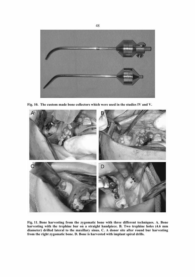
48
Fig. 10. The custom made bone collectors which were used in the studies IV and V.
Fig. 11. Bone harvesting from the zygomatic bone with three different techniques. A. Bone harvesting with the trephine bur on a straight handpiece. B. Two trephine holes (4.6 mm diameter) drilled lateral to the maxillary sinus. C. A donor site after round bur harvesting from the right zygomatic bone. D. Bone is harvested with implant spiral drills.

49
4.4 The volume of bone graft harvested from the zygomatic bone
4.4.1 Bone graft volume measurements in cadaver specimens
The zygomatic bone was harvested bilaterally from all cadavers, producing a total of 40 surgical sites. The numbers of harvested bone cores were recorded. All bone was stored in plastic bags, which were sealed and marked.
After weighing with a scale (Denver Instrument M-220D, Max 31 g, min 1 mg, d1=0.01mg), the bone graft samples were particulated with a small rongeur. The bone graft was placed into a 3 ml syringe and compressed with digital forces (Fig. 12). The volume of this measurement was recorded as Volume 1. A glass tube with 0.1 ml calibrations was used for water displacement volumetry. The bone graft was placed into the tube, and 3 ml of distilled water was added (Fig. 13). The result of this measurement was recorded as Volume 2.
After the bone graft harvesting, nine cadaver skulls (specimens 12-20) were examined with a CT scan. The entire zygomatic bone was evaluated using CT scans in three different planes in order to assess the completeness of bone harvesting.
Fig. 12. The particulated bone from the zygoma compressed into a syringe for volumetric measurement (Volume 1).

50
Fig. 13. The bone placed into a glass measure and 3 ml of distilled water added for displacement volumetric analysis (Volume 2).
4.4.2 Bone graft volume measurement in clinical operations
The volume of bone harvested from each zygoma was measured using a syringe (Fig. 14) or a scaled plastic cup. Bone from the implant preparation sites was also used for recipient site bone grafting but not included in the bone graft volume measurements.
Fig. 14. Particulated bone graft packed into a syringe for volumetric analysis. After the measurement the graft is injected on to gauze and prepared for grafting.
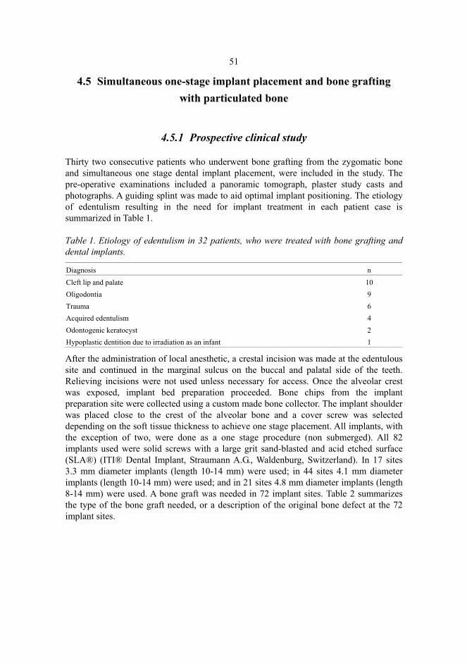
51
4.5 Simultaneous one-stage implant placement and bone grafting with particulated bone
4.5.1 Prospective clinical study
Thirty two consecutive patients who underwent bone grafting from the zygomatic bone and simultaneous one stage dental implant placement, were included in the study. The pre-operative examinations included a panoramic tomograph, plaster study casts and photographs. A guiding splint was made to aid optimal implant positioning. The etiology of edentulism resulting in the need for implant treatment in each patient case is summarized in Table 1.
Table 1. Etiology of edentulism in 32 patients, who were treated with bone grafting and dental implants.
Diagnosis n Cleft lip and palate 10 Oligodontia 9 Trauma 6 Acquired edentulism 4 Odontogenic keratocyst 2 Hypoplastic dentition due to irradiation as an infant 1
After the administration of local anesthetic, a crestal incision was made at the edentulous site and continued in the marginal sulcus on the buccal and palatal side of the teeth. Relieving incisions were not used unless necessary for access. Once the alveolar crest was exposed, implant bed preparation proceeded. Bone chips from the implant preparation site were collected using a custom made bone collector. The implant shoulder was placed close to the crest of the alveolar bone and a cover screw was selected depending on the soft tissue thickness to achieve one stage placement. All implants, with the exception of two, were done as a one stage procedure (non submerged). All 82 implants used were solid screws with a large grit sand-blasted and acid etched surface (SLA®) (ITI® Dental Implant, Straumann A.G., Waldenburg, Switzerland). In 17 sites 3.3 mm diameter implants (length 10-14 mm) were used; in 44 sites 4.1 mm diameter implants (length 10-14 mm) were used; and in 21 sites 4.8 mm diameter implants (length 8-14 mm) were used. A bone graft was needed in 72 implant sites. Table 2 summarizes the type of the bone graft needed, or a description of the original bone defect at the 72 implant sites.

52
Table 2. Bone graft use and recipient defects at 72 implant sites.
Description of recipient site n
Apical fenestration of implant 22 Apical fenestration and marginal palatal bone defect 4 Apical fenestration and gap in an implant socket 2 Lack of cancellous bone in an implant socket 1 Marginal buccal exposure of implant 29 Marginal buccal and palatal exposure of implant 3 Marginal buccal exposure and osteotome sinus augmentation 4 Osteotome sinus augmentation procedure 5 Sinus floor augmentation procedure 2
The bone graft collected from the zygomatic bone and implant osteotomies was dried between layers of gauze producing a mouldable particulate graft which could be adapted easily to irregularly shaped bony defects. Bony defects around the implants were covered with tightly packed bone chips. Marginal and apical defects were grafted if at least 3 mm of the implant’s threaded SLA® surface was exposed. In situations where the maxillary sinus was pneumatized and reduced the posterior maxillary alveolar height to 6-8 mm, the implant site was prepared with osteotomes (Straumann A.G., Waldenburg, Switzerland) for partial sinus floor elevation as described by Summers (Summers 1994c). The sinus floor was augmented with a particulated bone graft before implant insertion. In two cases where implant stability was not achieved primarily, the bone graft was used to fill the gap between the alveolar bone and implant and also to graft the apical fenestration. In one case where the implant socket had a lack of cancellous bone the bone graft was packed into the socket before implant insertion. In two implant sites where the maxillary sinus was pneumatized and reduced the alveolar height to less than 6 mm, a standard sinus floor augmentation through a lateral window approach with simultaneous one stage implant placement was performed (Boyne & James 1980, Lundgren et al. 1996). The implant sites were closed with resorbable interrupted sutures (Vicryl Rapid®, Ethicon, France).
A minimum of 4 months was permitted to elapse following placement before the implants were restored. During restoration, all implants were subjected to a torque of 35 Ncm using a hand-held torque wrench (Straumann A.G., Waldenburg, Switzerland). Panoramic tomographs and photographs were taken following restoration. Patients were followed after prosthodontic restoration for possible implant failures and other complications.
4.6 Statistical analysis
In study I the mean values for each drill and collecting sieve were calculated. In study II the mean values of the harvests were calculated and Analysis of Variance was used to analyze the mean harvests. The results were also evaluated with Tukey-HSD test with significance level 0.050.

53
In the bone graft volumetric measurements (study III) an arithmetic mean of volumes and weights were calculated for each cadaver of the harvests and measurements of the left and right sides (Volume 1, Volume 2, and weight of the bone harvests). The mean values of each cadaver were used to calculate the mean values and empirical standard deviations of the whole series. The measured dimensions of the zygomatic bone from cadaver operations and from CT scans were used to calculate the mean values and standard deviations.
In study V the mean values and the standard deviations were calculated from volumes of bone harvested from each zygoma. The mean values and standard deviations in study V were calculated for the duration of swelling and for the duration of the pain medication used.

5 Results
5.1 Development and comparison of bone collectors and bone harvesting drills
In study I using the finest-pore size, the sieve clogged quickly in approximately of 45 seconds with bone powder and chips, and suction became impossible. The mean weights of three consecutive harvests of bone chips over periods of about 45 seconds varied from 0.71 to 1.25 g, using various burs. Fewest bone chips during 60 seconds of drilling were found on the sieve with the largest holes. The mean harvest varied between 0.14 and 0.59 g. Most bone chips were obtained with the sieve with 0.50 mm diameter holes. The mean harvest using this sieve with different drills was 1.27 to 1.64 g. The difference between drills was small but HM 77 SX 060 and HM 77 G 070 were the best in this study. Drills HM 77 GX 060 and 77 080 were also quite effective, especially when the sieve with 0.50 mm diameter mesh was used (Fig. 15).
Bone chips obtained with drill HM 77 SX 060 ranged in diameter from 400 to 500 µm, those using bur HM 77 GX from 200 to 300 µm, those using bur HM 77 G 070 from 300 to 400 µm, and those using bur 77 080 from 200 to 400 µm.
In study II the results of five test rounds is displayed in Figure 16, showing the mean harvests of each collector. The gain with the OCT® (Quality Aspirators, Duncanville, Texas, USA) ranged between 0.85 to 1.25 g (mean 1.03 g), but the sieve became quite easily blocked and suction became impossible. In two test rounds the OCT® collector became clogged in 20 seconds. The mean weight of five consecutive bone harvests with the Frios® (Friatec, Mannheim, Germany) was 0.84 g. The Frios® became blocked four times during 30 seconds of drilling. The harvests ranged from 0.35 to 1.45 g.
Using the sieve with smaller diameter pores (0.36 mm) in Model I, the mean harvest was 1.47 g. Harvests ranged from 1.18 to 1.98 g. With this sieve the bone collector clogged once during the tests. Most chips were obtained with Model I with the larger pore diameter (0.50 mm). The mean harvest with this sieve was 1.48 g and harvests ranged from 1.12 to 1.88 g. The mean harvest with Model II was 1.06 g and the range was from 0.58 to 1.64 g.

55
Analysis of variance shows the differences between the different collectors. The mean values of the Model I (with large and small net) and the Frios® bone collector differs significantly by the Tukey-HSD test. The other collectors can not be ranked by this test.
Fig. 15. Mean harvests of bone chips using different burs and sieves (Study I). Each bar is the mean of three tests.
Fig. 16. The results of the tests are shown with rings and the mean of the five rounds are marked with a line (Study II).

56
5.2 Safety and morbidity of intra-oral zygomatic bone harvesting procedure
5.2.1 Cadaver study
5.2.1.1 Pre-operative CT scan and intra-operative measurements
Study III permitted the following measurements from the cadaver model. The average clinical height of the zygomatic bone at the level of the trephine holes was 22.5 mm ± 2.4, and the distance from the trephine holes to the orbital rim was 6.3 mm ± 0.9 and that to the inferior zygomatic rim 4.4 mm ± 0.7 (Specimen 1-20). The height of the zygoma in the CT-scanned specimens was 21.2 mm ± 2.7. The mean length and thickness of the zygoma in the CT scans were 32.2 mm ± 4.6 and 9 mm ± 1.7, respectively (Specimen 12-20) (Table 3).
Table 3. Mean dimensions of zygomatic bone as measured clinically in 20 cadavers (40 sites) and with CT scans in 9 cadavers (18 sites).
Measurement direction Clinical measurement (n=20) CT measurement (n=9) Height 22.5 mm ± 2.4 21.2 mm ± 2.7 Thickness - 9 mm ± 1.7 Length - 32.2 mm ± 4.6 mm
5.2.1.2 Post-operative clinical observations and CT scan measurements
In study III clinically observed perforations into the maxillary sinus during zygomatic bone harvesting occurred at 15 of the 40 sites, and this was confirmed with both post-operative CT scans (18 sites, specimens 12-20) and clinical examinations (40 sites, specimens 1-20). No perforations through the orbital floor were noted. The average distance from the zygomatic donor site to the orbital floor was 5.6 mm ± 1.9, as measured from CT scans. Seven minor perforations into the infratemporal fossa occurred during curettage, but none of these perforations penetrated through the periosteum (Table 4). The CT scans at two zygomatic sites showed the distance from the donor site to the zygomaticofacial nerve to be 1-2 mm. The authors felt that a neurosensory deficit could be caused by harvesting bone in such close proximity to the zygomaticofacial nerve. Safe margins to the infraorbital nerve were preserved in all cases (Table 5).

57
Table 4. Perforations into neighboring structures during harvesting of trephine cores. Analysis of clinical observations and CT scans in study III.
Location Perforation Percentage without perforation Maxillary sinus 15 62.5 % Infratemporal fossa 7 82.5 % Orbit 0 100 %
Table 5. Damage to the infraorbital (ION) and zygomaticofacial nerves (ZFN) during the harvesting of trephine cores from the zygoma. The analysis was made from CT scans (n=18 sites).
Nerve Potential nerve damage Percentage without potential nerve damage ION 0 100 % ZFN 2 89 %
5.2.2 Clinical study
Study IV introduced the clinical technique in three patients and the results were favourable enough to stimulate further prospective research. A total of 33 zygomatic donor sites were used in 32 patients in study V. In one case bone was harvested bilaterally from both zygomas to perform bilateral sinus floor augmentation procedures. The only intra-operative complication encountered in this study group was perforation of the maxillary sinus, which occurred in 11 zygomas (33 %). The diameter of the perforations ranged from 2 to 5 mm. None of these patients experienced any post-operative problems due to perforation. Sinus perforation occurred in both of the two zygomas (100 %) in which a trephine bur was used for harvesting, in 8 of the 22 zygomas (36 %) in which a round bur was used for harvesting, and in 1 of the 9 zygomas (11 %) in which implant spiral drills were used for harvesting. Patients who underwent zygomatic bone harvesting under local anesthesia tolerated the procedure well, and none of them experienced any pain intra-operatively.
At the first follow-up visit, the sensory nerve function in the distribution of the infraorbital and zygomaticofacial nerves was normal in all patients. Eight of the patients (25 %) developed post-operative bruising over the donor site area. The mean duration of post-operative swelling as reported by the patients was 4.5 days (range: 0 to 12 days; SD 2.34). Post-operative pain medication was used for a mean duration of 4 days (range: 1 to 9 days; SD 2.08). There were no post-operative infections in any of the bone graft recipient or donor sites. After 3-4 weeks there was no soreness of the donor sites when the zygoma area was palpated extra-orally, and the donor defect was not palpable.

58
5.3 The volume of bone graft harvested from the zygomatic bone
5.3.1 Cadaver study
The average number of harvested trephine cores at each of the 40 sites was 3.2 ± 1.2. The volume of the harvested zygomatic bone ranged from 0.2 to 1.4 ml (Volume 1 = volume measured with the syringe) and from 0.2 to 1.3 ml (Volume 2 = volume measured with the water displacement method). The average volumes were 0.59 ml and 0.53 ml, respectively. The weight of the graft ranged from 0.32 to 1.72 g, and the average weight was 0.72 g. The volumes and weights of the bone harvested from the zygomatic bone are listed in Table 6.
The CT scans showed that it would have been possible to harvest more bone from 12 of the 18 zygomatic donor sites. The residual bone for harvesting was usually available in the inferior part of the zygoma.
5.3.2 Clinical study
Together with relatively minimal contribution from the implant osteotomies, the zygoma yielded sufficient quantities of bone to complete the required reconstructions in all 32 cases. The average zygomatic bone graft volume was 0.90 ml (SD 0.30) (Table 6). The graft volumes ranged between 0.5 to 1.5 ml from each zygomatic donor sites.
Table 6. Volumes and weights of harvests from zygomatic bone in cadaver and clinical study (mean=x, standard deviation=SD, Volume 1=measured with the syringe, Volume 2=water displacement method).
Mean ± SD Clinical bone graft volume
Cadaver bone graft (Volume 1)
Cadaver bone graft (Volume 2)
Cadaver bone graft weight
X ± SD 0.90 ml ± 0.30 0.59 ml ± 0.25 0.53 ml ± 0.26 0.72 g ± 0.32
5.4 Survival of dental implants with simultaneously placed particulated bone grafts
Zygomatic bone grafting was employed at 72 of the 82 implant sites in this series. Three of the grafted implants lacked primary stability. At restoration, two of the three implants lacking primary stability were found to be loose. Success rates of the implants are summarized in Table 7. The mean follow-up time after the surgery was 26.9 months (18-30 months) and after the prosthetic loading was 19.4 months (11-23 months). During follow-up time there were no other implant losses.

59
Grafted sites healed remarkably well without signs of irritation from the bone graft to the soft tissues, and no obvious signs of graft resorption were noted during the follow-up period.
Table 7. Survival rates of the dental implants after the mean follow-up time of 19.4 months (range: 11 to 23 months) after prosthetic loading.
Implants Osseointegrated implants (%) Failed implants (%)
Implants with grafts (n=72) 70 (97.2 %) 2 (2.8 %) Implants not requiring grafts (n=10) 10 (100 %) 0 (0 %) Total (n=82) 80 (97.6 %) 2 (2.4%)

6 Discussion
6.1 Development and comparison of bone collectors and bone harvesting drills
The main object of testing bone collectors with different sieves and drills was to find an optimal combination of drill and bone collector to collect bone from the intra-oral donor sites. The bone slurry from the collector can be used for example in sinus floor and alveolar augmentation procedures (Boyne & James 1980, Lundgren et al. 1996, Widmark & Ivanoff 2000). Harvesting of such grafts from intra-oral donor sites has several advantages. The graft is derived from membranous bone, which may not resorb as quickly as endochondral bone grafts (Koole et al. 1989, Sindet-Pedersen & Enemark 1990, Scott & Hightower 1991, Manson 1994). Harvesting bone from the extra-oral donor sites such as iliac crest has several disadvantages compared to intra-oral bone harvesting. Pain, delay in ambulation, blood loss, hematoma, seroma and paresthesia have been reported and harvesting bone from the iliac crest requires general anesthesia (Marx & Morales 1988, Sindet-Pedersen & Enemark 1990). Intra-oral bone graft harvesting can be performed under local anaesthesia and complications and post-operative discomfort as seen after harvesting from distant sites such as the iliac crest do not occur (Sindet-Pedersen & Enemark 1990, Lundgren et al. 1996, Misch 1997, Clavero & Lundgren 2003). The nature and the origin of the bone graft may also have some influence on the graft survival. Lundgren used particulated bone harvested from the mandibular symphysis for maxillary sinus floor augmentation. They found an increased net volume fraction of bone over a healing period of 6 months without loading of the implants. They hypothesized that particulation of the graft is also a factor with a positive influence on the preservation of the graft (Lundgren et al. 1996).
The results of these studies show that bone chips can effectively be harvested using bone collectors. The bone collectors are connected to a conventional suction device and they can be re-sterilized. In study I the best harvest was obtained using the sieve with a medium-sized mesh (0.5 × 0.5 mm). The sieve with the finest mesh (0.355 × 0.355 mm) can be recommended when small amounts of bone chips are needed, for example to fill

61
space under a guided bone regeneration membrane. The sieve with the finest pore size can also be used to collect bone chips from the implant osteotomies.
The drills tested in study I produced relatively similar amounts of bone chips with the different sieves. The drill HM 77 SX 060 (Meisinger, Germany) produced the most bone chips with the tested sieves. This drill is made of hard metal and its cutting blades are bigger than in the other tested drills. The histology revealed that the mean size of bone chips with this drill were larger (400-500 µm) than with the other drills. The drill HM 77SX 060 was therefore chosen for use in the study II.
In study II the same bone collector as described in study I was used. The best combination of stainless steel sieves and drill were used in a study where this Model I was compared to two commercially available bone collectors. Also a new custom made model with a different chamber design and a cone shaped sieve (Model II) was included in the study. Comparison of four different bone collectors in an in vitro study showed that all collectors are usable in clinical situations but their effectiveness varies. The OCT® collected bone chips and powder effectively, because the slots in its sieve are very small. The problem with this collector was that it became clogged quite easily. The best indication for the OCT® could be during implant bed preparation and when only a small amount of bone is needed.
The Frios® collected bone chips quite effectively but it also became clogged quickly. The advantage of this collector is that the collected bone can easily be removed from the sieve and the collector can be quickly reconnected. The Frios® is easy to use but the disposable filter makes its use more expensive than the use of custom made models whose filters can be re-sterilized.
One potential problem occurs when bone is harvested, and there is concomitant bleeding in the operative field. Suctioning blood will clog some bone collectors fairly quickly and bone procurement becomes minimal (Young et al. 2002b). Young et al. have tested the Frios® and the OCT® in a clinical study which involved 38 patients. The patients underwent bone collection with the Frios® bone collector or the OCT® according to a randomisation sequence. During surgery, the Frios® bone trap became blocked once and the OCT® 11 times. In all cases where blockage occurred, excess blood coagulum was apparent. All the samples that were collected by the Frios® bone trap contained bone and coagulum, with a mean proportion of 90.6% bone. With regard to the OCT®, one sample contained no bone and two samples contained only trace amounts of bone; the remaining samples contained a mean proportion of 67.3% bone. The pore size affects both clinical performance and the histological composition of the debris collected, and this might have important implications if the trapped debris is used as an augmentation material (Young et al. 2002b). In study II there was no blood in the operative field but still the OCT® was found to become blocked quickly. The Frios® also became blocked quickly in this study (study II), probably because the volume of the harvested bone was fairly large.
During intra-oral bone harvesting there is always a risk for bacterial contamination of the graft and especially with these suction connected bone collectors it is possible to contaminate the graft by aspirating saliva during the harvesting procedure. The contamination from saliva does not necessarily affect bone graft survival but it should be avoided. During the bone harvesting it is advisable to have a second suction line available for cleaning the mouth of saliva and blood (Young et al. 2001, 2002a, Blay et al. 2003).

62
Pre-operative chlorhexidine mouthrinse has been shown to reduce the yield of bacterial contaminants in bone graft collected with bone collectors (Young et al. 2002a).
The main indication for the use of these bone collectors is the harvesting of bone chips for dental implant related surgery. Preparation of the implant bed and simultaneous alveoloplasty produce bone chips which are produced under ideal conditions, with copious irrigation and sharp burs. The amounts of bone obtained with these collectors make their use favourable for smaller bone grafting operations, for example alveolar ridge augmentation, sinus floor augmentation and to cover exposed implant surfaces (Haessler et al. 1995, Widmark & Ivanoff 2000, Blay et al. 2003). If more bone is needed harvesting can be repeated several times at different donor sites. The amount of bone obtained in study I during one minute drilling was estimated to be sufficient for a unilateral sinus floor augmentation.
Model I with two different nets was the most effective collector in the in vitro studies. The volume of the chamber in this collector is bigger compared to the commercial ones and the sieve diameter is larger with a choice of two sizes. Model I can probably aspirate irrigation and bone chips with the same efficiency during the whole test period. In Model II the slot diameter in the sieve are too large (0.8 × 0.8 mm) and only the bigger bone chips and spicules are harvested. Model II could be usable in harvesting bone from the iliac crest using a minimally invasive approach.
After these in vitro studies the custom made collectors were developed further and they were used in the later parts of this study. The custom made collector, used in a prospective clinical study (V) (Fig. 17), have the same sieve pore size as in Model I, 0.50 mm and 0.355 mm, and these sieves can be used as separate sieves or stacked in combination at the same time inside the collector. The chamber size is also bigger in the revised collector used in study V than in Model I in studies I and II. The sieve used in study V was also modified to be bowl shaped.
Fig. 17. The bone collector opened and showing two bowl-shaped collecting sieves with different pore sizes (0.50 and 0.355 mm). The sieves can be used in a stacked arrangement in the collector at the same time.

63
6.2 Intra-oral zygomatic bone graft harvesting
To the author’s knowledge, the use of zygomatic bone as donor tissue for alveolar bone grafting has not been previously reported. In 1985 Wolford and Cooper described a technique to harvest a cortical bone block from the zygomatic eminence and arch during Le Fort I osteotomy. They found it to be easy to harvest a 1 cm by 1.5 cm graft from this area without an untoward esthetic or functional deficit in the donor site (Wolford & Cooper 1985).
As noted in studies IV and V the surgical approach to the zygomatic bone area is rather simple thru a short incision of the maxillary vestibule. The surgeon must identify the opening of the parotid duct and during dissection the buccal fat pad should be avoided. A round bur can be used to harvest bone by initially grinding on the most lateral portion of the anterior maxillary wall. Once the sinus border is identified by trimming the bone of the anterior maxillary wall down to it, bone harvesting can be continued lateral to the maxillary sinus. A fibre optic light could be useful as a guide in identifying the maxillary sinus, when this procedure is performed clinically, although no such light was used in this study.
In cadavers the bone harvesting was done with the trephine drill (Nobel Biocare, Gothenburg, Sweden) and the same drill was used in the first two clinical operations. In both of these operations, the trephine drill created small perforations to the maxillary sinus. Even though the perforations did not cause any post-operative problems for the patients the harvesting technique was modified. After the first two patients, the bone harvesting was accomplished with round burs. The round bur harvesting was found easier than trephine drill harvesting, especially in cases where the zygomatic bone was smaller in size. The third technique for bone graft harvesting in this study was to use implant spiral drills. These drills seem to produce larger diameter bone chips, which are efficiently collected with the bone collector. Also the spiral drills produced fewer perforations to the maxillary sinus than the other two techniques.
6.3 Safety and morbidity of zygomatic bone harvesting
The safety and complications of harvesting bone material from the zygomatic bone were evaluated in study III and V. Complications during the cadaver harvesting at the 40 sites included 15 perforations into the maxillary sinus and 7 perforations into the infratemporal fossa. All perforations into the maxillary sinus and the infratemporal fossa were less than 5 mm in diameter. In the clinical study, the perforation to the maxillary sinus occurred in 33 % of the donor sites. During the maxillary osteotomies large perforations of the maxillary sinus occurs and these usually heal without problems. Perforation of the sinus membrane during sinus floor augmentation generally does not cause infection if proper antibiotic coverage is used (van den Bergh et al. 1998, Raghoebar et al. 1999).
Nine cadaver skulls could be analysed with pre- and post-operative CT scans. None of the infratemporal fossa perforations breached the periosteum, making potential haemorrhage from this area unlikely. The infraorbital and zygomaticofacial nerves were intact on all scanned specimens. The zygomaticotemporal nerve area was also examined

64
from the CT scans, but this nerve is located so superiorly that its injury is unlikely with this harvesting approach. The distance from the donor area to the zygomaticofacial nerve, which might be most susceptible to damage, was over 5 mm in most cases. At two donor sites, however, the distance to the zygomaticofacial nerve was only 1-2 mm. These two sites were therefore judged to involve a risk for injury. The zygomaticofacial nerve is a sensory nerve, and damage to it could result in paresthesia of the malar skin area. No perforations through the orbital floor occurred, and the average distance between the donor site and the orbital floor was 5.6 mm.
The prospective clinical study (study V) examined intra-operative complications, donor site morbidity, and patient discomfort during the post-operative healing phase after simultaneous bone grafting with zygomatic bone and dental implant placement. Patients needed pain medication for a mean time of four days, and generally, it was difficult for them to decide which of the sites was more painful, the donor or recipient site. The usual finding after surgery was moderate swelling which resolved with a mean duration of 4.5 days. Patients did not demonstrate any paresthesias or altered sensations in the donor site area.
The mandibular symphysis is the largest intra-oral donor site, but the major disadvantage with its use is the potential for post-operative altered sensation of the teeth and chin area. In a recent study 43 % of the patients (n=21) reported a decreased sensibility in the symphyseal donor site. In 7 patients the paresthesia persisted, and 4 of these 7 patients also reported meteorotropism (weather-dependent discomfort) (Raghoebar et al. 2001a). Nkenke et al. confirmed these findings in a prospective study in which 8 of 20 patients had sensory disturbances on the chin area one month following chin bone harvesting. Sensory disturbances resolved in most cases during 12 months of follow-up, but in one patient paresthesia, and in two patients hypoaesthesia and hypalgesia, remained until the twelfth month (Nkenke et al. 2001). Clavero and Lundgren had similar results in the mandibular symphysis donor area. They compared mandibular symphysis and ramus areas and found much less morbidity and complications in the mandibular ramus donor sites (Clavero and Lundgren 2003). The mandibular ramus area has been shown to be a reliable source of cortical bone and fairly large defects can be grafted with bone blocks harvested from the ramus with minimal morbidity and few complications (Misch 1997, Nkenke et al. 2002, Clavero & Lundgren 2003).
Possible complications in harvesting ascending ramus bone are the injuries to molar teeth and mandibular fracture. It is possible to harvest a bicortical bone block from the mandibular symphysis but it cannot be recommended, because this may cause more post-operative complications including excessive bleeding, fracture of the mandible and sleep apnea by loss of the genioglossus muscle insertion. Although these complications have not been reported, it must not be forgotten that such injuries may occur during the operation or the post-operative period. These problems can be minimized if the surgeon has a clear understanding of mandibular anatomy.
According to the results of studies III and V the complications and morbidity are minimal after zygomatic bone harvesting and the zygomatic bone can be regarded as a safe intra-oral bone harvesting donor site. The only intra-operative complication in this clinical study was perforation to maxillary sinus in 33 % of the zygomatic sites and none of these patients experienced any post-operative problems. The safest technique with respect to the maxillary sinus perforation was bone harvesting with implant spiral drills.

65
When implants are placed in the maxilla and bone grafting is needed, it is more convenient and potentially less morbid harvest the graft from the neighbouring zygomatic area than, for example, from the mandible.
6.4 The volume of bone harvested from the zygomatic bone
In the cadaver model used in this study, the average yield of bone harvested at the zygomatic site was measured to be 0.59 ml (Volume 1 = measured with the syringe) and 0.53 ml (Volume 2 = measured with the water displacement method). In a clinical study the average zygomatic bone graft volume was 0.90 ml. Previous studies have not quantified the bone volumes required to reconstruct alveolar defects prior to or simultaneously with the placement of dental implants. Using a hypothetical implant site, it was assumed that the alveolar bone required to house an implant fixture (3.5-4.2 mm in diameter) should be 7 mm in mesio-distal length and 6 mm in bucco-lingual width. Although the length of an implant can be chosen to vary within 6-14 mm, the implant needed for a single tooth restoration could be assumed to be 10 mm in length. For the purposes of this study, a small defect was defined as a single tooth area with a narrow ridge with or without adequate ridge height. The maximum dimensions of this defect were assumed to be 7 mm in length, 5 mm in width and 12 mm in height. Based on these estimated dimensions, the authors calculated that a bone graft volume of 0.42 ml would be needed to fill a small defect (0.7 cm x 0.5 cm x 1.2 cm = 0.42 cm3 = 0.42 ml). A medium-sized defect was defined as having the length of 2 to 3 teeth. The dimensions of this defect were assumed to be 14 to 21 mm in length, 5 mm in width and 12 mm in height. This corresponds to a bone volume of 0.84-1.26 ml.
Volumes 1 and 2, in the cadaver study III are close to the volume required to fill a small alveolar defect. The largest volume in the range of harvested sites was 1.4 ml, which could fill even medium-sized defects. Eleven out of the 40 harvested sites in study III produced more than 0.8 ml of bone and would fill the defects of two implant sites. In the clinical part of the study (studies IV and V), the zygoma yielded sufficient quantities of bone to complete the required reconstructions in all 32 patient cases. The mean volume of the zygomatic bone harvest was 0.9 ml (SD 0.30). In the study group a 0.9 ml graft was sufficient to reconstruct the alveolar defects around 2-3 implants. Bilateral sinus floor augmentation for one implant on each side required 1.5 ml of bone from each zygoma in addition to 0.6 ml of bone from implant osteotomies.
The zygomatic bone is a rather large donor site. The measurements made during the surgical procedures and from the CT scans indicate that the average thickness of the zygomatic bone was 9 mm, ranging from 6 to 12 mm. The average length of the zygomatic bone was 32.2 mm (CT scan) and the average height 21.2 mm (CT scan) or 22.5 mm (clinical measurement). In a study by Nkenke et al. the anatomy of zygomatic bone was evaluated for zygomaticus implant placement. Thirty cadaver zygomatic bone specimens were examined by quantitative CT scans. The average medio-lateral thickness was less pronounced for the female group (7.60 mm) than in male group (8.00 mm), which corresponds well to the current study. The average anterior-posterior length was about 25 mm. The zygomatic bones in this study were separated from the facial skeleton

66
and this probably accounts for the difference between Nkenke´s study group and the group in the current study (study III) (Nkenke et al. 2003).
Residual, non-harvested bone at the cadaver donor sites was estimated from CT scans, to see if it would have been possible to harvest more bone without damaging the surrounding tissues. Twelve out of eighteen sites showed that larger harvests could have been possible. The remaining bone was found mostly in the inferior part of the zygoma. For technical reasons, the authors were not able to use a suction-operated bone collector during the cadaver harvesting procedures. It was estimated, however, that at least 20-25 % more bone could have been harvested by using suction traps during the drilling and curettage.
The cadavers used in this study were from an elderly population being moderately atrophic, and their bone exhibited early signs of deterioration. The bone harvesting surgeries were done during two sessions, and the yield from the zygomatic bone harvest sites was remarkably better during the second session (specimens 12-20). The average Volume 1 of the first session (specimens 1-11) was 0.44 ml and that of the second session 0.76 ml. This can be explained by the researchers’ learning curve. In addition, the quality of the cadaver specimens was better in the second session.
In a clinical study by Misch the average volume of a mandibular symphysis graft was 1.74 ml, and a retromolar graft was 0.9 ml (Misch 1997). The average bone volume from the zygoma of 0.9 ml (study V) compares favorably to a retromolar bone graft, and is roughly half the volume of the symphysis. Montazem and co-workers measured the quantity of bone in the mandibular symphysis in dentate adult cadavers. The average monocortical bone harvest was 4.84 ml when a 5 mm safety margin from the donor site to the tooth apices, the mental foramen and the inferior border of the mandible was maintained (Montazem et al. 2000). In another study, 26 mandibles with mixed dentitions were examined on CT scans to assess the volume of the donor site in the mandibular symphysis. The quantification gave an average bone volume of 1 ml, if the buccal and lingual cortices were included (Bähr & Coulon 1996). Because the amount of bone harvested from the zygoma is less than from mandibular donor sites, the mandibular donor sites can be used in larger alveolar reconstructions and zygoma used in cases where only moderate amounts of bone are needed.
The cadaver study showed that yield of the bone graft from zygomatic bone is sufficient to reconstruct an alveolar bone defect of at least one or two dental implant sites. This finding was confirmed in a clinical part of this project (studies IV and V). The quantity of harvested zygomatic bone grafts was sufficient in all patient cases to allow the placement of all the planned dental implants.
6.5 Survival of dental implants with simultaneously placed particulated bone grafts
In this study all the defects in the alveolar bone and around dental implants were too large for grafting only with the bone chips from the implant preparation sites. Two of the bone grafted implants failed, yielding a survival rate of 97.2 % for bone grafted implants and 97.6 % for the whole study group. Both of the failed implants lacked primary stability at

67
the time of placement. One of the failed implants had been placed in an edentulous maxillary site using an osteotome sinus floor augmentation. The five remaining implants placed in this patient’s maxilla osseointegrated succesfully, and the prosthodontic treatment was carried out as planned. The other failed implant had been placed into a bone grafted mandibular implant socket lacking in cancellous bone. The failure in osseointegration was noted 12 weeks post-operatively. That site was re-implanted six weeks after the removal of the failed implant and was successfully restored with a crown. There were no other implant losses during the mean follow-up time of 19.4 months after prosthetic loading.
When a particulated bone graft is used, the bone should be packed firmly into to the defect. The graft is stable when blood has created a clot type adhesion to the graft. The incision is easy to close without tension, because the bone chips adapts by the pressure of the overlying soft tissues. The particulated bone is supported by the walls of the bony defect and the underlying dental implants. In this study barrier membranes were not used and the grafts were covered with intact periosteum. Good results with similar techniques have been presented (Widmark & Ivanoff 2000, Blay et al. 2003). Widmark and Ivanoff covered exposed implant surfaces with bone chips from a bone collector without a membrane. The mean bone gain was 81 % in patients with a marginal defect and 82 % in patients with a fenestration defect (Widmark & Ivanoff 2000). Blay confirmed these findings in their study (Blay et al. 2003).
Grafted implant sites healed remarkably well without signs of irritation of the soft tissues, and no obvious signs of graft resorption were noted during the follow-up period. Due to a study design to use bone grafts simultaneously with one-stage dental implants, it was not possible to monitor healing of the bone graft using a second-stage implant exposure or re-entry operation. The post-operative evaluation of the bone grafts was done by palpating the grafted areas. Good healing and bone graft incorporation of exposed implant surfaces has been shown previously with a two-stage technique (Widmark & Ivanoff 2000, Blay et al. 2003).
The clinical data presented show that particulated bone grafts harvested from zygomatic bone and used with simultaneously placed one stage dental implants are an effective and safe method of treating resorbed edentulous alveolar ridges in partially edentulous patients.
6.6 Clinical implications and future prospects
The results of the use of particulated bone grafts in this study were promising. Bone augmentation with a particulated bone graft is a reliable technique when there is a defect with supporting bony walls, for example in the sinus floor augmentation. The results have been good when the exposed implant surface is covered with a particulated bone graft from a suction trap. The surface of a dental implant stabilizes the graft material and might enhance its healing potential (Widmark & Ivanoff 2000, Blay et al. 2003). When a particulated bone graft is used in a defect without supporting bony walls and simultaneous implant placement, the graft is occasionally seen to resorb during the healing period. This has been noted in some patient cases treated after the present study

68
and it might be advisable to cover the particulated graft with a GBR membrane. A further study, in which a particulated bone graft is covered with a resorbable collagen membrane, has already been started. This two-stage approach will allow to monitor the healing and resorption rate of the bone graft.
The harvested graft material from the zygomatic bone is corticocancellous in nature but most of its proportion is cortical bone. Lindholm et al. have shown that such a cortical graft from the zygoma includes living cells and it is possible to grow vital bone cells in culture media in vitro (Lindholm et al. 2003). Corticocancellous bone chips from a bone collector have also been shown to be able to preserve vital bone cells with osteogenic capacity (Blay et al. 2003). The future scenario would be to preserve some bone cells with a bone collector from an oral surgical procedure, such as wisdom tooth extraction, and culture and storage the graft for the possible future use. This would make it possible to do a bone grafting procedure later with a cultured autogenous bone graft which will be both osteogeneic and osteoconductive, and also immunologically safe. In the study of Lindholm et al. it was noted that if the bone graft was harvested from an implant osteotomy site the bacteria in the bone slurry destroyed the bone cell culture. The graft obtained from the zygomatic bone through a “keyhole” approach did not have similar infection associated problems and the bone cell culturing was more uneventful (Lindholm et al. 2003). Other studies have also noted that when harvesting a bone graft for this kind of purposes it is crucial to keep the saliva out of the harvested graft by using two suction lines (Young et al. 2001, Blay et al. 2003).
The bone collectors used in clinical studies have been modified from the models used in studies I and II. The most recent collector is equipt with a valve, which can close the suction through the collector. This shut-off valve may influence bone cell survival because the graft is not desiccated by continuous airflow (Blay et al. 2003). A further study concerning the efficiency of the latest bone collector models used in studies IV and V has been completed and submitted for publication. The aim of that particular study was to compare the performance of two custom made bone collectors and four other already available bone collectors.
The low morbidity of intra-oral bone harvesting compared to extra-oral donor sites has been well described (Sindet-Pedersen & Enemark 1990, Jensen & Sindet-Pedersen 1991). The complications and morbidity of the mandibular symphysis over the other intra-oral donor sites have been highlighted (Raghoebar et al. 2001a, Nkenke et al. 2001, Clavero & Lundgren 2003), although some other opinions exist (Cordaro et al. 2002, Cordaro 2003). From the current literature it can be stated, that the mandibular ramus and retromolar area is probably the best choice of intra-oral donor sites for alveolar augmentation performed with cortical block grafts. With regards to the current study, it shows that the zygomatic bone is a safe donor site with minimal morbidity, convenient surgical access and reliable amount of bone, when a corticocancellous particulated bone graft is needed in dental implant related surgery.

7 Summary and conclusions
The following conclusions can be made from the results of the present studies: 1. The bone collectors used and tested in these studies are suitable and efficient in
dental implant related bone grafting surgery. The bone collectors designed during these studies are more efficient and they have a larger capacity with less blockage during bone harvesting when compared to the two other bone collectors.
2. A new bone graft harvesting donor site, the zygomatic bone, has been introduced in this study. A new intra-oral bone harvesting technique was found suitable for dental implant related bone grafting surgery.
3. According to these studies the complications and morbidity are minimal after zygomatic bone harvesting. The zygomatic bone can be regarded as a safe intra-oral bone harvesting donor site.
4. The quantity of bone harvested from the zygomatic bone was found sufficient to augment a deficient alveolar ridge of one to three dental implant sites. In the clinical study the yield of the zygomatic bone graft was adequate in all 32 patient cases.
5. The clinical data presented show that particulated bone grafts harvested from the zygomatic bone and used with simultaneously placed one stage dental implants are an effective and safe method of treating resorbed edentulous alveolar ridges in partially edentulous patients.

References
Adell R, Lekholm U, Grondahl K, Branemark PI, Lindstrom J & Jacobsson M (1990) Reconstruction of severely resorbed edentulous maxillae using osseointegrated fixtures in immediate autogenous bone grafts. Int J Oral Maxillofac Implants 5: 233-246.
Alberius P, Dahlin C & Linde A (1992) Role of osteopromotion in experimental bone grafting to the skull: a study in adult rats using a membrane technique. J Oral Maxillofac Surg 50: 829-834.
Albrektsson T & Albrektsson B (1978) Microcirculation in grafted bone. A chamber technique for vital microscopy of rabbit bone transplants. Acta Orthop Scand 49: 1-7.
Albrektsson T (1980a) The healing of autologous bone grafts after varying degrees of surgical trauma. A microscopic and histochemical study in the rabbit. J Bone Joint Surg Br 62: 403-410.
Albrektsson T (1980b) In vivo studies of bone grafts. The possibility of vascular anastomoses in healing bone. Acta Orthop Scand 51: 9-17.
Albrektsson T (1980c) Repair of bone grafts. A vital microscopic and histological investigation in the rabbit. Scand J Plast Reconstr Surg 14: 1-12.
von Arx T, Hardt N & Wallkamm B (1996) The TIME technique: a new method for localized alveolar ridge augmentation prior to placement of dental implants. Int J Oral Maxillofac Implants 11: 387-394.
Atwood DA (1971) Reduction of residual ridges: A major oral disease entity. J Prosthet Dent 26: 266-279.
Bagatin M (1987) Reconstruction of orbital defects with autogenous bone from mandibular symphysis. J Craniomaxillofac Surg 15: 103-105.
Bassett CAL, Creighton DK & Stinchfield FE (1961) Contributions of endosteum, cortex and soft tissues to osteogenesis. Surg Gynecol Obstet 112: 145.
Becker W, Dahlin C, Lekholm U, Bergstrom C, van Steenberghe D, Higuchi K & Becker BE (1999) Five-year evaluation of implants placed at extraction and with dehiscences and fenestration defects augmented with ePTFE membranes: results from a prospective multicenter study. Clin Implant Dent Relat Res 1: 27-32.
Bedrossian E, Tawfilis A & Alijanian A (2000) Veneer grafting: a technique for augmentation of the resorbed alveolus prior to implant placement. A clinical report. Int J Oral Maxillofac Implants 15: 853-858.
Berarducci JP & Joseph RW (1994) A bone collection suction device for use during endosseous implant placement. J Oral Maxillofac Surg 52: 1090-1091.
van den Bergh JP, ten Bruggenkate CM, Krekeler G & Tuinzing DB (1998) Sinus floor elevation and grafting with autogenous iliac crest bone. Clin Oral Implants Res 9: 429-435.
Berry RL, Edwards RC & Paxton MC (1994) Nasal augmentation using the mandibular coronoid as an autogenous graft: report of case. J Oral Maxillofac Surg 52: 633-688.
Blay A, Tunchel S & Sendyk WR (2003) Viability of autogenous bone grafts obtained by using bone collectors: histological and microbiological study. Pesqui Odontol Bras 17: 234-240.

71Block MS & Kent JN (1997) Sinus augmentation for dental implants: the use of autogenous bone. J
Oral Maxillofac Surg 55: 1281-1286. Block MS, Kent JN, Kallukaran FU, Thunthy K & Weinberg R. (1998) Bone maintenance 5 to 10
years after sinus grafting. J Oral Maxillofac Surg 56: 706-714. Bonutti PM, Cremens MJ & Miller BG (1998) Formation of structural grafts from cancellous bone
fragments. Am J Orthop 27: 499-502. Borstlap WA, Heidbuchel KLWM, Freihofer HPM & Kuijpers-Jagtman A-M (1990) Early
secondary bone grafting of alveolar cleft defects. J Craniomaxillofac Surg 18: 201- . Boyne PJ & James RA (1980) Grafting of the maxillary sinus floor with autogenous marrow and
bone. J Oral Surg 38: 613-616. Boyne PJ (1992) Bone grafts and bone substitutes. 291-298. In: Habal MB, Reddi AH.
Maxillofacial surgery. Philadelphia, WB Saunders Company. Breine U & Branemark PI (1980) Reconstruction of alveolar jaw bone. An experimental and
clinical study of immediate and preformed autologous bone grafts in combination with osseointegrated implants. Scand J Plast Reconstr Surg 14: 23-48.
ten Bruggenkate CM, Kraaijenhagen HA, van der Kwast WA, Krekeler G & Oosterbeek HS (1992) Autogenous maxillary bone grafts in conjunction with placement of I.T.I. endosseous implants. A preliminary report. Int J Oral Maxillofac Surg 21: 81-84.
Brånemark PI, Hansson BO, Adell R, Breine U, Lindstrom J, Hallen O & Ohman A (1977) Osseointegrated implants in the treatment of the edentulous jaw. Experience from a 10-year period. Scand J Plast Reconstr Surg 16 (suppl.): 1-132.
Burchardt H (1983) The biology of bone graft repair. Clin Orthop 174 : 28-42. Buser D, Brägger U, Lang NP & Nyman S (1990) Regeneration and enlargement of jaw bone using
guided bone regeneration. Clin Oral Implants Res 1: 22-32. Buser D, Dula K & Belser U (1993) Localized ridge augmentation using guided bone regeneration.
I. Surgical procedure in the maxilla. Int J Periodont Prost Dent 13: 29-45. Buser D, Dula K & Belser U (1994) Localized ridge augmentation using guided bone regeneration.
13. In: Buser D, Dahlin C, Schenk RK. Guided bone regeneration in implant dentisty. Chicago. Quintessence.
Buser D, Dula K & Belser U (1995) Localized ridge augmentation using guided bone regeneration. II. Surgical procedure in the mandible. Int J Periodont Prost Dent 15: 10-29.
Buser D, Dula K, Hirt HP & Schenk R (1996) Lateral ridge augmentation using autografts and barrier membranes: A clinical study with 40 partially edentulous patients. J Oral Maxillofac Surg 54: 420-432.
Bähr W & Coulon J-P (1996) Limits of the mandibular symphysis as a donor site for bone grafts in early secondary cleft palate osteoplasty. Int J Oral Maxillofac Surg 25: 389-393.
Capelli M (2003) Autogenous bone graft from the mandibular ramus: a technique for bone augmentation. Int J Periodontics Restorative Dent 23: 277-285.
Cawood JI & Howell RA (1988) A classification of the edentulous jaws. Int J Oral Maxillofac Surg 17: 232-236.
Cawood JI & Howell RA (1991) Reconstructive preprosthetic surgery. I. Anatomical considerations. Int J Oral Maxillofac Surg 20: 75-82.
Cawood JI, Stoelinga PJ & Brouns JJ (1994) Reconstruction of the severely resorbed (Class VI) maxilla. A two-step procedure. Int J Oral Maxillofac Surg 23: 219-225.
Chin M & Toth BA (1996) Distraction osteogenesis in maxillofacial surgery using internal devices: review of five cases. J Oral Maxillofac Surg 54: 45-53.
Chin M (1999) Distraction osteogenesis for dental implants. Atlas Oral Maxillofac Surg Clin North Am 7: 41-63.
Choung P-H & Kim S-G (2001) The coronoid process for paranasal augmentation in the correction of midfacial concavity. Oral Surg Oral Med Oral Pathol 91: 28-33.
Clavero J & Lundgren S (2003) Ramus or chin grafts for maxillary sinus inlay and local onlay augmentation: Comparison of donor site morbidity and complications. Clin Implant Dent Relat Res 5: 154-160.
Collins TA & Nunn W (1994) Autogenous veneer grafting for improved esthetics with dental implants. Compendium Contin Educ Dent 15: 370-376.

72Cook SD & Rueger DC (1996) Osteogenic protein-1: Biology and applications. Clin Orthop 324:
29-38. Copeland M & Meisner J (1991) Maxillary antral bone grafts for repair of orbital fractures. J
Craniofac Surg 2: 18-21. Cordaro L, Amade DS & Cordaro M (2002) Clinical results of alveolar ridge augmentation with
mandibular block bone grafts in partially edentulous patients prior to implant placement. Clin Oral Implants Res 13: 103-111.
Cordaro L (2003) Bilateral augmentation of the maxillary sinus floor with particulated mandible. Report of a technique and preliminary results. Clin Oral Implants Res 14: 201-206.
Dahlin C, Linde A & Gottlow J (1988) Healing of bone defects by guided tissue regeneration. Plast Reconstr Surg 81: 672-676.
Dahlin C, Gottlow J & Linde A (1990) Healing of maxillary and mandibular bone defects using membrane technique. Scand J Plast Reconstr Hand Surg 24: 13-19.
Dahlin C, Lekholm U & Linde A (1991a) Membrane-induced bone augmentation at titanium implants. A report on ten fixtures followed from 1 to 3 years after loading. Int J Periodontics Restorative Dent 11: 273-281.
Dahlin C, Andersson L & Linde A (1991b) Bone augmentation at fenestrated implants by an osteopromotive membrane technique. A controlled clinical study. Clin Oral Implants Res 2: 159-165.
Danesh-Meyer MJ & Brice DM (2000) Implant site development using orthodontic extrusion: a case report. N Z Dent J 96: 18-22.
Dayoub MB (1981). A new filter for osseous coagulum collection. J Periodontol 52: 45-46. Ellis E 3rd & Sinn DP (1993) Use of homogenous bone in maxillofacial surgery. J Oral Maxillofac
Surg 51: 1181-1193. Freihofer HPM & Kuijpers-Jagtman A-M (1989) Early secondary osteoplastic closure of the
residual alveolar cleft in combination with orthodontic treatment. J Craniomaxillofac Surg 17 (suppl.): 26.
Friedlaender GE (1987) Bone grafts. The basic science rationale for clinical applications. J Bone Joint Surg 69A: 786-790.
Frost HM & Jee WSS (1994) Perspectives: A vital biomechanical model of the endochondral ossification mechanism. Anatomical Record 240: 435-446.
Furst IM, Austin P, Pharoah M & Mahoney J (2001) The use of computed tomography to define zygomatic complex position. J Oral Maxillofac Surg 59: 647-654.
Gaggi A, Schultes G & Kärcher H (1999) Distraction implants – A new possibility for the augmentative treatment of the edentulous atrophic mandible: Case report. Br J Oral Maxillofac Surg 37: 481-485.
Gaggi A, Schultes G & Kärcher H (2000) Vertical alveolar ridge distraction with prosthetic treatable distractors: a clinical investigation. Int J Oral Maxillofac Implants 15: 701-710.
Ganz SD (1997) Mandibular tori as a source for onlay bone graft augmentation: a surgical procedure. Pract Periodontics Aesthet Dent 9: 973-982.
Girdler NM & Hosseini M (1992) Orbital floor reconstruction with autogenous bone harvested from the mandibular lingual cortex. Br J Oral Maxillofac Surg 30:36-38.
Goldring SR & Goldring MB (1996) Cytokines and skeletal physiology. Clin Orthop 324: 13-23. Gungormus M & Yavuz MS (2002) The ascending ramus of the mandible as a donor site in
maxillofacial bone grafting. J Oral Maxillofac Surg 60: 1316-1318. Haessler D, Zöller V & Vizethum F. (1995) Autogene Knochentransplantation mit Hilfe eines
Spankollektors - Eine Methodenbeschreibung. Implantologie 4: 315-322. Hahn J (1999) Clinical uses of osteotomes. J Oral Implantol 25: 23-29. Heiple KG, Chase SW & Herndon CH (1963) A comparative study of the healing process
following different types of bone transplantation. J Bone Joint Surg 45A: 1593-1612. Hendy CW, Smith KG & Robinson PP (1994) Surgical anatomy of the buccal nerve. Br J Oral
Maxillofac Surg 34: 457-460. Hirsch JM & Ericsson I (1991) Maxillary sinus augmentation using mandibular bone grafts and
simultaneous installation of implants. Clin Oral Implants Res 2: 91-96.

73Hofer O & Mehnert H (1964) Eine neue methode zur rekonstruktion des alveolarkammes. Dtsch
Zahn Mund-Kieferheilkd 41: 353. Honig JF (1996) The coronoid process as a new donor source for autogenous bone grafts for
reconstructing orbital and midface defects. Bull Group Int Rech Sci Stomatol Odontol 39: 49-55.
Hurley LA, Stinchfield FE & Bassett CAL (1959) The role of soft tissues in osteogenesis. An experimental study of canine spine fusions. J Bone Joint Surg Am 41: 1243-1254.
Ilizarov GA (1989) The tension-stress effect on the genesis and growth of tissues. Part I: The influence of stability of fixation and soft tissue preservation. Clin Orthop 238: 249-281.
Jensen J & Sindet-Pedersen S (1991) Autogenous mandibular bone grafts and osseointegrated implants for reconstruction of the severely atrophied maxilla. J Oral Maxillofac Surg 49: 1277-1287.
Jensen J, Sindet-Pedersen S & Oliver AJ (1994) Varying treatment strategies for reconstruction of maxillary atrophy with implants: results in 98 patients. J Oral Maxillofac Surg 52: 210-216.
Jensen OT, Shulman LB, Block MS & Iacono VJ (1998) Report of the Sinus Consensus Conference of 1996. Int J Oral Maxillofac Implants 13: 11-45.
Kahle W, Leonhardt H & Platzer W (1986) Head and neck: Skull. 276-311. In: Platzer W. Colour atlas and textbook of human anatomy, Vol 1: Locomotor system. Stuttgart, Georg Thieme Verlag.
Kahnberg KE, Nystrom E & Bartholdsson L (1989) Combined use of bone grafts and Branemark fixtures in the treatment of severely resorbed maxillae. Int J Oral Maxillofac Implants 4: 297-304.
Kainulainen V, Sàndor G, Oikarinen K & Clokie C (2003a) Useful intra-oral bone harvesting sites for osseous reconstruction in oral and maxillofacial surgery. Oral Health, May: 10-24.
Kainulainen V, Sàndor G, Caminiti M, Oikarinen K & Clokie C (2003b) Useful extra-oral bone harvesting sites for osseous reconstruction in oral and maxillofacial surgery. Oral Health, May: 29-39.
Keller EE, Van Roekel NB, Desjardins RP & Tolman DE (1987) Prosthetic-surgical reconstruction of the severely resorbed maxilla with iliac bone grafting and tissue-integrated prostheses. Int J Oral Maxillofac Implants 2: 155-65.
Keller EE, Tolman D & Eckert S (1998) Endosseous implant and autogenous bone graft reconstruction of mandibular discontinuity: a 12-year longitudinal study of 31 patients. Int J Oral Maxillofacial Implants 13: 767-780.
Khoury F (1999) Augmentation of the sinus floor with mandibular bone block and simultaneous implantation: a 6-year clinical investigation. Int J Oral Maxillofac Implants 14: 557-564.
Klein C, Papageorge M, Kovacs A & Carchidi JE (2001) Initial experiences using a new implant based distraction system for alveolar ridge augmentation. Int J Oral Maxillofac Surg 30: 167-169.
Koole R, Bosker H & van der Dussen FN (1989) Late secondary autogenous bone grafting in cleft patients comparing mandibular (ectomesenchymal) and iliac crest (mesenchymal) grafts. J Craniomaxillofac Surg 17: 28-30.
Koole R, Visser WJ, Klein WR & Suiker AM (1991) A comparative investigation on autologous mandibular and iliac crest bone grafts. An experimental study in sheep. J Craniomaxillofac Surg 19: 133-143.
Krekeler G, ten Bruggenkate CM & Oosterbeck HS (1993) Verbesserung des implantatbettes durch augmentation mit autologem knochen. Z Zahnärztl Implatol 9: 231.
Kusiak JF, Zins JE & Whitaker LA (1985) The early revascularization of membranous bone. Plast Reconstr Surg 76: 510-514.
Laskin JL & Edwards DM (1977) Immediate reconstruction of an orbital complex fracture with autogenous mandibular bone. J Oral Surg 35: 749-751.
Lazzara R (1989) Immediate implant placement into extraction sites: surgical and restorative advantages. Int J Periodontics Restorative Dent 9: 332-343.
Lekholm U, Wannfors K, Isaksson S & Adielsson B (1999) Oral implants in combination with bone grafts. A 3-year retrospective multicenter study using the Branemark implant system. Int J Oral Maxillofac Surg 28: 181-187.

74Lekovic V, Camargo PM, Klokkevold PR, Weinlaender M, Kenney EB, Dimitrijevic B & Nedic M
(1998) Preservation of alveolar bone in extraction sockets using bioabsorbable membranes. J Periodontol 69: 1044-1049.
Li KK & Schwartz HC (1996) Mandibular body bone in facial plastic and reconstructive surgery. Laryngoscope 106: 504-506.
Lin KY, Bartlett SP, Yaremchuk MJ, Fallon M, Grossman RF & Whitaker LA (1990) The effect of rigid fixation on the survival of onlay bone grafts: an experimental study. Plast Reconstr Surg 86: 449-456.
Lindholm TC, Peel SAF, Clokie CML & Sàndor GKB (2003) Cortical bone grafts used to culture bone cells to be used for increasing efficacy of bone morphogenetic proteins in tissue engineered bone substitutes. J Oral Maxillofac Surg 61(Suppl. 1): 74.
Liström RD & Symington JM (1988) Osseointegrated dental implants in conjunction with bone grafts. Int J Oral Maxillofac Surg 17: 116-118.
Lu M & Rabie AB (2003) Microarchitecture of rabbit mandibular defects grafted with intramembranous or endochondral bone shown by micro-computed tomography. Br J Oral Maxillofac Surg 41: 385-391
Lundgren S, Moy P, Johansson C & Nilsson H (1996) Augmentation of the maxillary sinus floor with particulated mandible: A histologic and histomorphometric study. Int J Oral Maxillofac Implants 11: 760-766.
Lundgren S, Nystrom E, Nilson H, Gunne J & Lindhagen O (1997) Bone grafting to the maxillary sinuses, nasal floor and anterior maxilla in the atrophic edentulous maxilla. A two-stage technique. Int J Oral Maxillofac Surg 26: 428-434.
Lustmann J & Lewinstein I (1995) Interpositional bone grafting technique to widen narrow maxillary ridge. Int J Oral Maxillofac Implants 10: 568-577.
Manson P (1994) Facial bone healing and bone grafts. Review. Clin Plast Surg 21: 331-348. Mantzikos T & Shamus I (1998) Case report: forced eruption and implant site development. Angle
Orthod 68: 179-186. Marx RE & Morales MJ (1988) Morbidity of bone harvest in major jaw reconstruction: A
randomized trial comparing the lateral anterior and posterior approaches to the ilium. J Oral Maxillofac Surg 48: 196-203.
Marx RE (1993) Philosophy and particulars of autogenous bone grafting. Atlas of the oral and maxillofacial surgery clinics of North America 5: 599-612.
Marx RE (1994) Clinical application of bone biology to mandibular and maxillary reconstruction. Clin Plast Surg 21: 377-392.
Marx RE, Shellenberger T, Wimsatt J & Correa P (2002) Severely resorbed mandible: predictable reconstruction with soft tissue matrix expansion (tent pole) grafts. J Oral Maxillofac Surg 60: 878-888.
Mattson T, Andersson K, Koendell PA & Lindskog S (1990) A longitudinal comparative histometric study of the biocompatibility of three local hemostatic agents. Int J Oral Maxillofac Surg 19: 47-50.
McCarthy JG, Schreiber J, Karp N, Thorne CH & Grayson BH (1992) Lengthening the human mandible by gradual distraction. Plast Reconstr Surg 89: 1-8.
McGrath CJ, Schepers SH, Blijdorp PA, Hoppenreijs TJ & Erbe M (1996) Simultaneous placement of endosteal implants and mandibular onlay grafting for treatment of the atrophic mandible. A preliminary report. Int J Oral Maxillofac Surg 25: 184-188.
Mintz SM, Ettinger A, Schmakel T & Gleason MJ (1998) Contralateral coronoid process bone grafts for orbital floor reconstruction: an anatomic and clinical study. J Oral Maxillofac Surg 56: 1140-1144.
Misch CE (1987) Maxillary sinus augmentation for endosteal implants: Organized alternative treatment plans. Int J Oral Implantol 4: 49-58.
Misch CM, Misch CE, Resnik R & Ismail YH (1992) Reconstruction of maxillary alveolar defects with mandibular symphysis grafts for dental implants: A preliminary procedural report. Int J Oral Maxillofac Implants 7: 360-366.
Misch CE & Dietsch F (1993) Bone grafting materials in implant dentistry. Implant Dent 2: 158-167.

75Misch CM & Misch CE (1995) The repair of localized severe ridge defects for implant placement
using mandibular bone grafts. Implant Dent 4: 261-267. Misch CM (1996) Ridge augmentation using mandibular ramus bone grafts for the placement of
dental implants: presentation of a technique. Pract Periodontics Aesthet Dent 8:127-135. Misch CM (1997) Comparison of intraoral donor sites for onlay grafting prior to implant
placement. Int J Oral Maxillofac Implants 12: 767-776. Misch CM & Misch CE (1999) Intraoral autogenous donor bone grafts for implant dentistry. 497-
508. In: Misch CE. Contemporary implant dentistry 2nd edition. St Louis, Missouri. Mosby. Montazem A, Valauri D, St-Hilaire H & Buchbinder D (2000) The mandibular symphysis as a
donor site in maxillofacial bone grafting: A quantitative anatomic study. J Oral Maxillofac Surg 58: 1368-1371.
Moy PK, Lundgren S & Holmes RE (1993) Maxillary sinus augmentation: histomorphometric analysis of graft materials for maxillary sinus floor augmentation. J Oral Maxillofac Surg 51: 857-862.
Mulliken JB, Kaban LB & Glowacki J (1984) Induced osteogenesis - the biological principle and clinical applications. J Surg Res 37: 487-496.
Mundy GR (1996) Regulation of bone formation by bone morphogenetic proteins and other growth factors. Clin Orthop 324: 24-28.
Muto T & Kanazawa M (1997) Mandibular reconstruction using the anterior part of ascending ramus: report of two cases. J Oral Maxillofac Surg 55: 1152-1156.
Nather A (2003) Biology of healing of large deep-frozen cortical bone allografts. 47-67. In: Phillips GO. Bone biology and healing. Singapore, World Scientific Publishing co.
Neuner O (1965) Sekundäre korrekturmöglichkeiten bei lippen-kiefer-gaumenspelt-patienten. Österr Z Stomatol 62: 268-282.
Nkenke M, Schultze-Mosgau S, Radespiel-Tröger M, Kloss F & Neukam FW (2001) Morbidity of harvesting of chin grafts: a prospective study. Clin Oral Implants Res 12: 495-502.
Nkenke M, Radespiel-Tröger M, Wiltfang J, Schultze-Mosgau S, Winkler G & Neukam FW (2002) Morbidity of harvesting of retromolar bone grafts: a prospective study. Clin Oral Implants Res 13: 514-521.
Nkenke M, Hahn M, Lell M, Wiltgang J, Shultze-Mosgau S, Stech B, Radespiel-Tröger M & Neukam FW (2003) Anatomic site evaluation of the zygomatic bone for dental implant placement. Clin Oral Implants Res 14: 72-29.
Nyman S, Lindhe J & Karring T (1989) Reattachment – new attachment. 450-476. In: Lindhe J. Textbook of clinical periodontology. Copenhagen. Munksgaard.
Nystrom E, Lundgren S, Gunne J & Nilson H (1997) Interpositional bone grafting and Le Fort I osteotomy for reconstruction of the atrophic edentulous maxilla. A two-stage technique. Int J Oral Maxillofac Surg 26: 423-427.
Oikarinen KS, Sàndor GKB, Kainulainen VT & Salonen-Kemppi M (2003) Augmentation of the narrow traumatized anterior alveolar ridge to facilitate dental implant placement. Dent Traumatol 19: 19-29.
Orsini G, Bianchi AE, Vinci R & Piattelli A (2003) Histologic evaluation of autogenous calvarial bone in maxilla onlay bone grafts: a report of 2 cases. Int J Oral Maxillofac Implants 18: 594-594.
Phillips JH & Rahn BA (1990) Fixation effects on membranous and endochondral onlay bone graft revascularization and bone deposition. Plast Reconstr Surg 85: 891-897.
Proussaefs P, Lozada J, Kleinman A & Rohrer MD (2002) The use of ramus autogenous block grafts for vertical alveolar ridge augmentation and implant placement: a pilot study. Int J Oral Maxillofac Implants 17: 238-248.
Raghoebar GM, Brouwer TJ, Reintsema H & Van Oort RP (1993) Augmentation of the maxillary sinus floor with autogenous bone for the placement of endosseous implants: a preliminary report. J Oral Maxillofac Surg 51: 1198-1203.
Raghoebar GM, Batenburg RH, Vissink A & Reintsema H (1996) Augmentation of localized defects of the anterior maxillary ridge with autogenous bone before insertion of implants. J Oral Maxillofac Surg 54: 1180-1185

76Raghoebar GM, Batenburg RH, Timmenga NM, Vissink A & Reintsema H (1999) Morbidity and
complications of bone grafting of the floor of the maxillary sinus for the placement of endosseous implants. Mund, Kiefer und Gesichtschirurgen 3 (suppl.): 65-69.
Raghoebar GM, Louwerse C, Kalk WW & Vissink A (2001a) Morbidity of chin bone harvesting. Clin Oral Implants Res 12: 503-507.
Raghoebar GM, Timmenga NM, Reintsema H, Stegenga B & Vissink A (2001b) Maxillary bone grafting for insertion of endosseous implants: results after 12-124 months. Clin Oral Implants Res 12: 279-286.
Richardson D & Cawood JI (1991) Anterior maxillary osteoplasty to broaden the narrow maxillary ridge. Int J Oral Maxillofac Surg 20: 342-348.
Riley EH, Lane JM, Urist MR, Lyons KM & Lieberman JR (1996) Bone morphogenetic protein-2: Biology and applications. Clin Orthop 324: 39-46.
Robinson RE (1970) The osseous coagulum for bone induction technique. A review. J Calif Dent Assoc 46: 18-27.
Rosen PS, Summers R, Mellado JR, Salkin LM, Shanaman RH, Marks MH & Fugazzotto PA (1999) The bone-added osteotome sinus floor elevation technique: multicenter retrospective report of consecutively treated patients. Int J Oral Maxillofac Implants 14: 853-858.
Sailer HF (1989) A new method of inserting endosseous implants in totally atrophic maxillae. J Craniomaxillofac Surg 17: 299-305.
Salama H & Salama M (1993) The role of orthodontic extrusive remodeling in the enhancement of soft and hard tissue profiles prior to implant placement: a systematic approach to the management of extraction site defects. Int J Periodontics Restorative Dent 13: 312-333.
Salata LZ, Rasmusson L & Kahnberg KE (2002) Effects of a mechanical barrier on the integration of cortical onlay bone grafts placed simultaneously with endosseous implant. Clin Implant Dent Relat Res 4: 60-68.
Savant TD, Smith KS, Sullivan SM & Owen WL (2001) Bone volume collected from dental implant sites during osteotomy. J Oral Maxillofac Surg 59: 905-907.
Scott CK & Hightower JA (1991) The matrix of endochondral bone differs from the matrix of intramembranous bone. Calcif Tissue Int 49: 349-354.
Seibert J & Nyman S (1990) Localized ridge augmentation in dogs: A pilot study using membranes and hydroxylapatite. J Periodontol 61: 157-165.
Sethi A & Kaus T (2001) Ridge augmentation using mandibular block bone grafts: preliminary results of an ongoing prospective study. Int J Oral Maxillofac Implants 16: 378-388.
Simion M, Baldoni M & Zaffe D (1992) Jaw bone enlargement using immediate implant placement associated with a split-crest technique and guided tissue regeneration. Int J Perio Rest Dent 12: 462-473.
Simion M, Dahlin C, Trisi P & Piattelli T (1994) Qualitative and quantitative comparative study on different filling materials used in bone tissue regeneration: a controlled clinical study. Int J Perio Rest Dent 14: 198-215.
Simion M, Jovanovic SA, Trisi P, Scarano A & Piattelli T (1998) Vertical ridge augmentation around dental implants using a membrane technique and autogenous bone or allografts in humans. Int J Periodontics Restorative Dent 18: 8-23.
Simion M, Jovanovic SA, Tinti C & Benfenati SP (2001) Long-term evaluation of osseointegrated implants inserted at the time or after vertical ridge augmentation. A retrospective study on 123 implants with 1-5 year follow-up. Clin Oral Implants Res 12: 35-45.
Sindet-Pedersen S & Enemark H (1988) Mandibular bone grafts for reconstruction of alveolar clefts. J Oral Maxillofac Surg 46: 533-537.
Sindet-Pedersen S & Enemark H (1990) Reconstruction of alveolar clefts with mandibular or iliac crest bone grafts: a comparative study. J Oral Maxillofac Surg 48: 554-556.
Smiler DG, Johnson PW, Lozada JL, Misch C, Rosenlicht JL, Tatum OH Jr & Wagner JR (1992) Sinus lift grafts and endosseous implants. Treatment of the atrophic posterior maxilla. Dent Clin North Am 36: 151-186.
Smith JD & Abramson M (1974) Membranous versus endochondral bone autografts. Arch Otolaryngol 99: 203-205.

77Summers RB (1994a) A new concept in maxillary implant surgery: The osteotome technique.
Compend Contin Educ Dent, 15: 152-160. Summers RB (1994b) The osteotome technique: part 2 - The ridge expansion osteotomy (REO)
procedure. Compend Contin Educ Dent 15: 422-434. Summers RB (1994c) The osteotome technique: part 3 - Less invasive methods of elevating the
sinus floor. Compend Contin Educ Dent 15: 698-708. Summers RB (1995) The osteotome technique: part 4 – Future site development. Compend Contin
Educ Dent 16: 1090-1098. Strietzel FP, Nowak M, Kuchler I & Friedmann A (2002) Peri-implant alveolar bone loss with
respect to bone quality after use of the osteotome technique: results of a retrospective study. Clin Oral Implants Res 13: 508-513.
Tallgren A (1972) The continuing reduction of the residual alveolar ridges in complete denture wearers: a mixed-longitudinal study covering 25 years. J Prosthet Dent 27: 120-132.
Tatum H jr (1986) Maxillary and sinus implant reconstructions. Dent Clin North Am 30: 207-229. Tawil G, El-Ghoule G & Mawla M (2001) Clinical evaluation of a bilayered collagen membrane
(Bio-Gide) supported by autografts in the treatment of bone defects around implants. Int J Oral Maxillofac Implants 16: 857-863.
Triplett RG & Schow SR (1996) Autologous bone grafts and endosseus implants: Complementary techniques. J Oral Maxillofac Surg 54: 486-494.
Urist MR & McLean F (1952) Osteogenic potency and new bone formation by induction in transplants to the anterior chamber of the eye. J Bone and Joint Surgery 34A: 443-476.
Urist MR (1965) Bone: formation by autoinduction. Science 150: 893-899. Urist MR, Mikulski AJ, Nakagawa M & Yen K (1977) A bone matrix calcification-initiator
noncollagenous protein. Am J Physiol 232: 115-127. Verhoeven JW, Ruijter J, Cune MS, Terlou M & Zoon M (2000) Onlay grafts in combination with
endosseous implants in severe mandibular atrophy: one year results of a prospective, quantitative radiological study. Clin Oral Implants Res 11: 583-594.
Vinzenz KG, Holle J, Wuringer E, Kulenkampff KJ & Plenk H Jr (1998) Revascularized composite grafts with inserted implants for reconstructing the maxilla: improved flap design and flap prefabrication. Brit J Oral Maxillofac Surg 36: 346-352.
Waite PD, Tejera TJ & Anucul B (1996) The stability of maxillary advancement using Le Fort I osteotomy with and without genial bone grafting. Int J Oral Maxillofac Surg 25: 264-267.
Watzek G, Zechner W, Crismani A & Zauza K (2000) A distraction abutment system for 3-dimensional distraction osteogenesis of the alveolar process: technical note. Int J Oral Maxillofac Implants 15: 731-737.
Weingart D, Strub JR & Schilli W (1993) Mandibular ridge augmentation with autogenous bone grafts and immediate implants: A 3-year longitudinal study. 105. In: Lill W, Spiekermann H, Watzek G. Proceedings of the 5th international congress on preprosthetic surgery.
Wheeler S (1997) Sinus augmentation for dental implants: the use of alloplastic materials. J Oral Maxillofac Surg 55: 1287-1293.
Widmark G & Ivanoff CJ (2000) Augmentation of exposed implant threads with autogenous bone chips: prospective clinical study. Clin Implant Dent Relat Res 2: 178-183.
Widmark G, Andersson B, Carlsson GE, Lindvall AM & Ivanoff CJ (2001) Rehabilitation of patients with severely resorbed maxillae by means of implants with or without bone grafts: a 3- to 5-year follow-up clinical report. Int J Oral Maxillofac Implants 16: 73-79.
Williamson RA (1996) Rehabilitation of the resorbed maxilla and mandible using autogenous bone grafts and osseointegrated implants. Int J Oral Maxillofac Implants 11: 476-488.
Wolford LM & Cooper RL (1985) Alternative donor sites for maxillary bone grafts. J Oral Maxillofac Surg 43: 471-472.
Wong RW & Rabie AB (1999) A quantitative assessment of the healing of intramembranous and endochondral autogenous bone grafts. Eur J Orthod 21: 119-126.
Wood RM & Moore DL (1988) Grafting of the maxillary sinus with intraorally harvested autogenous bone prior to implant placement. Int J Oral Maxillofac Implants 3: 209-214.
Yim C-J (2003) The application of growth factors to enhance bone healing. 31-45. In: Phillips GO (Ed.). Bone biology and healing. Singapore, World Scientific Publishing co.

78Ylimaz S, Efeoglu E & Kilic AR (1998) Alveolar ridge reconstruction and/or preservation using
root form bioglass cones. J Clin Periodontol 25: 832-839. Youmans RD & Russell EA (1969) The coronoid process: a new donor source for autogenous bone
grafts. Oral Surg Oral Med Oral Pathol 27: 422-428. Young MP, Carter DH, Worthington H, Korachi M & Drucker DB (2001) Microbial analysis of
bone collected during implant surgery: a clinical and laboratory study. Clin Oral Implants Res 12: 95-103.
Young MP, Korachi M, Carter DH, Worthington HV, McCord JF & Drucker DB (2002a) The effects of an immediately pre-surgical chlorhexidine oral rinse on the bacterial contaminants of bone debris collected during dental implant surgery. Clin Oral Implants Res 13: 20-29.
Young MP, Worthington HV, Lloyd RE, Drucker DB, Sloan P & Carter DH (2002b) Bone collected during dental implant surgery: a clinical and histological study. Clin Oral Implants Res 13: 298-303.
Zide MF (2000) Autogenous bone harvest and bone compacting for dental implants. Compend Contin Educ Dent 21: 585-590.
Zins JE & Whitaker LA (1979) Membranous versus endochondral bone autografts: Implications for craniofacial reconstruction. Surg Forum 30: 521-523.
Zins JE & Whitaker LA (1983) Membranous versus endochondral bone: implications for craniofacial reconstruction. Plast Reconstr Surg 72: 778-785.
Zuccati G & Bocchieri A (2003) Implant site development by orthodontic extrusion of teeth with poor prognosis. J Clin Orthod 37: 307-311.