Embed Size (px)
Citation preview
Safe surgical technique: intramedullary nailfixation of tibial shaft fractures
Zelle and Boni Patient Safety in Surgery (2015) 9:40 DOI 10.1186/s13037-015-0086-1
REVIEW Open Access
Safe surgical technique: intramedullary nailfixation of tibial shaft fracturesBoris A. Zelle1* and Guilherme Boni2
Abstract
Statically locked, reamed intramedullary nailing remains the standard treatment for displaced tibial shaft fractures.Establishing an appropriate starting point is a crucial part of the surgical procedure. Recently, suprapatellar nailing inthe semi-extended position has been suggested as a safe and effective surgical technique. Numerous reductiontechiques are available to achieve an anatomic fracture alignment and the treating surgeon should be familiar withthese maneuvers. Open reduction techniques should be considered if anatomic fracture alignment cannot beachieved by closed means. Favorable union rates above 90 % can be achieved by both reamed and unreamedintramedullary nailing. Despite favorable union rates, patients continue to have functional long-term impairments.In particular, anterior knee pain remains a common complaint following intramedullary tibial nailing. Malrotationremains a commonly reported complication after tibial nailing. The effect of postoperative tibial malalignment onthe clinical and radiographic outcome requires further investigation.
Keywords: Tibia, Fracture, Intramedullary, Nail, Knee pain
BackgroundIntramedullary nail fixation remains the treatment ofchoice for unstable and displaced tibial shaft fractures inthe adult [1]. The goals of surgical treatment are toachieve osseous union and to restore length, alignment,and rotation of the fractured tibia. Intramedullary nailingcarries the advantage of minimal surgical dissection withappropriate preservation of blood supply to the fracture.Moreover, the surgical implant offers appropriate bio-mechanical fracture stabilization and acts as a load shar-ing device allowing for early postoperative mobilization.Recent advances in nail design and reduction techniqueshave expanded the indications for intramedullary nailfixation to include proximal and distal third tibialfractures.As of today, intramedullary nail fixation represents a
well-described and commonly performed surgical pro-cedure for both the community orthopaedic surgeon aswell as the subspecialized orthopaedic trauma surgeon.Despite its popularity, intramedullary nail fixation of dis-placed tibial shaft fractures remains challenging and is
associated with multiple potential pitfalls. The surgicaltechnique continues to evolve and numerous recent in-vestigations have contributed significant advances in thisarea. The goal of this article is to describe the currentconcepts of intramedullary nail fixation of tibial shaftfractures and to summarize recent developments in thisfield.
Evaluation and initial managementIn younger patients, tibial shaft fractures are frequentlythe result of high-energy injuries and patients must beevaluated for associated injuries according to AdvancedTrauma Life Support (ATLS) guidelines. The injuredlower extremity must be examined in a thorough fash-ion. Injuries to the surrounding skin and soft tissues,such as fracture blisters, skin abrasions, burns, ecchym-osis or skin tenting, must be recorded and documented.Open fractures must be identified and appropriatetetanus update and antibiotics should be initiated im-mediately upon the initial presentation. A comprehen-sive neurovascular examination must be performed anddocumented.The evaluating surgeon should maintain a high suspi-
cion for an associated compartment syndrome and serialclinical examinations are required in these patients.
* Correspondence: [email protected] of Orthopaedic Surgery, Division of Orthopaedic Traumatology,University of Texas Health Science Center at San Antonio, 7703 Floyd Curl Dr,MC-7774, San Antonio, TX 78229, USAFull list of author information is available at the end of the article
© 2015 Zelle and Boni. Open Access This article is distributed under the terms of the Creative Commons Attribution 4.0International License (http://creativecommons.org/licenses/by/4.0/), which permits unrestricted use, distribution, andreproduction in any medium, provided you give appropriate credit to the original author(s) and the source, provide a link tothe Creative Commons license, and indicate if changes were made. The Creative Commons Public Domain Dedication waiver(http://creativecommons.org/publicdomain/zero/1.0/) applies to the data made available in this article, unless otherwise stated.
Zelle and Boni Patient Safety in Surgery (2015) 9:40 DOI 10.1186/s13037-015-0086-1

Recent investigations have shown that in diaphysealtibial fractures the rate of associated compartment syn-drome may be as high as 11.5 % [2]. In particular, theyounger patient population seems to be at increased riskfor the development of a compartment syndrome [2, 3].The diagnosis of a compartment syndrome should bebased on clinical findings including pain, use of narcotics,neurovascular changes, swelling of the muscle compart-ments, and pain increase with passive toe stretch. Thus,compartment syndrome remains a clinical diagnosisand a thorough documentation of the clinical examin-ation is crucial. Measuring of intracompartmental pres-sures through a pressure needle (Fig. 1) has beensuggested as a useful tool and may play a role, in particu-lar in the obtunded patient when the availability of clinicaldata points is limited [4–6]. In order to obatin reliabledata, the intracompartmental pressures should be mea-sured in all four muscle compartments and in different lo-cations within the respective muscle compartments. Adifferential pressure (diastolic blood pressure minus com-partment pressure) of less than 30 mmHg has been sug-gested to be indicative of a compartment syndrome [4, 6].It is important to recognize that diastolic blood pressures
typically drop during the surgery and that the preoperativediastolic blood pressure should be considered for calculat-ing the differential pressure [7]. Recent investigations havesuggested intracompartmental pressure monitoring as apotentially useful tool for diagnosing acute compartmentsyndrome with an estimated sensitivity of 94 % and a spe-cificity of 98 % [5]. However, given the potentially devas-tating consequences of a missed compartment syndrome,we strongly emphasize that the diagnosis of a compart-ment syndrome should be based on clinical exam findings.In our opinion, the use of intracompartmental pressuremeasurements should be reserved for special situations,such as the obtunded patient or when clinical data pointsare equivocal.The radiographic evaluation of patients with tibial
shaft fractures should include standard anteroposteriorand lateral radiographs of the injured tibia along withdedicated radiographs of the adjacent knee and anklejoint. Associated tibial plateau fractures should be fur-ther evaluated using computer tomography (CT) scans.Similarly, CT scans of the ankle may be required inorder to identify and depict fracture lines extending intothe tibial plafond as well as associated noncontiguousankle injuries.
Pitfalls
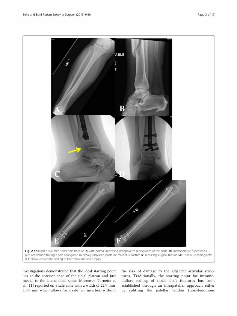
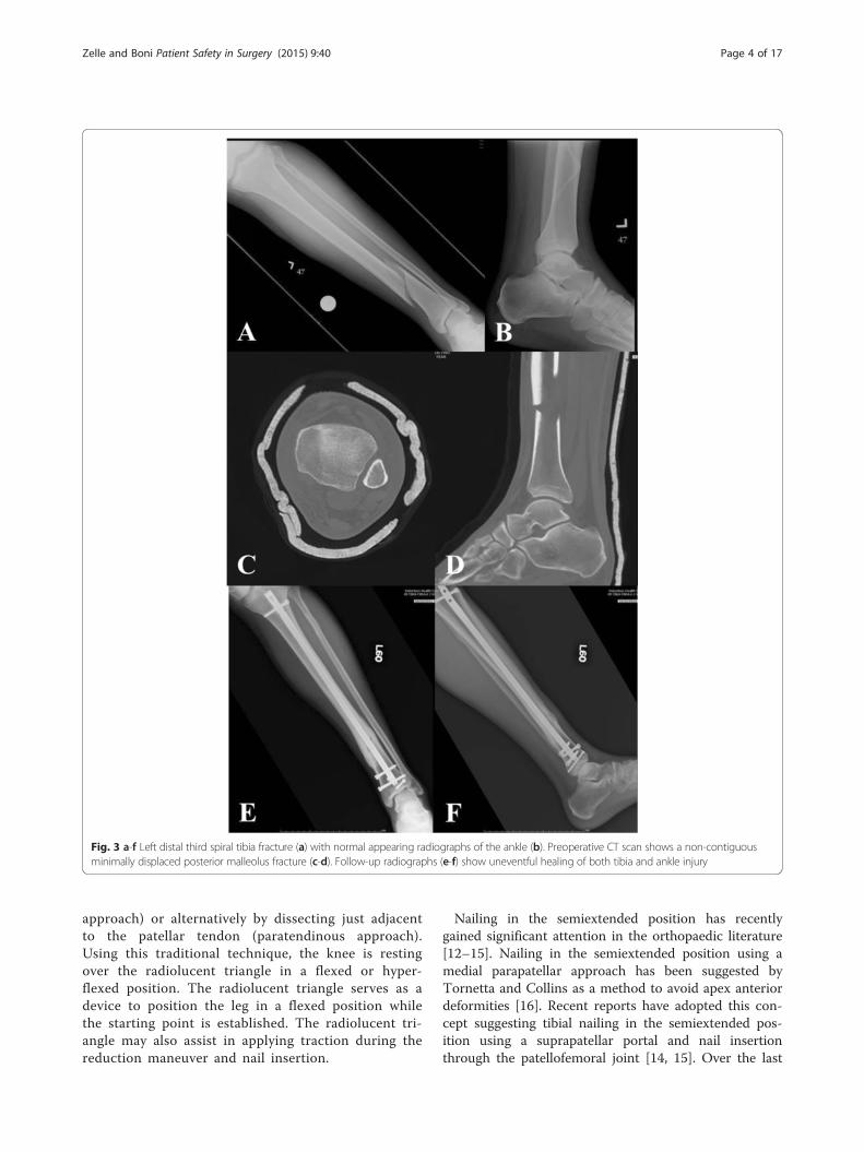
In particular, distal third tibia fractures have beenreported to have a high rate of noncontiguousassociated ankle fractures [8]. Using routine CT scans,Purnell et al. [8] reported that 43 % of distal thirdtibia fractures had associated ankle fractures. Themajority of these associated ankle fractures were foundto require surgical treatment. The most commonlyobserved fracture pattern was characterized by aspiral distal third tibial shaft fracture associated witha minimally or non-displaced posterior malleolusfractures (Fig. 2a-f ). Furthermore, the authors of thisinvestigation reported that due to the minmaldisplacement of the associated ankle fracture, only45 % of these injuries were identified on the plainradiographs of the ankle by a fellowship-trainedorthopaedic traumatologist [8]. Therefore, routineCT scans of the ankle should be given a strongconsideration in the presence of distal third tibialshaft fractures (Fig. 3a-f ).
Surgical considerationsTibial nail starting pointEstablishing an accurate starting point continues to playa crucial role in any intramedullary nailing procedure.Research studies have provided important informationon the anatomic location of the ideal starting point forintramedullary nailing of tibia fractures [9–11]. These
Fig. 1 Compartment pressure measurement of the right leg anteriormuscle compartment with a pressure needle
Zelle and Boni Patient Safety in Surgery (2015) 9:40 Page 2 of 17
investigations demonstrated that the ideal starting pointlies at the anterior edge of the tibial plateau and justmedial to the lateral tibial spine. Moreover, Tornetta etal. [11] reported on a safe zone with a width of 22.9 mm± 8.9 mm which allows for a safe nail insertion without
the risk of damage to the adjacent articular struc-tures. Traditionally, the starting point for intrame-dullary nailing of tibial shaft fractures has beenestablished through an infrapatellar approach eitherby splitting the patellar tendon (transtendinous
Fig. 2 a-f Right distal third spiral tibia fracture (a) with normal appearing preoperative radiographs of the ankle (b). Intraoperative fluoroscopicpictures demonstrating a non-contiguous minimally displaced posterior malleolus fracture (c) requiring surgical fixation (d). Follow-up radiographs(e-f) show uneventful healing of both tibia and ankle injury
Zelle and Boni Patient Safety in Surgery (2015) 9:40 Page 3 of 17
approach) or alternatively by dissecting just adjacentto the patellar tendon (paratendinous approach).Using this traditional technique, the knee is restingover the radiolucent triangle in a flexed or hyper-flexed position. The radiolucent triangle serves as adevice to position the leg in a flexed position whilethe starting point is established. The radiolucent tri-angle may also assist in applying traction during thereduction maneuver and nail insertion.
Nailing in the semiextended position has recentlygained significant attention in the orthopaedic literature[12–15]. Nailing in the semiextended position using amedial parapatellar approach has been suggested byTornetta and Collins as a method to avoid apex anteriordeformities [16]. Recent reports have adopted this con-cept suggesting tibial nailing in the semiextended pos-ition using a suprapatellar portal and nail insertionthrough the patellofemoral joint [14, 15]. Over the last
Fig. 3 a-f Left distal third spiral tibia fracture (a) with normal appearing radiographs of the ankle (b). Preoperative CT scan shows a non-contiguousminimally displaced posterior malleolus fracture (c-d). Follow-up radiographs (e-f) show uneventful healing of both tibia and ankle injury
Zelle and Boni Patient Safety in Surgery (2015) 9:40 Page 4 of 17
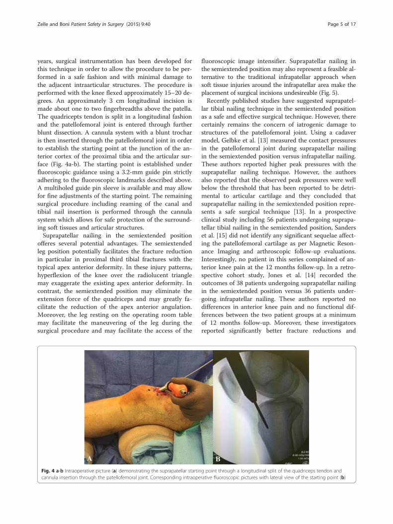
years, surgical instrumentation has been developed forthis technique in order to allow the procedure to be per-formed in a safe fashion and with minimal damage tothe adjacent intraarticular structures. The procedure isperformed with the knee flexed approximately 15–20 de-grees. An approximately 3 cm longitudinal incision ismade about one to two fingerbreadths above the patella.The quadricepts tendon is split in a longitudinal fashionand the patellofemoral joint is entered through furtherblunt dissection. A cannula system with a blunt trocharis then inserted through the patellofemoral joint in orderto establish the starting point at the junction of the an-terior cortex of the proximal tibia and the articular sur-face (Fig. 4a-b). The starting point is established underfluoroscopic guidance using a 3.2-mm guide pin strictlyadhering to the fluoroscopic landmarks described above.A multiholed guide pin sleeve is available and may allowfor fine adjustments of the starting point. The remainingsurgical procedure including reaming of the canal andtibial nail insertion is performed through the cannulasystem which allows for safe protection of the surround-ing soft tissues and articular structures.Suprapatellar nailing in the semiextended position
offeres several potential advantages. The semiextendedleg position potentially facilitates the fracture reductionin particular in proximal third tibial fractures with thetypical apex anterior deformity. In these injury patterns,hyperflexion of the knee over the radiolucent trianglemay exaggerate the existing apex anterior deformity. Incontrast, the semiextended position may eliminate theextension force of the quadriceps and may greatly fa-cilitate the reduction of the apex anterior angulation.Moreover, the leg resting on the operating room tablemay facilitate the maneuvering of the leg during thesurgical procedure and may facilitate the access of the
fluoroscopic image intensifier. Suprapatellar nailing inthe semiextended position may also represent a feasible al-ternative to the traditional infrapatellar approach whensoft tissue injuries around the infrapatellar area make theplacement of surgical incisions undesireable (Fig. 5).Recently published studies have suggested suprapatel-
lar tibial nailing technique in the semiextended positionas a safe and effective surgical technique. However, therecertainly remains the concern of iatrogenic damage tostructures of the patellofemoral joint. Using a cadavermodel, Gelbke et al. [13] measured the contact pressuresin the patellofemoral joint during suprapatellar nailingin the semiextended position versus infrapatellar nailing.These authors reported higher peak pressures with thesuprapatellar nailing technique. However, the authorsalso reported that the observed peak pressures were wellbelow the threshold that has been reported to be detri-mental to articular cartilage and they concluded thatsuprapatellar nailing in the semiextended position repre-sents a safe surgical technique [13]. In a prospectiveclinical study including 56 patients undergoing suprapa-tellar tibial nailing in the semiextended position, Sanderset al. [15] did not identify any significant sequelae affect-ing the patellofemoral cartilage as per Magnetic Reson-ance Imaging and arthroscopic follow-up evaluations.Interestingly, no patient in this series complained of an-terior knee pain at the 12 months follow-up. In a retro-spective cohort study, Jones et al. [14] recorded theoutcomes of 38 patients undergoing suprapatellar nailingin the semiextended position versus 36 patients under-going infrapatellar nailing. These authors reported nodifferences in anterior knee pain and no functional dif-ferences between the two patient groups at a minimumof 12 months follow-up. Moreover, these investigatorsreported significantly better fracture reductions and
Fig. 4 a-b Intraoperative picture (a) demonstrating the suprapatellar starting point through a longitudinal split of the quadriceps tendon andcannula insertion through the patellofemoral joint. Corresponding intraoperative fluoroscopic pictures with lateral view of the starting point (b)
Zelle and Boni Patient Safety in Surgery (2015) 9:40 Page 5 of 17

more precise starting points in the suprapatellar nailinggroup. These promising data suggest that suprapatellartibial nailing in the semiextended position represents asafe surgical technique and appropriate clinical andradiographic outcomes can be achieved using this ap-proach. However, future clinical trials are required tofurther study the advantages and disadvantages of supra-patellar nailing and to evaluate the long-term outcomesassociated with this technique.
Reduction techniquesPlacement of the tibial nail alone does not result inadequate fracture reduction and appropriate fracturealignment must be maintained throughout the ream-ing process and nail placement. While application oflongitudinal traction typically results in improvedfracture alignment through ligamentotaxis, the simpleapplication of manual traction by itself may not al-ways achieve an anatomic fracture alignment. Variousclosed, minimal invasive, and open reduction maneu-vers have been described and should be in the sur-geons armamentarium.
Technical trick
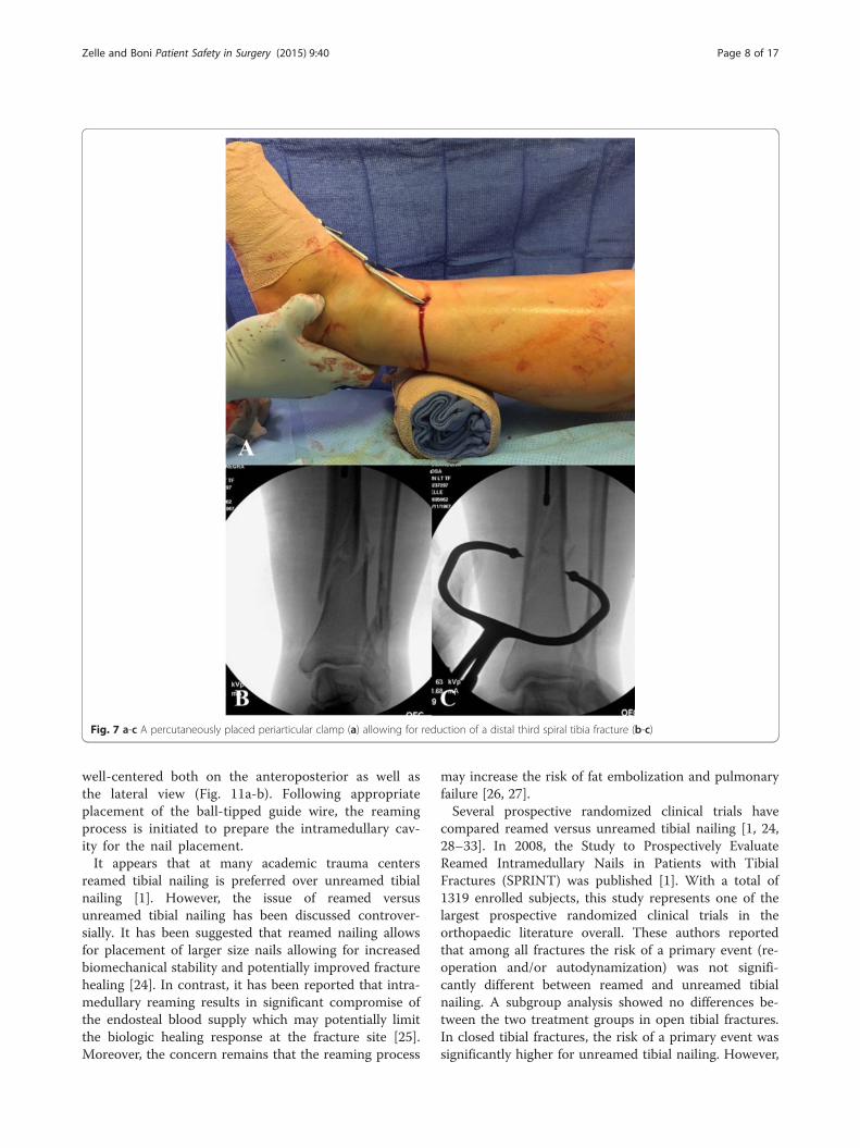
Closed reduction maneuvers can be facilitated bywidely available reduction tools, such as the F-tool.The F-tool is a an F-shaped radiolucent reductiondevice that will allow for correction of varus/valgusangulation as well as correction of medial/lateraltranslation (Fig. 6a-d). However, due to significantpressure on the tissues prolonged application of thisreduction device should be avoided. Certain fracturesare also amenable to placement of percutaneouslyplaced reduction clamps. In particular, spiral andoblique fractures lean themselves towards placementof percutaneous clamps. These clamps can beapplied in a soft tissue friendly manner throughsmall stab incisions (Fig.7a-c). The type of the clampand the location of the surgical incisions should bestrategically chosen in order to minimize anyprolonged soft tissue compromise from clampplacement (Fig.8a-b).
The universal distractor can be used as an additionalreduction tool [17]. The universal distractor may assistin maintaining length and alignment. Careful attentionmust be paid to the placement of the Schanz pins. Theseare placed from the medial side into the proximal anddistal fragment away from the planned position of thetibial nail. Moreover, the proximal Schanz pin can beplaced in a position that mimics the position of aproximal blocking screw [17]. This may become par-ticularly useful when seeking fracture reduction inproximal tibia fractures with the typical apex anteriordeformity. Similar to the universal distractor, two-pinexternal fixation can be used to obtain and maintainlength and alignment during intramedullary nailing oftibial shaft fractures [18]. When using this technique,the pin placement should follow the same principlesas with the use of the universal distractor.In some instances closed and minimal invasivive
reduction techniques remain insufficient in obtainingan anatomic fracture alignment. In these cases, openreduction techniques with respectful handling of thesurrounding soft tissues should be considered [19, 20].Open reduction techniques allow for surgical reductionunder direct visualization. Potential disadvantages ofopen reduction techniques include the additional sur-gical dissection which in may potentially increase therisk of surgical site infection. Moreover, the additionalstripping of the blood supply to the fracture site maypotentially increase the risk of subsequent fracturenonunion. However, retrospective cohort studies havenot shown any increased risk of surgical site infectionor fracture nonunion with the use of open reductiontechniques [19, 20].
Fig. 5 Intraoperative picture demonstrating the soft tissue injury tothe infrapatellar area as an indication for suprapatellar nailing in thesemiextended position
Zelle and Boni Patient Safety in Surgery (2015) 9:40 Page 6 of 17
Technical trick
Open reduction maneuvers do not only allow forplacement of appropriate surgical reduction clamps,but also provide the opportunity to apply a small- ormini-fragment plate at the fracture site in order toachieve and maintain fracture reduction during theintramedullary nailing procedure [17, 21]. The platesare secured to the proximal and distal fracturefragments using unicortical screws. The plate is thenmaintained throughout the reaming procedure andplacement of the intramedullary tibial nail. Followingnail placement the plate can be removed oralternatively be left in situ in order to enhance thestability of the fixation construct (Fig. 9a-e). If thesurgeon chooses to leave the plate in situ, theunicortical screws should be exchanged againstbicortical screws. Unicortical plating or “reductionplating” has been suggested as a safe and effectivetechnique and should be considered for select cases oftibial shaft that require an open approach to achievean acceptable fracture reduction [17, 21].
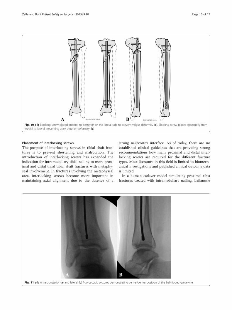
Blocking screws (or “poller” screws) have been popu-larized by Krettek et al. [22]. The purpose of blockingscrews is to narrow the canal in the metaphyseal areaand to substitute a deficient cortex. Therefore, blockingscrews are useful tools in fractures with metaphyseal in-volvement. The blocking screws are placed prior to thereaming process and nail placement. Blocking screws aretypically placed in the short, articular fragment and onthe concave side of the deformity. For instance, thetypical deformity of a proximal third tibia fracture ischaracterized by a valgus- and apex anterior deformity.
In order to overcome the valgus deformity, a blockingscrew can be placed in an anterior to posterior directioninto the lateral portion of the proximal fracture fragment(i.e. on the concave side of the deformity). This blockingscrew is used to guide the nail medially and thus pre-vents a valgus angulation. Similarly, the apex anteriordeformity can be overcome by a blocking screw that isplaced in a medial to lateral direction in the posteriorportion of the proximal fragment (i.e. on the concaveside of the deformity) (Fig. 10a-b). Krettek et al. [22] re-ported on 21 tibial fractures treated with intramedullarytibial nailing plus blocking screws. These authors re-ported favorable clinical and radiological outcomes andno complications related to the placement of blockingscrews. Ricci et al. [23] reported on 12 patients undero-ing tibial nailing in conjunction with blocking screws.All but one patient went on to fracture union. The au-thors reported only one patient with an angular deform-ity of more than 5 degrees. This patient was found tohave a postoperative valgus angulation of 10 degrees.However, this patient had not undergone blocking screwplacement to control for valgus angulation.
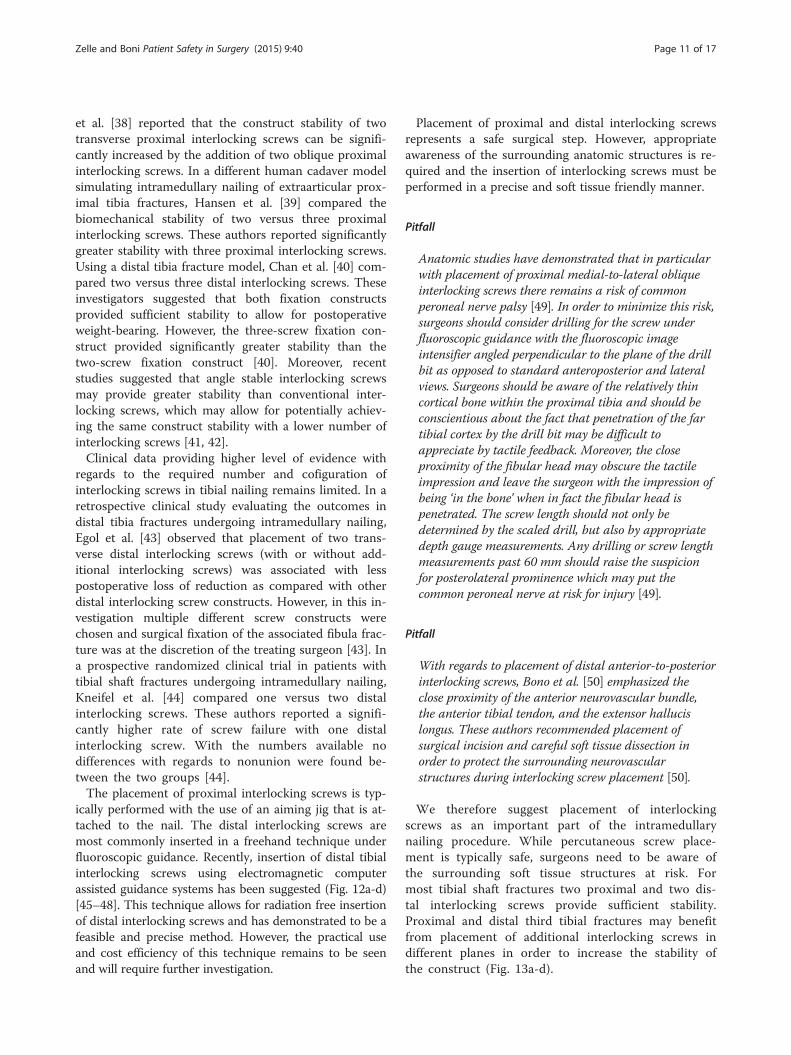
Reaming of the intramedullary canalUpon successful completion of the fracture reduction,the intramedullary cavity is prepared for the place-ment of the tibial nail. A ball-tipped guide wire istypically inserted into the tibial canal and across thefracture site. The reamers as well as the tibial nail arepassed over the ball-tipped guide wire. Therefore, it isvery important to confirm on fluoroscopic imagesthat the ball-tipped guide wire is positioned appropri-ately. In particular, it is crucial to confirm that on thelevel of the ankle joint, the ball-tipped guide wire is
Fig. 6 a-d The F-tool (a) allowing for reduction of a medially translated tibia fracture (b-d)
Zelle and Boni Patient Safety in Surgery (2015) 9:40 Page 7 of 17
well-centered both on the anteroposterior as well asthe lateral view (Fig. 11a-b). Following appropriateplacement of the ball-tipped guide wire, the reamingprocess is initiated to prepare the intramedullary cav-ity for the nail placement.It appears that at many academic trauma centers
reamed tibial nailing is preferred over unreamed tibialnailing [1]. However, the issue of reamed versusunreamed tibial nailing has been discussed controver-sially. It has been suggested that reamed nailing allowsfor placement of larger size nails allowing for increasedbiomechanical stability and potentially improved fracturehealing [24]. In contrast, it has been reported that intra-medullary reaming results in significant compromise ofthe endosteal blood supply which may potentially limitthe biologic healing response at the fracture site [25].Moreover, the concern remains that the reaming process
may increase the risk of fat embolization and pulmonaryfailure [26, 27].Several prospective randomized clinical trials have
compared reamed versus unreamed tibial nailing [1, 24,28–33]. In 2008, the Study to Prospectively EvaluateReamed Intramedullary Nails in Patients with TibialFractures (SPRINT) was published [1]. With a total of1319 enrolled subjects, this study represents one of thelargest prospective randomized clinical trials in theorthopaedic literature overall. These authors reportedthat among all fractures the risk of a primary event (re-operation and/or autodynamization) was not signifi-cantly different between reamed and unreamed tibialnailing. A subgroup analysis showed no differences be-tween the two treatment groups in open tibial fractures.In closed tibial fractures, the risk of a primary event wassignificantly higher for unreamed tibial nailing. However,
Fig. 7 a-c A percutaneously placed periarticular clamp (a) allowing for reduction of a distal third spiral tibia fracture (b-c)
Zelle and Boni Patient Safety in Surgery (2015) 9:40 Page 8 of 17

this difference was largely driven by the least importantoutcomes, dynamization and autodynamization. More-over, the authors reported that the treating surgeons hadrelatively more experience with reamed tibial nailing.With regards to adverse events, the authors recorded asignificantly higher death rate in reamed tibial nailing.The investigators noted that blinded adjudicators clas-sified all deaths as unrelated to the intramedullarynailing procedure [1]. Subsequent meta-analyses aswell as a Cochrane review were published with theintent to obtain pooled results from the above men-tioned randomized clinical trials [34–37]. The results
of these meta-analyses were mostly dominated by theresults from the SPRINT study [2] due to its largesample size. Therefore, the results of the above men-tioned meta-analyses [34–37] were overall in line withthe results from the SPRINT study [1] and mostlyconfirmed its findings.We suggest that most surgeons in North America pre-
fer reamed intramedullary tibial nailing over unreamednailing. However, both reamed and unreamed intrame-dullary nailing can be suggested as acceptable standardtechniques and good outcomes can be achieved withboth of these methods.
Fig. 9 a-e Open tibia fracture with significant comminution and bone loss (a). A unicortical plate was applied through the traumatic wound toachieve fracture reduction (b). The plate was maintained throughout the reaming process and nail placement (c). Following successful nailstabilization, the plate was removed (d-e)
Fig. 8 a-b In same patient, a percutaneously placed pointed reduction clamp (a) resulted in significant soft tissue compromise (b) which requiredchanging to a different clamp
Zelle and Boni Patient Safety in Surgery (2015) 9:40 Page 9 of 17
Placement of interlocking screwsThe purpose of interlocking screws in tibial shaft frac-tures is to prevent shortening and malrotation. Theintroduction of interlocking screws has expanded theindication for intramedullary tibial nailing to more prox-imal and distal third tibial shaft fractures with metaphy-seal involvement. In fractures involving the metaphysealarea, interlocking screws become more important inmaintaining axial alignment due to the absence of a
strong nail/cortex interface. As of today, there are noestablished clinical guidelines that are providing strongrecommendations how many proximal and distal inter-locking screws are required for the different fracturetypes. Most literature in this field is limited to biomech-anical investigations and published clinical outcome datais limited.In a human cadaver model simulating proximal tibia
fractures treated with intramedullary nailing, Laflamme
Fig. 11 a-b Anteroposterior (a) and lateral (b) fluoroscopic pictures demonstrating center/center position of the ball-tipped guidewire
Fig. 10 a-b Blocking screw placed anterior to posterior on the lateral side to prevent valgus deformity (a). Blocking screw placed posteriorly frommedial to lateral preventing apex anterior deformity (b)
Zelle and Boni Patient Safety in Surgery (2015) 9:40 Page 10 of 17
et al. [38] reported that the construct stability of twotransverse proximal interlocking screws can be signifi-cantly increased by the addition of two oblique proximalinterlocking screws. In a different human cadaver modelsimulating intramedullary nailing of extraarticular prox-imal tibia fractures, Hansen et al. [39] compared thebiomechanical stability of two versus three proximalinterlocking screws. These authors reported significantlygreater stability with three proximal interlocking screws.Using a distal tibia fracture model, Chan et al. [40] com-pared two versus three distal interlocking screws. Theseinvestigators suggested that both fixation constructsprovided sufficient stability to allow for postoperativeweight-bearing. However, the three-screw fixation con-struct provided significantly greater stability than thetwo-screw fixation construct [40]. Moreover, recentstudies suggested that angle stable interlocking screwsmay provide greater stability than conventional inter-locking screws, which may allow for potentially achiev-ing the same construct stability with a lower number ofinterlocking screws [41, 42].Clinical data providing higher level of evidence with
regards to the required number and cofiguration ofinterlocking screws in tibial nailing remains limited. In aretrospective clinical study evaluating the outcomes indistal tibia fractures undergoing intramedullary nailing,Egol et al. [43] observed that placement of two trans-verse distal interlocking screws (with or without add-itional interlocking screws) was associated with lesspostoperative loss of reduction as compared with otherdistal interlocking screw constructs. However, in this in-vestigation multiple different screw constructs werechosen and surgical fixation of the associated fibula frac-ture was at the discretion of the treating surgeon [43]. Ina prospective randomized clinical trial in patients withtibial shaft fractures undergoing intramedullary nailing,Kneifel et al. [44] compared one versus two distalinterlocking screws. These authors reported a signifi-cantly higher rate of screw failure with one distalinterlocking screw. With the numbers available nodifferences with regards to nonunion were found be-tween the two groups [44].The placement of proximal interlocking screws is typ-
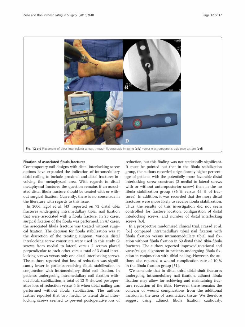
ically performed with the use of an aiming jig that is at-tached to the nail. The distal interlocking screws aremost commonly inserted in a freehand technique underfluoroscopic guidance. Recently, insertion of distal tibialinterlocking screws using electromagnetic computerassisted guidance systems has been suggested (Fig. 12a-d)[45–48]. This technique allows for radiation free insertionof distal interlocking screws and has demonstrated to be afeasible and precise method. However, the practical useand cost efficiency of this technique remains to be seenand will require further investigation.
Placement of proximal and distal interlocking screwsrepresents a safe surgical step. However, appropriateawareness of the surrounding anatomic structures is re-quired and the insertion of interlocking screws must beperformed in a precise and soft tissue friendly manner.
Pitfall
Anatomic studies have demonstrated that in particularwith placement of proximal medial-to-lateral obliqueinterlocking screws there remains a risk of commonperoneal nerve palsy [49]. In order to minimize this risk,surgeons should consider drilling for the screw underfluoroscopic guidance with the fluoroscopic imageintensifier angled perpendicular to the plane of the drillbit as opposed to standard anteroposterior and lateralviews. Surgeons should be aware of the relatively thincortical bone within the proximal tibia and should beconscientious about the fact that penetration of the fartibial cortex by the drill bit may be difficult toappreciate by tactile feedback. Moreover, the closeproximity of the fibular head may obscure the tactileimpression and leave the surgeon with the impression ofbeing ‘in the bone’ when in fact the fibular head ispenetrated. The screw length should not only bedetermined by the scaled drill, but also by appropriatedepth gauge measurements. Any drilling or screw lengthmeasurements past 60 mm should raise the suspicionfor posterolateral prominence which may put thecommon peroneal nerve at risk for injury [49].
Pitfall
With regards to placement of distal anterior-to-posteriorinterlocking screws, Bono et al. [50] emphasized theclose proximity of the anterior neurovascular bundle,the anterior tibial tendon, and the extensor hallucislongus. These authors recommended placement ofsurgical incision and careful soft tissue dissection inorder to protect the surrounding neurovascularstructures during interlocking screw placement [50].
We therefore suggest placement of interlockingscrews as an important part of the intramedullarynailing procedure. While percutaneous screw place-ment is typically safe, surgeons need to be aware ofthe surrounding soft tissue structures at risk. Formost tibial shaft fractures two proximal and two dis-tal interlocking screws provide sufficient stability.Proximal and distal third tibial fractures may benefitfrom placement of additional interlocking screws indifferent planes in order to increase the stability ofthe construct (Fig. 13a-d).
Zelle and Boni Patient Safety in Surgery (2015) 9:40 Page 11 of 17
Fixation of associated fibula fracturesContemporary nail designs with distal interlocking screwoptions have expanded the indication of intramedullarytibial nailing to include proximal and distal fractures in-volving the metaphyseal area. With regards to distalmetaphyseal fractures the question remains if an associ-ated distal fibula fracture should be treated with or with-out surgical fixation. Currently, there is no consensus inthe literature with regards to this issue.In 2006, Egol et al. [43] reported on 72 distal tibia
fractures undergoing intramedullary tibial nail fixationthat were associated with a fibula fracture. In 25 cases,surgical fixation of the fibula was performed. In 47 cases,the associated fibula fracture was treated without surgi-cal fixation. The decision for fibula stabilization was atthe discretion of the treating surgeon. Various distalinterlocking screw constructs were used in this study (2screws from medial to lateral versus 2 screws placedperpendicular to each other versus total of 3 distal inter-locking screws versus only one distal interlocking screw).The authors reported that loss of reduction was signifi-cantly lower in patients receiving fibula stabilization inconjunction with intramedullary tibial nail fixation. Inpatients undergeoing intramedullary nail fixation with-out fibula stabilization, a total of 13 % showed postoper-ative loss of reduction versus 4 % when tibial nailing wasperformed without fibula stabilization. The authorsfurther reported that two medial to lateral distal inter-locking screws seemed to prevent postoperative loss of
reduction, but this finding was not statistically significant.It must be pointed out that in the fibula stabilizationgroup, the authors recorded a significantly higher percent-age of patients with the potentially more favorable distalinterlocking screw construct (2 medial to lateral screwswith or without anteroposterior screw) than in the nofibula stabilization group (86 % versus 45 % of frac-tures). In addition, it was recorded that the more distalfractures were more likely to receive fibula stabilization.Thus, the results of this investigation did not seemcontrolled for fracture location, configuration of distalinterlocking screws, and number of distal interlockingscrews [43].In a prospective randomized clinical trial, Prasad et al.
[51] compared intramedullary tibial nail fixation withfibula fixation versus intramemedullary tibial nail fix-ation without fibula fixation in 60 distal third tibia-fibulafractures. The authors reported improved rotational andvarus/valgus alignment in patients undergoing fibula fix-ation in conjunction with tibial nailing. However, the au-thors also reported a wound complication rate of 10 %in the fibula fixation group [51].We conclude that in distal third tibial shaft fractures
undergoing intramedullary nail fixation, adjunct fibulafixation may allow for achieving and maintaining frac-ture reduction of the tibia. However, there remains theconcern of wound complications from the additionalincision in the area of traumatized tissue. We thereforesuggest using adjunct fibula fixation cautiously.
Fig. 12 a-d Placement of distal interlocking screws through fluoroscopic imaging (a-b) versus electromagnetic guidance system (c-d)
Zelle and Boni Patient Safety in Surgery (2015) 9:40 Page 12 of 17

Contemporary tibial nail designs typically provide differ-ent options for placement of stable distal interlockingscrew constructs minimizing the risk for postoperativeloss of reduction. Additional plate fixation of the fibulashould be reserved for associated unstable injuries to theankle joint or when it is felt that anatomic tibial align-ment cannot be achieved without direct reduction of theassociated fibula fracture.
OutcomesGood outcomes and reproducible results can be achievedwith intramedullary nail fixation of tibial shaft fractures.The reported union rates of intramedullary tibial nailingvary among different studies. With contemporary im-plants and appropriate surgical techniques, union ratesabove 90 % can be expected [34–37]. Tibial shaft fracturesthat fail to heal following intramedullary nail fixationtypically respond well to exchange reamed nailingprocedures [52].
Despite favorable union rates that can be achievedwith intramedullary nail fixation of tibial shaft fractures,patients continue to have functional long-term sequelaefollowing this procedure. Outcome evaluations at oneyear after surgery demonstrated that as many as 44 % ofpatients continued to have functional limitations withregards to their injured lower extremity [53]. Moreover,it has been reported that at one year after surgery asmany as 47 % of patients continue to report work-related disability [54]. Other follow-up studies recordedthat at approximately two years after intramedullary nailfixation, almost 20 % of patients had not yet returned totheir previous occupation and almost 30 % had not yetreturned to their previous level of recreation [31]. In along-term outcome study including 56 patients aftertibial nailing with a median follow-up of 14 years,Lefaivre et al. [55] reported that the SF-36 and theShort Musculoskeletal Functional Assessment (SMFA)scores were not statistically different from reference
Fig. 13 a-d Segmental tibia fracture (a-b) treated with intramedullary nailing with two distal and three proximal interlocking screws. Follow-upradiographs (c-d) demonstrate uneventful healing
Zelle and Boni Patient Safety in Surgery (2015) 9:40 Page 13 of 17
population norms. However, 73.2 % of patients self-reported at least moderate knee pain and 33.9 % ofpatients self-reported complaints of swelling. Thephysical examination showed decreased range of mo-tion of the ankle joint in 42.4 % of examined patientswhile 93.9 % of patients demonstrated full range ofmotion of the knee joint. Atrophies of the calf and/orthe quadriceps muscles were observed in 27.3 % ofpatients. Radiographic evidence of osteoarthritis ofthe knee and/or ankle joint was found in 35.4 % ofpatients despite the absence of radiographic tibialmalalignment [55].These data indicate that patients undergoing intrame-
dullary tibial nailing continue to have remarkable func-tional limitations in the long-term. Surgeons should beaware of these issues and counsel patients accordingly.
Anterior knee painAnterior knee pain is a commonly reported complicationafter intramedullary nailing of tibial shaft fractures[55–62]. A comprehensive review with pooled datafrom publications including the years 1990 until 2005suggested that postoperative knee pain may occur inapproximately 47 % of patients following intramedul-lary nailing [60]. The exact etiology of anterior kneepain following tibial nailing is not fully understood.Potentially contributing factors may include traumaticand iatrogenic damage to intraarticular structures, in-juries to the infrapatellar branch of the saphenousnerve, thigh muscle weakness secondary to pain-relatedneuromuscular reflex inhibition, fat pad fibrosis leading toimpingement, reactive patellar tendonitis, bending strainexerted by the nail on the proximal part of the tibial bone,and proximal protrusion of the nail [10, 57, 58, 60, 63, 64].As of today, it must be assumed that the reason for post-operative knee pain is multifactorial and that the differentabove-named factors may be contributing to this problemat varying degrees.In an attempt to address the etiology of anterior knee
pain after intramedullary nailing, transtendinous ap-proaches have been compared to paratendinous ap-proaches. Previous studies suggested that transtendinousmay be associated with a higher incidence of postopera-tive knee pain [61]. However, prospective randomizedclinical data has not shown any significant difference be-tween the transtendinous and paratendinous approach[65–67]. In a prospective randomized clinical trial in-cluding fifty patients undergoing intramedullary tibialnailing, Toivanen et al. [65] did not find any significantdifferences in the functional outcomes of the transtendi-nous versus paratendinous approach at an averagefollow-up of 3.2 years. In a subsequent follow-up studyusing the same patient population, these authors re-ported on the long-term results with an eight year
follow-up [66]. At eight year follow-up, there were nosignificant differences between the two approaches. Ofnote, these investigators also observed a significant de-crease in anterior knee pain over time. While 69 % ofpatients complained of anterior knee pain at 3.2 yearsafter surgery, a total of 29 % complained of knee pain atthe eight year follow-up [66].The effect of elective hardware removal in order to ad-
dress anterior knee pain following intramedullary tibialnailing remains uncertain. Court-Brown et al. [56] re-ported marked or complete relief of anterior knee painin 60 out of 62 patients who underwent elective tibialnail removal due to persistent anterior knee pain follow-ing intramedullary tibial nailing. In contrast, Keating etal. [61] reported on 49 patients undergoing tibial nail re-moval due to persistent anterior knee pain. These au-thors reported complete relief in approximately 45 %,partial relief in approximately 35 %, and no improve-ment in approximately 20 % of patients. Therefore, theindication for tibial nail removal in the treatment ofpostoperative anterior knee pain remains poorly defined.We suggest considering a tibial nail removal only in pa-tients with persistent anterior knee pain if a mechanicaletiology, such as nail protrusion or prominent interlock-ing screws, can be identied. However, in symptomaticpatients with appropriately placed hardware, the benefitof a tibial nail removal remains questionable.With regards to postoperative anterior knee pain, in-
triguing results have been reported in preliminary clin-ical investigations of suprapatellar tibial nailing in thesemiextended position. Jones et al. [14] reported nostatistical differences with regards to anterior knee painbetween patients undergoing suprapatellar versus infra-patellar nailing. However, the authors reported that therewas trend toward greater symptomatic knee pain in theinfrapatellar group. Furthermore, Sanders et al. [15]reported on 56 consecutive patients undergoing supra-patellar nailing in the semiextended position. These au-thors did not identify any patients with postoperativeanterior knee pain at 12 months follow-up except onepatient who presented with peri-incisional pain aroundthe knee [15]. While these preliminary data seem en-couraging, there remains the theoretical concern of iat-rogenic cartilage damage to the patellofemoral jointassociated with this procedure [12, 13]. Therefore, largerclinical investigations with long-term follow-up periodsare necessary in order to substantiate the impact of supra-patellar nailing on postoperative anterior knee pain.
Effect of postoperative malalignmentPosttraumatic osteoarthritis remains an important con-cern following treatment of tibial shaft fractures withintramedullary nailing. Biomechanical studies have dem-onstrated that tibial malalignment may result in significant
Zelle and Boni Patient Safety in Surgery (2015) 9:40 Page 14 of 17

changes of contact pressures in the adjacent ankle andknee joint [68]. However, the clinical effects of tibial mala-lignment on clinical and functional outcomes continue tobe controversial.Clinical investigations evaluating the long-term clinical
and radiographic outcomes after tibial shaft fractureshave provided conflicting data with regards to the seque-lae of tibial malalignment. In a long-term outcome studywith an average of 29 years of follow-up, Merchant andDietz [69] did not find any significant correlationbetween tibial malalignment and malfunction of theadjacent ankle or knee joint [69]. In contrast, otherinvestigators reported significant correlations betweentibial malalignment and ankle and/or knee malfunc-tion [70, 71]. Similarly, the relationship between tibialmalalignment and radiographic signs of posttraumaticarthritis of the adjacent joints remains controversial.While some authors [72] reported a significant correlationbetween tibial malalignment and radiographic signs ofposttraumatic arthritis other investigations did not showany significant correlations between these two variables[69, 70]. Of note, most of these long-term data were de-rived from patient populations in which the majority ofsubjects were treated nonoperatively with casting and/orbracing [69–72] and these data may not be extrapolatedto patients undergoing intramedullary tibial nailing.Reports on postoperative malalignment following intra-
medullary tibial nailing remain limited and the reportedcase numbers are low [73, 74]. It must be assumed thatwith contemporary implants and appropriate surgicaltechniques the rates of malangulation are rather low ascompared to nonoperative treatment. However, postoper-ative malrotation remains a commonly reported concernspecific to intramedullary tibial nailing. Unfortunately, theintraoperative assessment of tibial rotation remains chal-lenging. As of today, there is no single one clinical orfluoroscopic method that has been established as the goldstandard for judging tibial rotation intraoperatively. Re-sults from sophisticated CT assessments recorded thatmalrotation following intramedullary tibial nailing may beas 19 % to 41 % [75–78]. In particular, external rotationdeformities seem to be more common than internal rota-tion deformities. In a series of 70 patients with an averagefollow-up of 58 months, Theriault et al. [78] reported thattibial malrotation did not show any significant correlationwith the functional outcomes. The authors also reportedthat clinical examinations to assess for postoperative mal-rotation were inaccurate and showed low correlations withCT assessments.In our opinion, malreduction remains a concern for the
long-term outcome of tibial shaft fractures undergoingtibial nailing. Despite the conflicting data with regards tothe relationship between malalignment and clinical andradiographic outcomes, we suggest that surgeons should
strive to achieve an anatomic fracture alignment in anattempt to control for this variable and to achieve the bestpossible outcome.
ConclusionsStatically locked, reamed intramedullary nailing remainsthe standard treatment for displaced tibial shaft frac-tures. A correct starting point remains a crucial part ofthe surgical procedure. Suprapatellar nailing in the semi-extended position has been suggested as a safe and ef-fective procedure and future studies are warranted tofurther evaluate the risks and benefits of this surgicalprocedure. The treating surgeon should be familiar withcontemporary reduction techniques. Open reductiontechniques should be considered if anatomic fracturealignment cannot be achieved by closed means. Favor-able union rates above 90 % can be achieved by bothreamed and unreamed intramedullary nailing. Despitefavorable union rates, patients continue to have func-tional long-term limitations. In particular, anterior kneepain remains a common complaint following intrame-dullary tibial nailing. In addition, malrotation remains acommonly reported concern after tibial nailing. As oftoday, no significant correlation between malrotationand functional outcome has been established in theliterature.
Competing interestsThe authors declare no conflicts of interests related to this manuscript. Allauthors confirm that they have no financial relationship to companiesinvolved in the marketing of the products described in the article.
Authors’ contributionsBAZ performed the surgeries, collected picture material, wrote the first draftof the article, and critically reviewed the final draft. GB participated in writingthe first draft of the article, contributed picture material, and criticallyreviewed the final draft. All authors approved the final version of the article.
Author details1Department of Orthopaedic Surgery, Division of Orthopaedic Traumatology,University of Texas Health Science Center at San Antonio, 7703 Floyd Curl Dr,MC-7774, San Antonio, TX 78229, USA. 2Department of Orthopaedics andTraumatology, Federal University of São Paulo, Rua Borges Lagoa, 783-50Andar, São Paulo 04038032, Brazil.
Received: 3 November 2015 Accepted: 22 November 2015
References1. Study to Prospectively Evaluate Reamed Intramedullary Nails in Patients
with Tibial Fractures Investigators, Bhandari M, Guyatt G, Tornetta III P,Schemitsch EH, Swiontkowski M, et al. Randomized trial of reamed andunreamed intramedullary nailing of tibial shaft fractures. J Bone Joint SurgAm. 2008;90:2567–78.
2. McQueen MM, Duckworth AD, Aitken SA, Sharma R, Court-Brown CM.Predictors of compartment syndrome after tibial fracture. J Orthop Trauma.2015. [Epub ahead of print].
3. Park S, Ahn J, Gee AO, Kuntz AF, Esterhai JL. Compartment syndrome intibial fractures. J Orthop Trauma. 2009;23:514–8.
4. McQueen MM, Court-Brown CM. Compartment monitoring in tibialfractures. The pressure threshold for decompression. J Bone Joint Surg (Br).1996;78:99–104.
Zelle and Boni Patient Safety in Surgery (2015) 9:40 Page 15 of 17
5. McQueen MM, Duckworth AD, Aitken SA, Court-Brown CM. The estimatedsensitivity and specificity of compartment pressure monitoring for acutecompartment syndrome. J Bone Joint Surg Am. 2013;95:673–7.
6. Whitesides Jr TE, Haney TC, Morimoto K, Harada H. Tissue pressuremeasurements as a determinant for the need of fasciotomy. Clin Orthop.1975;113:43–51.
7. Kakar S, Firoozabadi R, McKean J, Tornetta 3rd P. Diastolic blood pressure inpatients with tibia fractures under anaesthesia: implications for thediagnosis of compartment syndrome. J Orthop Trauma. 2007;21:99–103.
8. Purnell GJ, Glass ER, Altman DT, Sciulli RL, Muffly MT, Altman GT. Results of acomputed tomography protocol evaluating distal third tibial shaft fracturesto assess noncontiguous malleolar fractures. J Trauma. 2011;71:163–8.
9. Buehler KC, Green J, Woll TS, Duwelius PJ. A technique for intramedullarynailing of proximal third tibia fractures. J Orthop Trauma. 1997;11:218–23.
10. McConnell T, Tornetta III P, Tilzey J, Casey D. Tibial portal placement: theradiographic correlate of the anatomic safe zone. J Orthop Trauma.2001;15:207–9.
11. Tornetta III P, Riina J, Geller J, Purban W. Intraarticular anatomic risks of tibialnailing. J Orthop Trauma. 1999;13:247–51.
12. Eastman J, Tseng S, Lo E, Li CS, Yoo B, Lee M. Retropatellar technique forintramedullary nailing of proximal tibia fractures: a cadaveric assessment.J Orthop Trauma. 2010;24:672–6.
13. Gelbke MK, Coombs D, Powell S, DiPasquale TG. Suprapatellar versusinfra-patellar intramedullary nail insertion of the tibia: a cadaveric model forcomparison of patellofemoral contact pressures and forces. J OrthopTrauma. 2010;24:665–71.
14. Jones M, Parry M, Whitehouse M, Mitchell S. Radiologic outcome andpatient-reported function after intramedullary nailing: a comparison of theretropatellar and infrapatellar approach. J Orthop Trauma. 2014;28:256–62.
15. Sanders RW, DiPasquale TG, Jordan CJ, Arrington JA, Sagi HC. Semiextendedintramedullary nailing of the tibia using a suprapatellar approach:radiographic results and clinical outcomes at a minimum of 12 monthsfollow-up. J Orthop Trauma. 2014;28:245–55.
16. Tornetta III P, Collins E. Semiextended position of intramedullary nailing ofthe proximal tibia. Clin Orthop Relat Res. 1996;328:185–9.
17. Nork SE, Barei DP, Schildhauer TA, Agel J, Holt SK, Schrick JL, et al.Intramedullary Nailing of proximal quarter tibial fractures. J Orthop Trauma.2006;20:523–8.
18. Wysocki RW, Kapotas JS, Virkus WW. Intramedullary nailing of proximal anddistal one-third tibial shaft fractures with intraoperative two-pin externalfixation. J Trauma. 2009;66:1135–9.
19. Bishop JA, Dikos GD, Mickelson D, Barei DP. Open reduction andintramedullary nail fixation of closed tibial fractures. Orthopedics.2012;35:e1631–1634.
20. Tang P, Gates C, Hawes J, Vogt M, Prayson MJ. Does open reductionincrease the chance of infection during intramedullary nailing of closedtibial shaft fractures? J Orthop Trauma. 2006;20:317–22.
21. Archdeacon MT, Wyrick JD. Reduction plating for provisional fracturefixation. J Orthop Trauma. 2006;20:206–11.
22. Krettek C, Stephan C, Schandelmaier P, Richter M, Pape HC, Miclau T. Theuse of poller screws in stabilizing tibial fractures treated with small diameterintramedullary nails. J Bone Joint Surg (Br). 1999;81:963–8.
23. Ricci WM, O'Boyle M, Borrelli J, Bellabarba C, Sanders R. Fractures of theproximal third of the tibial shaft treated with intramedullary nails andblocking screws. J Orthop Trauma. 2001;15:264–70.
24. Anglen JO, Blue JM. A comparison of reamed and unreamed nailing of thetibia. J Trauma. 1995;39:351–5.
25. Klein MP, Rahn BA, Frigg R, Kessler S, Perren SM. Reaming versus non-reamingin medullary nailing: interference with cortical circulation of the canine tibia.Arch Orthop Trauma Surg. 1990;109:314–6.
26. Pape HC, Auf’m’Kolk M, Paffrath T, Regel G, Sturm JA, Tscherne H. Primaryintramedullary femur fixation in multiple trauma patients with associatedlung contusion: a cause of posttraumatic ARDS? J Trauma. 1993;34:540–7.
27. Pape HC, Zelle BA, Hildebrand F, Giannoudis PV, Krettek C, van Griensven M.Reamed femoral nailing in sheep: does irrigation and aspiration ofintramedullary contents alter the systemic response? J Bone Joint Surg Am.2005;87:2515–22.
28. Blachut PA, O’Brien PJ, Meek RN, Broekhuyse HM. Interlockingintramedullary nailing with and without reaming for the treatment ofclosed fractures of the tibial shaft. A prospective, randomized study. J BoneJoint Surg Am. 1997;79:640–6.
29. Court-Brown CM, Will E, Christie J, McQueen MM. Reamed or unreamednailing for closed tibial fractures: a prospective study in Tscherne C1fractures. J Bone Joint Surg (Br). 1996;78:580–3.
30. Finkemeier CG, Schmidt AH, Kyle RF, Templeman DC, Varecka TF. Aprospective, randomized study of intramedullary nails inserted with andwithout reaming for the treatment of open and closed fractures of thetibial shaft. J Orthop Trauma. 2000;14:187–93.
31. Keating FE, O’Brien PJ, Blachut PA, Meek RN, Broekhuyse HM. Lockingintramedullary nailing with and without reaming for open fractures of thetibial shaft. A prospective randomized study. J Bone Joint Surg Am.1997;79:334–41.
32. Larsen LB, Madsen JE, Høiness PR, Øvre S. Should insertion of intramedullarynails for tibial fractures be with or without reaming? A prospective,randomized study with 3.8 years’ follow-up. J Orthop Trauma. 2004;18:144–9.
33. Nassif JM, Gorczyca JT, Cole JK, Pugh KJ, Pienkowski D. Effect of acutereamed versus unreamed intramedullary nailing on compartment pressurewhen treating closed tibial shaft fractures: a randomized prospective study.J Orthop Trauma. 2000;14:554–8.
34. Duan X, Al-Qwbani M, Zeng Y, Zhang W, Xiang Z. Intramedullarynailing for tibial shaft fractures in adults. Cochrane Database Syst Rev.2012;1:CD008241.
35. Shao Y, Zou H, Chen S, Shan J. Meta-analysis of reamed versus unreamedintramedullary nailing for open tibial fractures. J Orthop Surg Res. 2014;9:74.
36. Xia L, Zhou J, Zhang Y, Mei G, Jin D. A meta-analysis of reamed versusunreamed intramedullary nailing for the treatment of closed tibial fractures.Orthopedics. 2014;37:e332–8.
37. Xue D, Zheng Q, Li H, Qian S, Zhang B, Pan Z. Reamed and unreamedintramedullary nailing for the treatment of open and closed tibial fractures:a subgroup analysis of randomised trials. Int Orthop. 2010;34:1307–13.
38. Laflamme GY, Heimlich D, Stephen D, Kreder HJ, Whyne CM. Proximal tibialfracture stability with intramedullary nail fixation using oblique interlockingscrews. J Orthop Trauma. 2003;17:496–502.
39. Hansen M, Blum J, Mehler D, Hessmann MH, Rommens PM. Double or tripleinterlocking when nailing proximal tibial fractures? A biomechanicalinvestigation. Arch Orthop Trauma Surg. 2009;129:1715–9.
40. Chan DS, Nayak AN, Blaisdell G, James CR, Denard A, Miles J, et al. Effect ofdistal interlocking screw number and position after intramedullary nailing ofdistal tibial fractures: a biomechanical study simulating immediateweight-bearing. J Orthop Trauma. 2015;29:98–104.
41. Gueorguiev B, Ockert B, Schwieger K, Wähnert D, Lawson-Smith M, WindolfM, et al. Angular stability potentially permits fewer locking screwscompared with conventional locking in intramedullary nailed distal tibiafractures: a biomechanical study. J Orthop Trauma. 2011;25:340–6.
42. Horn J, Linke B, Höntzsch D, Gueorguiev B, Schwieger K. Angle stableinterlocking screws improve construct stability of intramedullary nailing ofdistal tibia fractures: a biomechanical study. Injury. 2009;40:767–71.
43. Egol KA, Weisz R, Hiebert R, Tejwani NC, Koval KJ, Sanders RW. Does fibularplating improve alignment after intramedullary nailing of distal metaphysealtibia fractures. J Orthop Trauma. 2006;20:94–103.
44. Kneifel T, Buckley R. A comparison of one versus two distal locking screwsin tibial fractures treated with unreamed tibial nails: a prospectiverandomized clinical trial. Injury. 1996;27:271–3.
45. Chan DS, Burris RB, Erdogan M, Sagi HC. The insertion of intramedullary naillocking screws without fluoroscopy: a faster and safer technique. J OrthopTrauma. 2013;27:363–6.
46. Langfitt MK, Halvorson JJ, Scott AT, Smith BP, Russell GB, Jinnah RH, et al. Distallocking using an electromagnetic field-guided computer-based real-timesystem for orthopaedic trauma patients. J Orthop Trauma. 2013;27:367–72.
47. Moreschini O, Petrucci V, Cannata R. Insertion of distal locking screws oftibial intramedullary nails: a comparison between the free-hand techniqueand the SURESHOT™ Distal Targeting System. Injury. 2014;45:405–7.
48. Stathopoulos I, Karampinas P, Evangelopoulos DS, Lampropoulou-AdamidouK, Vlamis J. Radiation-free distal locking of intramedullary nails: evaluation ofa new electromagnetic computer-assisted guidance system. Injury.2013;44:872–5.
49. Jones BG, Mehin R, Young D. Anatomical study of the placement ofproximal oblique locking screws in intramedullary tibial nailing. J Bone JointSurg (Br). 2007;89:1495–7.
50. Bono CM, Sirkin M, Sabatino CT, Reilly MC, Tarkin I, Behrens FF.Neurovascular and tendinous damage with placement of anteroposteriordistal locking bolts in the tibia. J Orthop Trauma. 2003;17:677–82.
Zelle and Boni Patient Safety in Surgery (2015) 9:40 Page 16 of 17
51. Prasad M, Yadav S, Sud A, Arora NC, Kumar N, Singh S. Assessment of therole of fibular fixation in distal-third tibia-fibula fractures and its significancein decreasing malrotation and malalignment. Injury. 2013;44:1885–191.
52. Zelle BA, Gruen GS, Klatt B, Haemmerle MJ, Rosenblum WJ, Prayson MJ.Exchange reamed nailing for aseptic nonunion of the tibia. J Trauma.2004;57:1053–9.
53. Skoog A, Söderqvist A, Törnkvist H, Ponzer S. One-year outcome after tibialshaft fractures: results of a prospective fracture registry. J Orthop Trauma.2001;15:210–5.
54. Ferguson M, Brand C, Lowe A, Gabbe B, Dowrick A, Hart M, et al. Outcomesof isolated tibial shaft fractures treated at level 1 trauma centres. Injury.2008;39:187–95.
55. Lefaivre KA, Guy P, Chan H, Blachut PA. Long-term follow-up of tibial shaftfractures treated with intramedullary nailing. J Orthop Trauma. 2008;22:525–9.
56. Court-Brown CM, Gustilo T, Shaw AD. Knee pain after intramedullary tibialnailing: its incidence, etiology, and outcome. J Orthop Trauma. 1997;11:103–5.
57. Devitt AT, Coughlan KA, Ward T, McCormick D, Mulcahy D, Felle P, et al.Patellofemoral contact forces and pressures during intramedullary tibialnailing. Int Orthop. 1998;22:92–6.
58. Hernigou P, Cohen D. Proximal entry for intramedullary nailing of the tibia:the risk of unrecognised articular damage. J Bone Joint Surg (Br).2000;82:33–41.
59. Karladani AH, Granhed H, Edshage B, et al. Displaced tibial shaft fractures: aprospective randomized study of closed intramedullary nailing versus casttreatment in 53 patients. Acta Orthop Scand. 2000;71:160–7.
60. Katsoulis E, Court-Brown C, Giannoudis PV. Incidence and aetiology ofanterior knee pain after intramedullary nailing of the femur and tibia.J Bone Joint Surg (Br). 2006;88:576–80.
61. Keating JF, Orfaly R, O’Brien PJ. Knee pain after tibial nailing. J OrthopTrauma. 1997;11:10–3.
62. Koval KJ, Clapper MF, Brumback RJ, Ellison Jr PS, Poka A, Bathon GH, et al.Complications of reamed intramedullary nailing of the tibia. J OrthopTrauma. 1991;5:184–9.
63. Mochida H, Kikuchi S. Injury to infrapatellar branch of saphenous nerve inarthroscopic knee surgery. Clin Orthop Relat Res. 1995;320:88–94.
64. Nyland J, Bealle DP, Kaufer H, Johnson DL. Long-term quadriceps femorisfunctional deficits following intramedullary nailing of isolated tibial fractures.Int Orthop. 2001;24:342–6.
65. Toivanen JA, Väistö O, Kannus P, Latvala K, Honkonen SE, Järvinen MJ.Anterior knee pain after intramedullary nailing of fractures of the tibial shaft.A prospective, randomized study comparing two different nail-insertiontechniques. J Bone Joint Surg Am. 2002;84:580–5.
66. Väistö O, Toivanen J, Kannus P, Järvinen M. Anterior knee pain afterintramedullary nailing of fractures of the tibial shaft: an eight-year follow-upof a prospective, randomized study comparing two different nail-insertiontechniques. J Trauma. 2008;64:1511–6.
67. Väistö O, Toivanen J, Paakkala T, Järvelä T, Kannus P, Järvinen M. Anteriorknee pain after intramedullary nailing of a tibial shaft fracture: an ultrasoundstudy of the patellar tendons of 36 patients. J Orthop Trauma.2005;19:311–6.
68. McKellop HA, Sigholm G, Redfern FC, Doyle B, Sarmiento A, Luck Sr JV. Theeffect of simulated fracture-angulations of the tibia on cartilage pressures inthe knee joint. J Bone Joint Surg Am. 1991;73:1382–91.
69. Merchant TC, Dietz FR. Long-term follow-up after fractures of the tibial andfibular shafts. J Bone Joint Surg Am. 1989;71:599–606.
70. Milner SA, Davis TR, Muir KR, Greenwood DC, Doherty M. Long-termoutcome after tibial shaft fracture: is malunion important? J Bone Joint SurgAm. 2002;84-A:971–80.
71. Puno RM, Vaughan JJ, Stetten ML, Johnson JR. Long-term effects of tibialangular malunion on the knee and ankle joints. J Orthop Trauma.1991;5:247–54.
72. van der Schoot DK, Den Outer AJ, Bode PJ, Obermann WR, van Vugt AB.Degenerative changes at the knee and ankle related to malunion of tibialfractures. 15-year follow-up of 88 patients. J Bone Joint Surg (Br).1996;78:722–5.
73. Boucher M, Leone J, Pierrynowski M, Bhandari M. Three-dimensionalassessment of tibial malunion after intramedullary nailing: a preliminarystudy. J Orthop Trauma. 2002;16:473–83.
74. Obremskey WT, Medina M. Comparison of intramedullary nailing of distalthird tibial shaft fractures before and after traumatologists. Orthopedics.2004;27:1180–4.
75. Prasad CV, Khalid M, McCarthy P, O’Sullivan ME. CT assessment of torsionfollowing locked intramedullary nailing of tibial fractures. Injury.1999;30:467–70.
76. Puloski S, Romano C, Buckley R, Powell J. Rotational malalignment of thetibia following reamed intramedullary nail fixation. J Orthop Trauma.2004;18:397–402.
77. Say F, Bülbül M. Findings related to rotational malalignment in tibialfractures treated with reamed intramedullary nailing. Arch Orthop TraumaSurg. 2014;134:1381–6.
78. Theriault B, Turgeon AT, Pelet S. Functional impact of tibial malrotationfollowing intramedullary nailing of tibial shaft fractures. J Bone Joint SurgAm. 2012;94:2033–9.
• We accept pre-submission inquiries
• Our selector tool helps you to find the most relevant journal
• We provide round the clock customer support
• Convenient online submission
• Thorough peer review
• Inclusion in PubMed and all major indexing services
• Maximum visibility for your research
Submit your manuscript atwww.biomedcentral.com/submit
Submit your next manuscript to BioMed Central and we will help you at every step:
Zelle and Boni Patient Safety in Surgery (2015) 9:40 Page 17 of 17


![Meta-analysis of plate fixation versus intramedullary fixation ......intramedullary fixation (IF), the common devices in clinics are Knowles pinning [14,15], elastic stable intramedullary](https://img.dokumen.tips/doc/110x75/60ec8dbb516bc21c1e0f6489/meta-analysis-of-plate-fixation-versus-intramedullary-fixation-intramedullary.jpg)