Embed Size (px)
Citation preview
Ruptured abdominal aortic aneurysm afterendovascular repairVictor M. Bernhard, MD,a R. Scott Mitchell, MD,b Jon S. Matsumura, MD,c David C. Brewster, MD,d
Maria Decker, MD,e Patrick Lamparello, MD,f Dieter Raithel, MD,g and Jack Collin, MA, MD, FRCS,h
Chicago, Ill; Stanford, Calif; Boston, Mass; Menlo Park, Calif; New York, NY; Nurnberg, Germany; Oxford, UK
Objective: The purpose of this study was to present the experience with aneurysm rupture after deployment ofGuidant/EVT (Guidant) endografts and review previously reported cases with other devices.Methods: Records from Guidant/EVT clinical trials and postmarket approval databases from February 1993 to August2000 were analyzed to identify patients with rupture and to extract pertinent data. Previously reported cases wereobtained with a Medline search.Results: Seven ruptures were found with Guidant/EVT devices. Five of these occurred among the 686 patients in USFood and Drug Administration protocols (group I) who were followed for a mean of 41.8 � 21.9 months and limited tothe subgroup of 93 first generation tube endografts. Two ruptures occurred in group II (3260 patients after marketapproval with limited follow-up), specifically in the subgroup of 166 patients who underwent treatment with secondgeneration tube grafts. No ruptures were found in patients with bifurcation or unilateral iliac implants followed for amean of 37.5 months. All ruptures were caused by distal aortic type I endoleaks on the basis of attachment systemfractures (first generation devices only), aortic neck dilatations, persistent primary endoleaks, migration, overlookedimaging abnormalities, refused reintervention, and poor patient selection. The mortality rate was 57% (4/7) overall andwas 50% for surgical repair (3/6). A literature search identified 40 additional ruptures related to other devices, for a totalof 47. All 44 that were documented with adequate data were caused by endoleaks (26 type I, 2 type II, 11 type III, and5 source not reported). Other contributing factors were graft module separation and graft wall deterioration. The overallmortality rate for the combined series was 50%, with an operative mortality rate of 41%.Conclusion: Postendograft AAA rupture is infrequent, although the true incidence rate is unclear because of inadequatefollow-up of individual device designs. Tube endografts should be limited to the rare patient with ideal anatomy, no otheralternatives, and at high risk for standard open repair. Prevention of aneurysm rupture requires long-term surveillancewith attention to subtle imaging abnormalities and the establishment of reliable follow-up protocols for specific devices.The outcome of postendograft aneurysm rupture is similar to that of rupture without prior endograft therapy. (J VascSurg 2002;35:1155-62.)
Since the initial description of endograft repair byParodi, Palmaz, and Barone,1 several reports have beenpublished of rupture after endograft implantation.2-18 Theprimary purpose of this study was to describe and analyze allruptures of abdominal aortic aneurysms (AAAs) after en-dograft repair with devices developed by EndoVascularTechnologies (EVT), Inc, part of Guidant Corporation.This experience has been compared with information
concerning ruptures after repair with other devices that hasbeen reported in peer-reviewed publications to estimate thefrequency and causes of postendograft rupture and to im-prove strategies for postimplant surveillance, management,and prevention.
MATERIALS AND METHODS
The records of all patients who received Guidant/EVTendografts from the beginning of clinical trials on February10, 1993, until August 31, 2000, were reviewed to identifyall aneurysm ruptures occurring after device implantation.The study included patients treated with the first genera-tion Endovascular Graft System and second generation(ANCURE) devices.
Guidant/EVT implants are fabricated from standardwoven Dacron grafts configured as tube, bifurcation, oraortouniiliac prostheses. They are unibody in design, withno additional modules. Self-expanding elgiloy metal at-tachment systems with hooks provide graft fixation to thearterial walls above and below the aneurysm. No additionallongitudinal or circumferential support exists, except forauxiliary stent deployment in graft limbs.
Patients were divided into two groups on the basis ofthe methods of data acquisition. Group I consisted of 686patients who underwent implant insertion in US Food and
From the Department of Surgery, University of Chicago School of Medicinea;Stanford University School of Medicineb; Northwestern University MedicalSchoolc; Massachusetts General Hospital and Harvard Medical Schoold;New York University Medical Centerf; Klinikum Sud Nurnbergg; The JohnRadcliff Hospital Oxfordh; and The Guidant Corporation.e
Competition of interest: Dr Bernhard is a consultant to The GuidantCorporation and owns shares of stock in the company; Dr Decker is anemployee of The Guidant Corporation and is a shareholder; Dr Mat-sumura has received fees as a consultant for clinical research from TheGuidant Corporation, Medtonic, Inc, and W. L. Gore and has receivedresearch support from Boston Scientific; and Dr Raithel has received feesas a consultant from The Guidant Corporation.
Additional material for this article may be found online at www.mosby.com/jvs.
Reprint requests: Victor M. Bernhard, MD, University of Chicago, Depart-ment of Surgery, SBRI J555, MC5028, 5841 S Maryland Ave, Chicago,IL 60637.
Copyright © 2002 by The Society for Vascular Surgery and The AmericanAssociation for Vascular Surgery.
0741-5214/2002/$35.00 � 0 24/1/123758doi:10.1067/mva.2002.123758
1155
Drug Administration (FDA)–approved clinical trials be-tween February 1993 and November 1998. Initially, 93tube and 10 bifurcation endografts of a first generation(Endograft System) design were implanted from February1993 until January 1995 when trials were suspended be-cause of attachment hook fractures. Trials were resumed 10months later with second generation devices (ANCURE)with modified attachment systems. This phase II investiga-tion included 242 patients with bifurcation endografts, 141with tube endografts, and 114 with unilateral aortoiliacendografts. In phase III, 86 patients received 9 tube and 77bifurcation ANCURE devices. Group I patients were mon-itored in a 5-year surveillance program mandated by theFDA as a condition for market approval. All availablerecords, including postimplant imaging studies, throughAugust 31, 2000, were evaluated to determine the timeinterval after implant, the cause of rupture, the findings atsurgery or autopsy, and the treatment and outcome.
Group II consisted of 3260 patients who received 166tube and 3094 bifurcation Ancure endografts. Of these,3108 patients underwent treatment after market approvalfrom September 28, 1999, until August 31, 2000, and 152underwent treatment in European centers with EuropeanCertification (CE mark) approval. These implants were notsubject to the stringent monitoring requirements man-dated for group I. Accrual of information depended onreports received by the company from implanting physi-cians and device user facilities under the FDA-monitoredMedical Device Reporting system and on the basis of anapproved device tracking system established by the com-pany. Group II patients with rupture were investigated bydirect contact with the implanting physicians.
A Medline literature search was conducted to identifyall peer-reviewed publications through December 2000that reported aneurysm rupture after endograft repair.Available information regarding time from implant to rup-ture, causative factors, surveillance imaging, the findings atsurgery or autopsy, treatment, outcome, and device typeand manufacturer were extracted and tabulated along withthe data from Guidant/EVT cases.
RESULTS
Aneurysm rupture occurred in seven patients withGuidant/EVT devices, five in group I and two in group II.Cases 4 and 5 were published previously in less detailedform.17,18
Case reports: group I
Case 1. Patient MZ. A first generation tube endograftwas implanted in the aorta of this 76-year-old white man toexclude a 6-cm AAA on July 8, 1993. Distal attachment siteleak, noted on discharge computed tomographic (CT)scan, closed spontaneously by 12 months. A lumbar leakwith aneurysm expansion was successfully treated withbranch embolization. The distal type I endoleak reappearedat 5 years with increasing AAA diameter. Insertion of stentsfailed, but the patient refused standard surgical repair. Rup-ture, confirmed with CT scan, occurred 85 months after
implant, at age 83 years. At surgery, supraceliac cross clampingwas required, and a bifurcation graft was inserted. The patientdied from sepsis and multiorgan failure on the 21st postoper-ative day. Dilatation of the distal neck and an attachment hooktip fracture that were noted after the endograft was excisedwere the apparent causes of endoleak and rupture.
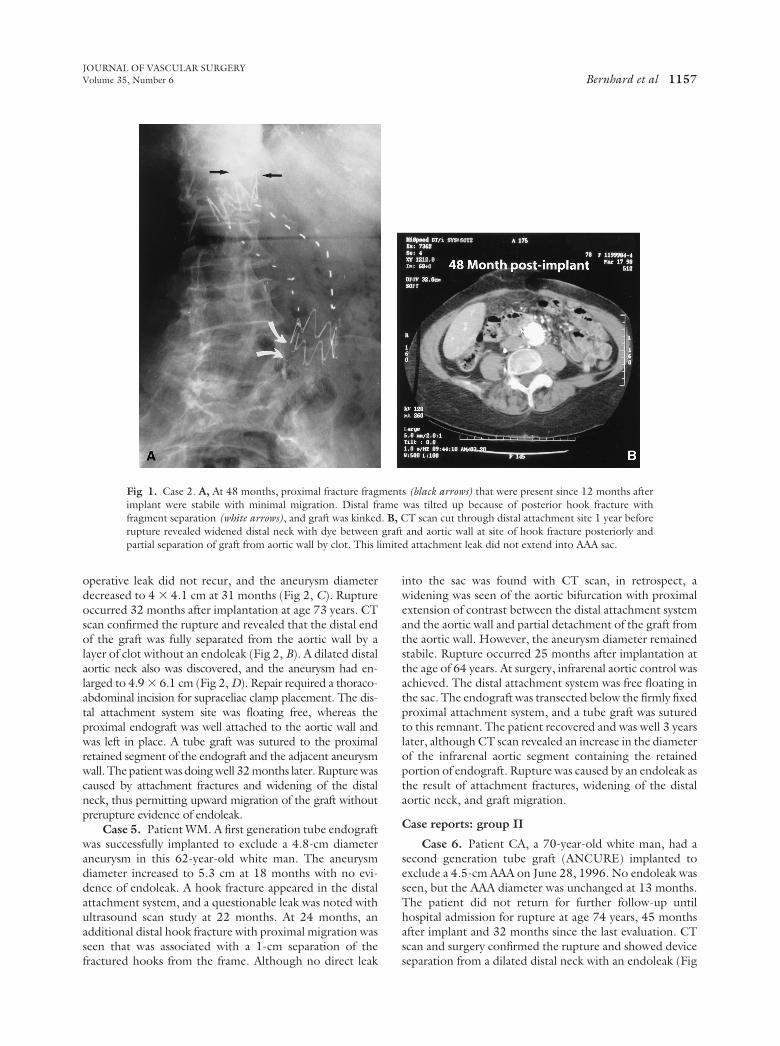
Case 2. Patient GW. On March 14, 1994, a firstgeneration tube endograft was deployed to exclude a 5-cmAAA in this 74-year-old white woman. A proximal leak,noted on the discharge CT scan, closed spontaneously. Anabdominal x-ray at 12 months revealed multiple fractures ofthe proximal attachment system without endoleak. At 48months, a distal attachment hook fracture was noted withupward displacement of the posterior portion of the attach-ment frame. (Fig 1, A). A distal endoleak, suggested withultrasound scan, was not confirmed with CT scan. How-ever, a retrospective review of scans obtained at 48 and 61months revealed widening of the distal neck with proximalextension of contrast between the attachment frame andthe aortic wall but not into the aneurysm sac (Fig 1, B). Amidgraft kink appeared at 4 years. The widest diameter ofthe AAA sac diminished to 2.9 cm. Rupture occurred 62months after implant, at age 79 years.
Infrarenal aortic control was achieved, after which anendoleak was shown on the posterior aspect of the distalattachment site. However, the endograft was firmly fixedproximally despite the fractures. The graft body was ligatedabove the endoleak, and the iliac arteries were ligated neartheir origins. A bifurcation graft was sutured to the retainedendograft with bypass to the distal iliac arteries. The patientdied with renal and respiratory failure on the 14th postop-erative day. Autopsy confirmed the operative findings.Rupture was caused by a distal type I leak that was related tohook fracture, a widened distal aortic neck, and migrationof the posterior aspect of the attachment system.
Case 3. Patient LW was a 61-year-old white manwhose 5-cm AAA was repaired with a first generation EVTtube endograft on July 6, 1994. A type I endoleak arisingfrom the distal attachment site was shown with ultrasoundscan 2 months after implant, and a distal attachment hookbreak was noted at 6 months. The leak was repeatedlyshown with ultrasound scan although not seen with aor-tography. The aneurysm diameter increased to 6 cm.Planned conversion to standard repair was postponed onthe basis of a myocardial infarction. Rupture occurred 19months after implant at age 63 years. The patient died withcardiac complications on the 2nd day after open repair. Thedistal attachment endoleak that had been present sinceimplantation was the cause of rupture. The hook fracturemay have been a contributing factor.
Case 4. Patient CM. The 5.9-cm AAA in this 70-year-old white man was treated with a first generation EVT tubegraft on October 6, 1994. A distal attachment site endoleakthat had been shown on completion arteriography closedspontaneously by 6 weeks. Distal attachment fractures werenoted at 22 and 26 months. In retrospect, progressiveseparation of the hooks from the distal frame was seen (Fig2, A), suggesting proximal migration. The immediate post-
JOURNAL OF VASCULAR SURGERYJune 20021156 Bernhard et al
operative leak did not recur, and the aneurysm diameterdecreased to 4 � 4.1 cm at 31 months (Fig 2, C). Ruptureoccurred 32 months after implantation at age 73 years. CTscan confirmed the rupture and revealed that the distal endof the graft was fully separated from the aortic wall by alayer of clot without an endoleak (Fig 2, B). A dilated distalaortic neck also was discovered, and the aneurysm had en-larged to 4.9 � 6.1 cm (Fig 2, D). Repair required a thoraco-abdominal incision for supraceliac clamp placement. The dis-tal attachment system site was floating free, whereas theproximal endograft was well attached to the aortic wall andwas left in place. A tube graft was sutured to the proximalretained segment of the endograft and the adjacent aneurysmwall. The patient was doing well 32 months later. Rupture wascaused by attachment fractures and widening of the distalneck, thus permitting upward migration of the graft withoutprerupture evidence of endoleak.
Case 5. Patient WM. A first generation tube endograftwas successfully implanted to exclude a 4.8-cm diameteraneurysm in this 62-year-old white man. The aneurysmdiameter increased to 5.3 cm at 18 months with no evi-dence of endoleak. A hook fracture appeared in the distalattachment system, and a questionable leak was noted withultrasound scan study at 22 months. At 24 months, anadditional distal hook fracture with proximal migration wasseen that was associated with a 1-cm separation of thefractured hooks from the frame. Although no direct leak
into the sac was found with CT scan, in retrospect, awidening was seen of the aortic bifurcation with proximalextension of contrast between the distal attachment systemand the aortic wall and partial detachment of the graft fromthe aortic wall. However, the aneurysm diameter remainedstabile. Rupture occurred 25 months after implantation atthe age of 64 years. At surgery, infrarenal aortic control wasachieved. The distal attachment system was free floating inthe sac. The endograft was transected below the firmly fixedproximal attachment system, and a tube graft was suturedto this remnant. The patient recovered and was well 3 yearslater, although CT scan revealed an increase in the diameterof the infrarenal aortic segment containing the retainedportion of endograft. Rupture was caused by an endoleak asthe result of attachment fractures, widening of the distalaortic neck, and graft migration.
Case reports: group II
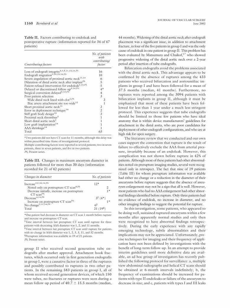
Case 6. Patient CA, a 70-year-old white man, had asecond generation tube graft (ANCURE) implanted toexclude a 4.5-cm AAA on June 28, 1996. No endoleak wasseen, but the AAA diameter was unchanged at 13 months.The patient did not return for further follow-up untilhospital admission for rupture at age 74 years, 45 monthsafter implant and 32 months since the last evaluation. CTscan and surgery confirmed the rupture and showed deviceseparation from a dilated distal neck with an endoleak (Fig
Fig 1. Case 2. A, At 48 months, proximal fracture fragments (black arrows) that were present since 12 months afterimplant were stabile with minimal migration. Distal frame was tilted up because of posterior hook fracture withfragment separation (white arrows), and graft was kinked. B, CT scan cut through distal attachment site 1 year beforerupture revealed widened distal neck with dye between graft and aortic wall at site of hook fracture posteriorly andpartial separation of graft from aortic wall by clot. This limited attachment leak did not extend into AAA sac.
JOURNAL OF VASCULAR SURGERYVolume 35, Number 6 Bernhard et al 1157

3, A). A suprarenal clamp was required, and the aneurysmwas successfully repaired with a tube graft. No evidence ofgraft or attachment system deterioration was found. Thecause of rupture was a type I endoleak arising from progres-sive dilatation of the distal aortic neck.
Case 7. Patient AK was a 93-year-old white womanwith a painful and tender 5-cm AAA. The patient refusedopen surgery and was a poor candidate for endograftplacement on the basis of a short (7-mm) proximal aortic
neck, a wide distal neck, and aneurysmal iliac arteries. Asecond generation (ANCURE) tube endograft was de-ployed through a left retroperitoneal conduit on January19, 2000. No endoleak was seen; nevertheless, the distalend of the graft appeared to be implanted in thrombus.Pain and tenderness disappeared, but the aneurysm re-mained pulsatile. Rupture, confirmed with CT scan with-out contrast, occurred at 6 months. The aneurysm diam-eter had increased to 7.8 cm, with wide separation of the
Fig 2. Case 4. A, Separation of distal fracture fragmentsfrom attachment frame was seen 31 months after implan-tation with development of bend in graft because of prox-imal migration of distal attachment site. B, Postrupture CTscan revealed dilated distal neck and separation of attach-ment system from aortic wall, surrounded by clot withoutendoleak. C, AAA diameter expanded from 4 � 4.1 cm 1month before rupture to 4.9 � 6.1 cm. D, At time ofrupture.
JOURNAL OF VASCULAR SURGERYJune 20021158 Bernhard et al
distal attachment system from the aortic neck and anendoleak. The patient refused intervention and diedshortly after admission. Rupture was caused by failure toachieve distal aortic attachment in a patient who wasanatomically a poor candidate for endograft therapy.
Review of published cases
We were able to collect 40 cases of ruptured AAA afterendograft repair that have been published in referencedjournals. The pertinent data relating to these ruptures plusthe seven patients with Guidant/EVT are fully described inan Appendix (online only). Bifurcation grafts had beeninserted in 30 patients, 12 patients received tube implants,and no information concerning the endograft configura-tion was recorded for five patients. The average age of the24 patients with available data was 73.8 years, and 22 ofthese patients (88%) were men. Endografts were providedby a diverse group of manufacturers (Appendix, onlineonly). The delay from implant to rupture ranged from 3days to 85 months, with a mean of 16.4 (� 16.8) months(median, 16 months).
The causes of rupture were described in varying detailfor 44 of the 47 reported cases (Table I). All were caused byendoleaks except for one patient with endotension that alsowas probably related to an underlying proximal endoleakon the basis of prerupture imaging that showed graft mi-gration. All type III endoleaks occurred in patients withmodular implants. Thirteen of the endoleaks were primary(ie, recognized at implantation), and 12 were type I.Among the primary type I endoleaks, rupture occurredwithin 1 month of implantation in five patients and within6 months in nine.
Factors that contributed to endoleak and rupture arelisted in Table II. In several instances, multiple problemsprevented an effective attachment seal. Postimplant aneu-rysm diameter was recorded in 21 of the 42 patients withmore than a 1-month follow-up period (Table III). Three
of the 13 patients with an increase in aneurysm diameterhad this change noted only with a postrupture CT scan. Ofthe nine patients with either no change in aneurysm diam-eter or smaller aneurysms, no postrupture imaging infor-mation was available in eight to determine whether a diam-eter change had occurred since the last prerupture CT scanhad been performed.
Abnormalities identified in follow-up imaging studiesand their times of appearance are recorded in Table IV. Anendoleak was not identified before rupture in 13 patients,but, in retrospect, a limited leak was identified in two ofthese patients. Only three patients had no evidence ofendoleak or any other imaging abnormality before rup-ture.4,14 Additional abnormalities were present in the re-maining 10 patients. However, almost half (11/23) wererecognized only after rupture had occurred (n � 4) or wereidentified in retrospect (n � 7).
Open surgical repair was performed in 40 of 47 patients(Table V). Suprarenal clamp application was required in73% (11/15 for whom information was available). Theperioperative mortality rate was 41%.
DISCUSSION
Ruptures after implantation of Guidant/EVT en-dografts have certain unique features. All occurred inpatients with tube grafts, and all were a consequence of atype I endoleak that developed at the distal aortic attach-ment site. Five occurred in the 93 patients who had tubegrafts that were followed for a mean of 41.8 months(median, 43 months) among the total of 103 patientswith first generation devices that were prone to developattachment mechanism fractures. The remaining tworuptures occurred in the subgroup of 166 patients of
Fig 3. Case 6. This CT scan showed ruptured aneurysm with dyeextravasation into perianeurysmal hematoma (circle 1). Distal aor-tic neck was dilated at level of lower end of attachment frame(upper arrow) and was source of distal endoleak (lower arrow).
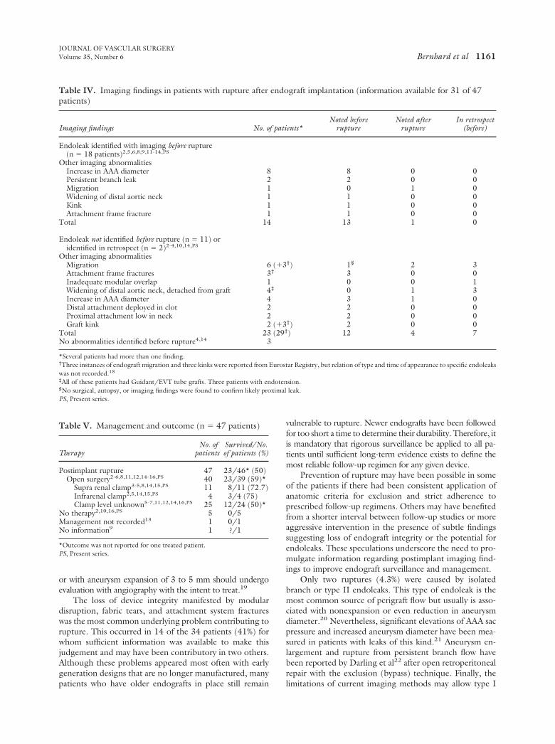
Table I. Causes of rupture (n � 44 of 47 patients; causenot reported in three patients)
Type and source of endoleaks No. of patients
Type I endoleak 27Proximal attachment3,5,9,10,12,14,16* 12†
Distal attachment 14Aorta2,5,16,17,18,PS 10†
Iliac12,14 4Site not reported13 1
Type II endoleak16 2Type III endoleak 11
Modular disconnection4,8,14,15 5 (2‡)Stent erosion through fabric11 1Details not reported16 5
Leak present, source not reported6,7 4
*Bibliographic references.†Three patients met criteria for endotension (AAA enlargement in absenceof detectable endoleak before AAA rupture). One had proximal leak shownat surgery.3 Another was classified as proximal leak on basis of knownmigration at proximal neck; however, no post rupture CT scan, surgery, orautopsy was found to verify this presumption.10 A third had initial increase inAAA diameter that remained stabile until rupture from distal aortic en-doleak. Endoleak was recognized in retrospect.PS
‡Associated fabric tear in Dacron graft wall and disruption of suturesattaching it to metal frame.PS, Present series.
JOURNAL OF VASCULAR SURGERYVolume 35, Number 6 Bernhard et al 1159
group II who received second generation tube en-dografts after market approval. Attachment hook frac-tures, which occurred only in first generation endograftsin group I, were a causative factor in three of the rupturesand possibly contributed to ruptures in two other pa-tients. In the remaining 583 patients in group I, all ofwhom received second generation devices, of which 150were tubes, no fractures or ruptures were seen during amean follow-up period of 40.7 � 15.5 months (median,
44 months). Widening of the distal aortic neck after endograftplacement was a significant issue, in addition to attachmentfracture, in four of the five patients in group I and was the onlycause of endoleak in one patient in group II. This problem hasbeen evaluated by Matsumura and Chaikof,17 who showedprogressive widening of the distal aortic neck over a 2-yearperiod after insertion of tube endografts.
Bifurcation endografts avoid the problems associatedwith the distal aortic neck. This advantage appears to beconfirmed by the absence of ruptures among the 433patients who received bifurcation and aortouniiliac im-plants in group I and have been followed for a mean of37.5 months (median, 41 months). Furthermore, noruptures were reported among the 3094 patients withbifurcation implants in group II, although it must beemphasized that most of these patients have been fol-lowed for less than 1 year under a much less stringentprotocol. This experience suggests that tube endograftsshould be limited to those few patients who have idealanatomy that is within device manufacturers’ guidelines forattachment in the distal aorta, who are poor candidates fordeployment of other endograft configurations, and who are athigh risk for open surgery.
The literature review that we conducted and our owncases support the contention that rupture is the result offailure to effectively exclude the AAA from arterial pres-sure, invariably because of an endoleak. However, thiscomplication was not shown before rupture in 42% ofpatients. Although most of these patients had other abnormal-ities noted on prerupture imaging studies, several were recog-nized only in retrospect. The fact that nine of 19 patients(Table III) for whom prerupture information was availablehad either no change or a reduction in the diameter of theiraneurysms before rupture suggests that the absence of aneu-rysm enlargement may not be a sign that all is well. However,most patients who had no AAA enlargement had other abnor-mal findings identified before rupture. Only three patients hadno evidence of endoleak, no increase in diameter, and noother imaging findings to suggest the potential for rupture.
In this investigation, some patients, who appeared tobe doing well, sustained ruptured aneurysms within a fewmonths after apparently normal studies and only thenwere recognized to have abnormal findings retrospec-tively. During the early experience with any rapidlyemerging technology, subtle abnormalities and theirimplications may not be appreciated. Unfortunately, pre-cise techniques for imaging and their frequency of appli-cation have not been defined by investigations with thebenefit of long-term follow-up. In an attempt to provideinterim guidelines until more definitive data are avail-able, an ad hoc group of investigators has recently pub-lished the following protocol for surveillance: a, multipleview abdominal radiographs and helical CT scans shouldbe obtained at 6-month intervals indefinitely; b, thefrequency of examinations should be increased for pa-tients with type II endoleaks or failure of the aneurysm todecrease in size; and c, patients with types I and III leaks
Table II. Factors contributing to endoleak andpostoperative rupture (information reported for 34 of 47patients)
Contributing factors
No. of patientswith
contributingfactor
Loss of endograft integrity4,5,8,11,14,16,PS 16Endograft migration5,10,14,16,PS 10Severe angulation of proximal aortic neck12,14 6Dilatation of distal aortic neck after implantPS 5Patient refused intervention for endoleak2,14,PS 5Delayed or discontinued follow-up3,14,PS 4*Surgical conversion delayed5,12,PS 4Poor patient selection 3
Wide distal neck lined with clot2,PS 2Iliac artery attachment site too wide12 1
Short proximal aortic neck14 2Error in deployment technique14 2Stiff graft body design14 2Proximal neck thrombus3 1Short distal aortic neck2 1Low graft implantation14 1AAA shrinkage14 1Total 63
*Two patients did not have CT scan for 11 months, although this delay waswithin prescribed time frame of investigational protocol.Multiple contributing factors were reported in several patients; two in sevenpatients, three in seven patients, and five in two patients.PS, Present series.
Table III. Changes in maximum aneurysm diameter inpatients followed for more than 30 days (informationrecorded for 21 of 42 patients)
Changes in diameter No. of patients
Increase3,5,10,14,PS 12*Noted only on postrupture CT scan8,PS 2Decrease initially, increase on postrupture
CT scanPS1*
Decrease4,14,PS 3† (4*)Increase on postrupture CT scanPS 1*
No change2,11,14,PS 6‡
Total 21 (19§)
*One patient had decrease in diameter on CT scan 1 month before ruptureand increase on postrupture CT scan.†Time interval between last prerupture CT scan until rupture for threepatients with decreasing AAA diameter was 1, 2, and 11 months.‡Time interval between last prerupture CT scan until rupture for patientswith no change in AAA diameter was 1, 2, 3, 3, 11, and 32 months.§Prerupture information was available in 19 of 21 patients.PS, Present series.
JOURNAL OF VASCULAR SURGERYJune 20021160 Bernhard et al
or with aneurysm expansion of 3 to 5 mm should undergoevaluation with angiography with the intent to treat.19
The loss of device integrity manifested by modulardisruption, fabric tears, and attachment system fractureswas the most common underlying problem contributing torupture. This occurred in 14 of the 34 patients (41%) forwhom sufficient information was available to make thisjudgement and may have been contributory in two others.Although these problems appeared most often with earlygeneration designs that are no longer manufactured, manypatients who have older endografts in place still remain
vulnerable to rupture. Newer endografts have been followedfor too short a time to determine their durability. Therefore, itis mandatory that rigorous surveillance be applied to all pa-tients until sufficient long-term evidence exists to define themost reliable follow-up regimen for any given device.
Prevention of rupture may have been possible in someof the patients if there had been consistent application ofanatomic criteria for exclusion and strict adherence toprescribed follow-up regimens. Others may have benefitedfrom a shorter interval between follow-up studies or moreaggressive intervention in the presence of subtle findingssuggesting loss of endograft integrity or the potential forendoleaks. These speculations underscore the need to pro-mulgate information regarding postimplant imaging find-ings to improve endograft surveillance and management.
Only two ruptures (4.3%) were caused by isolatedbranch or type II endoleaks. This type of endoleak is themost common source of perigraft flow but usually is asso-ciated with nonexpansion or even reduction in aneurysmdiameter.20 Nevertheless, significant elevations of AAA sacpressure and increased aneurysm diameter have been mea-sured in patients with leaks of this kind.21 Aneurysm en-largement and rupture from persistent branch flow havebeen reported by Darling et al22 after open retroperitonealrepair with the exclusion (bypass) technique. Finally, thelimitations of current imaging methods may allow type I
Table V. Management and outcome (n � 47 patients)
TherapyNo. of
patientsSurvived/No.of patients (%)
Postimplant rupture 47 23/46* (50)Open surgery2-6,8,11,12,14-16,PS 40 23/39 (59)*
Supra renal clamp3-5,8,14,15,PS 11 8/11 (72.7)Infrarenal clamp2,5,14,15,PS 4 3/4 (75)Clamp level unknown5-7,11,12,14,16,PS 25 12/24 (50)*
No therapy2,10,16,PS 5 0/5Management not recorded13 1 0/1No information9 1 ?/1
*Outcome was not reported for one treated patient.PS, Present series.
Table IV. Imaging findings in patients with rupture after endograft implantation (information available for 31 of 47patients)
Imaging findings No. of patients*Noted before
ruptureNoted after
ruptureIn retrospect
(before)
Endoleak identified with imaging before rupture(n � 18 patients)2,5,6,8,9,11-14,PS
Other imaging abnormalitiesIncrease in AAA diameter 8 8 0 0Persistent branch leak 2 2 0 0Migration 1 0 1 0Widening of distal aortic neck 1 1 0 0Kink 1 1 0 0Attachment frame fracture 1 1 0 0
Total 14 13 1 0
Endoleak not identified before rupture (n � 11) oridentified in retrospect (n � 2)2-4,10,14,PS
Other imaging abnormalitiesMigration 6 (�3†) 1§ 2 3Attachment frame fractures 3† 3 0 0Inadequate modular overlap 1 0 0 1Widening of distal aortic neck, detached from graft 4‡ 0 1 3Increase in AAA diameter 4 3 1 0Distal attachment deployed in clot 2 2 0 0Proximal attachment low in neck 2 2 0 0Graft kink 2 (�3†) 2 0 0
Total 23 (29†) 12 4 7No abnormalities identified before rupture4,14 3
*Several patients had more than one finding.†Three instances of endograft migration and three kinks were reported from Eurostar Registry, but relation of type and time of appearance to specific endoleakswas not recorded.18
‡All of these patients had Guidant/EVT tube grafts. Three patients with endotension.§No surgical, autopsy, or imaging findings were found to confirm likely proximal leak.PS, Present series.
JOURNAL OF VASCULAR SURGERYVolume 35, Number 6 Bernhard et al 1161
and type III leaks to masquerade as type II leaks. Therefore,type II endoleaks should be carefully monitored over thelong term with a plan for aggressive intervention in theevent of aneurysm expansion or graft migration.
Primary type I endoleaks have a tendency to rupture early,which supports Chuter’s recommendation to treat primaryleaks without delay.12 However, there were not enough casesand no reliable denominators for the various implant config-urations in our collective series to determine which attach-ment site is more vulnerable, and no information was reportedcorrelating leak severity with the likelihood for early rupture.
The frequency of rupture is difficult to assess becausemany cases were reported as isolated anecdotes or from sub-groups of patients with specific endograft designs that were nolonger used even in subsequent patients in the same series. Forinstance, five ruptures, 4.9%, were seen in the 103 patientswith first generation EVT grafts in group I (median follow-upperiod, 42 months), but none was seen in the next 583patients in this group who received second generation grafts(median follow-up period, 41 months). The remaining tworuptures occurred among the 3260 patients who had limitedfollow-up in group II and were confined to the subgroup of166 patients who received tube grafts. Harris et al16 calculateda rupture rate of 1.4% during the first year and 0.6% for thesecond year with life table analysis of the patients from theEurostar Registry, which included early and late generationsof devices from multiple sources. Similarly, Zarins, White, andFogarty14 noted a 0.4% risk for rupture during the first yearand a 2.6% risk during the second year after repair with twovariants of the AneuRx device (Medtronic AVE, Santa Rosa,Calif). Prospective long-term evaluation of specific devices willbe necessary to determine the reliability of endograft exclusionto prevent rupture.
The outcome of rupture after endograft repair is similarto that expected for patients without prior endografts whenthe overall experience reported herein is considered andthat reported from the Eurostar,16 AneuRx,14 and Guidantdatabases. Although the number of cases is small, suprare-nal clamp application did not appear to correlate with ahigher mortality rate.
REFERENCES
1. Parodi JC, Palmaz JC, Barone HD. Transfemoral intraluminal graftimplantation for abdominal aortic aneurysms. Ann Vasc Surg 1991;5:494-9.
2. Lumsden AB, Allen RC, Chaikof EL, Resnikoff M, Moritz MW, Ger-hard H, et al. Delayed rupture of aortic aneurysms following endovas-cular stent grafting. Am J Surg 1995;170:174-8.
3. Torsello GB, Klenk E, Kasprzak B, Umscheid T. Rupture of abdominalaortic aneurysm previously treated by endovascular stentgraft. J VascSurg 1998;28:184-7.
4. Alimi YS, Chakfe N, Rivoal E, Slimane KK, Valerio N, Riepe G, et al.Rupture of an abdominal aortic aneurysm after endovascular graftplacement and aneurysm size reduction. J Vasc Surg 1998;28:178-83.
5. May J, White GH, Waugh R, Chaufour X, Stephen MS, Yu W, et al.Rupture of abdominal aortic aneurysms: a concurrent comparison of
outcome of those occurring after endoluminal repair versus thoseoccurring de novo. Eur J Vasc Endovasc Surg 1999;18:344-8.
6. May J, White GH, Harris JP. Techniques for surgical conversion ofaortic endoprosthesis. Eur J Vasc Endovasc Surg 1999;18:284-9.
7. Riepe G, Heilberger P, Umschied T, Chakfe N, Raithel D, Stelter W, etal. Frame dislocation of body middle rings in endovascular stent tubegrafts. Eur J Vasc Endovasc Surg 1999;17:28-34.
8. Krohg-Sorensen K, Brekke M, Drolsum A, Kvernebo K. Periprostheticleak and rupture after endovascular repair of abdominal aortic aneu-rysm: the significance of device design for long-term results. J Vasc Surg1999;29:1152-8.
9. Parodi JC. Endovascular repair of abdominal aortic aneurysms andother arterial lesions. J Vasc Surg 1995;21:549-57.
10. Walker SR, Macierewicz J, MacSweeney ST, Gregson RHS, WhitakerSC, Wenham PW, et al. Mortality rates following repair of abdominalaortic aneurysms. J Endovasc Surg 1999;6:233-8.
11. Breek JC, Hamming JF, Lohle PNM, Lampmann LEH, van BergeHenigouwen DP. Spontaneous perforation of an aortic endoprosthesis.Eur J Vasc Endovasc Surg 1999;18:174-5.
12. Chuter TAM, Risberg B, Hopkinson BR, Wendt G, Scott AP, WalkerPJ, et al. Clinical experience with a bifurcated endovascular graft forabdominal aortic aneurysm repair. J Vasc Surg 1996;24:655-66.
13. Wain RE, Marin M, Ohki T, Sanchez LA, Lyon RT, Rozenblit A, et al.Endoleaks after endovascular graft treatment of aortic aneurysms: clas-sification, risk factors and outcome. J Vasc Surg 1998;27:69-80.
14. Zarins C, White RA, Fogarty TJ. Aneurysm rupture after endovascularrepair using the AneuRx stent graft. J Vasc Surg 2000;31:960-70.
15. Politz JK, Newman VS, Stewart MT. Late abdominal aortic aneurysmrupture after AneuRx repair. A report of three cases. J Vasc Surg2000;31:599-606.
16. Harris PL, Vallabhaneni SR, Desgranges P, Becquemin JP, van Mar-rewijk C, Laheij RJF. Incidence and risk factors of late rupture, conver-sion, and death after endovascular repair of infrarenal aortic aneurysms:the EUROSTAR experience. J Vasc Surg 2000;32:739-48.
17. Matsumura JS, Chaikof EL. Continued expansion of aortic necks afterendovascular repair of abdominal aortic aneurysms. J Vasc Surg 1998;28:422-31.
18. Brewster DC, Geller SC, Kaufman JA, Cambria RP, Gertler JP, LaMu-raglia GM, et al. Initial experience with endovascular aneurysm repair:comparison of early results with outcome of conventional open repair. JVasc Surg 1998;27:992-1005.
19. Eskandari MK, Yao JST, Pearce WH, Rutherford RB, Veith FJ, HarrisP, et al. Surveillance after endoluminal repair of abdominal aorticaneurysms. Cardiovasc Surg 2001;9:469-71.
20. Resch T, Ivancev K, Lindh M, Nyman U, Brunkwall L, Malina M, et al.Persistent collateral perfusion of abdominal aortic aneurysm after endo-vascular repair does not lead to progressive change in aneurysm diam-eter. J Vasc Surg 1998;28:242-9.
21. Velazquez OC, Baum RA, Carpenter JP, Golden MA, Cohn M, PyeronA, et al. Relationship between preoperative patency of inferior mesen-teric artery and subsequent occurrences of type II endoleak in patientsundergoing endovascular repair of abdominal aortic aneurysms. J VascSurg 2000;32:777-88.
22. Darling RC III, Ozsvath KK, Chang BB, Kreinberg PB, Paty PSK,Lloyd WE, et al. The incidence, natural history, and outcome ofsecondary intervention for persistent collateral flow in the excludedabdominal aortic aneurysm. J Vasc Surg 1999;30:968-76.
Submitted May 1, 2001; accepted Dec 10, 2001.
Please see the related commentary by Dr Elliot L.Chaikof on pages 1299-300.
Additional material for this article may be found online atwww.mosby.com/jvs.
JOURNAL OF VASCULAR SURGERYJune 20021162 Bernhard et al