Embed Size (px)
Citation preview
895
Acta Pharmacologica Sinica 2006 Jul; 27 (7): 895–900
©2006 CPS and SIMM
Full-length article
Role of inositol 1,4,5-trisphosphate receptors in α1-adrenergic receptor-induced cardiomyocyte hypertrophy1
Da-li LUO2,3,5, Jian GAO2, Xiao-mei LAN3, Gang WANG4, Sheng WEI4, Rui-ping XIAO4, Qi-de HAN3
2Department of Pharmacology, School of Chemical Biology & Pharmaceutical Sciences, Capital University of Medical Sciences, Beijing100069, China; 3Institute of Cardiovascular Science at Health Science Center, Peking University, Beijing 100083, China; 4Institute ofMolecular Medicine and College of Life Science, Peking University, Beijing 100871, China
AbstractAim: Intracellular Ca2+ plays pivotal roles in diverse cellular functions, includinggene transcription that underlies cardiac remodeling during stress responses.However, the role of inositol 1,4,5-trisphosphate receptors (IP3Rs) in the media-tion of cardiac intracellular Ca2+ and hypertrophic growth remains elusive. Priorwork with neonatal rat ventricular myocytes suggests that activation of IP3Rsmay be linked to α1 adrenergic receptor (α1AR) increased stereotyped Ca2+ sparkoccurrence and global Ca2+ oscillations. Thus, we hypothesized that Ca2+ releasethrough IP3Rs was necessary for α1AR-stimulated cardiac hypertrophy. Methods:We used myoinositol 1,4,5-trisphosphate hexakis (butyryloxymethyl) ester (IP3BM),a membrane-permeant ester of IP3, to activate IP3Rs directly, and Fluo 4/AM tomeasure intracellular Ca2+ signaling. Results: IP3BM (10 µmol·L-1) mimicked theeffects of phenylephrine, a selective agonist of α1AR, in increments in local Ca2+
spark release (especially in the perinuclear area) and global Ca2+ transientfrequencies. More importantly, IP3R inhibitors, 2-aminoethoxydiphenyl borateand Xestospongin C, abolished the IP3BM-induced Ca2+ responses, and signifi-cantly suppressed α1AR-induced cardiomyocyte hypertrophy assayed by cellsize, [3H] leucine incorporation and atrial natriuretic factor gene expression, dur-ing sustained (48 h) phenylephrine stimulation. Conclusion: These results,therefore, provide cellular mechanisms that link IP3R signaling to α1AR-stimulatedgene expression and cardiomyocyte hypertrophy.
Key wordsinositol 1,4,5-trisphosphate receptors; Ca2+
sparks; α1 adrenergic stimulation; cardiachypert rophy
1 Project supported by the National NaturalScience Foundation of China (No 30470692).5 Correspondence to Dr Da-li LUO.Ph n 86-10-8391-1519.Fax 86-10-8391-1520.E-mail [email protected]
Received 2006-04-18Accepted 2006-05-17
doi: 10.1111/j.1745-7254.2006.00382.x
IntroductionIn response to mechanical and neurohumoral stimuli, car-
diac muscle undergoes adaptive hypertrophic growth, char-acterized by increases in myocyte size, protein synthesisand re-expression of fetal cardiac genes in order to maintaincardiac output. Although initially beneficial, sustained car-diac hypertrophy can be deleterious because of increasedrisk for the development of heart failure and lethal arrhythmias[1,2].
It has been demonstrated in vivo and in vitro that, byactivating phospholipase C (PLC), Gq protein-coupled re-ceptors (GqCRs) relay the signals of mechanical overloadand neurohumoral factors, such as catecholamine, angio-tensin-II and endothelin-1, to initiating cellular hypertrophic
response[3,4]. PLC catalyzes the cleavage of polyphosphoino-sitide into dual signaling molecules, inositol 1,4,5-trisphosphate (IP3) and diacylglycerol (DAG), which, in turn,activate downstream protein kinases and phosphatases, suchas Ca2+/calmodulin (CaM)-dependent calcineurin[5,6] and CaMkinases (CaMKs)[7,8], and protein kinase C (PKC)[9]. Thesesignaling pathways then act independently or crosstalk witheach other, converging on the cellular hypertrophic re-sponses[2,9,10]. Of these, elevation in intracellular Ca2+ con-centration is implicated as an important factor to initiate andperpetuate GqCR-mediated cardiac hypertrophy via CaM/carcineurin- and CaMK-mediated activation of transcriptionfactors, such as nuclear factor of activated T-cells (NFAT)and myocyte enhancer factor-2 (MEF2)[5–9]. Understanding
896
Acta Pharmacologica Sinica ISSN 1671-4083Luo DL et al
the regulation of intracellular Ca2+ signaling by the PLC-IP3-IP3 receptor (IP3R) pathway is thus crucial to understandingcellular mechanisms responsible for GqCR-mediated cardiachypertrophy.
In the heart, the main source of Ca2+ in excitation-con-traction is controlled by ryanodine receptors (RyRs), whereasthe role of IP3Rs, which express much less than RyRs, re-mained obscure[11]. Recently, however, IP3Rs have beenfound to be abundantly expressed in the embryonic heart[12,13]
and in the adult heart under conditions of heart failure andchronic arrhythmias[14,15]. This implicates the roles of IP3R-mediated Ca2+ signaling in cardiac development or remodel-ing in response to stress. Therefore, the present study wasaimed to determine IP3R regulation of intracellular Ca2+
signaling. In particular, we intended to delineate possibleroles for IP3R Ca2+ signaling in hypertrophic growth inducedby sustained stimulation of a prototypical GqCR, α1 adrener-gic receptor (α1AR), in ventricular myocytes from the devel-oping heart.
Materials and methodsIsolation and culture of myocytes Neonatal rat ventricu-
lar myocytes (NRVMs) were isolated from 1 to 2-d-oldSprague-Dawley rats by enzymatic digestion with 0.1%trypsin (Hyclone) and 0.03% collagenase (WorthingtonBiochemical), as described in a previous study[16]. After in-cubating the cell-containing supernatant for 1.5 h to removefibroblasts, cells were plated onto laminin-treated 35-mmdishes at a density of 1.0×10³−1.2×10³ cells/mm2 unless speci-fied otherwise. The culture medium, Dulbecco’s modifiedEagle’s medium (DMEM) and Medium 199 (4:1) containing10% fetal bovine serum, 4 mmol·L-1 L-glutamine, 100 U/mLpenicillin and streptomycin, and 0.1 mmol·L-1 5-bromo-2-deoxyuridine (Roche Molecular Biochemicals), was replacedat 42 h with serum-free medium, and cells were further cul-tured for 6 h. Cultured cells exhibited >95% positive stainingfor α-actinin, and began spontaneous contraction 24–48 hinto culture.
Reverse transcription-polymerase chain reaction Myocytes were lysed with TRIzol reagent (Invitrogen) and clari-fied by centrifugation. Analysis of mRNA levels for atrialnatriuretic factor (ANF) and 18 s were performed with prim-ers designed to detect rat gene products. ANF detectionused primers 5'-TCCCAGGCCATATTGGAGCA-3' and 5'-CAGCGAGAGCCCTCAGT-3', generating a 306 bp fragm ent.Detection of mRNA 18 s used primers 5'-TGCAGCCCCGGA-CATCTAAG-3' and 5'-GGAAGGGCACCACCAGGAGT-3',generating a 317 bp fragment. RT-PCR reactions were per-formed with 2 µg of total RNA and followed by 30 cycles of
PCR amplification.Confocal Ca2+ imaging NRVMs were loaded with 4 µmol/L
Fluo-4/AM (Molecular Probes) in culture medium at 37 oC for30 min and then were washed with HEPES-buffered saltsolution (mmol·L-1: NaCl 135, KCl 5, MgCl2 1, CaCl2 1.8,HEPES 10 and glucose 11, with pH 7.4 adjusted by NaOH)for 20 min. Confocal images of Fluo-4 fluorescence (excitationat 488 nm and emission detection at >515 nm) were obtainedusing Leica SP2 inverted microscope equipped with a 63×oilimmersion objective (NA 1.4). Time-lapsed (xy, 1.63 s/frame)or linescan (xt, 2 ms/line, 0.15 µm/pixel) images were obtainedwith 1.5-µm axial resolution. Image data analysis used cus-tomer-devised routines coded in the Interactive Data Lan-guage (IDL, Research System). All experiments were per-formed at room temperature (22–24 oC).
Assay of cellular hypertrophy After 6-h culture in se-rum-free DMEM, NRVMs at a density of 1×10³/mm2 werestimulated with 10 µmol·L-1 phenylephrine (PE), a selectiveα1AR agonist, for 48 h to induce cell hypertrophy. In experi-ments with pharmacological pretreatment, designated re-agents were applied 10–30 min prior to PE application andthen kept in culture medium. Cell areas were measured byplanimetry with 3−5 frames/dish captured at ×40 magnifica-tion (Leica software) for >100 cells in each treatment. Rela-tive rates of protein synthesis were determined by incuba-tion of myocytes with 2 µCi·mL-1 [3H] leucine (Amersham,UK) for 6 h and then incubated in 10% trichloroacetic acidfor 30 min on ice to precipitate the protein. The precipitateswere washed twice with cold water and then were solubilizedin 0.1% SDS+0.3 mol·L-1 NaOH at 37 oC for 1 h. [3H]leucineincorporation was then quantified for radioactivity (BeckmanLS 1801) from triplicate aliquots of each sample.
Materials 2-Aminoethoxydiphenylborate (2-APB) andxestospongin C (Xe C) were from Calbiochem. Ryanodineand phenylephrine (PE) were purchased from Sigma-Aldrich.Myo-inositol 1,4,5-trisphosphate hexakis (butyryloxymethyl)ester (IP3BM) was synthesized as described in a previousstudy (purity >95%)[17].
Statistical analysis The data were analyzed and pre-sented as mean±SEM. When appropriate, statistical com-parison was carried out with two-way paired or unpairedStudent’s t-test. The accepted level of significance wasP<0.05.
ResultsIP3BM-induced potentiation of Ca2+ oscillations In a pre-
vious study (submitted to PNAS) we identified that IP3 for-mation and spontaneous global Ca2+ transients were in-creased in a dose-dependent manner after α1AR stimulation
Http://www.chinaphar.com Luo DL et al
897
with PE. Additionally, local Ca2+ sparks and waves, espe-cially in the perinuclear area, were enhanced significantlyupon PE stimulation, concomitant with the enriched distri-bution of IP3Rs in this area. More importantly, inhibition ofIP3Rs with membrane permeable IP3R inhibitors, 2-APB andXe C, significantly inhibited these PE effects, suggesting aninvolvement of IP3R activation for the PE-enhanced Ca2+ sig-naling in the developing cardiomyocytes.
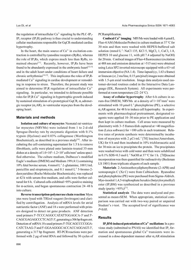
To confirm this hypothesis, we applied IP3BM, a mem-brane permeant ester of IP3 to activate IP3Rs directly[17], by-passing α1AR. First, we used a type of non-excitable cell,HEK293 cells, that dominantly express IP3Rs as the intracel-lular Ca2+ release channels to identify specific properties ofthis synthesized compound[18]. As reported[17], exposure ofIP3BM (2–25 µmol·L-1) for 5 min induced dose-dependentintracellular Ca2+ release in a Ca2+-free medium, with approxi-mately 70% of maximal Ca2+ release seen at a concentrationof 10 µmol·L-1 (data not shown). Thus, IP3BM (10 µmol·L-1)stimulating NRVMs continuously for 6 min was used through-out the following study. Figure 1A shows that the frequencyof spontaneous Ca2+ oscillations in minolayer NRVMs wasincreased in the presence of IP3BM, from 5.40±0.36 min-1 incontrol (n=12) to 14.50±1.38 min-1 (n=7, P<0.01 vs control).
Pretreatment of cells with 2-APB 4 µmol·L-1 or Xe C 10 µmol·L-1
for 10 min robustly inhibited IP3BM-induced potentiation ofCa2+ oscillations, whereas ryanodine, a RyRs inhibitor, at aconcentration of 30 µmol·L-1 failed to block the IP3BM effect(Figures 1B−D). These findings, therefore, provide directevidence for the IP3R signaling pathway involved in α1ARpotentiation of Ca2+ oscillations.
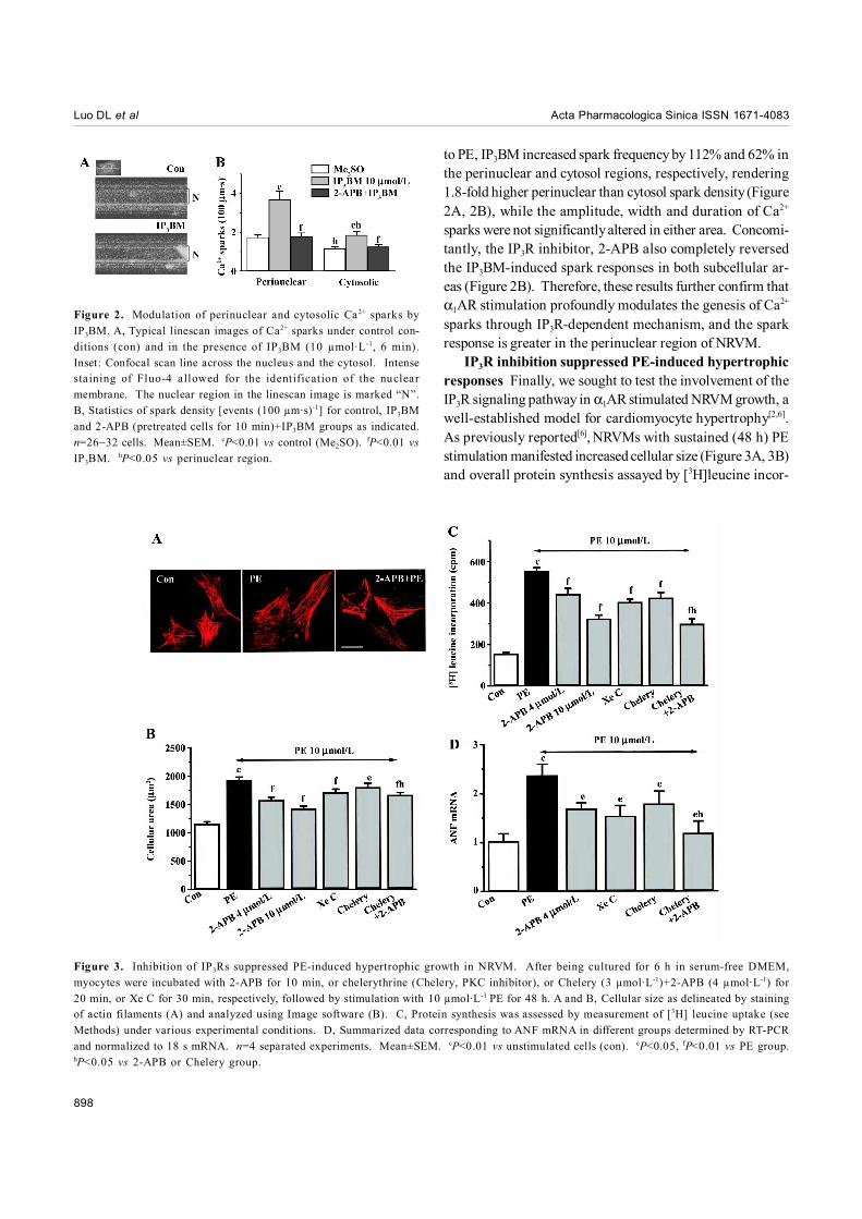
Subcellular responses of Ca2+ sparks to IP3BM stimu-lation Ca2+ sparks are thought to constitute the elementaryevents of Ca2+ waves and global Ca2+ transients[19]. Stimula-tion of NRVMs with PE (10 µmol·L-1) could increase the sparkproduction by 163% and 86.3% in the perinuclear and cyto-sol regions, respectively. To further identify the underlyingmolecular mechanism, we then tested this local Ca2+ responseto IP3BM using high-resolution linescan measurement acrossthe nucleus of a single NRVM. We defined “perinuclearsparks” as sparks whose centers were within the nucleusand its 1-µm flanking regions seen in linescan images (Figure2A); the rest were referred to as “cytosolic sparks”.
In IP3BM-unstimulated myocytes, cytosolic sparks oc-curred at a space-time density of 1.15±0.11 (100 µm·s)-1, andperinuclear sparks displayed a 51% higher density (1.74±0.12(100 µm·s)-1, n=32 cells, P<0.05 vs cytosolic events). Similar
Figure 1. Effect of IP3BM on spontaneous Ca2+ transients. A, Spontaneous Ca2+ oscillations prior to and 6 min after IP3BM 10 µmol·L-1
treatment in myocytes loaded with the Ca2+ indicator Fluo-4. B and C, Representative effects of 2-APB (B) and ryanodine (Rya, C) on basaland IP3BM-induced global Ca2+ oscillations. D, Effects of pretreatment with 2-APB, Rya and Xe C (at 4, 30 and 10 µmol·L-1 for 10, 10 and 30min, respectively) on IP3BM-induced spontaneous Ca2+ transients. Data are presented as percentage of vehicle control (Me2SO, 0 .1%).n=6−8 experiments. Mean±SEM. cP<0.01 vs Me2SO. fP<0.01 vs IP3BM group.
898
Acta Pharmacologica Sinica ISSN 1671-4083Luo DL et al
to PE, IP3BM increased spark frequency by 112% and 62% inthe perinuclear and cytosol regions, respectively, rendering1.8-fold higher perinuclear than cytosol spark density (Figure2A, 2B), while the amplitude, width and duration of Ca2+
sparks were not significantly altered in either area. Concomi-tantly, the IP3R inhibitor, 2-APB also completely reversedthe IP3BM-induced spark responses in both subcellular ar-eas (Figure 2B). Therefore, these results further confirm thatα1AR stimulation profoundly modulates the genesis of Ca2+
sparks through IP3R-dependent mechanism, and the sparkresponse is greater in the perinuclear region of NRVM.
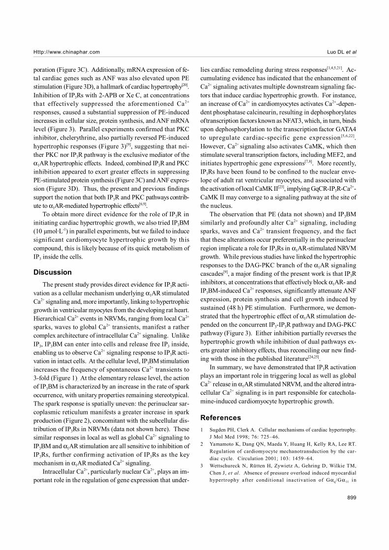
IP3R inhibition suppressed PE-induced hypertrophicresponses Finally, we sought to test the involvement of theIP3R signaling pathway in α1AR stimulated NRVM growth, awell-established model for cardiomyocyte hypertrophy[2,6].As previously reported[6], NRVMs with sustained (48 h) PEstimulation manifested increased cellular size (Figure 3A, 3B)and overall protein synthesis assayed by [3H]leucine incor-
Figure 2. Modulation of perinuclear and cytosolic Ca2+ sparks byIP3BM. A, Typical linescan images of Ca2+ sparks under control con-ditions (con) and in the presence of IP3BM (10 µmol·L-1, 6 min).Inset: Confocal scan line across the nucleus and the cytosol. Intensesta ining of Fluo-4 a llowed for the identification of the nuclearmembrane. The nuclear region in the linescan image is marked “N”.B, Statistics of spark density [events (100 µm·s)-1] for control, IP3BMand 2-APB (pretreated cells for 10 min)+IP3BM groups as indicated.n=26−32 cells. Mean±SEM. cP<0.01 vs control (Me2SO). fP<0.01 vsIP3BM. hP<0.05 vs perinuclear region.
Figure 3. Inhibition of IP3Rs suppressed PE-induced hypertrophic growth in NRVM. After being cultured for 6 h in serum-free DMEM,myocytes were incubated with 2-APB for 10 min, or chelerythrine (Chelery, PKC inhibitor), or Chelery (3 µmol·L-1)+2-APB (4 µmol·L-1) for20 min, or Xe C for 30 min, respectively, followed by stimulation with 10 µmol·L-1 PE for 48 h. A and B, Cellular size as delineated by stainingof actin filaments (A) and analyzed using Image software (B). C, Protein synthesis was assessed by measurement of [3H] leucine uptake (seeMethods) under various experimental conditions. D, Summarized data corresponding to ANF mRNA in different groups determined by RT-PCRand normalized to 18 s mRNA. n=4 separated experiments. Mean±SEM. cP<0.01 vs unstimulated cells (con). eP<0.05, fP<0.01 vs PE group.hP<0.05 vs 2-APB or Chelery group.
Http://www.chinaphar.com Luo DL et al
899
poration (Figure 3C). Additionally, mRNA expression of fe-tal cardiac genes such as ANF was also elevated upon PEstimulation (Figure 3D), a hallmark of cardiac hypertrophy[20].Inhibition of IP3Rs with 2-APB or Xe C, at concentrationsthat effectively suppressed the aforementioned Ca2+
responses, caused a substantial suppression of PE-inducedincreases in cellular size, protein synthesis, and ANF mRNAlevel (Figure 3). Parallel experiments confirmed that PKCinhibitor, chelerythrine, also partially reversed PE-inducedhypertrophic responses (Figure 3)[9], suggesting that nei-ther PKC nor IP3R pathway is the exclusive mediator of theα1AR hypertrophic effects. Indeed, combined IP3R and PKCinhibition appeared to exert greater effects in suppressingPE-stimulated protein synthesis (Figure 3C) and ANF expres-sion (Figure 3D). Thus, the present and previous findingssupport the notion that both IP3R and PKC pathways contrib-ute to α1AR-mediated hypertrophic effects[6,9].
To obtain more direct evidence for the role of IP3R ininitiating cardiac hypertrophic growth, we also tried IP3BM(10 µmol·L-1) in parallel experiments, but we failed to inducesignificant cardiomyocyte hypertrophic growth by thiscompound, this is likely because of its quick metabolism ofIP3 inside the cells.
DiscussionThe present study provides direct evidence for IP3R acti-
vation as a cellular mechanism underlying α1AR stimulatedCa2+ signaling and, more importantly, linking to hypertrophicgrowth in ventricular myocytes from the developing rat heart.Hierarchical Ca2+ events in NRVMs, ranging from local Ca2+
sparks, waves to global Ca2+ transients, manifest a rathercomplex architecture of intracellular Ca2+ signaling. UnlikeIP3, IP3BM can enter into cells and release free IP3 inside,enabling us to observe Ca2+ signaling response to IP3R acti-vation in intact cells. At the cellular level, IP3BM stimulationincreases the frequency of spontaneous Ca2+ transients to3-fold (Figure 1). At the elementary release level, the actionof IP3BM is characterized by an increase in the rate of sparkoccurrence, with unitary properties remaining stereotypical.The spark response is spatially uneven: the perinuclear sar-coplasmic reticulum manifests a greater increase in sparkproduction (Figure 2), concomitant with the subcellular dis-tribution of IP3Rs in NRVMs (data not shown here). Thesesimilar responses in local as well as global Ca2+ signaling toIP3BM and α1AR stimulation are all sensitive to inhibition ofIP3Rs, further confirming activation of IP3Rs as the keymechanism in α1AR mediated Ca2+ signaling.
Intracellular Ca2+, particularly nuclear Ca2+, plays an im-portant role in the regulation of gene expression that under-
lies cardiac remodeling during stress responses[1,4,5,21]. Ac-cumulating evidence has indicated that the enhancement ofCa2+ signaling activates multiple downstream signaling fac-tors that induce cardiac hypertrophic growth. For instance,an increase of Ca2+ in cardiomyocytes activates Ca2+-depen-dent phosphatase calcineurin, resulting in dephosphorylatesof transcription factors known as NFAT3, which, in turn, bindsupon dephosphorylation to the transcription factor GATA4to upregulate cardiac-specific gene expression[5,6,22].However, Ca2+ signaling also activates CaMK, which thenstimulate several transcription factors, including MEF2, andinitiates hypertrophic gene expressions[7,8]. More recently,IP3Rs have been found to be confined to the nuclear enve-lope of adult rat ventricular myocytes, and associated withthe activation of local CaMK II[23], implying GqCR-IP3R-Ca2+-CaMK II may converge to a signaling pathway at the site ofthe nucleus.
The observation that PE (data not shown) and IP3BMsimilarly and profoundly alter Ca2+ signaling, includingsparks, waves and Ca2+ transient frequency, and the factthat these alterations occur preferentially in the perinuclearregion implicate a role for IP3Rs in α1AR-stimulated NRVMgrowth. While previous studies have linked the hypertrophicresponses to the DAG-PKC branch of the α1AR signalingcascades[9], a major finding of the present work is that IP3Rinhibitors, at concentrations that effectively block α1AR- andIP3BM-induced Ca2+ responses, significantly attenuate ANFexpression, protein synthesis and cell growth induced bysustained (48 h) PE stimulation. Furthermore, we demon-strated that the hypertrophic effect of α1AR stimulation de-pended on the concurrent IP3-IP3R pathway and DAG-PKCpathway (Figure 3). Either inhibition partially reverses thehypertrophic growth while inhibition of dual pathways ex-erts greater inhibitory effects, thus reconciling our new find-ing with those in the published literature[24,25].
In summary, we have demonstrated that IP3R activationplays an important role in triggering local as well as globalCa2+ release in α1AR stimulated NRVM, and the altered intra-cellular Ca2+ signaling is in part responsible for catechola-mine-induced cardiomyocyte hypertrophic growth.
References1 Sugden PH, Clerk A. Cellular mechanisms of cardiac hypertrophy.
J Mol Med 1998; 76: 725–46.2 Yamamoto K, Dang QN, Maeda Y, Huang H, Kelly RA, Lee RT.
Regulation of cardiomyocyte mechanotransduction by the car-diac cycle. Circulation 2001; 103: 1459–64.
3 Wettschureck N, Rütten H, Zywietz A, Gehring D, Wilkie TM,Chen J, et al. Absence of pressure overload induced myocardialhypertrophy after conditional inactivation of Gαq/Gα11 in
900
Acta Pharmacologica Sinica ISSN 1671-4083Luo DL et al
cardiomyocytes. Nat Med 2001; 7: 1236–40.4 Sabri A, Wilson BA, Steinberg SF. Dual actions of the Gαq ago-
nist pasteurella multocida toxin to promote cardiomyocyte hy-pertrophy and enhance apoptosis susceptibility. Circ Res 2002;90: 850–7.
5 Crabtree GR. Generic signals and specific outcomes: signalingthrough Ca2+, calcineurin, and NF-AT. Cell 1999; 96: 611–4.
6 Taigen T, De Windt LJ, Lim HW, Molkentin JD. Targetedinhibition of calcineurin prevents agonist-induced cardiomyocytehypertrophy. Proc Natl Acad Sci USA 2000; 97: 1196–201.
7 Blaeser F, Ho N, Prywes R, Chatila TA. Ca2+-dependent geneexpression mediated by MEF2 transcription factors. J Biol Chem2000; 275: 197–209.
8 Akazawa H, Komuro I. Roles of cardiac transcription factors incardiac hypertrophy. Circ Res 2003; 92: 1079–88.
9 Muth JN, Bodi I, Lewis W, Varadi G, Schwartz A. A Ca2+-depen-dent transgenic model of cardiac hypertrophy: a role for proteinkinase Calpha. Circulation 2001; 103: 140–7.
1 0 De Windt LJ, Lim HW, Haq S, Force T, Molkentin JD. Calcineurinpromotes protein kinase C and c-Jun NH2-terminal kinase acti-vation in the heart. Cross-talk between cardiac hypertrophicsignaling pathways. J Biol Chem 2000; 275: 13571–9.
1 1 Marks AR. Cardiac intracellular calcium release channels−role inheart failure. Circ Res 2000; 87: 8–11.
1 2 Rosemblit N, Moschella MC, Ondriasa E, Gutstein DE, OndriasK, Marks AR. Intracellular calcium release channel expressionduring embryogenesis. Dev Biol 1999; 206: 163–77.
1 3 Kolossov E, Fleischmann BK, Liu Q, Bloch W, Viatchenko-Karpinski S, Manzke O, et al, Functional characteristics of EScell-derived cardiac precursor cells identified by tissue-specificexpression of the green fluorescent protein. J Cell Biol 1998;143: 2045–56.
1 4 Go LO, Moschella MC, Watras J, Handa KK, Fyfe BS, Marks AR.Differential regulation of two types of intracellular calcium re-lease channels during end-stage heart failure. J Clin Invest 1995;95: 888–94.
1 5 Ai X, Curran JW, Shannon TR, Bers DM, Pogwizd SM. Ca2+/
calmodulin-dependent protein kinase modulates cardiac ryanodinereceptor phosphorylation and sarcoplasmic reticulum Ca2+ leakin heart failure. Circ Res 2005; 97: 1314–22.
1 6 Chesley A, Lundberg MS, Asai T, Xiao RP, Ohtani S, Lakatta EG,et al. The beta(2)-adrenergic receptor delivers an antiapoptoticsignal to cardiac myocytes through G(i)-dependent coupling tophosphatidylinositol 3'-kinase. Circ Res 2000; 87: 1172–9.
1 7 Li W, Schultz C, Llopis J, Tsien RY. Membrane-permeant estersof inositol polyphosphates, chemical syntheses and biologicalapplications. Tetrahedron 1997; 53: 12017–40.
1 8 Venkatachalam K, van Rossum DB, Patterson RL, Ma HT, GillDL. The cellular and molecular basis of store-operated calciumentry. Nat Rev Mol Cell Biol 2002; 4: 263–72.
1 9 Cheng H, Lederer WJ, Cannel MB. Calcium sparks: the elemen-tary events underlying excitation-contraction coupling in heartmuscle. Science 1993; 262: 740–4.
2 0 Wang Y, Huang S, Sah VP, Ross J Jr, Brown JH, Han J, et al.Cardiac muscle cell hypertrophy and apoptosis induced by dis-tinct members of the p38 mitogen-activated protein kinase family.J Biol Chem 1998; 273: 2161–8.
2 1 Frey N, McKinsey TA, Olson EN. Decoding calcium signalsinvolved in cardiac growth and function. Nat Med 2000; 6:1221–7.
2 2 Molkentin JD, Lu JR, Antos CL, Markham B, Richardson J,Robbins J, et al. A calcineurin-dependent transcriptional path-way for cardiac hypertrophy. Cell 1998; 93: 215–28.
2 3 Bare DJ, Kettlun CS, Liang M, Ber DM, Mignery GA. Cardiactype 2 inositol 1 ,4 ,5-trisphosphate receptor: interaction andmodulation by calcium/calmodulin-dependent protein kinase II.J Biol Chem 2005; 280: 15912–20.
2 4 Wu GY, Toyokawa T, Hahn H, Dorn GW. Epsilon proteinkinase C in pathological myocardial hypertrophy. Analysis bycombined transgenic expression of translocation modifiers andGalphaq. J Biol Chem 2000; 275: 29927–30.
2 5 Pan J, Fukuda K, Saito M, Matsuzaki J, Kodama H, Sano M, et al.Mechanical stretch activates the JAK/STAT pathway in ra tcardiomyocytes. Circ Res 1999; 84: 1127–36.


![Cell cycle-coupled [Ca oscillations in mouse zygotes and ... · Cell cycle-coupled [Ca2+] i oscillations in mouse zygotes and function of the inositol 1,4,5-trisphosphate receptor-1](https://img.dokumen.tips/doc/110x75/5fb285c478c1117d6b731391/cell-cycle-coupled-ca-oscillations-in-mouse-zygotes-and-cell-cycle-coupled.jpg)


![PerkinElmer Life Sciences, Inc. · 2015-04-17 · PerkinElmer Life Sciences, Inc. Inositol-1,4,5-Trisphosphate [3H] Radioreceptor Assay Kit Catalog Number NEK064 For Laboratory Use](https://img.dokumen.tips/doc/110x75/5e6df5ebde16fe011c3cbedc/perkinelmer-life-sciences-inc-2015-04-17-perkinelmer-life-sciences-inc-inositol-145-trisphosphate.jpg)