Embed Size (px)
Citation preview
ROCK inhibition and CNTF interact on intrinsicsignalling pathways and differentially regulate survivaland regeneration in retinal ganglion cellsPaul Lingor,1,2,� LarsTo« nges,1,� Nicole Pieper,1Christina Bermel,1 Elisabeth Barski,1,2 Veronique Planchamp1,3
and Mathias Ba« hr1,2
1Department of Neurology,Georg-August-University Go« ttingen, University Medicine,Waldweg 33, 37073 Go« ttingen,2DFG Research Center for Molecular Physiology of the Brain (CMPB),Go« ttingen,Germany and 3European ResearchTraining Network (RTN) ‘Nervous System Repair’�These authors contributed equally to this work.
Correspondence to: Paul Lingor, MD, Department of Neurology,Georg-August-University Go« ttingen, University Medicine,Waldweg 33, 37073 Go« ttingen,Germany.E-mail: [email protected]
Functional regeneration in the CNS is limited by lesion-induced neuronal apoptosis and an environment inhibit-ing axonal elongation. A principal, yet unresolved question is the interaction between these two major factors.We thus evaluated the role of pharmacological inhibition of rho kinase (ROCK), a key mediator of myelin-derived axonal growth inhibition and CNTF, a potent neurotrophic factor for retinal ganglion cells (RGC), inmodels of retinal ganglion cell apoptosis and neurite outgrowth/regeneration in vitro and in vivo. Here, weshow for the first time that the ROCK inhibitor Y-27632 significantly enhanced survival of RGC in vitro andin vivo. In vitro, the co-application of CNTF and Y-27632 potentiated the effect of either substance alone.ROCK inhibition resulted in the activation of the intrinsic MAPK pathway, and the combination of CNTF andY-27632 resulted in even more pronounced MAPK activation.While CNTF also induced STAT3 phosphorylation,the additional application of ROCK inhibitor surprisingly diminished the effects of CNTF on STAT3 phosphory-lation. ROCK activity was also decreased in an additive manner by both substances. In vivo, both CNTF and Y-27632 enhanced regeneration of RGC into the non-permissive optic nerve crush model and additive effectswere observed after combination treatment. Further evaluation using specific inhibitors delineate STAT3 asa negative regulator of neurite growth and positive regulator of cell survival, while MAPK and Akt supportneurite growth. These results show that next to neurotrophic factors ROCK inhibition by Y-27632 potentlysupports survival of lesioned adult CNS neurons. Co-administration of CNTF and Y-27632 results in additiveeffects on neurite outgrowth and regeneration. The interaction of intracellular signalling pathways may,however, attenuate more pronounced synergy and has to be taken into account for future treatmentstrategies.
Keywords: retinal ganglion cells; CNTF; rho kinase; axotomy; regeneration
Abbreviations: ANOVA=analysis of variance; BSA=bovine serum albumin; CNTF=ciliary neurotrophic factor;CTB=choleratoxin subunit-B; DAPI=4,6-diamidino-2-phenylindole; DMEM=Dulbecco’s modified Eagle’s medium;EBSS=Earle’s balanced salt solution; FCS= fetal calf serum; GAP=growth associated protein; HRP=horse radishperoxidase; JAK= janus kinase; MAG=myelin-associated glycoprotein; MAPK=mitogen-activated protein kinase;MTT=3-(4,5-dimethylthiazol-2-yl)-2,5-diphenyltetrazoliumbromide; OMgp=oligodendrocyte-myelin glycoprotein;PBS=phosphate-buffered saline; PFA=paraformaldehyde; PKC=protein kinase C; PKN=protein kinase N; RGC=retinalganglion cell; ROCK=rho kinase; SDS^PAGE=sodium dodecyl sulphate ^polyacrylamide gel electrophoresis; STAT=signaltransducer and activator of transcription
Received July 5, 2007. Revised October 18, 2007. Accepted October 26, 2007. Advance Access publication December 5, 2007
doi:10.1093/brain/awm284 Brain (2008), 131, 250^263
� The Author (2007). Published by Oxford University Press on behalf of the Guarantors of Brain. All rights reserved. For Permissions, please email: [email protected]
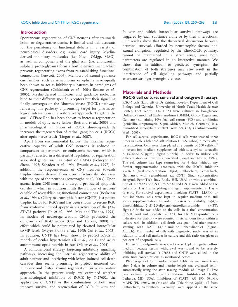
IntroductionSpontaneous regeneration of CNS neurons after traumaticlesion or degenerative demise is limited and this accountsfor the persistence of functional deficits in a variety ofneurological disorders, e.g. spinal cord injury. Myelin-derived inhibitory molecules (i.e. Nogo, OMgp, MAG),as well as components of the glial scar (i.e. chondroitinsulphate proteoglycans) form a hostile environment, whichprevents regenerating axons from re-establishing functionalconnections (Fawcett, 2006). Members of axonal guidancecue families, such as semaphorins or ephrins have equallybeen shown to act as inhibitory substrates in paradigms ofCNS regeneration (Goldshmit et al., 2004; Benson et al.,2005). Myelin-derived inhibitors and guidance moleculesbind to their different specific receptors but their signallingfinally converges on the Rho/rho kinase (ROCK) pathway,rendering this pathway a promising target for pharmaco-logical intervention in a restorative approach. Targeting thesmall GTPase Rho has been shown to increase regenerationin models of optic nerve lesion (Bertrand et al., 2005) andpharmacological inhibition of ROCK dose-dependentlyincreases the regeneration of retinal ganglion cells (RGCs)after optic nerve crush (Lingor et al., 2007).Apart from environmental factors, the intrinsic regen-
erative capacity of adult CNS neurons is reduced incomparison to peripheral or embryonic neurons, which ispartially reflected in a differential regulation of regenerationassociated genes, such as c-Jun or GAP43 (Schreyer andSkene, 1993; Schaden et al., 1994; Broude et al., 1997). Inaddition, the responsiveness of CNS neurons towardstrophic stimuli derived from growth factors also decreaseswith the age of the neurons (Avwenagha et al., 2003). Uponaxonal lesion CNS neurons undergo a protracted apoptoticcell death which in addition limits the number of neuronscapable of re-establishing functional connections (Berkelaaret al., 1994). Ciliary neurotrophic factor (CNTF) is a potenttrophic factor for RGCs and has been shown to rescue RGCfrom axotomy-induced apoptosis via activation of the JAK/STAT3 pathway (Ip et al., 1993; Mey and Thanos, 1993).In models of neuroregeneration, CNTF promoted theoutgrowth of RGC axons (Cui and Harvey, 2000), aneffect which could be potentiated by elevated intracellularcAMP levels (Meyer-Franke et al., 1995; Cui et al., 2003).In addition, CNTF has been shown to protect RGCs inmodels of ocular hypertension (Ji et al., 2004) and acuteautoimmune optic neuritis in rats (Maier et al., 2004).A combinatorial strategy targeting inhibitory signalling
pathways, increasing the intrinsic regenerative ability ofadult neurons and interfering with lesion-induced cell deathcascades thus seems promising to protect neuronal cellnumbers and foster axonal regeneration in a restorativeapproach. In the present study, we examined whetherpharmacological inhibition of ROCK by Y-27632, theapplication of CNTF or the combination of both mayimprove survival and regeneration of RGCs in vitro and
in vivo and which intracellular survival pathways aretriggered by each substance alone or by their interactions.Our results show that the theoretical distinction betweenneuronal survival, afforded by neurotrophic factors, andaxonal elongation, regulated by the Rho/ROCK pathway,cannot be maintained in a strict sense, since bothparameters are regulated in an interactive manner. Weshow, that in addition to predicted synergism, thecombination of both strategies may also result in theinterference of cell signalling pathways and partiallyattenuate stronger synergistic effects.
Materials and MethodsRGC-5 cell culture, survival and outgrowth assaysRGC-5 cells (kind gift of Dr Krishnamoorthy, Department of CellBiology and Genetics, University of North Texas Health Science
Center, Fort Worth, TX, USA) were cultured in low-glucoseDulbecco’s modified Eagle’s medium (DMEM; Gibco, Eggenstein,Germany) containing 10% fetal calf serum (FCS) and antibiotics
(penicillin, streptomycin; Sigma-Aldrich, Munich, Germany) in ahumidified atmosphere at 37�C with 5% CO2 (Krishnamoorthyet al., 2001).For cell-survival experiments, RGC-5 cells were washed three
times in Eagle’s balanced salt solution (EBSS; Gibco) directly aftertrypsinization. Cells were then plated at a density of 500 cells/cm2
in serum-free medium supplemented with succinyl concanavalinA (sConA; 50 mg/ml; Sigma-Aldrich) in order to induce celldifferentiation as previously described (Seigel and Notter, 1992).
The cell culture was kept serum-free for 6 days without anyfurther supplementation (control), with the ROCK inhibitorY-27632 (final concentration 10 mM; Calbiochem, Schwalbach,Germany), with recombinant rat CNTF (final concentration
50 ng/ml, PeproTech Inc., Rocky Hill, USA) or with a combina-tion of Y-27632 and CNTF. Y-27632 and CNTF were added to theculture on Day 1 after plating and again supplemented at Day 4
in vitro. For survival experiments involving STAT3-, MAPK- orAkt inhibitors, cells were kept in regular medium with fullserum supplementation. In order to assess cell viability, 3-(4,5-
dimethylthiazol-2-yl)-2,5-diphenyltetrazoliumbromide (MTT;Sigma-Aldrich) was added to the cells in a final concentrationof 500 mg/ml and incubated at 37�C for 1 h. MTT-positive cells
indicative for viability were counted in six random fields within aculture well. In addition, cell death was quantified after nuclearstaining with DAPI (4,6-diamidino-2-phenylindole) (Sigma-
Aldrich). The number of cells with fragmented nuclei was set inrelation to total cell number in culture and this ratio was given asper cent of apoptotic cells.For neurite outgrowth assays, cells were kept in regular culture
medium because serum withdrawal was found to be severelyinhibiting cell survival. Y-27632 and CNTF were added in the
same final concentrations as mentioned before.Photographs of four random visual fields per well were taken
after 2 days in culture and neurite length was evaluated semi-
automatically using the axon tracing module of ‘Image J’ (FreeJava software provided by the National Institutes of Health,Bethesda, MD, USA). Inhibitors of STAT3 (AG 490; 50 mM),MAPK (PD 98059, 50 mM) and Akt (Triciribine, 2 mM), all from
Calbiochem, Schwalbach, Germany, were applied at the same
ROCK inhibition and CNTF for RGC regeneration Brain (2008), 131, 250^263 251
time as Y-27632 and CNTF in neurite outgrowth and cell-deathexperiments.All cell culture experiments were performed in triplicate and
repeated at least three times.
Primary RGC culture, survivaland outgrowth assaysFor immunopurified rat RGC cultures, Wistar rat pups weresacrificed on postnatal Day 7–8. RGCs were purified according toa two-step panning protocol for Thy-1 to 499.5% purity asdescribed previously (Barres et al., 1988). Cells were culturedin serum-free neurobasal medium (Gibco), supplemented withB-27 supplement, pyruvate (Sigma-Aldrich), glutamine, cysteine,triiodothyronine, Sato (BSA, transferrin, progesterone, putrescine,sodium selenite) (Gibco), forskolin (final concentration 10 mM),human BDNF (final concentration 50 ng/ml) (Tebu, Offenbach,Germany), insulin (final concentration 5 mg/ml) (Sigma-Aldrich)and CNTF (final concentration 10 ng/ml) (Tebu). Cell culturesused for growth factor deprivation studies were initially plated infull medium after preparation and then were switched 24 h afterplating to medium as described in the following paragraphs.For survival assays primary RGC were plated at a density of
5000/well on 96well-plates (Sarstedt, Numbrecht, Germany)coated with poly-d-lysine. After incubation for 24 h in fullmedium the cells were deprived of neurotrophins by mediumchange and cultures were supplemented with ROCK inhibitorY-27632 (final concentration 10 mM), CNTF (final concentration50 ng/ml) or both. Viability was assessed after 2 days by countingMTT-positive RGC in six random microscopic fields within aculture well after addition of MTT (final concentration 500 mg/ml)and incubation at 37�C for 1 h.For outgrowth assays, cells were first grown in full culture
medium for 1 day and then withdrawn only from CNTF becausedeprivation of all neurotrophins resulted in a severely reducedcell survival rate. Neurite elongation was evaluated at Day 3in vitro after 2 days of treatment with Y-27632 (final concentra-tion of 10 mM) and/or recombinant rat CNTF (final concentrationof 50 ng/ml). Photographs of four random visual fields perculture well were taken and neurite length was evaluated semi-automatically using the axon tracing module of ‘Image J’(National Institutes of Health, Bethesda, MD, USA). All cellculture experiments have been performed in triplicate andrepeated at least three times.
Axotomy of the optic nerve, optic nervecrush and peripheral nerve graftAll animal experiments were carried out according to theregulations of the local animal research council and legislationof the State of Lower Saxony. In all animal experiments adultfemale Wistar rats (200–250 g; Charles River, Sulzfeld, Germany)were used.The anaesthesia was carried out by intraperitoneal injection
of chloral hydrate (420mg/kg body weight). A similar surgicalapproach was used in all three experimental paradigms to accessthe optic nerve: the skin was incised close to the superior orbitalrim and the orbita was opened leaving the supraorbital vein intact.The intraorbital glands were moved aside and the superiorextraocular muscles detached from their tendinous insertionpoints. The eye was then rotated in the ventral direction and
the optic nerve exposed by longitudinal incision of the opticnerve sheath.For axotomy experiments, the optic nerve was then transected
�2mm from the posterior eye pole taking care not to damagethe retinal blood supply. A 2� 2mm piece of gel foam(Braun, Melsungen, Germany) was soaked in FluoroGold(Hydroxystilbamidine; Bio-Trend, Cologne, Germany) andplaced on the optic nerve stump in order to retrogradely labelRGC. On Day 14 post-axotomy, animals were sacrificed by CO2
inhalation and the eyes were extracted. The cornea, the lens andthe vitreous body were removed, and the remaining eye cupcontaining the retina was fixed in 4% paraformaldehyde in PBS,pH 7.4 (PFA) for 1 h. Retinae were then extracted and flat-mounted in glycerol-PBS (1 : 1) on glass slides. The number ofFluoroGold-positive RGC was determined by fluorescence micro-scopy (Zeiss-Axioplan, Oberkochen, Germany) using a UV filter(365/420 nm). Three fields of 62 500 mm2 were counted in eachretinal quadrant (at eccentricities of one-sixth, one-half and five-sixths of the retinal radius). RGC counts were performedindependently by two different investigators according to ablinded protocol. Axotomy groups consisted of the followinganimal numbers: PBS: n= 4, Y-27632: n= 6, CNTF: n= 5,combination: n=6.For optic nerve crush experiments, the optic nerve was ligated
using a 10/0 suture (Ethicon, Johnson–Johnson, Livingston, UK)for 30 s, resulting in a complete transection of all RGC axons.The suture was removed and the operative access closed. Rats weresacrificed on Day 28 of the study by CO2 inhalation andimmediately perfused by transcardial injection of 250ml PBS and200ml 4% PFA. The eye and the optic nerve were removeden bloc from the orbit. The cornea, the lens and the vitreous bodywere removed and the remaining eye cup containing the retinaand the adjacent optic nerve were post-fixed in 4% PFA for 1 h.The tissue was then dehydrated in 30% sucrose overnight and keptat –20�C until further processing. Longitudinal sections (16 mm)of the optic nerve were prepared using a Leica cryostat andcollected on gelatine-coated glass slides. For evaluation of RGCaxon regeneration slides were immunostained for GAP-43 andphotomicrographs were taken using a fluorescence microscope(Axiovert 35, Zeiss, Germany). The number of regenerating axonsat designated distances from the crush was evaluated using acounting grid superimposed on the photomicrograph. Optic nervecrush groups consisted of the following animal numbers: PBS:n=7, Y-27632: n= 7, CNTF: n=6, combination: n= 4.For peripheral nerve autograft experiments the optic nerve was
transected �2mm from the posterior eye pole. A piece of 2.5 cmlength of the sciatic nerve was dissected from the left leg andgrafted to the optic nerve stump using a 10/0 suture. The distalpart of the sciatic nerve was placed in an osseous canal milled inadvance on the surface of the skull.Labelling of regenerating axons was achieved by intravitreal
injection of 3 ml Cy3-labeled choleratoxin subunit-B (CTB;Molecular Probes, Eugene, Oregon) into the vitreous chamber1 day before sacrificing the animal. Rats were sacrificed on Day 28of the study by CO2 inhalation and immediately perfused bytranscardial injection of 250ml PBS and 200ml 4% PFA. The eye,the optic nerve and the peripheral nerve graft were removeden bloc from the orbit. The cornea, the lens and the vitreous bodywere removed and the remaining eye cup containing the retina,the adjacent optic nerve and the graft were post-fixed in 4% PFAfor 1 h. The tissue was then dehydrated in 30% sucrose overnight
252 Brain (2008), 131, 250^263 P. Lingor et al.
and kept at –20�C until further processing. Longitudinal sectionsof the optic nerve stump and the adjacent graft (16 mm) wereprepared using a Leica cryostat and collected on gelatine-coatedglass slides. Evaluation of CTB fluorescence indicative forregenerating axons was performed directly from non-processedslides using a fluorescence microscope (Axiovert 35, Zeiss,Germany) and a filter for Cy3-fluorescence.For quantification of regenerating axon numbers, seven
regularly spaced sections in each graft were evaluated using agrid superimposed on the photomicrograph. Numbers of CTB-positive axons crossing perpendicular grid lines were counted atmultiples of 1mm from the grafting point until the end of thegraft. For each section the sum of all counted axons was calculatedand indicated as cumulative number of regenerating axons.Peripheral nerve graft groups consisted of the following animal
numbers: PBS: n=6, Y-27632: n= 5, CNTF: n= 4, combination:n= 5.After all surgical procedures retinal blood supply was verified by
fundoscopy and animals with persistent retinal ischaemia wereexcluded. All eyes were checked for cataracts induced by lensinjury after repeated intravitreal injections and animals wereexcluded from the study in case of cataract formation.
Dosage and application of Y-27632 andCNTF for in vivo experimentsAll intravitreal injections were performed using a glass microelec-trode connected to a Hamilton precision syringe, puncturing theeye at the cornea–sclera junction. Care was taken to not puncturethe lens. Vehicle or test substance measuring 3.5 ml were injectedat each injection time point. For ROCK inhibition, a 2.9mMsolution of Y-27632 in PBS was injected, resulting in anapplication of �3.3 mg of Y-27632 per injection. The concentrationof Y-27632 used in the present study was based on data from aprevious study where we have evaluated the dose-dependenteffects of Y-27632 on RGC regeneration (Lingor et al., 2007).For CNTF injections 1.5 mg of CNTF were administered in PBS.For combination treatments �3.3 mg of Y-27632 and 1.5 mg ofCNTF were administered in 3.5 ml PBS solution. In all experi-mental paradigms substance injections were performed threetimes: directly after surgery (Day 0), on Day 3 and Day 6 aftersurgery.
Western blotsRGC-5 cells were washed three times in EBSS after trypsinizationand kept in serum-free medium supplemented with concanavalinA on Day 1 in culture. Lysates were prepared on Day 3 after30min and 240min of treatment with the ROCK inhibitorY-27632 (final concentration 10 mM), with recombinant rat CNTF(final concentration 50 ng/ml) or with a combination of Y-27632and CNTF. For inhibition of MAPK signalling PD-98059 wasadded to the cell culture at a final concentration of 50mM. Thelysis buffer was composed of 10mM HEPES (pH 7.2), 142mMKCl, 5mM MgCl2, 1mM EGTA and 1% IGEPAL plus proteaseinhibitors (‘Complete tablets’, Roche). The protein content ofthe cell lysate samples was determined using the bicinchoninicacid assay (Pierce, Rockford, IL, USA) and equal amounts ofprotein (20mg) were loaded in each lane for a sodium dodecylsulphate–polyacrylamide gel electrophoresis (SDS–PAGE).Proteins were then transferred to a nitrocellulose membrane andblocked with 5% BSA in Tris-buffered saline/Tween-20 (TBS-T)
for 1 h. Membranes were then incubated with primary antibodies(anti-phospho-STAT3 (1 : 1000), anti-STAT3 (1:1000), anti-phospho-Akt (1:1000), anti-Akt (1 : 1000), anti-phospho-MAPK(1 : 1000), anti-MAPK (1 : 1000), (all Cell Signaling Technologies,Danvers, MA, USA) anti-ROCK-II (Dilution 1 : 250), anti-RhoA(Dilution 1 : 500) (both Santa Cruz, Heidelberg, Germany) for 24 h
at 4�C in TBS-T and 2% BSA or with anti-beta Tubulin (1 : 5000,Sigma, Munich, Germany) for 1 h at room temperature in TBS-Tand 5% BSA. This was followed by incubation with correspondinghorseradish peroxidase-coupled secondary antibodies (1 : 2000,for 1 h at room temperature; Dianova, Hamburg, Germany).ECL-Plus reagent (Amersham, Arlington Heights, IL, USA) wasapplied on the membrane and the chemiluminescence wasvisualized and quantified using a Fluor-S-max imager (Bio-Rad,Munich, Germany).
ImmunohistochemistryRetinal sections were dehumidified at 37�C for 1 h and antigenretrieval was performed for 4 h in TBS-T (pH 9.0) at 60�C.Unspecific binding was blocked by application of 10% new-borngoat or horse serum, and primary antibodies (anti-ROCK-II,1:250, sc-1851 (C20) Santa-Cruz; anti-GAP43, 1:500, Abcam) wereapplied at 4�C overnight. Secondary antibody (Alexa 546-labelledanti-goat, 1:200 or Cy2-labelled anti-rabbit; Dianova) was applied1:300 for 45min at room temperature. The sections were nuclearcounter-stained with DAPI (4,6-diamidino-2-phenylindole)
(Sigma-Aldrich) and mounted in Moviol (Hoechst, Frankfurt,Germany).
ROCK activity assaySimilar to western blot experiments, RGC-5 cells were cultured for3 days in serum-free medium supplemented with concanavalin Aon Day 1 and then treated for 30 or 240min with the ROCK
inhibitor Y-27632 (final concentration 10 mM), with recombinantrat CNTF (final concentration 50 ng/ml) or with a combination ofY-27632 and CNTF. The lysis buffer was composed of 20mMTris–HCl (pH 7.4), 1% Triton X-100 plus protease inhibitors(‘Complete tablets’, Roche). The protein content of the cell lysatesamples was determined using the bicinchoninic acid assay(Pierce). The ROCK activity assay was performed according themanufacturer’s instructions (CycLex, Nagano, Japan). Briefly,equal amounts of protein (50 ng) were applied to the assay platewells and incubated with ATP-containing kinase reaction bufferat 30�C for 30min. After washing and incubation with theHRP, conjugated antibody detection was performed at roomtemperature for 60min. After another washing step, the tetra-methylbenzidine containing substrate reagent was added and
incubated at room temperature for 10min until the reaction wasstopped with 0.5N sulphuric acid. The absorbance was measuredin a Tecan spectrophotometric plate reader (Tecan, Crailsheim,Germany) at a 450 nm single wavelength.
StatisticsAll in vitro experiments and western blots were reproduced at leastin triplicate. Animal groups were as indicated in the accordingmethods section. Differences between groups were consideredstatistically significant according to a one-way ANOVA followedby a parametric multiple comparison test (Dunnett test).
ROCK inhibition and CNTF for RGC regeneration Brain (2008), 131, 250^263 253
Significances were indicated with �P50.05, ��P50.01,���P50.001, unless otherwise stated.A comprehensive summary of all antibodies and inhibitors used
in this study is given in Supplementary Table S1.
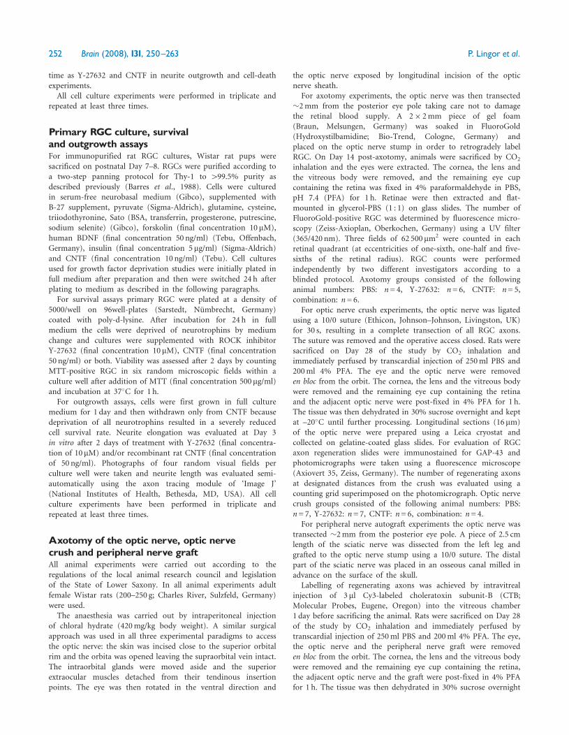
ResultsROCK inhibition potently increasesRGC survival in vitroIn order to examine the effect of ROCK inhibition on thesurvival of RGCs, we employed models of serum-deprivedRGC-5 cells and neurotrophin-deprived primary RGCs.We compared the effect of ROCK inhibition on cell survivalwith the application of a saturating amount of CNTF andthe combination of both substances. After 6-day serumdeprivation only 8.0%� 1.4% of the initial RGC-5 popula-tion were still viable (by MTT exclusion test) and77.8%� 2.8% showed fragmented apoptotic nuclei (DAPIstaining) in control cultures. Addition of the ROCK-inhibitor Y-27632 (10mM) to the culture medium increasedthe number of MTT-positive cells and decreased thenumber of cells with fragmented nuclei (25.3%� 3.5%and 53.8%� 1.8%, respectively). CNTF substitutioninduced a similar effect (27.4%� 1.7% and 53.1%� 3.0%,respectively). The combination of both substances resultedin even more pronounced survival (35.4%� 2.5% and42.8%� 2.4%, respectively), which was significantly higherthan in cultures treated with either substance alone (Fig. 1Aand B). Serum-deprived control cultures showed cellswith retracted processes and a tendency to form aggregates.Cells treated with Y-27632 exhibited less pronouncedmorphological signs of degeneration, which was similar toCNTF-treated cultures. Cultures treated with the combina-tion of Y-27632 and CNTF most closely resembled the non-serum deprived control, although numbers of survivingcells and apoptotic nuclei did not reach the equivalentnumbers of the control group (Fig. 1C).We compared the results obtained in the cell line with a
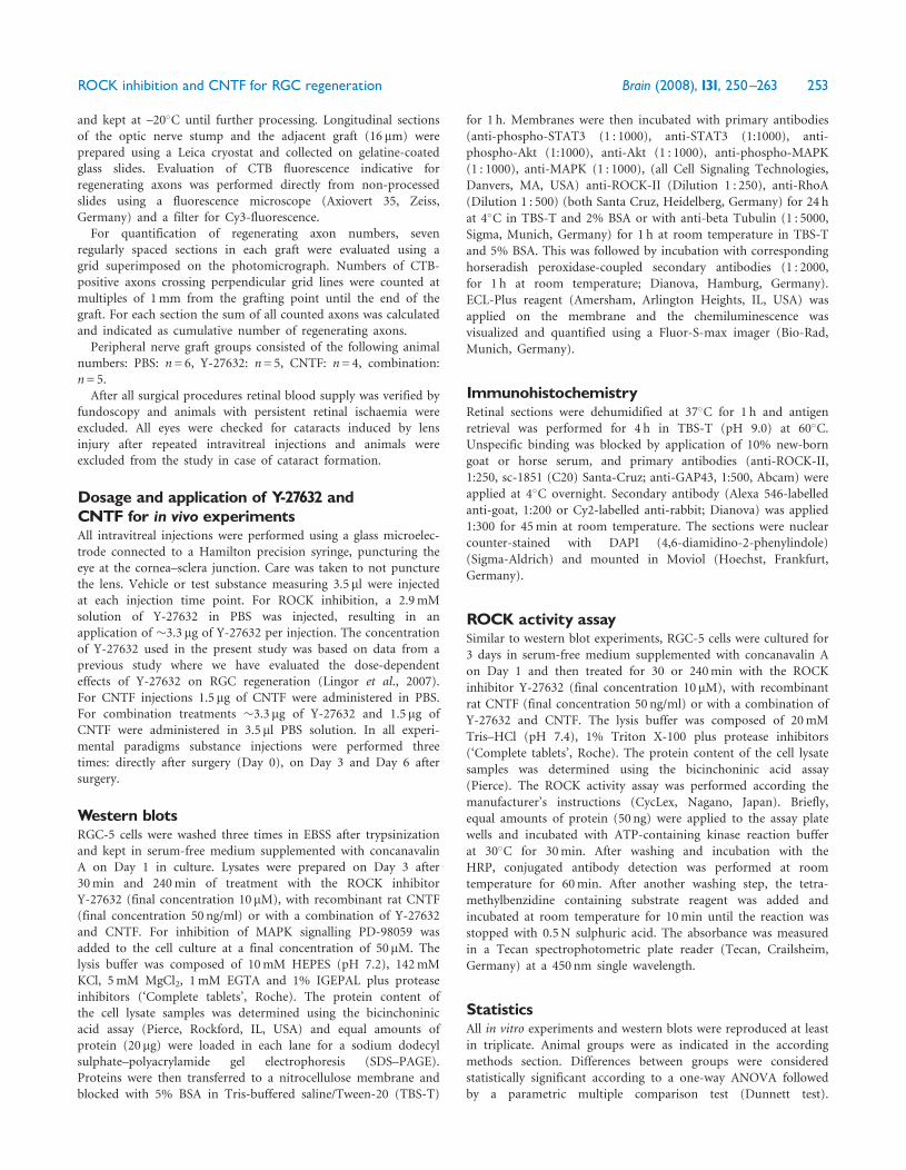
primary culture of RGCs which were neurotrophin-deprived for induction of apoptotic cell death. In controlcultures, only 33.5%� 2.2% of the cells were viable after 2days in culture (by MTT exclusion test) and 62.0%� 6.0%showed fragmented apoptotic nuclei (DAPI staining).Similar to the results obtained in the RGC-5 cell lineapplication of the ROCK inhibitor enhanced survivalafter neurotrophin deprivation (36.8%� 2.2%) thenumber of cells with fragmented nuclei was markedlydecreased (51.8%� 2.0%). Supplementation with CNTFsignificantly increased survival of primary RGC neurons(64.0%� 8.3%) and decreased the fragmentation of nuclei(44.6%� 1.9%). The combination of CNTF and Y-27632again resulted in increased survival and decreased nuclearfragmentation, although the additive effect of the combina-tion treatment was less pronounced compared to RGC-5cells (69.1%� 4.5% and 41.6%� 1.0%, respectively)(Fig. 2A–C).
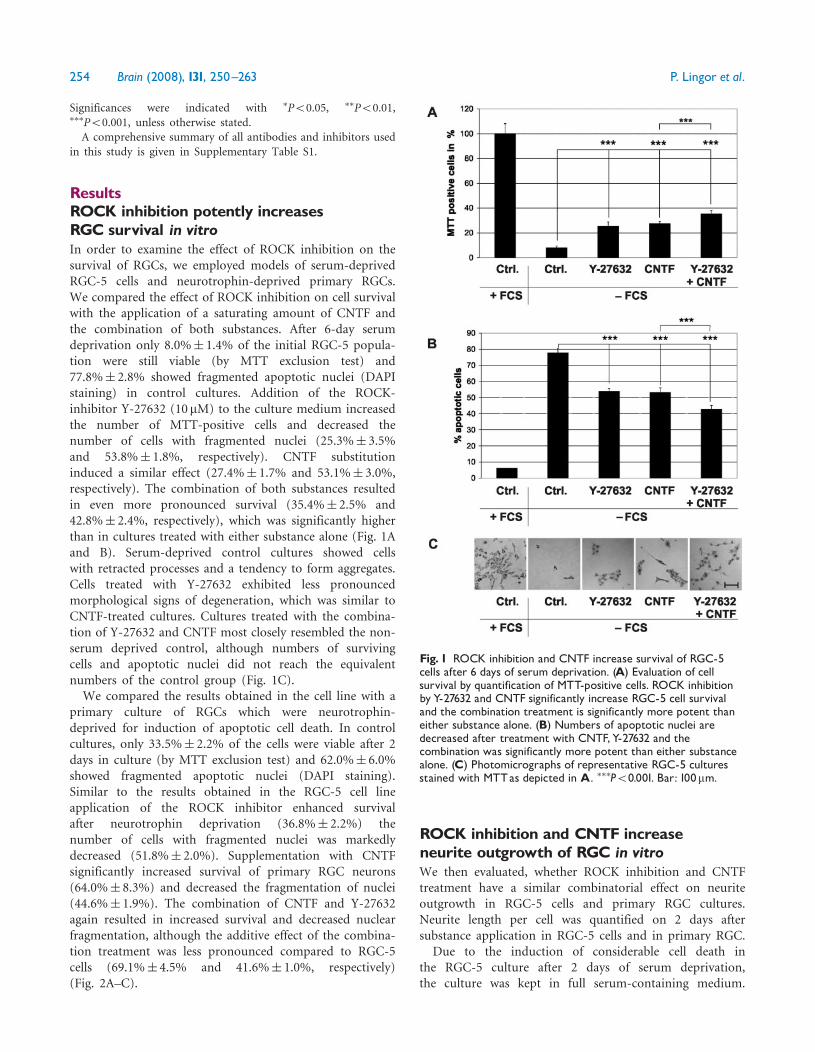
ROCK inhibition and CNTF increaseneurite outgrowth of RGC in vitroWe then evaluated, whether ROCK inhibition and CNTFtreatment have a similar combinatorial effect on neuriteoutgrowth in RGC-5 cells and primary RGC cultures.Neurite length per cell was quantified on 2 days aftersubstance application in RGC-5 cells and in primary RGC.
Due to the induction of considerable cell death inthe RGC-5 culture after 2 days of serum deprivation,the culture was kept in full serum-containing medium.
Fig. 1 ROCK inhibition and CNTF increase survival of RGC-5cells after 6 days of serum deprivation. (A) Evaluation of cellsurvival by quantification of MTT-positive cells. ROCK inhibitionby Y-27632 and CNTF significantly increase RGC-5 cell survivaland the combination treatment is significantly more potent thaneither substance alone. (B) Numbers of apoptotic nuclei aredecreased after treatment with CNTF,Y-27632 and thecombination was significantly more potent than either substancealone. (C) Photomicrographs of representative RGC-5 culturesstained with MTTas depicted in A. ���P50.001. Bar: 100mm.
254 Brain (2008), 131, 250^263 P. Lingor et al.
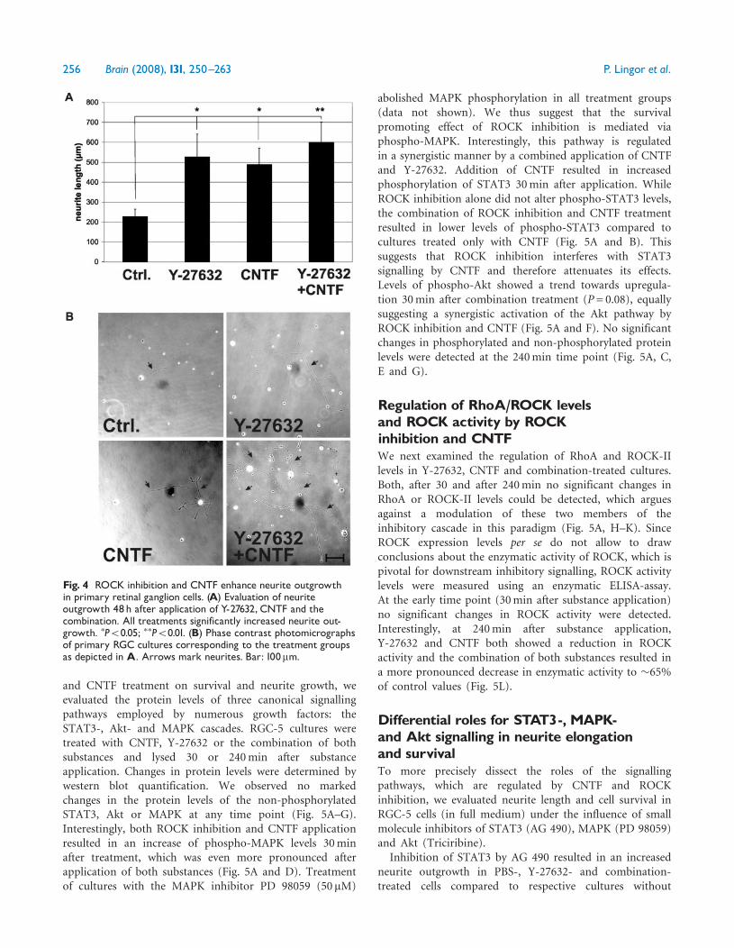
After 2 days of treatment with Y-27632, RGC-5 cellsshowed a significantly enhanced mean neurite outgrowth(34.6mm� 2.0 mm) compared to non-treated RGC-5 cells(24.2mm� 1.8 mm). Supplementation of the culturemedium by CNTF showed also increased neurite lengths(30.7mm� 1.5 mm). The combination treatment withY-27632 and CNTF resulted in significantly greater lengthof outgrowing neurites compared to treatment with eithersubstance alone (49.1 mm� 3.8 mm) (Fig. 3A and B).Primary RGCs cultured in CNTF-free medium displayed
a significantly increased neurite outgrowth after
supplementation with Y-27632 (526.2 mm� 115.2 mm) orCNTF (489.1 mm� 83.0 mm) as compared to control cells(228.1 mm� 36.1 mm). Supplementation of both Y-27632and CNTF enhanced neurite outgrowth to an even largerextent (600.6 mm� 100.9 mm), though the additional effectwas less pronounced than in the RGC-5 culture (Fig. 4Aand B).
Regulation of intrinsic survival pathwaysby ROCK inhibition and CNTFIn order to elucidate the intracellular signalling cascadesinvolved in the combinatorial effects of ROCK inhibition
Fig. 2 ROCK inhibition and CNTF increase survival of growthfactor-deprived primary retinal ganglion cells. (A) Survival after2 days of growth factor deprivation evaluated by quantification ofMTT-positive cells.Y-27632, CNTF and the combination of allsignificantly increased cell survival. (B) Decreased numbers ofapoptotic nuclei after application of CNTF,Y-27632 or thecombination of both. (C) Photomicrographs of primary RGCcorresponding to the treatment groups as depicted inA. �P50.05;���P50.001. Bar: 100mm.
Fig. 3 ROCK inhibition and CNTF enhance neurite outgrowthin RGC-5 cells. (A) Evaluation of neurite outgrowth 48h afterapplication of Y-27632, CNTF and the combination. All treatmentssignificantly increase neurite outgrowth.The dual treatmentdemonstrates an additive effect compared to the single treatment.��P50.01; ���P50.001. (B) Phase contrast photomicrographs ofrepresentative cultures corresponding to the treatment groups asdepicted in A. Bar: 50mm.
ROCK inhibition and CNTF for RGC regeneration Brain (2008), 131, 250^263 255
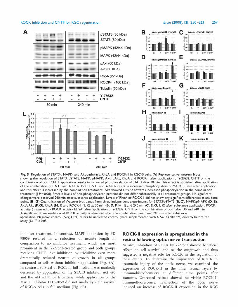
and CNTF treatment on survival and neurite growth, weevaluated the protein levels of three canonical signallingpathways employed by numerous growth factors: theSTAT3-, Akt- and MAPK cascades. RGC-5 cultures weretreated with CNTF, Y-27632 or the combination of bothsubstances and lysed 30 or 240min after substanceapplication. Changes in protein levels were determined bywestern blot quantification. We observed no markedchanges in the protein levels of the non-phosphorylatedSTAT3, Akt or MAPK at any time point (Fig. 5A–G).Interestingly, both ROCK inhibition and CNTF applicationresulted in an increase of phospho-MAPK levels 30minafter treatment, which was even more pronounced afterapplication of both substances (Fig. 5A and D). Treatmentof cultures with the MAPK inhibitor PD 98059 (50mM)
abolished MAPK phosphorylation in all treatment groups(data not shown). We thus suggest that the survivalpromoting effect of ROCK inhibition is mediated viaphospho-MAPK. Interestingly, this pathway is regulatedin a synergistic manner by a combined application of CNTFand Y-27632. Addition of CNTF resulted in increasedphosphorylation of STAT3 30min after application. WhileROCK inhibition alone did not alter phospho-STAT3 levels,the combination of ROCK inhibition and CNTF treatmentresulted in lower levels of phospho-STAT3 compared tocultures treated only with CNTF (Fig. 5A and B). Thissuggests that ROCK inhibition interferes with STAT3signalling by CNTF and therefore attenuates its effects.Levels of phospho-Akt showed a trend towards upregula-tion 30min after combination treatment (P= 0.08), equallysuggesting a synergistic activation of the Akt pathway byROCK inhibition and CNTF (Fig. 5A and F). No significantchanges in phosphorylated and non-phosphorylated proteinlevels were detected at the 240min time point (Fig. 5A, C,E and G).
Regulation of RhoA/ROCK levelsand ROCK activity by ROCKinhibition and CNTFWe next examined the regulation of RhoA and ROCK-IIlevels in Y-27632, CNTF and combination-treated cultures.Both, after 30 and after 240min no significant changes inRhoA or ROCK-II levels could be detected, which arguesagainst a modulation of these two members of theinhibitory cascade in this paradigm (Fig. 5A, H–K). SinceROCK expression levels per se do not allow to drawconclusions about the enzymatic activity of ROCK, which ispivotal for downstream inhibitory signalling, ROCK activitylevels were measured using an enzymatic ELISA-assay.At the early time point (30min after substance application)no significant changes in ROCK activity were detected.Interestingly, at 240min after substance application,Y-27632 and CNTF both showed a reduction in ROCKactivity and the combination of both substances resulted ina more pronounced decrease in enzymatic activity to �65%of control values (Fig. 5L).
Differential roles for STAT3-, MAPK-and Akt signalling in neurite elongationand survivalTo more precisely dissect the roles of the signallingpathways, which are regulated by CNTF and ROCKinhibition, we evaluated neurite length and cell survival inRGC-5 cells (in full medium) under the influence of smallmolecule inhibitors of STAT3 (AG 490), MAPK (PD 98059)and Akt (Triciribine).
Inhibition of STAT3 by AG 490 resulted in an increasedneurite outgrowth in PBS-, Y-27632- and combination-treated cells compared to respective cultures without
Fig. 4 ROCK inhibition and CNTF enhance neurite outgrowthin primary retinal ganglion cells. (A) Evaluation of neuriteoutgrowth 48h after application of Y-27632, CNTF and thecombination. All treatments significantly increased neurite out-growth. �P50.05; ��P50.01. (B) Phase contrast photomicrographsof primary RGC cultures corresponding to the treatment groupsas depicted in A. Arrows mark neurites. Bar: 100mm.
256 Brain (2008), 131, 250^263 P. Lingor et al.
inhibitor treatment. In contrast, MAPK inhibition by PD
98059 resulted in a reduction of neurite length in
comparison to no inhibitor treatment, which was most
prominent in the Y-27632-treated group and both groups
receiving CNTF. Akt inhibition by triciribine even more
dramatically reduced neurite outgrowth in all groups
compared to cells without inhibitor application (Fig. 6A).
In contrast, survival of RGCs in full medium was markedly
decreased by application of the STAT3 inhibitor AG 490
and the Akt inhibitor triciribine, while application of the
MAPK inhibitor PD 98059 did not markedly alter survival
of RGC-5 cells in full medium (Fig. 6B).
ROCK-II expression is upregulated in theretina following optic nerve transectionIn vitro, inhibition of ROCK by Y-27632 showed beneficialeffects on cell survival and neurite outgrowth and thussuggested a negative role for ROCK in the regulation ofthese events. To determine the importance of ROCK intraumatic injury of the optic nerve, we examined theexpression of ROCK-II in the inner retinal layers byimmunohistochemistry at different time points afteraxotomy. Untreated retinae showed no visible ROCK-IIimmunofluorescence. Transection of the optic nerveinduced an increase of ROCK-II expression in the RGC
Fig. 5 Regulation of STAT3-, MAPK- and Akt-pathways, RhoA and ROCK-II in RGC-5 cells. (A) Representative western blotsshowing the regulation of STAT3, pSTAT3, MAPK, pMAPK, Akt, pAkt, RhoA and ROCK-II after application of Y-27632, CNTF or thecombination of both.CNTF application results in increased phosphorylation of STAT3 after 30min.This effect is abolished after applicationof the combination of CNTF and Y-27632. Both CNTF and Y-27632 result in increased phosphorylation of MAPK 30min after applicationand this effect is increased by the combination treatment. Akt showed a trend towards increased phosphorylation in the combinationtreatment (x P=0.08). Protein levels of non-phosphorylated proteins did not differ substantially in all treatment groups. No significantchanges were observed 240min after substance application. Levels of RhoA or ROCK-II did not show any significant differences at any timepoint. (B ^G) Quantification of Western blot bands from three independent experiments for STAT3/pSTAT3 (B,C), MAPK/pMAPK (D, E),Akt/pAkt (F,G), RhoA (H, I) and ROCK-II (J,K) at 30min (B,D, F,H, J) and 240min (C, E,G, I,K) after substance application. ROCKactivity (measured by ROCK activity ELISA) after application of Y-27632, CNTF or the combination of both after 30 and 240min.A significant downregulation of ROCK activity is observed after the combination treatment 240min after substanceapplication. Negative control (Neg. Ctrl.) refers to untreated control lysate supplemented withY-27632 (200 nM) directly before theassay (L). �P50.05.
ROCK inhibition and CNTF for RGC regeneration Brain (2008), 131, 250^263 257
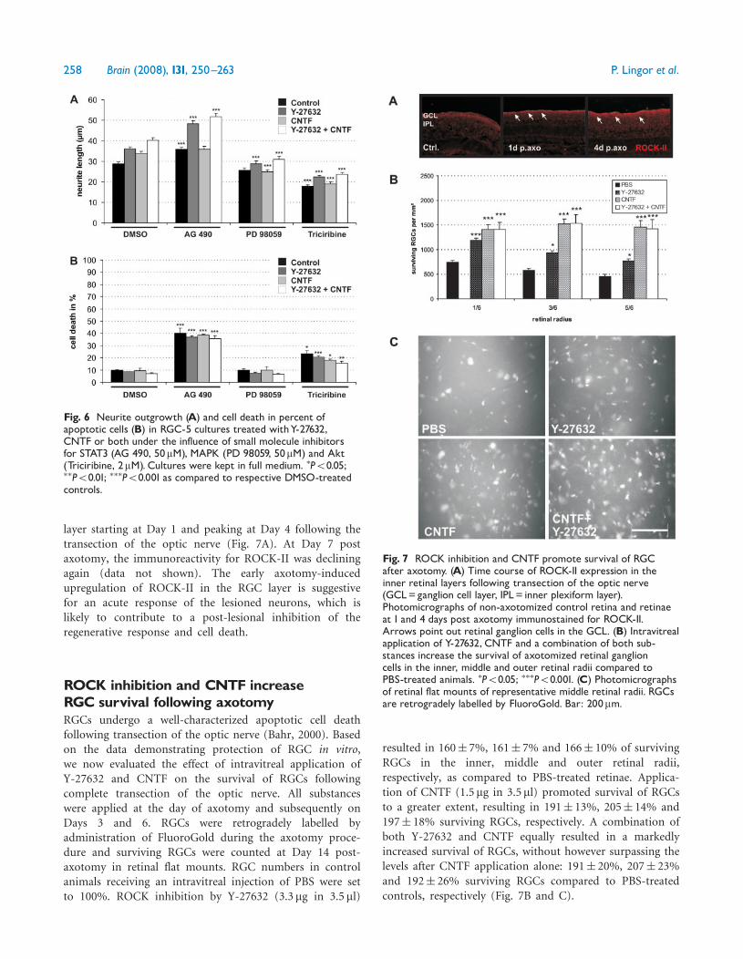
layer starting at Day 1 and peaking at Day 4 following thetransection of the optic nerve (Fig. 7A). At Day 7 postaxotomy, the immunoreactivity for ROCK-II was decliningagain (data not shown). The early axotomy-inducedupregulation of ROCK-II in the RGC layer is suggestivefor an acute response of the lesioned neurons, which islikely to contribute to a post-lesional inhibition of theregenerative response and cell death.
ROCK inhibition and CNTF increaseRGC survival following axotomyRGCs undergo a well-characterized apoptotic cell deathfollowing transection of the optic nerve (Bahr, 2000). Basedon the data demonstrating protection of RGC in vitro,we now evaluated the effect of intravitreal application ofY-27632 and CNTF on the survival of RGCs followingcomplete transection of the optic nerve. All substanceswere applied at the day of axotomy and subsequently onDays 3 and 6. RGCs were retrogradely labelled byadministration of FluoroGold during the axotomy proce-dure and surviving RGCs were counted at Day 14 post-axotomy in retinal flat mounts. RGC numbers in controlanimals receiving an intravitreal injection of PBS were setto 100%. ROCK inhibition by Y-27632 (3.3mg in 3.5 ml)
resulted in 160� 7%, 161� 7% and 166� 10% of survivingRGCs in the inner, middle and outer retinal radii,respectively, as compared to PBS-treated retinae. Applica-tion of CNTF (1.5 mg in 3.5 ml) promoted survival of RGCsto a greater extent, resulting in 191� 13%, 205� 14% and197� 18% surviving RGCs, respectively. A combination ofboth Y-27632 and CNTF equally resulted in a markedlyincreased survival of RGCs, without however surpassing thelevels after CNTF application alone: 191� 20%, 207� 23%and 192� 26% surviving RGCs compared to PBS-treatedcontrols, respectively (Fig. 7B and C).
Fig. 7 ROCK inhibition and CNTF promote survival of RGCafter axotomy. (A) Time course of ROCK-II expression in theinner retinal layers following transection of the optic nerve(GCL=ganglion cell layer, IPL= inner plexiform layer).Photomicrographs of non-axotomized control retina and retinaeat 1 and 4 days post axotomy immunostained for ROCK-II.Arrows point out retinal ganglion cells in the GCL. (B) Intravitrealapplication of Y-27632, CNTF and a combination of both sub-stances increase the survival of axotomized retinal ganglioncells in the inner, middle and outer retinal radii compared toPBS-treated animals. �P50.05; ���P50.001. (C) Photomicrographsof retinal flat mounts of representative middle retinal radii. RGCsare retrogradely labelled by FluoroGold. Bar: 200mm.
Fig. 6 Neurite outgrowth (A) and cell death in percent ofapoptotic cells (B) in RGC-5 cultures treated withY-27632,CNTF or both under the influence of small molecule inhibitorsfor STAT3 (AG 490, 50mM), MAPK (PD 98059, 50mM) and Akt(Triciribine, 2mM). Cultures were kept in full medium. �P50.05;��P50.01; ���P50.001 as compared to respective DMSO-treatedcontrols.
258 Brain (2008), 131, 250^263 P. Lingor et al.
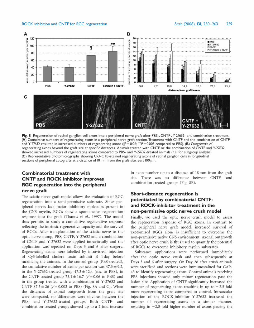
Combinatorial treatment withCNTF and ROCK inhibitor improvesRGC regeneration into the peripheralnerve graftThe sciatic nerve graft model allows the evaluation of RGCregeneration into a semi-permissive substrate. Since per-ipheral nerves lack major inhibitory molecules present inthe CNS myelin, RGCs show a spontaneous regenerationresponse into the graft (Thanos et al., 1997). The modelthus permits to study a cumulative regenerative responsereflecting the intrinsic regenerative capacity and the survivalof RGCs. After transplantation of the sciatic nerve to theoptic nerve stump, PBS, CNTF, Y-27632 and a combinationof CNTF and Y-27632 were applied intravitreally and theapplication was repeated on Days 3 and 6 after surgery.Regenerating axons were labelled by intravitreal injectionof Cy3-labelled cholera toxin subunit B 1 day beforesacrificing the animals. In the control group (PBS-treated),the cumulative number of axons per section was 47.3� 9.2,in the Y-27632-treated group 47.3� 12.4 (n.s. to PBS), inthe CNTF-treated group 73.1� 16.7 (P= 0.06 to PBS) andin the group treated with a combination of Y-27632 andCNTF 87.5� 26 (P= 0.003 to PBS) (Fig. 8A and C). Whenthe distances of axonal outgrowth from the graft sitewere compared, no differences were obvious between thePBS- and Y-27632-treated groups. Both CNTF- andcombination-treated groups showed up to a 2-fold increase
in axon number up to a distance of 18mm from the graftsite. There was no difference between CNTF- andcombination-treated groups (Fig. 8B).
Short-distance regeneration ispotentiated by combinatorial CNTF-and ROCK-inhibitor treatment in thenon-permissive optic nerve crush modelFinally, we used the optic nerve crush model to assessthe regeneration response of RGC axons. In contrast tothe peripheral nerve graft model, increased survival ofaxotomized RGCs alone is insufficient to overcome thenon-permissive native CNS environment. Axonal outgrowthafter optic nerve crush is thus used to quantify the potentialof RGCs to overcome inhibitory myelin substrates.
Substance applications were performed immediatelyafter the optic nerve crush and then subsequently atDays 3 and 6 after surgery. On Day 28 after crush animalswere sacrificed and sections were immunostained for GAP-43 to identify regenerating axons. Control animals receivingPBS injections showed only minor regeneration past thelesion site. Application of CNTF significantly increased thenumber of regenerating axons resulting in up to �2.5-foldmore regenerating axons compared to control. Intraocularinjection of the ROCK-inhibitor Y-27632 increased thenumber of regenerating axons in a similar manner,resulting in �2.5-fold higher number of axons passing the
Fig. 8 Regeneration of retinal ganglion cell axons into a peripheral nerve graft after PBS-, CNTF-, Y-27632- and combination treatment.(A) Cumulative numbers of regenerating axons in a peripheral nerve graft section. Treatment with CNTF and the combination of CNTFand Y-27632 resulted in increased numbers of regenerating axons (xP=0.06; ��P=0.003 compared to PBS). (B) Outgrowth ofregenerating axons beyond the graft site at specific distances. Animals treated with CNTF or the combination of CNTF and Y-27632showed increased numbers of regenerating axons compared to PBS- and Y-27632-treated animals (n.s. for subgroup analysis)(C) Representative photomicrographs showing Cy3-CTB-stained regenerating axons of retinal ganglion cells in longitudinalsections of peripheral autografts at a distance of 10mm from the graft site. Bar: 100mm.
ROCK inhibition and CNTF for RGC regeneration Brain (2008), 131, 250^263 259
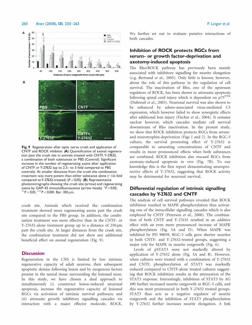
crush site. Animals which received the combinationtreatment showed more regenerating axons past the crushsite compared to the PBS group. In addition, the combi-nation treatment was more effective than in the CNTF- orY-27632-alone treatment group up to a distance of 250 mmpast the crush site. At larger distances from the crush site,the combination treatment did not show any additionalbeneficial effect on axonal regeneration (Fig. 9).
DiscussionRegeneration in the CNS is limited by low intrinsicregenerative capacity of adult neurons, their subsequentapoptotic demise following lesion and by exogenous factorspresent in the neural tissue surrounding the lesioned axon.In this study, we have chosen a dual approach tosimultaneously (i) counteract lesion-induced neuronalapoptosis, increase the regenerative capacity of lesionedRGCs via activation of intrinsic survival pathways; and(ii) attenuate growth inhibitory signalling cascades viainteraction with a major effector molecule, ROCK.
We further set out to evaluate putative interactions ofboth cascades.
Inhibition of ROCK protects RGCs fromserum- or growth factor-deprivation andaxotomy-induced apoptosisThe Rho/ROCK pathway has previously been mainlyassociated with inhibitory signalling for neurite elongation(e.g. Bertrand et al., 2005). Only little is known, however,about the role of this pathway in the regulation of cellsurvival. The inactivation of Rho, one of the upstreamregulators of ROCK, has been shown to attenuate apoptosisfollowing spinal cord injury which is dependent on p75NTR
(Dubreuil et al., 2003). Neuronal survival was also shown tobe enhanced by adeno-associated virus-mediated C3expression, which however failed to show synergistic effectsafter additional lens injury (Fischer et al., 2004). It remainsunclear however, which cascades mediate cell survivaldownstream of Rho inactivation. In the present study,we show that ROCK inhibition protects RGCs from serum-and neurotrophin-deprivation (Figs 1 and 2). In the RGC-5culture, the survival promoting effect of Y-27632 iscomparable to saturating concentrations of CNTF andresults in more pronounced effects when both substancesare combined. ROCK inhibition also rescued RGCs fromaxotomy-induced apoptosis in vivo (Fig. 7B). To ourknowledge this is the first report demonstrating neuropro-tective effects of Y-27632, suggesting that ROCK activitymay be detrimental for neuronal survival.
Differential regulation of intrinsic signallingcascades byY-27632 and CNTFThe analysis of cell survival pathways revealed that ROCKinhibition resulted in MAPK phosphorylation thus activat-ing one of the intracellular signalling cascades which is alsoemployed by CNTF (Peterson et al., 2000). The combina-tion of both CNTF and Y-27632 resulted in an additiveeffect with an even more pronounced increase of MAPKphosphorylation (Fig. 5A and D). When MAPK wasinhibited by PD 98059, RGC-5 cells grew shorter neuritesin both CNTF- and Y-27632-treated groups, suggesting amajor role for MAPK in neurite outgrowth (Fig. 6).
Levels of pSTAT3 were not markedly altered byapplication of Y-27632 alone (Fig. 5A and B). However,when cultures were treated with a combination of Y-27632and CNTF, phosphorylation of STAT3 was markedlyreduced compared to CNTF-alone treated cultures suggest-ing that ROCK inhibition results in the attenuation of theSTAT3 response. Interestingly, inhibition of STAT3 by AG490 further increased neurite outgrowth in RGC-5 cells, andthis was most pronounced in both Y-27632 treated groups.STAT3 thus acts as a negative regulator of neuriteoutgrowth and the inhibition of STAT3 phosphorylationby Y-27632 further increases neurite elongation. A link
Fig. 9 Regeneration after optic nerve crush and application ofCNTF and ROCK inhibitor. (A) Quantification of axonal regenera-tion past the crush site in animals treated with CNTF,Y-27632,a combination of both substances or PBS (Control). Significantincrease in the number of regenerating axons after applicationof CNTF orY-27632 (up to 2.5- to 3-fold compared to PBScontrols). At smaller distances from the crush the combinationtreatment was more potent than either substance alone (�1.6 -foldcompared toY-27632-treated; xP50.05). (B) Representativephotomicrographs showing the crush site (arrow) and regeneratingaxons by GAP-43 immunofluorescence (arrow heads). �P50.05;��P50.01; ���P50.001. Bar: 100mm.
260 Brain (2008), 131, 250^263 P. Lingor et al.
between RhoA and STAT3 signalling has been previouslyestablished in mouse fibroblasts, where RhoA has beenshown to signal via Rho kinase and STAT3 (Debidda et al.,2005).Combination treatment by Y-27632 and CNTF also
exerted additive effect on p-Akt levels (Fig. 5A and F).Inhibition of Akt by Triciribine resulted in shorter neuritesand increased cell death in RGC-5 cells (Fig. 6). Akt thuspromotes neurite elongation and cell survival and thispathway is equally employed by Y-27632 and CNTF.Taken together, inhibition of ROCK in our paradigm
attenuates some of the CNTF-mediated effects (via STAT3)while other pathways are triggered in an additive manner(MAPK and Akt). Based on the fact that STAT3 activationis a major promoter of RGC survival in several models ofaxonal lesion, the cross-talk of Y-27632 with STAT3phosphorylation may be responsible for the lack of anadditive effect on RGC survival in the combinationtreatment group, as observed in our axotomy modelin vivo (Fig. 7B and C). On the other hand, inhibition ofSTAT3 signalling increases the effects of Y-27632 on neuriteelongation. In addition to specific effects of Y-27632 onROCK, non-specific effects of Y-27632 on other kinaseshave also to be considered: we have previously shownthat among several tested kinase activities the Ki values ofY-27632 were lowest for ROCK-II, but activities of PKNand PKC delta were also inhibited to a lesser extent(Lingor et al., 2007). Since PKC delta has been implicatedin ERK 1/2 activation (Choi et al., 2006), its inhibition mayinterfere with survival mediating signals.Vice versa, the application of Y-27632 and CNTF was
diminishing ROCK activity in culture more than eithersubstance alone (Fig. 5L). Thus, in addition to the effectson survival, the combination of CNTF and Y-27632 is ableto modulate the growth inhibitory pathway in an additivemanner.
Upregulation of ROCK is a specificaxotomy-induced response in RGCMechanical lesions in the CNS, such as crush or axotomy,result in an exposure of damaged axons to inhibitorysubstrates. We thus asked, whether axotomy may lead to anupregulation of mediators of inhibitory signalling, such asROCK. In our study, immunohistochemical analysis ofretinae at different time points after axotomy revealed anupregulation of ROCK-II with a maximum at Day 4following optic nerve lesion (Fig. 7A). ROCK-II upregula-tion was not observed in other retinal layers, implicatingthat this is a specific response of RGCs to axonal lesion. Inhypoglossal nerves, upstream activators of ROCK, likeRhoA, and other Rho family GTPases, like Rac1, Cdc42 andTC10, have equally been shown to be upregulated afteraxotomy (Tanabe et al., 2000). This adds members of theRho/ROCK pathway to the list of lesion-induced moleculesin CNS neurons, such as the immediate-early genes c-Jun
or c-Fos (Hull and Bahr, 1994). Upregulation of ROCK-IIper se does not implicate its activation, but it suggests agreater availability of this protein for a subsequentactivation step, which is similar for the upregulation ofc-Jun after optic nerve axotomy (Hull and Bahr, 1994).Downregulation of c-Jun by siRNA-injection into the opticnerve stump was therefore able to rescue RGCs fromaxotomy-induced apoptosis (Lingor et al., 2005). Similarly,increased expression of ROCK-II following nerve lesionmay argue for its participation in regeneration-inhibitorycascades, but it may also reflect a compensatory mechanismin an inefficient regenerative attempt. We therefore askedthe question whether lesion-induced upregulation ofROCK-II may play a detrimental role for RGC regenerationand whether inhibition of ROCK activity by Y-27632 hadbeneficial effects for survival and regeneration in vivo.
RGC regeneration in vivo is enhanced byaY-27632/CNTF combination treatmentThe peripheral nerve graft model is a semi-permissivemodel for regeneration: although peripheral nerves do notexpress Nogo-A (Pot et al., 2002), they show expression ofother inhibitory molecules, e.g. Sema-III or MAG, similarto those found in the CNS (Pasterkamp et al., 1998; Guptaet al., 2006). Unlike into the native optic nerve, regenerat-ing axons regrow into a peripheral nerve graft even atcontrol conditions (Fig. 8). It is thus not surprising, thatinhibition of ROCK by Y-27632 alone did not significantlyalter the regeneration response in this model. However,co-application of Y-27632 and CNTF increased the numberof regenerating axons into the peripheral nerve graft with atrend to be more potent than CNTF alone, suggesting abeneficial interaction of CNTF and Y-27632 even in thesemi-permissive environment (Fig. 8).
The effect on regeneration was more pronounced in theoptic nerve crush model where animals treated with acombination of Y-27632 and CNTF showed a strongerregeneration at distances up to 250 mm from the crush sitecompared to CNTF- or Y-27632-alone treatments (Fig. 9).Both the STAT3 and the MAPK pathway have been recentlysuggested to affect regeneration of RGCs (Kretz et al., 2005;Rios-Munoz et al., 2005). Our results suggest that axonalregeneration of RGC in vivo depends less on STAT3 andmore on MAPK signalling, since the co-application ofY-27632 and CNTF still resulted in beneficial effects. This issupported by the in vitro data using specific inhibitors forSTAT3 (AG 490) and MAPK (PD 98059) (Fig. 6).
In general, protective and pro-regenerative effects of thecombination treatment appear to be more pronouncedin vitro than in vivo in this study. While we used RGC-5cells and primary RGCs of P7-8 pups for in vitroexperiments, all in vivo studies were performed on adultrats. In comparison to embryonic and postnatal RGCs theregenerative potential of adult RGCs is markedly lower(Chierzi et al., 2005; Verma et al., 2005). Our results thus
ROCK inhibition and CNTF for RGC regeneration Brain (2008), 131, 250^263 261
additionally support the concept that in the adult CNS,the response to pro-regenerative cues, in our case CNTFand Y-27632, is decreased.In summary, our data shows beneficial effects of ROCK
inhibition by Y-27632 and CNTF on both survival andneurite outgrowth/regeneration with additive effects onboth parameters compared to each substance alone. Inter-estingly, ROCK inhibition by Y-27632 results in MAPK andAkt phosphorylation, thus employing a signalling cascadecommonly triggered by neurotrophic factors for themediation of cell survival. We suggest that the combinator-ial effects are mediated via synergistic activation of theMAPK and Akt pathways, while inhibition of STAT3phosphorylation by Y-27632 may account for a partialattenuation of a beneficial effect on cell survival. ROCKclearly represents an independent pharmacological targetnext to RhoA: besides being activated by RhoA, othermolecules have been implicated to directly activate ROCK(Fu et al., 1998; Shirao et al., 2002) and RhoA on the otherhand may activate molecules different from ROCK (Ridley,2006). From the translational point of view, the regulationof ROCK by small molecule pharmacological inhibitorsbears another major advantage: in contrast to RhoA inhi-bitors, ROCK inhibitors are already at present in clinicaluse facilitating their putative therapeutic application forCNS injury (Wettschureck and Offermanns, 2002; Lai andFrishman, 2005). Although a combinatorial approachtargeting apoptosis and inhibitory pathways holds promisefor an increased functional regeneration, a cross-talk ofintracellular pathways may differentially enhance and at thesame time limit beneficial effects, which has to be takeninto account in the development of further therapeuticstrategies.
AcknowledgementsP.L. and M.B. were supported by the DFG Research Centerfor Molecular Physiology of the Brain (CMPB), Gottingen.V.P. holds a Ph.D. studentship of the European ResearchTraining Network (RTN) ‘Nervous System Repair’, MarieCurie Actions of the European Union. We thank AlexandraMarten for expert technical assistance and Christoph Dohmfor critically reading the manuscript.
ReferencesAvwenagha O, Campbell G, Bird MM. The outgrowth response of the
axons of developing and regenerating rat retinal ganglion cells in vitro
to neurotrophin treatment. J Neurocytol 2003; 32: 1055–75.
Bahr M. Live or let die – retinal ganglion cell death and survival during
development and in the lesioned adult CNS. Trends Neurosci 2000; 23:
483–90.
Barres BA, Silverstein BE, Corey DP, Chun LL. Immunological,
morphological, and electrophysiological variation among retinal gang-
lion cells purified by panning. Neuron 1988; 1: 791–803.
Benson MD, Romero MI, Lush ME, Lu QR, Henkemeyer M, Parada LF.
Ephrin-B3 is a myelin-based inhibitor of neurite outgrowth. Proc Natl
Acad Sci USA 2005; 102: 10694–9.
Berkelaar M, Clarke DB, Wang YC, Bray GM, Aguayo AJ. Axotomy results
in delayed death and apoptosis of retinal ganglion cells in adult rats.
J Neurosci 1994; 14: 4368–74.
Bertrand J, Winton MJ, Rodriguez-Hernandez N, Campenot RB,
McKerracher L. Application of Rho antagonist to neuronal cell bodies
promotes neurite growth in compartmented cultures and regeneration
of retinal ganglion cell axons in the optic nerve of adult rats. J Neurosci
2005; 25: 1113–21.
Broude E, McAtee M, Kelley MS, Bregman BS. c-Jun expression in adult
rat dorsal root ganglion neurons: differential response after central or
peripheral axotomy. Exp Neurol 1997; 148: 367–77.
Chierzi S, Ratto GM, Verma P, Fawcett JW. The ability of axons to
regenerate their growth cones depends on axonal type and age, and is
regulated by calcium, cAMP and ERK. Eur J Neurosci 2005; 21:
2051–62.
Choi BH, Hur EM, Lee JH, Jun DJ, Kim KT. Protein kinase Cdelta-
mediated proteasomal degradation of MAP kinase phosphatase-1
contributes to glutamate-induced neuronal cell death. J Cell Sci 2006;
119: 1329–40.
Cui Q, Harvey AR. CNTF promotes the regrowth of retinal ganglion cell
axons into murine peripheral nerve grafts. Neuroreport 2000; 11:
3999–4002.
Cui Q, Yip HK, Zhao RC, So KF, Harvey AR. Intraocular elevation of
cyclic AMP potentiates ciliary neurotrophic factor-induced regeneration
of adult rat retinal ganglion cell axons. Mol Cell Neurosci 2003; 22:
49–61.
Debidda M, Wang L, Zang H, Poli V, Zheng Y. A role of STAT3 in Rho
GTPase-regulated cell migration and proliferation. J Biol Chem 2005;
280: 17275–85.
Dubreuil CI, Winton MJ, McKerracher L. Rho activation patterns after
spinal cord injury and the role of activated Rho in apoptosis in the
central nervous system. J Cell Biol 2003; 162: 233–43.
Fawcett JW. Overcoming inhibition in the damaged spinal cord.
J Neurotrauma 2006; 23: 371–83.
Fischer D, Petkova V, Thanos S, Benowitz LI. Switching mature
retinal ganglion cells to a robust growth state in vivo: gene
expression and synergy with RhoA inactivation. J Neurosci 2004; 24:
8726–40.
Fu X, Gong MC, Jia T, Somlyo AV, Somlyo AP. The effects of the Rho-
kinase inhibitor Y-27632 on arachidonic acid-, GTPgammaS-, and
phorbol ester-induced Ca2+-sensitization of smooth muscle. FEBS Lett
1998; 440: 183–7.
Goldshmit Y, Galea MP, Wise G, Bartlett PF, Turnley AM. Axonal
regeneration and lack of astrocytic gliosis in EphA4-deficient mice.
J Neurosci 2004; 24: 10064–73.
Gupta R, Rummler LS, Palispis W, Truong L, Chao T, Rowshan K, et al.
Local down-regulation of myelin-associated glycoprotein permits axonal
sprouting with chronic nerve compression injury. Exp Neurol 2006; 200:
418–29.
Hull M, Bahr M. Regulation of immediate-early gene expression in rat
retinal ganglion cells after axotomy and during regeneration through a
peripheral nerve graft. J Neurobiol 1994; 25: 92–105.
Ip NY, McClain J, Barrezueta NX, Aldrich TH, Pan L, Li Y, et al. The
alpha component of the CNTF receptor is required for signaling and
defines potential CNTF targets in the adult and during development.
Neuron 1993; 10: 89–102.
Ji JZ, Elyaman W, Yip HK, Lee VW, Yick LW, Hugon J, et al. CNTF
promotes survival of retinal ganglion cells after induction of ocular
hypertension in rats: the possible involvement of STAT3 pathway. Eur J
Neurosci 2004; 19: 265–72.
Kretz A, Happold CJ, Marticke JK, Isenmann S. Erythropoietin promotes
regeneration of adult CNS neurons via Jak2/Stat3 and PI3K/AKT
pathway activation. Mol Cell Neurosci 2005; 29: 569–79.
Krishnamoorthy RR, Agarwal P, Prasanna G, Vopat K, Lambert W,
Sheedlo HJ, et al. Characterization of a transformed rat retinal ganglion
cell line. Brain Res Mol Brain Res 2001; 86: 1–12.
262 Brain (2008), 131, 250^263 P. Lingor et al.
Lai A, Frishman WH. Rho-kinase inhibition in the therapy of
cardiovascular disease. Cardiol Rev 2005; 13: 285–92.
Lingor P, Koeberle P, Kugler S, Bahr M. Down-regulation of apoptosis
mediators by RNAi inhibits axotomy-induced retinal ganglion cell death
in vivo. Brain 2005; 128: 550–8.
Lingor P, Teusch N, Schwarz K, Mueller R, Mack H, Bahr M, et al.
Inhibition of Rho kinase (ROCK) increases neurite outgrowth on CSPG
in vitro and axonal regeneration in the adult optic nerve in vivo.
J Neurochem 2007; 103: 181–9.
Maier K, Rau CR, Storch MK, Sattler MB, Demmer I, Weissert R, et al.
Ciliary neurotrophic factor protects retinal ganglion cells from
secondary cell death during acute autoimmune optic neuritis in rats.
Brain Pathol 2004; 14: 378–87.
Mey J, Thanos S. Intravitreal injections of neurotrophic factors support the
survival of axotomized retinal ganglion cells in adult rats in vivo. Brain
Res 1993; 602: 304–17.
Meyer-Franke A, Kaplan MR, Pfrieger FW, Barres BA. Characterization
of the signaling interactions that promote the survival and growth of
developing retinal ganglion cells in culture. Neuron 1995; 15: 805–19.
Pasterkamp RJ, Giger RJ, Verhaagen J. Regulation of semaphorin III/
collapsin-1 gene expression during peripheral nerve regeneration. Exp
Neurol 1998; 153: 313–27.
Peterson WM, Wang Q, Tzekova R, Wiegand SJ. Ciliary neurotrophic
factor and stress stimuli activate the Jak-STAT pathway in retinal
neurons and glia. J Neurosci 2000; 20: 4081–90.
Pot C, Simonen M, Weinmann O, Schnell L, Christ F, Stoeckle S, et al.
Nogo-A expressed in Schwann cells impairs axonal regeneration after
peripheral nerve injury. J Cell Biol 2002; 159: 29–35.
Ridley AJ. Rho GTPases and actin dynamics in membrane protrusions and
vesicle trafficking. Trends Cell Biol 2006; 16: 522–9.
Rios-Munoz W, Soto I, Duprey-Diaz MV, Blagburn J, Blanco RE.
Fibroblast growth factor 2 applied to the optic nerve after axotomy
increases Bcl-2 and decreases Bax in ganglion cells by activating the
extracellular signal-regulated kinase signaling pathway. J Neurochem
2005; 93: 1422–33.
Schaden H, Stuermer CA, Bahr M. GAP-43 immunoreactivity and axon
regeneration in retinal ganglion cells of the rat. J Neurobiol 1994; 25:
1570–8.
Schreyer DJ, Skene JH. Injury-associated induction of GAP-43 expression
displays axon branch specificity in rat dorsal root ganglion neurons.
J Neurobiol 1993; 24: 959–70.
Seigel GM, Notter MF. Lectin-induced differentiation of transformed
neuroretinal cells in vitro. Exp Cell Res 1992; 199: 240–7.
Shirao S, Kashiwagi S, Sato M, Miwa S, Nakao F, Kurokawa T, et al.
Sphingosylphosphorylcholine is a novel messenger for Rho-kinase-
mediated Ca2+ sensitization in the bovine cerebral artery: unimportant
role for protein kinase C. Circ Res 2002; 91: 112–9.
Tanabe K, Tachibana T, Yamashita T, Che YH, Yoneda Y, Ochi T, et al.
The small GTP-binding protein TC10 promotes nerve elongation in
neuronal cells, and its expression is induced during nerve regeneration
in rats. J Neurosci 2000; 20: 4138–44.
Thanos S, Naskar R, Heiduschka P. Regenerating ganglion cell axons in the
adult rat establish retinofugal topography and restore visual function.
Exp Brain Res 1997; 114: 483–91.
Verma P, Chierzi S, Codd AM, Campbell DS, Meyer RL, Holt CE, et al.
Axonal protein synthesis and degradation are necessary for efficient
growth cone regeneration. J Neurosci 2005; 25: 331–42.
Wettschureck N, Offermanns S. Rho/Rho-kinase mediated signaling in
physiology and pathophysiology. J Mol Med 2002; 80: 629–38.
ROCK inhibition and CNTF for RGC regeneration Brain (2008), 131, 250^263 263