Embed Size (px)
Citation preview
152 Urology Annals | Apr - Jun 2014 | Vol 6 | Issue 2
Robot-assisted laparoscopic augmentation ileocystoplasty in a tubercular bladder
Prem Nath Dogra, Subodh K. Regmi, Prabhjot Singh, Girdhar Bora, A. K. Saini, Sandeep Aggarwal1
Department of Urology and 1Surgery, AIIMS, New Delhi, India
New Horizon
INTRODUCTION
The involvement of the urinary bladder in patients with genitourinary tuberculosis is characteristic with the appearance of thimble bladder in the latter stages. Anti‑tubercular therapy can help to limit the inflammatory processes within the bladder to a certain extent. However, in a small capacity thimble bladder, the performance of augmentation cystoplasty becomes a must in order to relieve the irritative lower urinary tract symptoms as well as prevent upper tract deterioration.[2]
Augmentation cystoplasty is commonly performed using a patch of the bowel to augment the urinary bladder. The most
commonly used segments of the bowel are ileum, sigmoid colon, stomach, the ileo‑caecal segment, and caecum.[2] It is general practice to perform it through the open approach owing to the technical complexity of the procedure.[3] The minimally invasive interest in this procedure started with the standard laparoscopic approach, which then paved way for the use of the robotics. The use of robotics has a unique advantage of providing greater degree of instrument freedom, and thus has been proposed to provide suturing advantages during the enterocystoplasty procedure.[4]
We present our experience with this technically demanding procedure in a patient of small capacity contracted bladder secondary to genitourinary tuberculosis. To our knowledge, this is the first application of robotic enterocystoplasty in a case of tubercular bladder.
CASE REPORT
A 43‑year‑old gentleman presented to us with a history of increasing frequency of micturition, nocturia, urgency, and urge incontinence for a period of 4 years. He had already
Some of the patients with genitourinary tuberculosis (GUTB) present to the urologist with small contracted bladders or with significant renal damage.[1] Additional reconstructive procedures are often required along with anti-tubercular treatment in these patients. These procedures commonly performed via the open approach, now have the advantage of minimally invasive approach provided by laparoscopic and robotic surgery. The technique of robot-assisted laparoscopic augmentation ileocystoplasty in a patient with a small contracted bladder due to GUTB will be described. The procedure was performed via a completely intra-corporeal technique using an ileal “cap” created from a 15 cm segment of distal ileum which was anastomosed to the urinary bladder bi-valved in the mid-sagittal plane. The procedure lasted for 420 minutes and the patient was discharged on postoperative day 5. At 6 month follow-up, the patient has no irritative urinary symptoms and voiding with insignificant post-void residual urine.
Key Words: Ileocystoplasty, robot, tubercular
Abstract
Address for Correspondence: Dr. Prabhjot Singh, Department of Urology, Ansari Nagar, AIIMS, New Delhi ‑ 110 029, India. E-mail: [email protected]: 24.03.2013, Accepted: 31.07.2013
Access this article onlineQuick Response Code:
Website:
www.urologyannals.com
DOI:
10.4103/0974-7796.130647
[Downloaded free from http://www.urologyannals.com on Wednesday, June 25, 2014, IP: 197.39.189.167] || Click here to download free Android application for thisjournal
Dogra, et al.: Robotic augmentation cystoplasty in tubercular bladder
Urology Annals | Apr - Jun 2014 | Vol 6 | Issue 2 153
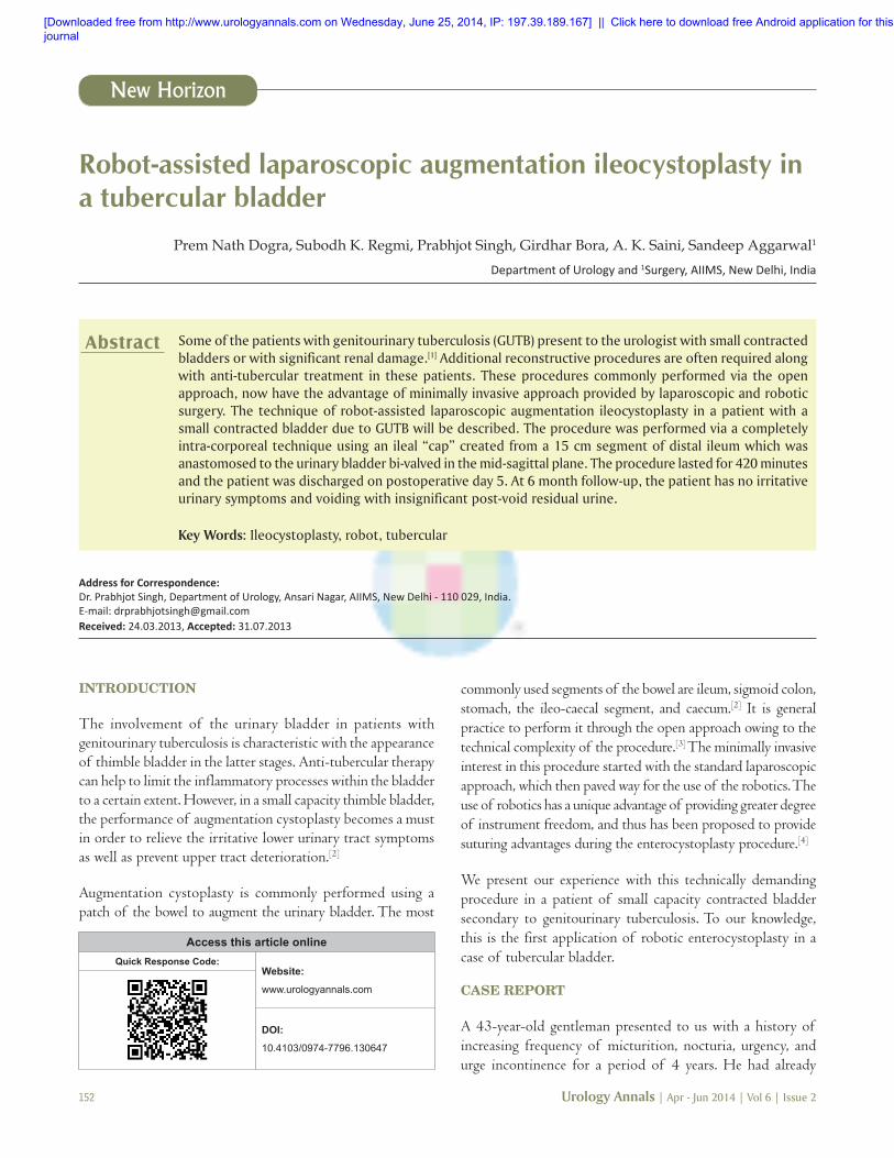
undergone transurethral resection of prostate at another center without any symptomatic improvement and was subsequently evaluated and found to have genitourinary tuberculosis based on positive urine AFB stain and urine for TB PCR and then put on anti‑tubercular therapy (ATT) for 6 months. Post ATT, the patient persisted to have these symptoms although to a lesser extent. A repeat urinary AFB stain was negative but a cystogram revealed a very low capacity (100 ml) urinary bladder [Figure 1]. An ultrasound and intravenous urogram (IVU) showed normal upper tracts. The patient was thus taken up for robot‑assisted laparoscopic augmentation ileocystoplasty.
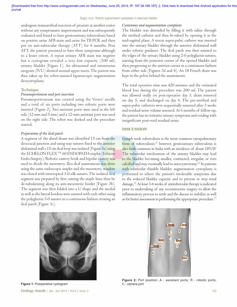
TechniquePneumoperitonem and port insertionPneumoperitoneum was created using the Verres’ needle and a total of six ports including two robotic ports were inserted [Figure 2]. Two assistant ports were used in the left side (12 mm and 5 mm) and a 12‑mm assistant port was used on the right side. The robot was docked and the procedure started.
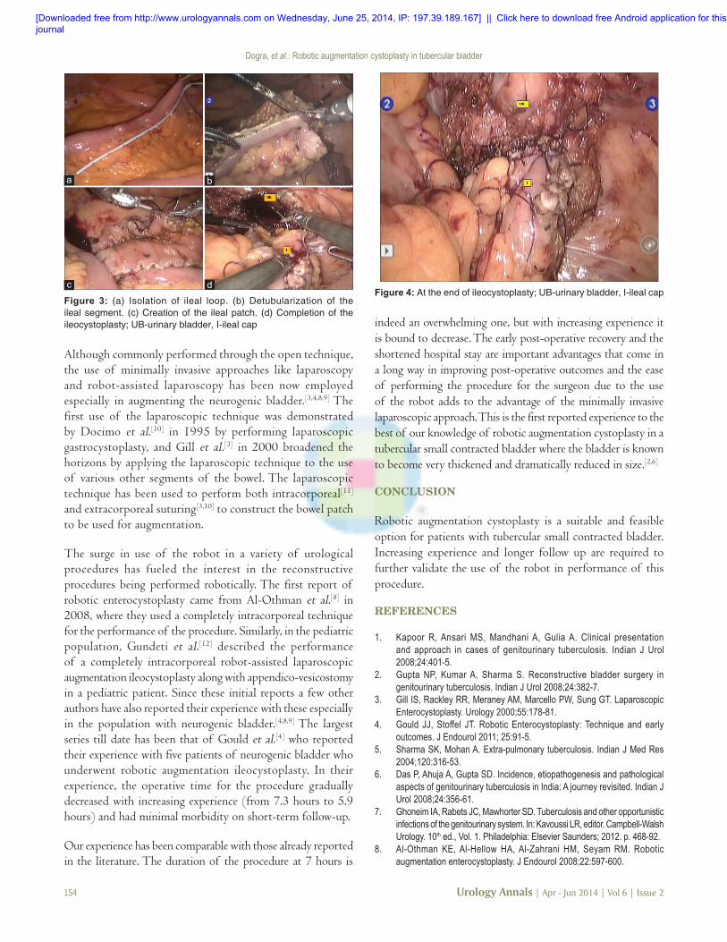
Preparation of the ileal patchA segment of the distal ileum was identified 15 cm from the ileoceacal junction and using stay sutures fixed to the anterior abdominal wall a 15 cm ileal loop was isolated [Figure 3a] using the ECHELON FLEX TM 60 ENDOPATH stapler (Ethicon Endo‑Surgery). Robotic cautery hook and bipolar cautery was used to divide the mesentery. Ileo‑ileal anastomosis was done using the same endoscopic stapler and the mesenteric window was closed with interrupted 3‑0 silk sutures. The isolated ileal segment was prepared by first cutting the staple lines then by de‑tubularizing along its anti‑mesenteric border [Figure 3b]. The segment was then folded into a U shape and the medial as well as the lateral borders were sutured with each other using the polyglactin 3‑0 sutures in a continuous fashion creating an ileal patch [Figure 3c].
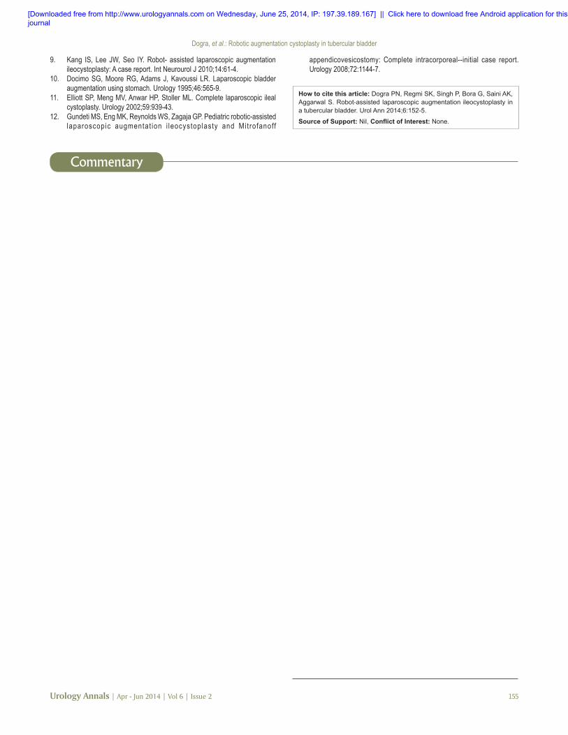
Cystostomy and augmentation cystoplastyThe bladder was distended by filling it with saline through the urethral catheter and then bi‑valved by opening it in the mid‑sagittal plane. A trocar supra‑pubic catheter was inserted into the urinary bladder through the anterior abdominal wall under robotic guidance. The ileal patch was then sutured to the edges of the urinary bladder using 2‑0 polyglactin sutures, starting from the posterior corner of the opened bladder and then progressing to the anterior corner in a continuous fashion from either side [Figures 3d and 4]. An 18 French drain was kept in the pelvis behind the anastomosis.
The total operative time was 420 minutes and the estimated blood loss during the procedure was 200 ml. The patient was allowed orally on post‑operative day 3, drain removed on day 5, and discharged on day 6. The per‑urethral and supra‑pubic catheters were sequentially removed after 3 weeks and residual urine volume assessed. At 6 months of follow‑up, the patient has no irritative urinary symptoms and voiding with insignificant post‑void residual urine.
DISCUSSION
Lymph node tuberculosis is the most common extrapulmonary form of tuberculosis;[5] however, genitourinary tuberculosis is also fairly common in India with an incidence of about 18%W The tubercular involvement of the urinary bladder may lead to the bladder becoming smaller, contracted, irregular, or even calcified and may eventually lead to autocystectomy.[6] In patients with tubercular thimble bladder, augmentation cystoplasty is performed to relieve the patient’s intolerable symptoms due to the reduced bladder capacity and to prevent or stop renal damage.[2] At least 3‑6 weeks of antitubercular therapy is indicated prior to undertaking of any reconstructive surgery to allow the inflammatory process to settle and the disease to stabilize as well as for better assessment in performing the appropriate procedure.[7]
Figure 1: Preoperative cystogramFigure 2: Port position: A ‑ assistant ports, R ‑ robotic ports, C ‑ camera port
[Downloaded free from http://www.urologyannals.com on Wednesday, June 25, 2014, IP: 197.39.189.167] || Click here to download free Android application for thisjournal
Dogra, et al.: Robotic augmentation cystoplasty in tubercular bladder
154 Urology Annals | Apr - Jun 2014 | Vol 6 | Issue 2
Although commonly performed through the open technique, the use of minimally invasive approaches like laparoscopy and robot‑assisted laparoscopy has been now employed especially in augmenting the neurogenic bladder.[3,4,8,9] The first use of the laparoscopic technique was demonstrated by Docimo et al.[10] in 1995 by performing laparoscopic gastrocystoplasty, and Gill et al.[3] in 2000 broadened the horizons by applying the laparoscopic technique to the use of various other segments of the bowel. The laparoscopic technique has been used to perform both intracorporeal[11] and extracorporeal suturing[3,10] to construct the bowel patch to be used for augmentation.
The surge in use of the robot in a variety of urological procedures has fueled the interest in the reconstructive procedures being performed robotically. The first report of robotic enterocystoplasty came from Al‑Othman et al.[8] in 2008, where they used a completely intracorporeal technique for the performance of the procedure. Similarly, in the pediatric population, Gundeti et al.[12] described the performance of a completely intracorporeal robot‑assisted laparoscopic augmentation ileocystoplasty along with appendico‑vesicostomy in a pediatric patient. Since these initial reports a few other authors have also reported their experience with these especially in the population with neurogenic bladder.[4,8,9] The largest series till date has been that of Gould et al.[4] who reported their experience with five patients of neurogenic bladder who underwent robotic augmentation ileocystoplasty. In their experience, the operative time for the procedure gradually decreased with increasing experience (from 7.3 hours to 5.9 hours) and had minimal morbidity on short‑term follow‑up.
Our experience has been comparable with those already reported in the literature. The duration of the procedure at 7 hours is
indeed an overwhelming one, but with increasing experience it is bound to decrease. The early post‑operative recovery and the shortened hospital stay are important advantages that come in a long way in improving post‑operative outcomes and the ease of performing the procedure for the surgeon due to the use of the robot adds to the advantage of the minimally invasive laparoscopic approach. This is the first reported experience to the best of our knowledge of robotic augmentation cystoplasty in a tubercular small contracted bladder where the bladder is known to become very thickened and dramatically reduced in size.[2,6]
CONCLUSION
Robotic augmentation cystoplasty is a suitable and feasible option for patients with tubercular small contracted bladder. Increasing experience and longer follow up are required to further validate the use of the robot in performance of this procedure.
REFERENCES
1. Kapoor R, Ansari MS, Mandhani A, Gulia A. Clinical presentation and approach in cases of genitourinary tuberculosis. Indian J Urol 2008;24:401‑5.
2. Gupta NP, Kumar A, Sharma S. Reconstructive bladder surgery in genitourinary tuberculosis. Indian J Urol 2008;24:382‑7.
3. Gill IS, Rackley RR, Meraney AM, Marcello PW, Sung GT. Laparoscopic Enterocystoplasty. Urology 2000;55:178‑81.
4. Gould JJ, Stoffel JT. Robotic Enterocystoplasty: Technique and early outcomes. J Endourol 2011; 25:91‑5.
5. Sharma SK, Mohan A. Extra‑pulmonary tuberculosis. Indian J Med Res 2004;120:316‑53.
6. Das P, Ahuja A, Gupta SD. Incidence, etiopathogenesis and pathological aspects of genitourinary tuberculosis in India: A journey revisited. Indian J Urol 2008;24:356‑61.
7. Ghoneim IA, Rabets JC, Mawhorter SD. Tuberculosis and other opportunistic infections of the genitourinary system. In: Kavoussi LR, editor. Campbell‑Walsh Urology. 10th ed., Vol. 1. Philadelphia: Elsevier Saunders; 2012. p. 468‑92.
8. Al‑Othman KE, Al‑Hellow HA, Al‑Zahrani HM, Seyam RM. Robotic augmentation enterocystoplasty. J Endourol 2008;22:597‑600.
Figure 4: At the end of ileocystoplasty; UB‑urinary bladder, I‑ileal capFigure 3: (a) Isolation of ileal loop. (b) Detubularization of the ileal segment. (c) Creation of the ileal patch. (d) Completion of the ileocystoplasty; UB‑urinary bladder, I‑ileal cap
a b
c d
[Downloaded free from http://www.urologyannals.com on Wednesday, June 25, 2014, IP: 197.39.189.167] || Click here to download free Android application for thisjournal
Dogra, et al.: Robotic augmentation cystoplasty in tubercular bladder
Urology Annals | Apr - Jun 2014 | Vol 6 | Issue 2 155
9. Kang IS, Lee JW, Seo IY. Robot‑ assisted laparoscopic augmentation ileocystoplasty: A case report. Int Neurourol J 2010;14:61‑4.
10. Docimo SG, Moore RG, Adams J, Kavoussi LR. Laparoscopic bladder augmentation using stomach. Urology 1995;46:565‑9.
11. Elliott SP, Meng MV, Anwar HP, Stoller ML. Complete laparoscopic ileal cystoplasty. Urology 2002;59:939‑43.
12. Gundeti MS, Eng MK, Reynolds WS, Zagaja GP. Pediatric robotic‑assisted laparoscopic augmentat ion i leocystoplasty and Mit rofanoff
appendicovesicostomy: Complete intracorporeal‑‑initial case report. Urology 2008;72:1144‑7.
How to cite this article: Dogra PN, Regmi SK, Singh P, Bora G, Saini AK, Aggarwal S. Robot-assisted laparoscopic augmentation ileocystoplasty in a tubercular bladder. Urol Ann 2014;6:152-5.
Source of Support: Nil, Conflict of Interest: None.
Other considerations for treating bladder tuberculosis
Commentary
Dear Editor,Regarding to the recently published manuscript about a case with genitourinary tuberculosis (GUTB) that successfully treated using robotic‑assisted cystoplasty, we would like to summarize some considerations about such these cases.
Kidney can be a primary organ affected by tuberculosis in urinary tract and bladder affected as second organ while GUTB is the third most common form of extra‑pulmonary tuberculosis.[1] Delay in diagnosis may lead to raise incidence of complications such as destruction and fibrosis of the urinary bladder. Bladder perforation is one of the cystitis complications, but we had seen more in some vulnerable people such as diabetic cases.[2] Bladder TB lesions could affect its function such away: 1‑decrease capacity of bladder, 2‑bladder contracture and lost its capacity and has little or no value for a urinary reservoir.
Impairment in bladder capacity induces some symptoms like frequency, nocturia, urgency, abdominal pain, and hematuria. These complications oblige surgeons for bladder augmentation. Moreover, surgeons should pay attention to long‑term complications of GUTB such as urinary stone and electrolyte disorders.[3]
Augmentation method for urinary bladder reconstruction can achieve from some part of GI such as stomach, ileum, and colon. Augmentation cystoscopy may have complication
for patients such as bladder perforation, and it will help to spread of infection in peritoneal space; nevertheless, perforation after surgery should be more attention. Before any operation on patients, spontaneous vesicle rupture secondary to bacterial cystitis in elderly patients may be accrued.[4] Also, surgeons should have a valid method to overruled TB infection in selected part of GI for augmentation.
Genitourinary tuberculosis may appear gross hematuria, which is similar to augmentation suture failure.[5] The tissue used in the procedure can be a source of many complications including abscess formation, enteric fistulas, bowel obstruction, and low fat‑soluble vitamins when stomach used for implantation, and maintain diarrhea when colon transfer in procedure. Also, it could have many disorders for urinary tract such as infection and renal impairment, blunting of the calyces, and papillary necrosis, urethra ulcers and after period of time cause for urethra necrosis due to narrow urethra.[4] Late surgical complications such as upper tract obstruction and perforation of the augmented bladder could make problem for patients. There are some method for genitourinary TB repair, but evidence showed that Robot‑assisted laparoscopic operation is method without risk of hand‑shaking of surgeons, high‑resolution, and had low risk for post‑operation complication.[6] Overall, due to low complications, device‑assisted surgery such as robot‑assisted laparoscopic and ureteroscopic procedures are better to use for initial evaluation before surgery in high‑risk patients with huge bladder lesions.
Amin Saburi1, Mahdi Safiabady2
Chemical Injuries Research Center, Baqiyatallah University of Medical Sciences, Tehran, & 1Atherosclerosis and Coronary Artery Research Centre, Birjand University of Medical Sciences, Birjand,
Iran 2Student Research Committee, Baqiyatallah University of Medical Sciences, Tehran, Iran
Access this article onlineQuick Response Code:
Website:
www.urologyannals.com
[Downloaded free from http://www.urologyannals.com on Wednesday, June 25, 2014, IP: 197.39.189.167] || Click here to download free Android application for thisjournal