Embed Size (px)
Citation preview

8ο Συνέδριο Επεμβατικής Καρδιολογίας και Ηλεκτροφυσιολογίας
Καρδιολογική Εταιρεία Βορείου Ελλάδος
Electra Palace, Θεσσαλονίκη, Σεπτέμβριος 2015
Δοκόπουλος Πέτρος
Ειδικευόμενος Β’ Κ/Δ Γ.Π.Ν. «Παπαγεωργίου»1
Right Heart Catheterization

ΣΥΓΚΡΟΥΣΗ ΣΥΜΦΕΡΟΝΤΩΝ(CONFLICTS OF INTEREST)
ΟΥΔΕΜΙΑ
Right Heart Catheterization2

STRUCTURE OF PRESENTATION
Right Heart Catheterization3
1. Definition - Indications - Contraindications -Technical -Possible sources of error
2. Complications
3. Principles of hemodynamics - Pressure Waveforms
4. Application in Pulmonary Arterial Hypertension
5. Interpretation measurement - results
6. Conclusions

Definition
Right Heart Catheterization4
Promoting catheters in the right
heart chambers and branches of
the pulmonary artery in order to:
1. Record pressures and their
waveforms
2. Measure cardiac output and the
vascular reseasants
3. Evaluate O2 saturation and
content –oxymetry run.
4. Angiographies
5. Interventional procedures

Indications
Right Heart Catheterization5
Diagnostic
Differentiation of various etiologies of shock and pulmonary
edema
Evaluation of pulmonary hypertension
Differentiation of pericardial tamponade from constrictive
pericarditis and restrictive cardiomyopathy
Diagnosis of intracardiac shunts
Therapeutic
Guide to fluid management and hemodynamic monitoring of
patients after surgery, complicated myocardial infarction,
patients in shock, heart failure

Contraindications
Right Heart Catheterization6
Absolute
Endocarditis (right cavities)
Clot / mass in the right cavities
Mechanical valves in the tricuspid / pulmonary
Relative
Haemorrhagic diathesis (INR> 2, platelet count < 50,000 )
Poor life expectancy
Poor cooperation / patient reluctance
Bioprosthetic valves
Recent installation PM / ICD
LBBB ( be possible pacing )
Contralateral pneumothorax

Catheter Swan-Ganz
These are 7F to 7.5F system catheters and are available as femoral vein insertion to continuous
cardiac output catheters
Right Heart Catheterization7

Technique (1)
Right Heart Catheterization8
The introduction of catheters made with the Seldinger technique,
comprising a vessel puncture and introduction guidewire

Technique (2)
Right Heart Catheterization9
- Venous access sites
Site of Insertion Distal to the Vena
Cava/RA junction
(cm)
Internal jugular vein 15 to 20
Subclavian vein 10 to 15
Antecubital vein (Right) 35 to 40
Antecubital vein (Left) 45 to 50
Femoral vein 25 to 30
J Am Coll Cardiol 2006;48:2546 –52

Technique (3)
Right Heart Catheterization10

Technique (4) – preparing the catheter
Right Heart Catheterization11

Technique (5) – inserting the catheter
Right Heart Catheterization12

Complications
Right Heart Catheterization13
Since probe
Thrombophlebitis
Thrombosis catheter
Bacteremia-Endocarditis
Ruptured balloon embolization
Since launching
Arrhythmias (PVC, NSVT, VF)
RBBB or PKKA
Valve Injury
Piercing PA or right ventricle of
the catheter
Since the puncture
Puncture artery
Pneumothorax
Nerve Injury / Horner’s
Embolization of air from the
promotion

14
Cardiac Cycle
Right Sided Pressures

Pressure Recordings (1)
Right Heart Catheterization15
Always record pressure at end expiration (except in patients on PEEP)
Under normal conditions, pressures will be lower in inspiration due to decrease in intrathoracic pressure
Before any pressure measurements are taken, it is imperative to perform zeroing and referencing of the system
For every inch the heart is offset from the reference point of the transducer, a 2mm Hg of error will be introduced. If the heart is lower than the transducer, the pressure will be erroneously low and if the heart is higher, the pressure will be erroneously high

Pressure Recordings (2)
Right Heart Catheterization16

Pressure Recordings (3)
Right Heart Catheterization17
Fast flush test/ Square wave testing The dynamic response of the pressure
monitoring system is determined by measuring the resonant frequency and the damping coefficient of the system using the fast flush test
Performed by briefly opening and closing the valve in the continuous flush device
This produces a square ware pattern on the oscilloscope, an initial steep rise followed by a plateau, followed by steep fall below baseline which is then followed by oscillations. The pattern determines optimal versus suboptimal damping

Intracardiac pressure waveforms during
passage through the heart
Right Heart Catheterization18

RA pressure waveforms (1)
Right Heart Catheterization19

RA pressure waveforms (2)
Right Heart Catheterization20
Effects of breathing in RAP
Normally :
Inspiration : ↓ intrathoracic
pressure →↓ RAP
Expiration : ↑ intrathoracic
pressure →↑ RAP

RA pressure waveforms (3)
Right Heart Catheterization21
Low RAP Hypovolemia
Improper zeroing of the tranducer
Elevated RAP Overload volume
Right heart failure
Valvular heart disease (TS, TR, PS, PR)
Attack infarction (ischemia RV, cardiomyopathy)
No heart disease (MS, MR, AS, AR, cardiomyopathy)
Increased pulmonary resistance (PE, COPD, primary pulmonary hypertension)
Tamponade
Myxoma

RV pressure waveforms (1)
Right Heart Catheterization22
RVESP
RVEDP
Rapid RV filling
Diastasis (RV)
Atrial contraction (RV)

RV pressure waveforms (2)
Right Heart Catheterization23
1. Systolic pressure overload
1. Pulmonary Hypertension
2. Pulmonary valve stenosis
3. Right ventricular outflow
obstruction
4. Increased pulmonary resistance
2. Low systolic pressure
1. Hypovolaemia
2. Cardiogenic shock
3. Tamponade
3. End-diastolic pressure overload
1. Hypervolaemia
2. Chronic heart failure
3. Reduced evendototita - Hypertrophy
4. Tamponade
5. Deficiency tricuspid valve
6. Constrictive pericarditis Decreased
end-diastolic pressure
4. Decreased end-diastolic pressure
1. Hypovolaemia
2. Narrowing tricuspid valve

PA pressure waveforms (1)
Right Heart Catheterization24
Systolic
Dichrotic notch
Diastolic

PA pressure waveforms (2)
Right Heart Catheterization25
Increased systolic pressure Primary pulmonary hypertension
Pulmonary disease
Failure of the mitral valve (MR / MS)
Chronic heart failure
Restrictive cardiomyopathy
Decreased systolic pressure Hypotension
Pulmonary valve stenosis
Pulmonary artery stenosis
Tricuspid valve stenosis
Tricuspid valve atresia
Ebstein’s anomaly

PCWP waveforms (1)
Right Heart Catheterization26

PCWP waveforms (3)
Right Heart Catheterization27
PCW tracing “approximates” actual LA tracing but is slightly
delayed since pressure wave is transmitted retrograde
through pulmonary veins
Normal LA pressure slightly higher
Baim DS and Grossman W. Cardiac Catheterization, Angiography, and Intervention. 5th Edition.
Baltimore: Williams and Wilkins, 1996

Abnormalities in PCWP Tracing (1)
Low mean pressure Elevated mean pressure
Right Heart Catheterization28
Hypovolemia
Improper zeroing of the transducer
Intravascular volume overload
Left ventricular failure Valvular disease (MS, MR,
AS, AR) Myocardial disease (LV
ischemia, cardiomyopathy) Left heart failure secondary
to HTN
Pericardial effusion with tamponade
Atrial myxoma

Abnormalities in PCWP Tracing (2)
Right Heart Catheterization29
Elevated a wave Mitral stenosis Decreased LV compliance due to LV failure / valve disease
Cannon a wave A-V asynchrony (3rd degree AVB, VT, V-pacer)
Elevated v wave MR LRV failure Ventricular septal defect
Equal a and v waves Tamponade Constrictive physiology
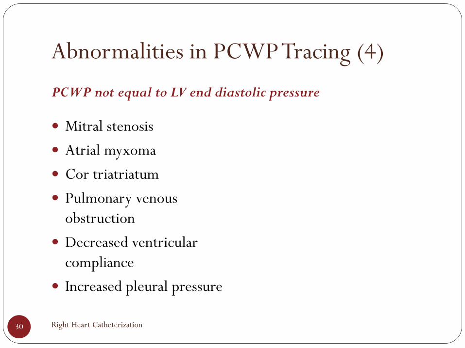
Abnormalities in PCWP Tracing (4)
PCWP not equal to LV end diastolic pressure
Right Heart Catheterization30
Mitral stenosis
Atrial myxoma
Cor triatriatum
Pulmonary venous
obstruction
Decreased ventricular
compliance
Increased pleural pressure

Pulmonary Hypertension
1. Important pathophysiological and clinical definitions
2. Haemodynamic definitions of Pulmonary Hypertension
3. Recommendations for RHC in PH
4. Recommendations for vasoreactivity testing
5. Risk assessment in PAH
6. Suggested assessment and timing for the follow-up of patients with PAH
Right Heart Catheterization31

Important pathophysiological and clinical definitions
2015 ESC/ERS Guidelines for the diagnosis and treatment of PH32

Haemodynamic definitions of Pulmonary Hypertension
2015 ESC/ERS Guidelines for the diagnosis and treatment of PH33

Recommendations for RHC in PH
2015 ESC/ERS Guidelines for the diagnosis and treatment of PH34

Recommendations for vasoreactivity testing
2015 ESC/ERS Guidelines for the diagnosis and treatment of PH35

Risk assessment in PAH
2015 ESC/ERS Guidelines for the diagnosis and treatment of PH36

Suggested assessment and timing for the follow-up of patients with PAH
2015 ESC/ERS Guidelines for the diagnosis and treatment of PH37

Cardiac output measurement
Right Heart Catheterization38
Definition Quantity of blood delivered to the systemic circulation
per unit time
Techniques Fick-Oxygen Method
Thermodilution

Cardiac output measurement
Fick Oxygen Method
39
Fick Principle: The total uptake or release of any substance by an organ is the
product of blood flow to the organ and the arteriovenous concentration
difference of the substance
In the absence of a shunt, systemic blood flow (Qs) is estimated by pulmonary
blood flow (Qp)

Cardiac output measurement
Fick Oxygen Method
Right Heart Catheterization40
Most accurate in low output states
The arteriovenous oxygen content difference (Ao – PA O2 content) can be
calculated (in ml oxygen) by the difference between the left ventricular oxygen
content and the Mixed venous (pulmonary artery) oxygen content
The value 1.36 is derived from the fact that 1 gram of hemoglobin, when 100%
saturated, combines with 1.36 ml of oxygen

Cardiac output measurement
Thermodilution Method
Right Heart Catheterization41
Principle:
injection of a given quantity of cold N / S to the central circulation
changes the temperature of blood distally.
Technique:
injecting 10ml N / S at room temperature within 2 seconds from the
proximal channel of the Swan-Ganz record temperature - time curve
catheterAn ideal thermodilution curve

Cardiac output measurement
Thermodilution Method
42 Baim DS and Grossman W. Cardiac Catheterization, Angiography, and Intervention. 5th Edition. Baltimore: Williams and Wilkins, 1996

Cardiac output measurement
Thermodilution Method
43 Love M, Lough ME, Bloomquist J. Cardiovascular laboratory assessment and diagnostic procedures. In: Thelan LA, Davie JK, Urden LD. Textbook of Critical Care
Nursing: Diagnosis and Management. St Louis, Mo: CV Mosby;1990:246
Normal CO state High CO state
Low CO state
Faulty injection
Imprecise measurement in tricuspid regurgitation
Overestimates cardiac output in low volume situations

Intracardiac Shunts (1)
Right Heart Catheterization44
Normally, pulmonary blood flow and systemic blood flow are equal
When there is an abnormal communication between intracardiac chambers or great vessels, blood flow is shunted either from the systemic circulation to the pulmonary circulation (left-to-right shunt), from the pulmonary circulation to the systemic circulation (right-to-left shunt), or in both directions (bidirectional shunt)

Intracardiac Shunts (2)
Oxymetric method
Right Heart Catheterization45
Blood sampling from various cardiac chambers for oxygen saturation determination
A left-to-right shunt is detected when there is a significant increase in blood oxygen saturation between two right-sided vessels or chambers (step up)
Despite its lack of sensitivity, clinically significant shunts are generally detected by this technique

“Oximetry run” in patient with..
..atrial septal defect ..ventricular septal defect
Right Heart Catheterization46

Shunt Detection & Measurement (2)
47
The flow ratio PBF/SBF (or QP/QS) is used
clinically to determine the significance of the
shunt
A ratio of less than 1.5 indicates a small left-to-right shunt
A ratio of 2.0 or more indicates a large left-to-right shunt and
generally requires repair in order to prevent future
pulmonary and/or right ventricular complications
A flow ratio of less than 1.0 indicates a net right-to-left shunt
Right Heart Catheterization
Key points
Right Heart Catheterization48
Right heart catheterization continues to be the gold standard in
diagnosing patients with elevated right heart pressures, although this
technique has complications
The development of noninvasive techniques has progressed, however
prospective clinical trials are lacking
It is important to note that the use of the pulmonary artery catheter
is a monitoring procedure and not a treatment

Right Heart Catheterization49