Embed Size (px)
Citation preview
Ann. rheum. Dis. (1970), 29, 617
Rheumatoid arthritis in Ugandan Africans
B. R. KANYEREZIDepartment of Medicine, Makerere University College Medical School,H. BADDELEYDepartment of Radiology, Mulago Hospital and Department of Radiodiagnosis, University of BristolD. KISUMBADepartment of Orthopaedics, Makerere University College Medical School
Until recently rheumatoid arthritis has been con-sidered to be rare in the tropics. Gelfand (1957), inhis extensive review of disease in Africans, statedthat he had rarely seen a true case in a native ofCentral Africa.Goodall (1956) described two atypical cases
amongst ninety patients with polyarthritis in Malawi.Since then eight cases have been reported from Kenya(Harries, 1962; Hall, 1966), three from Liberia(Hijmans, Valkenburg, Muller, and Gratama, 1964),39 from Uganda (Kanyerezi, 1969), and 71 fromWestern Nigeria (Greenwood, 1969).
Lawrence, Bremner, Ball, and Burch (1966) con-ducted a survey of rheumatoid arthritis in Jamaica(a sub-tropical zone) and concluded that the disease'occurs at least as frequently as in a Caucasianpopulation in the United Kingdom'.
Material and methods120 new patients attended the Arthritis Clinic, MulagoHospital, Kampala, over a period of one year, and 65of them were diagnosed as cases of rheumatoid arthritis.The Rome diagnostic criteria (Kellgren, Jeffrey, and
Ball, 1963) were used to classify these cases, so that thefindings may be compared easily with those of a pro-jected population survey. The authors have been con-servative in including cases in this analysis and this isreflected in the relatively small number of 'probable' cases.Patients with a past history of urethritis alone were notexcluded, as venereal infection is so very common inUganda. However, five young men with a history of co-existent urethritis and arthritis were excluded as cases ofReiter's disease.
All patients were examined by one of us (B.R.K.) andall had radiographs taken of the hands and wrists. Foreconomic reasons, other joints were radiographed onlywhen the patient had symptoms relating to those joints.Radiological changes were graded from 0 to 4 initially(0 = none, 1 = doubtful, 2 = mild, 3= moderate, 4 =severe), but in order to fit in with the Rome Criteria thecases were also divided into positive and negative groups,Grades 0 to 1 being regarded as negative and Grades 2to 4 as positive. Those patients classified as radiologically
positive had at least two marginal erosions in addition tojuxta-articular porosis or other changes.At least one slide latex test was performed on all 65
patients and search was made for L.E.-cells in nearly allof them.
Findings
Of the 65 cases, 30 were male and 35 female, giving amale: female ratio 1: 1 2. Before 30 years of agethere were more male cases but after 30 years thefemale cases predominated (Fig. 1).
male UL female8
F
6
, -.....d _ _ _ E1=
'I
10 20 30 40 50 60 70+
Age (decades)FIG. 1 Age distribution of Ugandan patients withrheumatoid arthritis
The duration of disease among these 65 casesranged from 4 months to 38 years (Table I), with amean of 3 years for males and 4 years for females.
Table I Duration of disease (yrs)
Duration No. of cases
Upto I 171-2 223-4 95-6 47-9 610 and over 7
copyright. on 12 June 2018 by guest. P
rotected byhttp://ard.bm
j.com/
Ann R
heum D
is: first published as 10.1136/ard.29.6.617 on 1 Novem
ber 1970. Dow
nloaded from
618 Annals of the Rheumatic Diseases
The mean age at onset was 32 5 years in males and37 years in females.
Criteria of diagnosis (Table II)Morning stiffness was complained of by all buttwo of these patients. 62 had pain, tenderness, andswelling of more than one joint. Of the three caseswith monoarticular disease, two had wrist involve-ment and the third had changes in an ankle joint.Subcutaneous nodules were palpated in six patientsbut none of these was biopsed to prove that theywere in fact 'rheumatoid' nodules. None of thesepatients had evidence of leprosy.
Table II Signs and symptoms (Rome criteria)
Symptoms No. ofcases
1 Morning stiffness 632 Pain/tenderness in at least one joint 653 Swelling in at least one joint 654 Swelling in at least one other joint 625 Symmetrical joint swelling 536 Subcutaneous nodules 67 X-ray changes 468 Positive agglutination test 35
ClassificationProbable = 3 or 4 criteriaDefinite = 5 or 6 criteriaClassical = 7 or 8 criteria
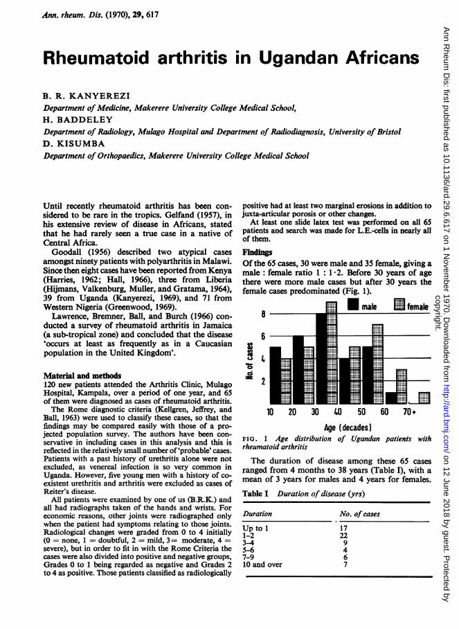
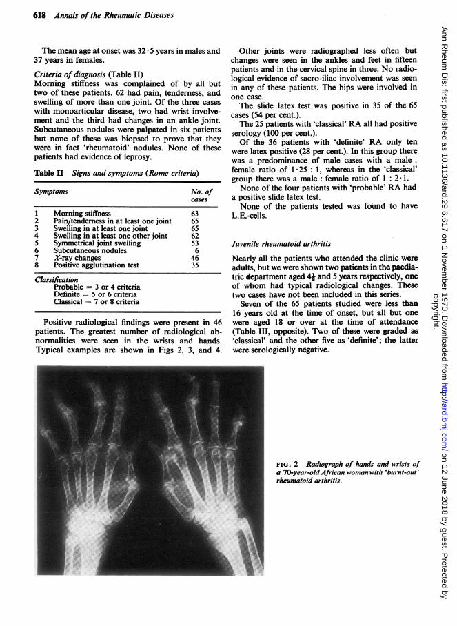
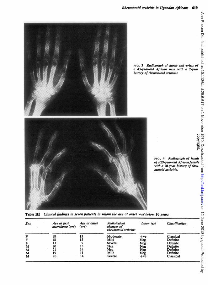
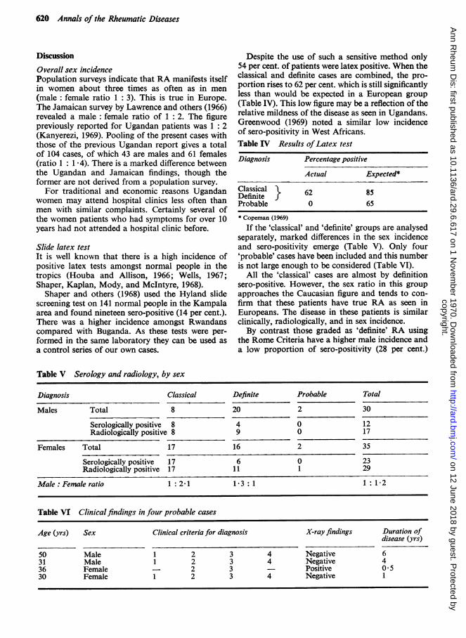
Positive radiological findings were present in 46patients. The greatest number of radiological ab-normalities were seen in the wrists and hands.Typical examples are shown in Figs 2, 3, and 4.
Other joints were radiographed less often butchanges were seen in the ankles and feet in fifteenpatients and in the cervical spine in three. No radio-logical evidence of sacro-iliac involvement was seenin any of these patients. The hips were involved inone case.The slide latex test was positive in 35 of the 65
cases (54 per cent.).The 25 patients with 'classical' RA all had positive
serology (100 per cent.).Of the 36 patients with 'definite' RA only ten
were latex positive (28 per cent.). In this group therewas a predominance of male cases with a male:female ratio of 1 25 :1, whereas in the 'classical'group there was a male : female ratio of 1 : 2 1.None of the four patients with 'probable' RA had
a positive slide latex test.None of the patients tested was found to have
L.E.-cells.
Juvenile rheumatoid arthritis
Nearly all the patients who attended the clinic wereadults, but we were shown two patients in the paedia-tric department aged 41 and 5 years respectively, oneof whom had typical radiological changes. Thesetwo cases have not been included in this series.
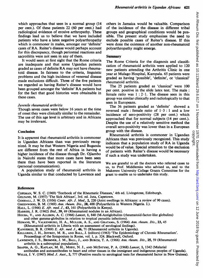
Seven of the 65 patients studied were less than16 years old at the time of onset, but all but onewere aged 18 or over at the time of attendance(Table III, opposite). Two of these were graded as'classical' and the other five as 'definite'; the latterwere serologically negative.
FIG. 2 Radiograph of hands and wrists ofa 70-year-old African woman with 'burnt-out'rheumatoid arthritis.
copyright. on 12 June 2018 by guest. P
rotected byhttp://ard.bm
j.com/
Ann R
heum D
is: first published as 10.1136/ard.29.6.617 on 1 Novem
ber 1970. Dow
nloaded from
Rheumatoid arthritis in Ugandan Africans 619
FIG. 3 Radiograph of hands and wrists ofa 43-year-old African man with a 2-yearhistory of rheumatoid arthritis
FIG. 4 Radiographo'f handsofa 29-year-old Africanfemalewith a 10-year history of rheu-matoid arthritis.
Table IMI Clinical findings in seven patients in whom the age at onset was below 16 years
Sex Age at first Age at-onset Radiological Latex test Classificationattendance (yrs) (yrs) changes of
rheumatoid arthritis
F 18 13 Moderate +ve ClassicalF 18 15 Mild Neg DefiniteF 13 9 Severe Neg DefiniteM 20 13 Neg Neg DefiniteM 21 14 Neg Neg DefiniteM 19 11 Mild Neg DefiniteM 26 14 Severe +ve Classical
copyright. on 12 June 2018 by guest. P
rotected byhttp://ard.bm
j.com/
Ann R
heum D
is: first published as 10.1136/ard.29.6.617 on 1 Novem
ber 1970. Dow
nloaded from
620 Annals of the Rheumatic Diseases
Discussion
Overall sex incidencePopulation surveys indicate that RA manifests itselfin women about three times as often as in men
(male: female ratio 1: 3). This is true in Europe.The Jamaican survey by Lawrence and others (1966)revealed a male: female ratio of 1: 2. The figurepreviously reported for Ugandan patients was 1: 2(Kanyerezi, 1969). Pooling of the present cases withthose of the previous Ugandan report gives a totalof 104 cases, of which 43 are males and 61 females(ratio 1 :1 *4). There is a marked difference betweenthe Ugandan and Jamaican findings, though theformer are not derived from a population survey.For traditional and economic reasons Ugandan
women may attend hospital clinics less often thanmen with similar complaints. Certainly several ofthe women patients who had symptoms for over 10years had not attended a hospital clinic before.
Slide latex testIt is well known that there is a high incidence ofpositive latex tests amongst normal people in thetropics (Houba and Allison, 1966; Wells, 1967;Shaper, Kaplan, Mody, and McIntyre, 1968).
Shaper and others (1968) used the Hyland slidescreening test on 141 normal people in the Kampalaarea and found nineteen sero-positive (14 per cent.).There was a higher incidence amongst Rwandanscompared with Buganda. As these tests were per-formed in the same laboratory they can be used asa control series of our own cases.
Despite the use of such a sensitive method only54 per cent. of patients were latex positive. When theclassical and definite cases are combined, the pro-portion rises to 62 per cent. which is still significantlyless than would be expected in a European group(Table IV). This low figure may be a reflection of therelative mildness of the disease as seen in Ugandans.Greenwood (1969) noted a similar low incidenceof sero-positivity in West Africans.Table IV Results ofLatex test
Diagnosis Percentage positive
Actual Expected*
Classical } 62 85Probable 0 65* Copeman (1969)
If the 'classical' and 'definite' groups are analysedseparately, marked differences in the sex incidenceand sero-positivity emerge (Table V). Only four'probable' cases have been included and this numberis not large enough to be considered (Table VI).
All the 'classical' cases are almost by definitionsero-positive. However, the sex ratio in this groupapproaches the Caucasian figure and tends to con-firm that these patients have true RA as seen inEuropeans. The disease in these patients is similarclinically, radiologically, and in sex incidence.By contrast those graded as 'definite' RA using
the Rome Criteria have a higher male incidence anda low proportion of sero-positivity (28 per cent.)
Table V Serology and radiology, by sex
Diagnosis Classical Definite Probable Total
Males Total 8 20 2 30
Serologically positive 8 4 0 12Radiologically positive 8 9 0 17
Females Total 17 16 2 35
Serologically positive 17 6 0 23Radiologically positive 17 11 1 29
Male: Female ratio 1 :2-1 1-3:1 1 :1-2
Table VI Clinical findings in four probable cases
Age (yrs) Sex Clinical criteria for diagnosis X-ray findings Duration ofdisease (yrs)
50 Male 1 2 3 4 Negative 631 Male 1 2 3 4 Negative 436 Female 2 3 Positive 0*530 Female 1 2 3 4 Negative 1
copyright. on 12 June 2018 by guest. P
rotected byhttp://ard.bm
j.com/
Ann R
heum D
is: first published as 10.1136/ard.29.6.617 on 1 Novem
ber 1970. Dow
nloaded from
Rheumatoid arthritis in Ugandan Africans 621
which approaches that seen in a normal group (14per cent.). Of these patients 22 (60 per cent.) hadradiological evidence of erosive arthropathy. Thesefindings lead us to believe that we have includedpatients who have a latex negative polyarthropathywhich is commoner in males, amongst our 'definite'cases of RA. Reiter's disease would perhaps accountfor this discrepancy, though periosteal reactions andsacroiliitis were not seen in any of them.
It would seem at first sight that the Rome criteriaare inadequate and that some Ugandan patientsgraded as cases of definite RA may not have rheuma-toid disease. In fairness to the criteria, linguisticproblems and the high incidence of venereal diseasemade exclusions difficult. Three of the five patientswe regarded as having Reiter's disease would havebeen grouped amongst the 'definite' RA patients butfor the fact that good histories were obtainable inthese cases.
Juvenile rheumatoid arthritisThough seven cases were below 16 years at the timeof onset they were clinically similar to the remainder.The use of this age level is arbitrary and in Africansmay be irrelevant.
ConclusionIt is apparent that rheumatoid arthritis is commonerin Ugandan Africans than was previously recog-nized. It may be that Western Nigeria and Bugandaare different from the rest of Africa in having ahigher incidence of the disease though Dr. Ojiamboin Nairobi states that more cases have been seenthere than have been reported in the literature(personal communication).A population study of rheumatoid arthritis in
Uganda similar to that conducted by Lawrence and
others in Jamaica would be valuable. Comparisonof the incidence of the disease in different tribalgroups and geographical conditions would be pos-sible. The present study emphasizes the need toexclude possible cases of Reiter's disease. If thiswere done the existence of another non-rheumatoidpolyarthropathy might emerge.
SummaryThe Rome Criteria for the diagnosis and classifi-cation of rheumatoid arthritis were applied to 120new patients attending the Arthritis Clinic in oneyear at Mulago Hospital, Kampala. 65 patients weregraded as having 'possible', 'definite', or 'classical'rheumatoid arthritis.The 25 patients graded as 'classical' were 100
per cent. positive in the slide latex test. The male:female ratio was 1 : 2 1. The disease seen in thisgroup was similar clinically and radiologically to thatseen in Europeans.The 36 patients graded as 'definite' showed a
reversed male : female ratio of 1 -3 : 1 and a lowincidence of sero-positivity (28 per cent.) whichapproached that for normal subjects (14 per cent.).Despite the use of a relatively sensitive method theoverall sero-positivity was lower than in a Europeangroup with the disease.Rheumatoid arthritis is commoner in Ugandan
Africans than was previously recognized. This studyindicates that a population study of RA in Ugandawould be of value. Special attention to the exclusionof patients with Reiter's disease would be necessaryif such a study was undertaken.
We are grateful to all the doctors who referred cases tous, to Prof. Middlemiss who advised us, and to theMakerere University College Grants Committee for thegrant to enable us to undertake this study.
ReferencesCOPEMAN, W. S. C. (1969) 'Textbook of the Rheumatic Diseases,' 4th ed. Livingstone, Edinburgh.GELFAND, M. (1957) 'The Sick African', 3rd. ed. Juta, Capetown.GOODALL, J. W. D. (1956) Centr. Afr. J. Med., 2, 220 (Joint swellings in Africans: a review of 90 cases).GREENWOOD, B. M. (1969) Ann. rheum. Dis., 28, 488 (Polyarthritis in Western Nigeria. I.).HALL, L. (1966) E. Afr. med. J., 43, 161 (Polyarthritis in Kenya).HARRIES, J. R. (1962) Ibid., 39, 69 (Rheumatoid nodules in an African).HOUBA, V., AND ALLISON, A. C. (1966) Lancet, 1, 848 (M-Antiglobulins (rheumatoid-factor-like globulins)
and other gamma-globulins in relation to tropical parasitic infections).HIjMANS, W., VALKENBURG, H. A., MULLER, A. S., AND GRATAMA, S. (1964) Ann. rheum. Dis., 23, 45
(Rheumatoid arthritis in Liberia with an assessment of serological findings).KANYEREzI, B. R. (1969) E. Afr. med. J., 46, 71 (Rheumatoid arthritis in Uganda).KELLGREN, J. H., JEFREY, M. R., AND BALL, J. (editors) (1963) 'The Epidemiology of Chronic Rheumatism'.
Proceedings of the Symposium in Rome, vol. 1, p. 324. Blackwell, Oxford.LAWRENCE, J. S., BREMNER, J. M., BALL, J., AND BURCH, T. A. (1966) Ann. rheum. Dis., 25, 59 (Rheumatoid
arthritis in a subtropical population).SHAPER, A. G., KAPLAN, M. H., MODY, N. J., AND McINTYRE, P. A. (1968) Lancet, 1, 1342 (Malarial
antibodies and autoantibodies to heart and other tissues in the immigrant and indigenous peoples of Uganda).WELLS, J. V. (1967) Med. J. Aust., 2, 777 (Positive results to serological tests for rheumatoid factor in New Guinea).
copyright. on 12 June 2018 by guest. P
rotected byhttp://ard.bm
j.com/
Ann R
heum D
is: first published as 10.1136/ard.29.6.617 on 1 Novem
ber 1970. Dow
nloaded from