Embed Size (px)
Citation preview

Volume 343 Number 23
·
1703
Review Articles
Advances in Immunology
I
AN
R. M
ACKAY
, M.D.,
AND
F
RED
S. R
OSEN
, M.D.,
Editors
ADVANCES IN IMMUNOLOGY
I
MMUNODEFICIENCY
D
ISEASES
C
AUSED
BY
D
EFECTS
IN
P
HAGOCYTES
J
ULIE
A. L
EKSTROM
-H
IMES
, M.D.,
AND
J
OHN
I. G
ALLIN
, M.D.
From the Laboratory of Host Defenses, National Institute of Allergy andInfectious Diseases, National Institutes of Health, Bethesda, Md. Addressreprint requests to Dr. Gallin at Bldg. 10, Rm. 2C146, 10 Center Dr., MSC1504, Bethesda, MD 20892-1504, or at [email protected].
©2000, Massachusetts Medical Society.
RIMARY phagocytic defects must be includedin the differential diagnosis of recurrent infec-tion and fever in a child and occasionally in an
adult. Early diagnosis is essential, because manifesta-tions of infection are usually blunted and rapid inter-vention can be lifesaving. In general, patients are iden-tified at a young age on the basis of their susceptibilityto normally nonpathogenic bacteria or fungi. In somecases, the infectious agents point to the disorder (Table1): catalase-positive microorganisms and aspergillosisspecies are characteristic of chronic granulomatous dis-ease,
1
and atypical mycobacteria suggest a defect in theinterferon-
g
–interleukin-12 axis.
2
These bacterial in-fections contrast with the viral and candida infectionsin deficiencies of T cells. Also suggestive is the failureof an infection to resolve with conventional treatment.
3
Other characteristic findings include recurrent infec-tions of the lungs, liver, and bone; aphthous ulcers;severe gingivitis; and in some disorders, periodontitis.Sepsis or meningitis is rare, but lymphadenopathy andhepatosplenomegaly are common.
Nearly all primary defects of phagocytes result froma mutation that affects the innate immune system.For example, leukocyte adhesion deficiency type 1 re-sults from the loss of an adhesion protein on neutro-phils, which causes leukocytosis owing to an impairedability of neutrophils to exit the circulation and trav-el to sites of infection.
4
Identification of the mutationsunderlying primary phagocytic disorders has provid-ed exciting revelations connecting molecular findingsto the clinical aspects of these diseases.
CONGENITAL NEUTROPENIAS
A defect in the life cycle of neutrophils can com-promise host defenses.
5
Severe neutropenia, defined
P
as an absolute neutrophil count of less than 500 cellsper cubic millimeter,
6-8
suppresses inflammation andincreases susceptibility to recurrent and severe bac-terial and fungal infections. Descriptions of patientswith infections and neutropenia appeared early thiscentury,
9
but in 1930, Roberts and Kracke showedthat neutropenia can precede infection.
10
Soon there-after, neutropenias were subdivided into asymptomaticneutropenias and those associated with bone marrowinsufficiency.
11
Cyclic Neutropenia
Cyclic neutropenia is an autosomal dominant dis-order in which cyclic hematopoiesis causes intervalsof neutropenia and susceptibility to opportunistic in-fection.
6,7
Recurrent, very severe neutropenia (an ab-solute neutrophil count of less than 200 cells per cubicmillimeter) that lasts 3 to 6 days of every 21-day pe-riod is typical. In about 30 percent of patients withcyclic neutropenia, however, the cycles range from 14to 36 days.
8
Patients are usually asymptomatic, butduring the period of severe neutropenia, aphthous ul-cers, gingivitis, stomatitis, and cellulitis may develop,and death from overwhelming infection occurs inabout 10 percent of patients.
8,12
Abdominal pain mustbe assessed aggressively because of the high frequen-cy of clostridium infections during the period of se-vere neutropenia.
12
During the periods of neutropeniathe bone marrow shows lack of maturation of granu-locyte precursors beyond myelocytes; moreover, thereis myeloid hyperplasia during the remainder of thecycle. Occasionally, there is a reduction in the sever-ity of neutropenia and the accompanying infectionsover time.
8,12
Mutations in the neutrophil elastase gene (
ELA2
)have been identified in patients with cyclic neutro-penia.
13
Neutrophil elastase is released from neutro-phils during inflammation and causes the destructionof tissues.
13,14
The mutations affect the catalytic siteof the enzyme, which can lead to the failure of in-hibitors to bind and thus inactivate elastase.
15
Micewith an inactivated neutrophil elastase gene have nor-mal numbers of neutrophils, but the ability of thesecells to kill pathogens is impaired.
16
Thus, the link be-tween neutrophil elastase and the cyclic changes inthe levels of neutrophils is unknown.
Mathematical models suggest that granulocyte col-ony-stimulating factor may modulate the control ofboth the numbers of circulating neutrophils and thedevelopment of hematopoietic stem cells into gran-ulocyte precursors.
17
Defects in granulocyte colony-stimulating factor signaling could destabilize normalsteady-state conditions and increase the numbers of

1704
·
December 7, 2000
The New England Journal of Medicine
circulating lymphocytes, reticulocytes, and platelets,as occurs in cyclic neutropenia.
17
Severe Congenital Neutropenia
Severe congenital neutropenia is characterized bysevere neutropenia (an absolute neutrophil count ofless than 500 cells per cubic millimeter), recurrentbacterial infections, and failure of myeloid cells to ma-ture from promyelocytes to myelocytes.
6-8
The dis-ease begins during the first year of life, and its infec-tious complications include cellulitis, perirectal abscess,
peritonitis, stomatitis, and meningitis, commonly asa result of infections with
Staphylococcus aureus
and
Burkholderia aeruginosa.
8
The numbers of circulat-ing monocytes and eosinophils are often increased.
8
Despite having increased plasma levels of granulocytecolony-stimulating factor, nearly all patients have a re-sponse to pharmacologic doses of recombinant gran-ulocyte colony-stimulating factor (filgrastim); neutro-phil counts rise, infection rates fall, and mortality isreduced.
18
Although severe congenital neutropenia was orig-
T
ABLE
1.
I
MMUNODEFICIENCY
D
ISEASES
C
AUSED
BY
D
EFECTS
IN
P
HAGOCYTES
.
D
ISEASE
M
OLECULAR
OR
G
ENETIC
D
EFECT
P
ATHOGENIC
O
RGANISMS
AND
S
ITES
A
FFECTED
C
LINICAL
P
RESENTATION
Severe chronic neutropeniaCyclic neutropenia Mutation in
ELA2,
encoding neutrophil elastase
Episodic bacterial infections, including those due to
Clostridium perfringens;
aphthous ulcers; gingivitis; stomatitis; cellulitis
21-Day oscillations in neutrophil, monocyte, platelet, and lymphocyte counts
Severe congenital neutropenia
Unknown
Staphylococcus aureus, Burkholderia aerugi-nosa;
cellulitis, perirectal abscess, stomati-tis, meningitis
Developmental arrest of bone marrow mye-loid cells at the promyelocyte stage; usually responsive to treatment with granulocyte colony-stimulating factor; increased risk of acute myelogenous leukemia and the myelodysplastic syndrome in some forms
Shwachman–Diamond syndrome
Unknown Infections involving the lungs, bone, skin, urinary tract
Cyclic or intermittent neutropenia, pancy-topenia; associated skeletal abnormalities; pancreatic insufficiency; recurrent infections of the sinuses, lungs, bones, skin, and urinary tract; increased risk of aplasia, myelodyspla-sia, and leukemia
Leukocyte adhesion defi-ciency
Type 1
Type 2
CD18
Carbohydrate fucosylation
Gram-negative enteric bacteria,
S. aureus,
candida species, aspergillus species
Gram-negative enteric bacteria,
S. aureus,
candida species, aspergillus species
Recurrent infections of skin, soft tissues, and respiratory and gastrointestinal tracts; peri-odontal disease; delayed separation of the umbilical cord
Recurrent infections of skin, soft tissues, and respiratory and gastrointestinal tracts; peri-odontal disease; delayed separation of the umbilical cord; growth retardation; dys-morphic features; neurologic deficits
Rac2 deficiency Deficiency of Rac2 Not reported Recurrent perirectal abscesses, poor wound healing, absence of pus at sites of infection, leukocytosis, and neutrophilia
Interferon-
g
and interleukin-12 defects
Interferon-
g
–receptor ligand-binding chain, interferon-
g
– receptor signaling chain, interleukin-12–receptor
b
1 chain, interleukin-12 p40 de-ficiency
Bacille Calmette–Guérin,
Mycobacterium avium
complex,
M. fortuitum, M. chelo-nae, M. smegmatis,
salmonella species
Infection with intracellular microorganisms with severe mycobacterial disease; onset in infancy, dissemination, and failure to form granulomas seen with autosomal recessive interferon-
g
–receptor defects; later-onset osteomyelitis is associated with autosomal dominant interferon-
g
–receptor defectsChronic granulomatous
disease of childhoodgp91
phox
(in X-linked chronic granulomatous disease)
p47
phox
p67
phox
p22
phox
Catalase-positive microorganisms:
S. aureus, B. cepacia,
aspergillus species, nocardia species,
Serratia marcescens
Abscess formation in the lungs, liver, brain, and bone; soft-tissue infection; gastrointes-tinal and urogenital obstruction from gran-ulomas
Myeloperoxidase deficiency Defects in
MPO
at chromosome 17, q11–21, q22–24, q21.3–23
Not usually associated with clinical disease Associated with disseminated candidiasis in patients with diabetes mellitus
Chédiak–Higashi syndrome Mutation in LYST, encoding a cytoplasmic protein involved in protein transport
S. aureus
, beta-hemolytic streptococcus Partial ocular and cutaneous albinism, periph-eral neuropathy, recurrent bacterial infec-tions, easy bruising, mild mental retarda-tion, severe periodontal disease
Neutrophil-specific granule deficiency
C/EBP
e
, encoding a transcrip-tion factor
S. aureus, S. epidermidis,
enteric bacteria Recurrent infections of skin and lungs, poor healing, bleeding diatheses
ADVANCES IN IMMUNOLOGY
Volume 343 Number 23
·
1705
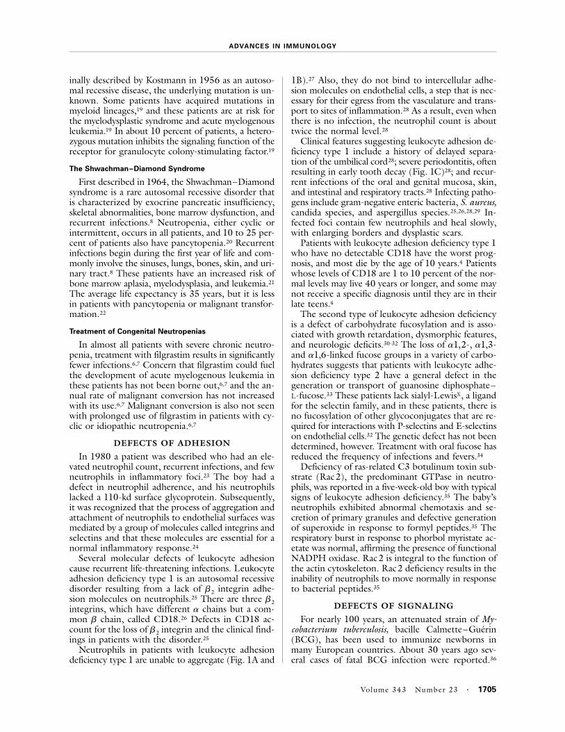
inally described by Kostmann in 1956 as an autoso-mal recessive disease, the underlying mutation is un-known. Some patients have acquired mutations inmyeloid lineages,
19
and these patients are at risk forthe myelodysplastic syndrome and acute myelogenousleukemia.
19
In about 10 percent of patients, a hetero-zygous mutation inhibits the signaling function of thereceptor for granulocyte colony-stimulating factor.
19
The Shwachman–Diamond Syndrome
First described in 1964, the Shwachman–Diamondsyndrome is a rare autosomal recessive disorder thatis characterized by exocrine pancreatic insufficiency,skeletal abnormalities, bone marrow dysfunction, andrecurrent infections.
8
Neutropenia, either cyclic orintermittent, occurs in all patients, and 10 to 25 per-cent of patients also have pancytopenia.
20
Recurrentinfections begin during the first year of life and com-monly involve the sinuses, lungs, bones, skin, and uri-nary tract.
8
These patients have an increased risk ofbone marrow aplasia, myelodysplasia, and leukemia.
21
The average life expectancy is 35 years, but it is lessin patients with pancytopenia or malignant transfor-mation.
22
Treatment of Congenital Neutropenias
In almost all patients with severe chronic neutro-penia, treatment with filgrastim results in significantlyfewer infections.
6,7
Concern that filgrastim could fuelthe development of acute myelogenous leukemia inthese patients has not been borne out,
6,7
and the an-nual rate of malignant conversion has not increasedwith its use.
6,7
Malignant conversion is also not seenwith prolonged use of filgrastim in patients with cy-clic or idiopathic neutropenia.
6,7
DEFECTS OF ADHESION
In 1980 a patient was described who had an ele-vated neutrophil count, recurrent infections, and fewneutrophils in inflammatory foci.
23
The boy had adefect in neutrophil adherence, and his neutrophilslacked a 110-kd surface glycoprotein. Subsequently,it was recognized that the process of aggregation andattachment of neutrophils to endothelial surfaces wasmediated by a group of molecules called integrins andselectins and that these molecules are essential for anormal inflammatory response.
24
Several molecular defects of leukocyte adhesioncause recurrent life-threatening infections. Leukocyteadhesion deficiency type 1 is an autosomal recessivedisorder resulting from a lack of
b
2
integrin adhe-sion molecules on neutrophils.
25
There are three
b
2
integrins, which have different
a
chains but a com-mon
b
chain, called CD18.
26
Defects in CD18 ac-count for the loss of
b
2
integrin and the clinical find-ings in patients with the disorder.
25
Neutrophils in patients with leukocyte adhesiondeficiency type 1 are unable to aggregate (Fig. 1A and
1B).
27
Also, they do not bind to intercellular adhe-sion molecules on endothelial cells, a step that is nec-essary for their egress from the vasculature and trans-port to sites of inflammation.
28
As a result, even whenthere is no infection, the neutrophil count is abouttwice the normal level.
28
Clinical features suggesting leukocyte adhesion de-ficiency type 1 include a history of delayed separa-tion of the umbilical cord
28
; severe periodontitis, oftenresulting in early tooth decay (Fig. 1C)
28
; and recur-rent infections of the oral and genital mucosa, skin,and intestinal and respiratory tracts.28 Infecting patho-gens include gram-negative enteric bacteria, S. aureus,candida species, and aspergillus species.25,26,28,29 In-fected foci contain few neutrophils and heal slowly,with enlarging borders and dysplastic scars.
Patients with leukocyte adhesion deficiency type 1who have no detectable CD18 have the worst prog-nosis, and most die by the age of 10 years.4 Patientswhose levels of CD18 are 1 to 10 percent of the nor-mal levels may live 40 years or longer, and some maynot receive a specific diagnosis until they are in theirlate teens.4
The second type of leukocyte adhesion deficiencyis a defect of carbohydrate fucosylation and is asso-ciated with growth retardation, dysmorphic features,and neurologic deficits.30-32 The loss of a1,2-, a1,3-and a1,6-linked fucose groups in a variety of carbo-hydrates suggests that patients with leukocyte adhe-sion deficiency type 2 have a general defect in thegeneration or transport of guanosine diphosphate–L-fucose.33 These patients lack sialyl-LewisX, a ligandfor the selectin family, and in these patients, there isno fucosylation of other glycoconjugates that are re-quired for interactions with P-selectins and E-selectinson endothelial cells.32 The genetic defect has not beendetermined, however. Treatment with oral fucose hasreduced the frequency of infections and fevers.34
Deficiency of ras-related C3 botulinum toxin sub-strate (Rac2), the predominant GTPase in neutro-phils, was reported in a five-week-old boy with typicalsigns of leukocyte adhesion deficiency.35 The baby’sneutrophils exhibited abnormal chemotaxis and se-cretion of primary granules and defective generationof superoxide in response to formyl peptides.35 Therespiratory burst in response to phorbol myristate ac-etate was normal, affirming the presence of functionalNADPH oxidase. Rac2 is integral to the function ofthe actin cytoskeleton. Rac2 deficiency results in theinability of neutrophils to move normally in responseto bacterial peptides.35
DEFECTS OF SIGNALING
For nearly 100 years, an attenuated strain of My-cobacterium tuberculosis, bacille Calmette–Guérin(BCG), has been used to immunize newborns inmany European countries. About 30 years ago sev-eral cases of fatal BCG infection were reported.36

1706 · December 7, 2000
The New England Journal of Medicine
Half the affected infants had a profound deficiencyof T cells as a result of severe combined immunode-ficiency, but the rest had no obvious immunologicdefect. Their problem was clarified by the study of sev-eral related children from a small, isolated fishing vil-lage on the island of Malta37 who had fatal infectionswith atypical mycobacteria that were not consideredto be pathogenic. The susceptibility gene in this kin-dred was mapped to chromosome 6 at the precisesite where the receptor for interferon-g is encoded.37
The interferon-g–interleukin-12 axis is critical fordefenses against intracellular microbes such as myco-bacteria, salmonella, and listeria (Fig. 2). Defects inthe ligand-binding chain of the interferon-g receptor,the signaling chain of the interferon-g receptor, theinterleukin-12 receptor, or interleukin-12 itself in-crease susceptibility to mycobacterial infection.2 Vari-ations in the clinical manifestations and severity of dis-ease and the presence or absence of a response totreatment with interferon gamma reflect the extent ofthe disruption of the interferon-g–interleukin-12 axis.2
The presence of a pathogen triggers the produc-tion of interleukin-12 by dendritic cells and macro-phages, which in turn induces the secretion of inter-feron-g by T cells and natural killer cells (Fig. 2).38
Interferon-g activates macrophages and neutrophils,causing them to produce tumor necrosis factor a andactivate NADPH oxidase, which promotes killing ofthe pathogen by increasing the production of hydro-gen peroxide.38 Interleukin-12 is also part of a feed-back control mechanism. It induces T cells to produceinterleukin-10, which suppresses the proliferation ofT cells and the production of interleukin-12 and inter-feron-g.39,40
The interferon-g receptor consists of a ligand-bind-ing chain and a signaling chain (also called the R1and R2 chains, respectively).2 Binding of interferon-gto the ligand-binding chain of the receptor causes itto link up with another such chain and leads to theaggregation of two interferon-g–receptor signalingchains.2 Mutations have been identified in the genesfor both chains of this receptor, with both autoso-mal recessive and autosomal dominant inheritance.Children with a mutation that causes complete lossof the ligand-binding chain have severe disease thatbegins in early infancy. The main features are dissem-inated atypical mycobacterial disease or fatal BCGinfection after vaccination,2,37,41 an inability to formgranulomas (Fig. 2), and the absence of a responseto high doses of interferon gamma. A mutation re-sulting in the partial loss of the ligand-binding chainof the interferon-g receptor causes less severe dis-ease, in which the capacity to form granulomas andresponsiveness to high doses of interferon gammaare not lost.42
Children with a different mutation of the interfer-on-g–receptor ligand-binding chain have milder dis-ease; nontuberculous mycobacterial infections devel-
Figure 1. Features of Leukocyte Adhesion Deficiency Type 1.In Panel A, normal neutrophils aggregate readily in response tophorbol myristate acetate (100 ng per milliliter) (¬500), where-as in Panel B, neutrophils from a patient with leukocyte adhe-sion deficiency type 1 fail to aggregate (¬500). Patients withleukocyte adhesion deficiency type 1 have periodontitis (arrowsin Panel C) with early tooth loss as part of a constellation of re-current infections involving the gastrointestinal, urogenital,and respiratory tracts. Panels A and B reprinted from Rotrosenand Gallin27 with the permission of the publisher.
A
B
C

ADVANCES IN IMMUNOLOGY
Volume 343 Number 23 · 1707
Figure 2. Interferon-g–Interleukin-12 Signal-Transduction Cascade.Interleukin-12, which is produced by macrophages and dendritic cells in response to the presence of a pathogen, binds to its re-ceptors on T cells and natural killer cells, inducing the release of interferon-g (IFN-g). Monocytes and macrophages bind interferon-g,resulting in the cross-linking of the interferon-g receptor; activation of the cells, with the production of hydrogen peroxide (H2O2);and the synthesis and release of tumor necrosis factor a and interleukin-12 (dimer of subunits p35 and p40). Mutations resultingin increased susceptibility to nontuberculous mycobacteria have been identified in the genes for both ligand-binding chain and thesignaling chain of the interferon-g receptor, the b1 chain and the b2 chain of the interleukin-12 receptor (the b2 chain is the signaltransducer), and the p40 subunit of interleukin-12. Panel A shows a resolving mycobacterial infection with normal granuloma for-mation in a lung-biopsy specimen from a patient with no known mutation in the interferon-g–interleukin-12 axis (hematoxylin andeosin, ¬20). Panel B shows a lung-biopsy specimen from a patient with an autosomal recessive mutation of the interferon-g–receptorligand-binding chain who was infected with nontuberculous mycobacteria (acid-fast Fite’s stain, ¬600). There are numerous myco-bacteria (red) within macrophages (blue). Panel C shows a contiguous section of lung from the same patient in which there is nogranuloma formation (hematoxylin and eosin, ¬200).
T cell
Natural 4killer cell
Macrophage
T cell
Natural 4killer cell
IFN-g–receptor>signaling chain
IFN-g–receptor >ligand-binding chain
IFN-g receptor >>
Interleukin-12>receptor
Tumor necrosis>factor\a
b1 chain
b2 chain
H2O+O2
H2O2
Dendritic cells
Macrophage
Interleukin-12>>
IFN-g
IFN-g
IFN-g
IFN-gp35p40
B CA

1708 · December 7, 2000
The New England Journal of Medicine
op in early childhood rather than infancy, and theyrespond to treatment with interferon gamma.2,43 In-terestingly, in one report 13 of 16 patients with thismutation had nontuberculous mycobacterial osteo-myelitis.32
A mutation in the gene for the receptor-signalingchain of interferon-g that eliminates signaling alsoincreases susceptibility to nontuberculous mycobac-terial disease. The clinical presentation resembles thatassociated with the complete loss of the interferon-g–receptor ligand-binding chain.44
Diminished production of interferon-g as a resultof abnormal regulation of interleukin-12 also increas-es susceptibility to disseminated nontuberculous my-cobacterial or BCG infection.2 The interleukin-12receptor has two chains, b1 and b2. Both are requiredfor high-affinity binding of interleukin-12; however,signal transduction is mediated by the b2 chain.45
The clinical effect of a mutation in the gene encodingthe b1 chain, which is associated with an increasedsusceptibility to nontuberculous mycobacterial dis-ease and salmonella infections,2,46,47 resembles that ofa defect in the gene for the interferon-g–receptorligand-binding chain, and patients with this mutationhave a response to interferon gamma therapy.2
Interleukin-12 also has two chains, a 35-kd and a40-kd chain. A mutation in the gene for the 40-kdchain increases susceptibility to mycobacterial dis-ease.48 Patients with a mutation in the gene for thischain or the b1 chain of the interleukin-12 receptorcan form mature granulomas, suggesting that they canproduce interferon-g in the absence of interleukin-12.As expected, patients with these mutations have a re-sponse to treatment with interferon gamma.2
DEFECTS OF INTRACELLULAR KILLING
The responses of phagocytes to pathogens includephagocytosis, proteolytic destruction within granules,and damage induced by hydroxyl radical, superoxide,and hydrogen peroxide generated by NADPH oxi-dase. Patients with defects in intracellular killing of mi-crobes have increased susceptibilities to specific patho-genic bacteria and fungi that result in atypical andoften muted inflammatory responses.
In 1954, Janeway and colleagues described chil-dren with elevated serum gamma globulin levels andrecurrent infections,49 some of whom were later shownto have chronic granulomatous disease. In 1957, fourboys with hypergammaglobulinemia, recurrent infec-tions of the lungs, lymph nodes, and skin, and gran-ulomatous lesions were described.50 An evaluation ofphagocytic function by the available methods did notreveal any defects, and the disorder was named “fatalgranulomatous disease of childhood.” In 1967, a spe-cific defect in the intracellular killing of bacteria wasidentified51 and traced to the oxidative metabolism ofphagocytes.52
The discovery of the protein components of the
NADPH oxidase apparatus was the direct conse-quence of studies of neutrophils from patients withchronic granulomatous disease. These neutrophilswere found to have defects in the generation of hy-drogen peroxide and in NADPH oxidase function.53-57
It became evident that chronic granulomatous dis-ease is a heterogeneous disorder caused by defects inany one of the four subunits of NADPH oxidase,58-68
the enzyme that initiates the process of forming hy-drogen peroxide (Fig. 3).69
The most common form of chronic granuloma-tous disease (present in approximately 70 percent ofpatients) is X-linked and is due to a mutation in thegene for the phagocyte oxidase cytochrome glyco-protein of 91 kd (gp91phox).70 The second most com-mon form is autosomal recessive and is due to a mu-tation in the gene for a cytosolic component of 47kd (p47phox).70
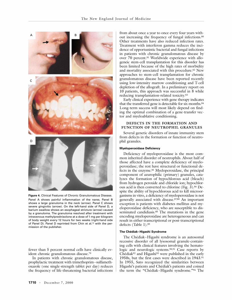
Chronic granulomatous disease is characterizedby recurrent infections with catalase-positive micro-organisms, which destroy their own hydrogen perox-ide, including S. aureus, Burkholderia cepacia, aspergil-lus species, nocardia species, and Serratia marcescens.1Infections with catalase-negative organisms, such asStreptococcus pneumoniae, are rare. The clinical man-ifestations include recurrent or persistent infectionsof the soft tissues, lungs, and other organs, despiteaggressive antibiotic therapy (Fig. 4).1,38,70 The appear-ance of fever and clinical signs of infection may bedelayed, requiring routine follow-up every four to sixmonths. Magnetic resonance imaging of the chest inasymptomatic children often reveals early pneumo-nia. Severe, resistant facial acne and painful inflam-mation of the nares are common (Fig. 4A). Severegingivitis (Fig. 4C) and aphthous ulcers are seen,but not periodontal disease (Fig. 1C), as is found inpatients with leukocyte adhesion deficiency type 1.In addition, there is excessive formation of granulo-mas in all tissues. These granulomas, which can ob-struct the genitourinary and gastrointestinal tracts,71,72
are exquisitely responsive to short courses of cortico-steroids (Fig. 4D).72 Chronic granulomatous diseasecan easily be diagnosed by a nitroblue tetrazoliumtest (Fig. 5)56 or by flow cytometry with dihydrorho-damine dye.73
Mycobacterial disease is uncommon in patientswith chronic granulomatous disease,74 but drainingskin lesions and lymphadenopathy can occur at thesite of BCG inoculation. Focal or miliary pulmonarydisease may arise as a result of infection with nontu-berculous mycobacteria, and it may be followed bypulmonary fibrosis.75 Intracellular killing of mycobac-teria is impaired in macrophages from patients withchronic granulomatous disease, demonstrating the es-sential role of NADPH oxidase in host defense againstmycobacteria.76
Children with X-linked chronic granulomatousdisease are more severely affected than children with

ADVANCES IN IMMUNOLOGY
Volume 343 Number 23 · 1709
autosomal mutations.70 In the X-linked form, the on-set is earlier, obstructive granulomas and infectionsare more frequent, and the mortality rate is higher.The reason for these differences is unknown. Interest-ingly, female carriers of X-linked chronic granuloma-tous disease, who have pronounced lyonization (inac-
tivation) of the X chromosome, have levels of NADPHoxidase activity that are only 10 percent of normallevels, and this feature protects them from infectionswith catalase-positive organisms and the infectioussequelae of chronic granulomatous disease77,78; occa-sional patients with extreme lyonization who have
Figure 3. Relation among the Components of NADPH Oxidase That Are Affected in Patients with Chronic Granulomatous Disease.The membrane-bound phagocyte oxidase components, the 91-kd glycoprotein (gp91phox) and the 22-kd protein (p22phox), interactwith the cytoplasmic components, the 47-kd protein (p47phox) and the 67-kd protein (p67phox). Glucose-6-phosphate dehydrogenase(G6PD) converts glucose-6-phosphate to 6-phosphogluconolactone, generating NADPH and a hydrogen ion from NADP+. NADPHoxidase catalyzes the monovalent reduction of O2 to superoxide anion (O¡•
2 ), with the subsequent conversion to hydrogen peroxide(H2O2) by superoxide dismutase. Neutrophil-derived myeloperoxidase (MPO) converts hydrogen peroxide to hypochlorous acid(HOCl– [bleach]), which is then converted to chlorine (Cl2). The genes for the components of NADPH oxidase, their chromosomallocations, and the frequency of mutations as a cause of chronic granulomatous disease are indicated in the box.
>
Approximate4Frequency (%)
67
533
5<0.01
Gene Chromosome
gp91 phox Xp21.1
p22 phox 16q24
p47 phox 7q11.23
p67 phox 1q25
G6PD Xq28
P
OH
H
NADPH+H+
H2O+O2
HOCl–4
Cl2
H2O2Electron
Glucose-6-phosphate>dehydrogenase
Glucose-6-phosphate 6-phosphogluconolactone
MPO, Cl–>
gp91phox
gp91phox
p22phox
p47phox
p67phox
p22phox
p47phox
p67phox
p22phox
p47phox
p67phox
p22phox
p47phox
p67phox
Cell4activation
Cell4activation
NADPH oxidase Superoxide>dismutase
NADP+
P
O
O2•>–>
1710 · December 7, 2000
The New England Journal of Medicine
fewer than 5 percent normal cells have clinically ev-ident chronic granulomatous disease.79
In patients with chronic granulomatous disease,prophylactic treatment with trimethoprim–sulfameth-oxazole (one single-strength tablet per day) reducesthe frequency of life-threatening bacterial infections
from about once a year to once every four years with-out increasing the frequency of fungal infections.80
Other treatments have also reduced infection rates.Treatment with interferon gamma reduces the inci-dence of opportunistic bacterial and fungal infectionsin patients with chronic granulomatous disease byover 70 percent.81 Worldwide experience with allo-geneic stem-cell transplantation for this disorder hasbeen limited because of the high rates of morbidityand mortality associated with this procedure.82 Newapproaches to stem-cell transplantation for chronicgranulomatous disease have been reported recentlyusing low-intensity marrow conditioning and T-celldepletion of the allograft. In a preliminary report on10 patients, this approach was successful in 8 whilereducing transplantation-related toxicity.83
Early clinical experience with gene therapy indicatesthat the transferred gene is detectable for six months.84
Long-term success will most likely depend on find-ing the optimal combination of a gene-transfer vec-tor and myeloablative conditioning.
DEFECTS IN THE FORMATION AND
FUNCTION OF NEUTROPHIL GRANULES
Several genetic disorders of innate immunity stemfrom defects in the formation or function of neutro-phil granules.
Myeloperoxidase Deficiency
Deficiency of myeloperoxidase is the most com-mon inherited disorder of neutrophils. About half ofthose affected have a complete deficiency of myelo-peroxidase; the rest have structural or functional de-fects in the enzyme.85 Myeloperoxidase, the principalcomponent of azurophilic (primary) granules, cata-lyzes the formation of hypochlorous acid (bleach)from hydrogen peroxide and chloride ion; hypochlor-ous acid is then converted to chlorine (Fig. 3).86 De-spite the ability of hypochlorous acid to kill microor-ganisms in vitro, a deficiency of myeloperoxidase is notgenerally associated with disease.87,88 An importantexception is patients with diabetes mellitus and my-eloperoxidase deficiency, who are susceptible to dis-seminated candidiasis.88 The mutations in the geneencoding myeloperoxidase are heterogeneous and canresult in either transcriptional or post-transcriptionaldefects (Table 1).89
The Chédiak–Higashi Syndrome
The Chédiak–Higashi syndrome is an autosomalrecessive disorder of all lysosomal granule-contain-ing cells with clinical features involving the hemato-logic and neurologic systems.90,91 Case reports byChédiak92 and Higashi93 were published in the early1950s, but the first cases were described in 1943.91
In 1955, Sato recognized the similarities betweenHigashi’s patients and Chédiak’s patients and coinedthe term the “Chédiak–Higashi syndrome.”94 The
Figure 4. Clinical Features of Chronic Granulomatous Disease.Panel A shows painful inflammation of the nares. Panel Bshows a large granuloma in the neck (arrow). Panel C showssevere gingivitis (arrow). On the left-hand side of Panel D, abarium swallow shows an esophageal stricture (arrow) causedby a granuloma. The granuloma resolved after treatment withintravenous methylprednisolone at a dose of 1 mg per kilogramof body weight every 12 hours for two weeks (right-hand sideof Panel D). Panel D reprinted from Chin et al.72 with the per-mission of the publisher.
A B
C
D

ADVANCES IN IMMUNOLOGY
Volume 343 Number 23 · 1711
clinical features of the Chédiak–Higashi syndromeinclude recurrent bacterial infections, especially ofS. aureus and beta-hemolytic streptococcus; periph-eral nerve defects (nystagmus and neuropathy); mildmental retardation; partial ocular and cutaneous al-binism; platelet dysfunction with easy bruising; andsevere periodontal disease.90,95 Patients have a mildneutropenia and normal immunoglobulin levels.90 Allcells containing lysosomes have giant granules (Fig.5B). In neutrophils, the large granules result fromthe abnormal fusion of primary (azurophilic) granuleswith secondary (specific) granules,96,97 and the fusionof the giant granules with phagosomes is delayed,
contributing to the impaired immunity.90,98 Hair alsohas giant inclusions (Fig. 5F).
The mutated gene in the Chédiak–Higashi syn-drome, LYST, encodes a cytoplasmic protein involvedin vacuolar formation, function, and transport of pro-teins.99,100 The neutrophils of patients with the Ché-diak–Higashi syndrome fail to orient themselves cor-rectly during chemotaxis as a result of a defect in theassembly of microtubules.101,102 The neutrophils alsolack the granule proteins elastase and cathepsin G.103
The response to infection is a blunted neutrophiliaand delayed diapedesis.90
In approximately 85 percent of patients with the
Figure 5. Diagnosis of Phagocytic Defect on the Basis of Light-Microscopical Findings.Panel A shows a peripheral-blood smear from a normal subject (Wright–Giemsa, ¬960). Panel B shows a peripheral-blood smearfrom a patient with the Chédiak–Higashi syndrome (Wright–Giemsa, ¬960) in which there are large perinuclear granules. Panel Cshows a peripheral-blood smear from a patient with neutrophil-specific granule deficiency in which the cytoplasm is pale (hyaline),no neutrophil-specific granules are present, and nuclei are notched and hyposegmented (Wright–Giemsa, ¬960). Panel D showsthe results of the nitroblue tetrazolium test in normal neutrophils: phagocytosis results in dark-blue staining of the cytoplasm(¬960). Panel E shows the results of the nitroblue tetrazolium test in neutrophils from a patient with chronic granulomatous disease:there is no phagocytosis and thus no dark-blue cytoplasmic staining (¬960). The left-hand side of Panel F shows a hair from apatient with the Chédiak–Higashi syndrome in which giant granules (arrows) are present, and the right-hand side of Panel F showsa hair from a normal subject (¬450).
A B C
D E F

1712 · December 7, 2000
The New England Journal of Medicine
Chédiak–Higashi syndrome, the disease culminatesin an often fatal infiltration of tissue by nonmalig-nant CD8+ T cells and macrophages, which requirestherapy with lympholytic agents.90 In patients wholive until their late 20s, a striking peripheral neurop-athy develops, which may be related to abnormal ax-onal transport as a result of the defect in microtu-bules. These patients become wheelchair-bound andusually die of infection in their early 30s.
Neutrophil-Specific Granule Deficiency
Neutrophil-specific granule deficiency is a rare butimportant disorder characterized by recurrent, severeinfections with S. aureus, S. epidermidis, and entericbacteria, primarily of the skin and lungs. The neu-trophils of affected patients lack specific (secondary)granules, the granules that have an important role ininflammation.104-109 Neutrophils with a deficiency ofspecific granules do not migrate normally, and theyhave atypical nuclear morphology (Fig. 5C).105 In ad-dition, neutrophils lack the primary-granule defen-sins,103 and a deficiency of eosinophil-specific granuleshas been described.110
Mice with an inactivated gene for the CCAAT/enhancer binding protein (C/EBPe) have a pheno-type that is similar to that of patients with neutro-phil-specific granule deficiency.111-113 This protein isa member of the basic zipper family of transcriptionfactors and is expressed nearly exclusively in mye-loid-lineage cells.114
CONCLUSIONS
Congenital phagocytic defects must be includedin the differential diagnosis of recurrent bacterial orfungal infections in a child or adult. The diagnosis isusually made during the first year of life, but leuko-cyte adhesion deficiency and chronic granulomatousdisease may not be diagnosed until adulthood. Thecausative agents are often common commensal or-ganisms of low pathogenicity, and certain microor-ganisms are associated with specific phagocytic de-fects. Infections with catalase-positive microorganismsare characteristic of chronic granulomatous disease,whereas infections with mycobacteria and other in-tracellular pathogens are typical of defects of the inter-feron-g–interleukin-12 axis. Severe candidiasis inpatients with diabetes suggests a deficiency of mye-loperoxidase.
In patients who are thought to have defects inphagocytes, examination of the peripheral-blood phag-ocytes is essential, and characteristic abnormalitiescan often be identified with the use of a few simplestains. Supportive treatment of infections must be ag-gressive and involve broad-spectrum antibiotics andsurgical drainage if necessary. Prophylactic treatmentwith interferon gamma is important in reducing therisk of infections in patients with chronic granulo-matous disease and in some patients with defects of
the interferon-g–interleukin-12 axis. Treatment withgranulocyte colony-stimulating factor is a promisingapproach for some patients with well-defined phag-ocytic defects. Since the clinical manifestations of in-fection are often blunted as a result of impaired in-flammation, diagnosis of phagocytic deficiencies andearly intervention to treat infectious complicationscan be lifesaving.
We are indebted to Drs. Susan E. Dorman and Philip Murphy fortheir critical reading of the manuscript, to Dr. Steven Holland forproviding tissue samples, and to Drs. Peter Bryant-Greenwood andStephen Hewitt for technical expertise and analysis of tissue sections.
REFERENCES
1. Gallin JI. Interferon-g in the management of chronic granulomatous disease. Rev Infect Dis 1991;13:973-8.2. Dorman SE, Holland SM. Interferon-g and interleukin-12 pathway de-fects and human disease. Cytokine Growth Factor Rev 2000;11:321-33.3. Malech HL, Nauseef WM. Primary inherited defects in neutrophil func-tion: etiology and treatment. Semin Hematol 1997;34:279-90.4. Anderson DC, Schmalsteig FC, Finegold MJ, et al. The severe and moderate phenotypes of heritable Mac-1, LFA-1 deficiency: their quantita-tive definition and relation to leukocyte dysfunction and clinical features. J Infect Dis 1985;152:668-89.5. Dale CD, Guerry D IV, Wewerka JR, Bull JM, Chusid MJ. Chronic neutropenia. Medicine (Baltimore) 1979;58:128-44.6. Welte K, Dale D. Pathophysiology and treatment of severe chronic neu-tropenia. Ann Hematol 1996;72:158-65.7. Welte K, Boxer LA. Severe chronic neutropenia: pathophysiology and therapy. Semin Hematol 1997;34:267-78.8. Bernini JC. Diagnosis and management of chronic neutropenia during childhood. Pediatr Clin North Am 1996;43:773-92.9. Brown PK. A fatal case of acute primary infectious pharyngitis with ex-treme leukopenia. Am Med 1902;3:649-51.10. Roberts SR, Kracke RR. Agranulocytosis: report of a case. JAMA 1930;95:780-7.11. Doan CA. The neutropenic state: its significance and therapeutic ra-tionale. JAMA 1932;99:194-202.12. Palmer SE, Stephens KB, Dale DC. Genetics, phenotype, and natural history of autosomal dominant cyclic hematopoiesis. Am J Med Genet 1996;66:413-22.13. Horwitz M, Benson KF, Person RE, Aprikyan AG, Dale DC. Muta-tions in ELA2, encoding neutrophil elastase, define a 21-day biological clock in cyclic haematopoiesis. Nat Genet 1999;23:433-6.14. Knight KR, Burdon JG, Cook L, Brenton S, Ayad M, Janus ED. The proteinase-antiproteinase theory of emphysema: a speculative analysis of re-cent advances into the pathogenesis of emphysema. Respirology 1997;2:91-5.15. Bode W, Mayer E Jr, Powers JC. Human leukocyte and porcine pan-creatic elastase: X-ray crystal structures, mechanism, substrate specificity, and mechanism-based inhibitors. Biochemistry 1989;28:1951-63.16. Belaaouaj A, McCarthy R, Baumann M, et al. Mice lacking neutrophil elastase reveal impaired host defense against gram negative bacterial sepsis. Nat Med 1998;4:615-8.17. Haurie C, Dale DC, Mackey MC. Cyclic neutropenia and other peri-odic hematological disorders: a review of mechanisms and mathematical models. Blood 1998;92:2629-40.18. Freedman MH. Safety of long-term administration of granulocyte col-ony-stimulating factor for severe chronic neutropenia. Curr Opin Hematol 1997;4:217-24.19. Touw IP, Dong F. Severe congenital neutropenia terminating in acute myeloid leukemia: disease progression associated with mutations in the granulocyte-colony stimulating factor receptor gene. Leuk Res 1996;20:629-31.20. Smith OP, Hann IM, Chessells JM, Reeves BR, Milla P. Haematolog-ical abnormalities in Shwachman-Diamond syndrome. Br J Haematol 1996;94:279-84.21. Dokal I, Rule S, Chen F, Potter M, Goldman J. Adult onset of acute myeloid leukaemia (M6) in patients with Shwachman-Diamond syndrome. Br J Haematol 1997;99:171-3.22. Nathan DG, Orkin SH, eds. Nathan and Oski’s hematology of infancy and childhood. 5th ed. Vol. 1. Philadelphia: W.B. Saunders, 1998:276-8.
ADVANCES IN IMMUNOLOGY
Volume 343 Number 23 · 1713
23. Crowley CA, Curnutte JT, Rosin RE, et al. An inherited abnormality of neutrophil adhesion: its genetic transmission and its association with a missing protein. N Engl J Med 1980;302:1163-8.24. Larson RS, Springer TA. Structure and function of leukocyte inte-grins. Immunol Rev 1990;114:181-217.25. Etzioni A, Doerschuk CM, Harlan JM. Of man and mouse: leukocyte and endothelial adhesion molecule deficiencies. Blood 1999;94:3281-8.26. Sanchez-Madrid F, Nagy JA, Robbins E, Simon P, Springer TA. A hu-man leukocyte differentiation antigen family with distinct a-subunits and a common b-subunit: the lymphocyte function-associated antigen (LFA-1), the C3bi complement receptor (OKM1/Mac-1), and the p150,95 mole-cule. J Exp Med 1983;158:1785-803.27. Rotrosen D, Gallin JI. Disorders of phagocyte function. Annu Rev Im-munol 1987;5:127-50.28. Brown E. Neutrophil adhesion and the therapy of inflammation. Sem-in Hematol 1997;34:319-26.29. Gallin JI. Leukocyte adherence-related glycoproteins LFA-1, Mo1, and p150,95: a new group of monoclonal antibodies, a new disease, and a pos-sible opportunity to understand the molecular basis of leukocyte adher-ence. J Infect Dis 1985;152:661-4.30. Etzioni A, Frydman M, Pollack S, et al. Recurrent severe infections caused by a novel leukocyte adhesion deficiency. N Engl J Med 1992;327:1789-92.31. Phillips ML, Schwartz BR, Etzioni A, et al. Neutrophil adhesion in leukocyte adhesion deficiency syndrome type 2. J Clin Invest 1995;96:2898-906.32. Marquardt T, Brune T, Luhn K, et al. Leukocyte adhesion deficiency II syndrome, a generalized defect in fucose metabolism. J Pediatr 1999;134:681-8.33. McDowell G, Gahl WA. Inherited disorders of glycoprotein synthesis: cell biological insights. Proc Soc Exp Biol Med 1997;215:145-57.34. Marquardt T, Luhn K, Srikrishna G, Freeze HH, Harms E, Vestweber D. Correction of leukocyte adhesion deficiency type II with oral fucose. Blood 1999;94:3976-85.35. Ambruso DR, Knall C, Abell AN, et al. Human neutrophil immuno-deficiency syndrome is associated with an inhibitory Rac2 mutation. Proc Natl Acad Sci U S A 2000;97:4654-9.36. Rosenberg EB, Kanner SP, Schwartzman RJ, Colsky J. Systemic infec-tion following BCG therapy. Arch Intern Med 1974;134:769-70.37. Newport MJ, Huxley CM, Huston S, et al. A mutation in the inter-feron-g–receptor gene and susceptibility to mycobacterial infection. N Engl J Med 1996;335:1941-9.38. Gallin JI, Farber JM, Holland SM, Nutman TB. Interferon-g in the management of infectious diseases. Ann Intern Med 1995;123:216-24.39. Meyaard L, Hovenkamp E, Otto SA, Meidema F. IL-12-induced IL-10 production by human T cells as a negative feedback for IL-12-induced immune responses. J Immunol 1996;156:2776-82.40. Gagro A, Gordon J. The interplay between T helper subset cytokines and IL-12 in directing human B lymphocyte differentiation. Eur J Immu-nol 1999;29:3369-79.41. Jouanguy E, Altare F, Lanhamedi S, et al. Interferon-g–receptor defi-ciency in an infant with fatal bacille Calmette–Guérin infection. N Engl J Med 1996;335:1956-61.42. Jouanguy E, Lamhamedi-Cherradi S, Altare F, et al. Partial interferon-g receptor 1 deficiency in a child with tuberculoid bacillus Calmette-Guerin infection and a sibling with clinical tuberculosis. J Clin Invest 1997;100:2658-64.43. Jouanguy E, Lanhamedi-Cherradi S, Lammas D, et al. A human IFNGR1 small deletion hotspot associated with dominant susceptibility to mycobacterial infection. Nat Genet 1999;21:370-8.44. Dorman SE, Holland SM. Mutation in the signal-transducing chain of the interferon-g receptor and susceptibility to mycobacterial infection. J Clin Invest 1998;101:2364-9.45. Gately MK, Renzetti LM, Magram J, et al. The interleukin-12/inter-leukin-12-receptor system: role in normal and pathologic immune respons-es. Annu Rev Immunol 1998;16:495-521.46. Altare F, Durandy A, Lammas D, et al. Impairment of mycobacterial immunity in human interleukin-12 receptor deficiency. Science 1998;280:1432-5.47. de Jong R, Altare F, Haagen IA, et al. Severe mycobacterial and Sal-monella infections in interleukin-12 receptor-deficient patients. Science 1998;280:1435-8.48. Altare F, Lammas D, Revy P, et al. Inherited interleukin 12 deficiency in a child with bacille Calmette-Guerin and Salmonella enteritidis dissem-inated infection. J Clin Invest 1998;102:2035-40.49. Janeway CA, Craig J, Davidson M, Downey W, Gitlin D, Sullivan JC. Hypergammaglobulinemia associated with severe recurrent and chronic nonspecific infection. Am J Dis Child 1954;88:388-9. abstract.50. Berendes H, Bridges RA, Good RA. A fatal granulomatosus of child-hood: the clinical study of a new syndrome. Minn Med 1957;40:309-12.
51. Quie PG, White JG, Holmes B, Good RA. In vitro bactericidal capac-ity of human polymorphonuclear leukocytes: diminished activity in chronic granulomatous disease of childhood. J Clin Invest 1967;46:668-79.52. Holmes B, Page AR, Good RA. Studies of the metabolic activity of leukocytes from patients with a genetic abnormality of phagocytic func-tion. J Clin Invest 1967;46:1422-32.53. Baehner RL, Nathan DG. Leukocyte oxidase: defective activity in chronic granulomatous disease. Science 1967;155:835-6.54. Baehner RL, Karnovsky ML. Deficiency of reduced nicotinamide-adenine dinucleotide oxidase in chronic granulomatous disease. Science 1968;162:1277-9.55. Klebanoff SJ, White LR. Iodination defect in the leukocytes of a pa-tient with chronic granulomatous disease of childhood. N Engl J Med 1969;280:460-6.56. Baehner RL, Nathan DG. Quantitative nitroblue tetrazolium test in chronic granulomatous disease. N Engl J Med 1968;278:971-6.57. Hamers MN, de Boer M, Meerhof LJ, Weening RS, Roos D. Comple-mentation in monocyte hybrids revealing genetic heterogeneity in chronic granulomatous disease. Nature 1984;307:553-5.58. Nunoi H, Rotrosen D, Gallin JI, Malech HL. Two forms of autosomal chronic granulomatous disease lack distinct neutrophil cytosol factors. Sci-ence 1988;242:1298-301.59. Curnutte JT, Scott PJ, Mayo LA. Cytosolic components of the respi-ratory burst oxidase: resolution of four components, two of which are miss-ing in complementing types of chronic granulomatous disease. Proc Natl Acad Sci U S A 1989;86:825-9.60. Clark RA, Malech HL, Gallin JI, et al. Genetic variants of chronic granulomatous disease: prevalence of deficiencies of two cytosolic compo-nents of the NADPH oxidase system. N Engl J Med 1989;321:647-52.61. Lomax KJ, Leto TL, Nunoi H, Gallin JI, Malech HL. Recombinant 47-kilodalton cytosol factor restores NADPH oxidase in chronic granulomatous disease. Science 1989;245:409-12. [Erratum, Science 1989;246:987.]62. Leto TL, Lomax KJ, Volpp BD, et al. Cloning of a 67-kD neutrophil oxidase factor with similarity to a noncatalytic region of p60c-src. Science 1990;248:727-30.63. Teahan C, Rowe P, Parker P, Totty N, Segal AW. The X-linked chronic granulomatous disease gene codes for the beta-chain of cytochrome b-245. Nature 1987;327:720-1.64. Segal AW. Absence of both cytochrome b-245 subunits from neutro-phils in X-linked chronic granulomatous disease. Nature 1987;326:88-91.65. Rotrosen D, Yeung CL, Leto TL, Malech HL, Kwong CH. Cyto-chrome b558: the flavin-binding component of the phagocyte NADPH oxidase. Science 1992;256:1459-62.66. Dinauer MC, Orkin SH, Brown R, Jesaitis AJ, Parkos CA. The glyco-protein encoded by the X-linked chronic granulomatous disease locus is a component of the neutrophil cytochrome b complex. Nature 1987;327:717-20.67. Royer-Pokora B, Kunkel LM, Monaco AP, et al. Cloning the gene for an inherited human disorder — chronic granulomatous disease — on the basis of its chromosomal location. Nature 1986;322:32-8.68. Dinauer MC, Pierce EA, Bruns GA, Curnutte JT, Orkin SH. Human neutrophil cytochrome b light chain (p22-phox): gene structure, chromo-somal location, and mutations in cytochrome-negative autosomal recessive chronic granulomatous disease. J Clin Invest 1990;86:1729-37.69. Roos D, van Zwieten R, Wijnen JT, et al. Molecular basis and enzy-matic properties of glucose 6-phosphate dehydrogenase volendam, leading to chronic nonspherocytic anemia, granulocyte dysfunction, and increased susceptibility to infections. Blood 1999;94:2955-62.70. Winkelstein JA, Marino MC, Johnston RB Jr, et al. Chronic granulo-matous disease: report on a national registry of 368 patients. Medicine (Baltimore) 2000;79:155-69.71. Walther MM, Malech H, Berman A, et al. The urological manifesta-tions of chronic granulomatous disease. J Urol 1992;147:1314-8.72. Chin TW, Stiehm ER, Falloon J, Gallin JI. Corticosteroids in treat-ment of obstructive lesions of chronic granulomatous disease. J Pediatr 1987;111:349-52.73. Vowells SJ, Sekhsaria S, Malech HL, Shalit M, Fleisher TA. Flow cy-tometric analysis of the granulocyte respiratory burst: a comparison study of fluorescent probes. J Immunol Methods 1995;178:89-97.74. Jacob CM, Pastorino AC, Azevedo AM, et al. Mycobacterium bovis dissemination (BCG strain) among immunodeficient Brazilian infants. J Invest Allergol Clin Immunol 1996;6:202-6.75. Donowitz GR, Mandell GL. Clinical presentation and unusual infec-tions in chronic granulomatous disease. In: Gallin JI, Fauci AS, eds. Ad-vances in host defense mechanisms. Vol. 3. Chronic granulomatous disease. New York: Raven Press, 1983:55-75.76. Lamhamedi-Cherradi S, de Chastellier C, Casanova JL. Growth of Mycobacterium bovis, bacille Calmette-Guerin, within human monocytes-macrophages cultured in serum-free medium. J Immunol Methods 1999;225:75-86.

1714 · December 7, 2000
The New England Journal of Medicine
77. Baehner RL, Johnston RB Jr, Nathan DG. Comparative study of the metabolic and bactericidal characteristics of severely glucose-6-phosphate dehydrogenase-deficient polymorphonuclear leukocytes and leukocytes from children with chronic granulomatous disease. J Reticuloendothel Soc 1972;12:150-69.78. Buescher ES, Alling DW, Gallin JI. Use of an X-linked human neutro-phil marker to estimate timing of lyonization and size of the dividing stem cell pool. J Clin Invest 1985;76:1581-4.79. Anderson-Cohen M, Roesler J, Holland SM, Fleischer TA, Malech HL. Severe phenotype of chronic granulomatous disease (CGD) present-ing in a female with a spontaneous mutation in gp91phox and non-random X-chromosome inactivation. Blood 1999;94:Suppl 1:208A. abstract.80. Margolis DH, Melnick DA, Alling DW, Gallin JI. Trimethoprim-sul-famethoxazole prophylaxis in the management of chronic granulomatous disease. J Infect Dis 1990;162:723-6.81. The International Chronic Granulomatous Disease Cooperative Study Group. A controlled trial of interferon gamma to prevent infection in chronic granulomatous disease. N Engl J Med 1991;324:509-16.82. Leung T, Chik K, Li C, Shing M, Yuen P. Bone marrow transplanta-tion for chronic granulomatous disease: long-term follow-up and review of literature. Bone Marrow Transplant 1999;24:567-70.83. Horwitz ME, Barrett AJ, Childs R, et al. Nonmyeloblative, T-cell deplet-ed allogeneic peripheral blood stem cell (PBSC) transplantation for pa-tients with chronic granulomatous disease. Blood 1999;94:Suppl:710a. ab-stract.84. Malech HL, Maples PB, Whiting-Theobald N, et al. Prolonged produc-tion of NADPH oxidase-corrected granulocytes after gene therapy of chron-ic granulomatous disease. Proc Natl Acad Sci U S A 1997;94:12133-8.85. Nauseef WM. Insights into myeloperoxidase biosynthesis from its in-herited deficiency. J Mol Med 1998;76:661-8.86. Klebanoff SJ. Myeloperoxidase-halide-hydrogen peroxide antibacterial system. J Bacteriol 1968;95:2131-8.87. Kuijpers TW, Weening RS, Roos D. Clinical and laboratory work-up of patients with neutrophil shortage or dysfunction. J Immunol Methods 1999;232:211-29.88. Lanza F. Clinical manifestation of myeloperoxidase deficiency. J Mol Med 1998;76:676-81.89. Nauseef WM, Brigham S, Cogley M. Hereditary myeloperoxidase de-ficiency due to a missense mutation of arginine 569 to tryptophan. J Biol Chem 1994;269:1212-6.90. Introne W, Boissy RE, Gahl WA. Clinical, molecular, and cell biolog-ical aspects of Chediak-Higashi syndrome. Mol Genet Metab 1999;68:283-303.91. Blume RS, Wolff SM. The Chediak-Higashi syndrome: studies in four patients and a review of the literature. Medicine (Baltimore) 1972;51:247-80.92. Chediak MM. Nouvelle anomalie leucocytaire de caractère constitu-tionnel et familial. Rev Hematol 1952;7:362-7.93. Higashi O. Congenital gigantism of peroxidase granules: the first case ever reported of qualitative abnormity of peroxidase. Tohoku J Exp Med 1954;59:315-32.94. Sato A. Chédiak and Higashi’s disease: probable identity of “a new leucocytal anomaly (Chédiak)” and “congenital gigantism of peroxidase granules (Higashi).” Tohoku J Exp Med 1955;61:201-10.95. Wolff SM. The Chediak-Higashi syndrome: studies of host defenses. Ann Intern Med 1972;76:293-306.96. White JG, Clawson CL. The Chédiak-Higashi syndrome: the nature of the giant neutrophil granules and their interactions with cytoplasm and foreign particulates. I. Progressive enlargement of the massive inclusions in mature neutrophils. II. Manifestations of cytoplasmic inquiry and seques-
tration. III. Interactions between giant organelles and foreign particulates. Am J Pathol 1980;98:151-96.97. Rausch PG, Pryzwansky KB, Spitznagel JK. Immunocytochemical identification of azurophilic and specific granule markers in the giant gran-ules of Chediak–Higashi syndrome neutrophils. N Engl J Med 1978;298:693-8.98. Root RK, Rosenthal AS, Balestra DJ. Abnormal bactericidal, metabol-ic, and lysosomal functions of Chediak-Higashi syndrome leukocytes. J Clin Invest 1972;51:649-65.99. Barbosa MD, Nguyen QA, Tchernev VT, et al. Identification of the homologous beige and Chediak-Higashi syndrome genes. Nature 1996;382:262-5. [Erratum, Nature 1997;385:97.]100. Nagle DL, Karim MA, Woolf EA, et al. Identification and mutation analysis of the complete gene for Chediak-Higashi syndrome. Nat Genet 1996;14:307-11.101. Clark RA, Kimball HR. Defective granulocyte chemotaxis in the Chediak-Higashi syndrome. J Clin Invest 1971;50:2645-52.102. Gallin JI, Klimerman JA, Padgett GA, Wolff SM. Defective mono-nuclear leukocyte chemotaxis in the Chediak-Higashi syndrome of hu-mans, mink, and cattle. Blood 1975;45:863-70.103. Ganz T, Metcalf JA, Gallin JI, Boxer LA, Lehrer RI. Microbicidal/cytotoxic proteins of neutrophils are deficient in two disorders: Chediak-Higashi syndrome and “specific” granule deficiency. J Clin Invest 1988;82:552-6.104. Wright DG, Gallin JI. Secretory responses of human neutrophils: exocytosis of specific (secondary) granules by human neutrophils during adherence in vitro and during exudation in vivo. J Immunol 1979;123:285-94.105. Gallin JI. Neutrophil specific granule deficiency. Annu Rev Med 1985;36:263-74.106. Breton-Gorius J, Mason DY, Buriot D, Vilde JL, Griscelli C. Lacto-ferrin deficiency as a consequence of a lack of specific granules in neutro-phils from a patient with recurrent infections: detection by immunoperox-idase staining for lactoferrin and cytochemical electron microscopy. Am J Pathol 1980;99:413-28.107. Komiyama A, Morosawa H, Nakahata T, Miyagawa Y, Akabane T. Abnormal neutrophil maturation in a neutrophil defect with morphologic abnormality and impaired function. J Pediatr 1979;94:19-25.108. Spitznagel JK, Cooper MR, McCall AE, DeChatelet LR, Welsh IRH. Selective deficiency of granules associated with lysozyme and lacto-ferrin in human polymorphs with reduced microbicidal capacity. J Clin In-vest 1972;51:93a. abstract.109. Strauss RG, Bove KE, Jones JF, Mauer AM, Fulginiti VA. An anom-aly of neutrophil morphology with impaired function. N Engl J Med 1974;290:478-84.110. Rosenberg HF, Gallin JI. Neutrophil-specific granule deficiency in-cludes eosinophils. Blood 1993;82:268-73.111. Lekstrom-Himes JA, Dorman SE, Kopar P, Holland SM, Gallin JI. Neutrophil-specific granule deficiency results from a novel mutation with loss of function of the transcription factor CCAAT/enhancer binding pro-tein e. J Exp Med 1999;189:1847-52.112. Yamanaka R, Barlow C, Lekstrom-Himes J, et al. Impaired granu-lopoiesis, myelodysplasia, and early lethality in CCAAT/enhancer binding protein epsilon-deficient mice. Proc Natl Acad Sci U S A 1997;94:13187-92.113. Lekstrom-Himes JA, Xanthopoulos KG. CCAAT/enhancer binding protein e is critical for effective neutrophil-mediated response to inflamma-tory challenge. Blood 1999;93:3096-105.114. Idem. Biological role of the CCAAT/enhancer-binding protein fam-ily of transcription factors. J Biol Chem 1998;273:28545-8.


![Advances in Immunology, Volume 108 - The Eyethe-eye.eu/public/Books/BioMed/Advances in Immunology [Vol 108] - … · Advances in IMMUNOLOGY VOLUME 108 Edited by FREDERICK W. ALT Howard](https://img.dokumen.tips/doc/110x75/60339a24cee62008450ddd8c/advances-in-immunology-volume-108-the-eyethe-eyeeupublicbooksbiomedadvances.jpg)


![Advances in Immunology [Vol 94] [AID for Immunoglobulin Diversity] - F. Alt, T. Honjo (AP, 2007) WW](https://img.dokumen.tips/doc/110x75/613caa5b9cc893456e1e9713/advances-in-immunology-vol-94-aid-for-immunoglobulin-diversity-f-alt-t.jpg)


![Advances in Immunology [Vol 03] - F. Dixon, J. Humphrey (AP, 1963) WW](https://img.dokumen.tips/doc/110x75/613caa5a9cc893456e1e970e/advances-in-immunology-vol-03-f-dixon-j-humphrey-ap-1963-ww.jpg)

![Advances in Immunology [Vol 1] - W. Taliaferro, et al., (AP, 1961) WW](https://img.dokumen.tips/doc/110x75/613caa599cc893456e1e970d/advances-in-immunology-vol-1-w-taliaferro-et-al-ap-1961-ww.jpg)
![Advances in Immunology [Vol 28] - F. Dixon, H. Kunkel (AP, 1979) WW](https://img.dokumen.tips/doc/110x75/613caa5a9cc893456e1e970f/advances-in-immunology-vol-28-f-dixon-h-kunkel-ap-1979-ww.jpg)
![Advances in Immunology [Vol 42] - F. Dixon (AP, 1988) WW](https://img.dokumen.tips/doc/110x75/613caa5b9cc893456e1e9710/advances-in-immunology-vol-42-f-dixon-ap-1988-ww.jpg)
![Advances in Immunology [Vol 109] - F. Alt (AP, 2011) WW](https://img.dokumen.tips/doc/110x75/613caa5c9cc893456e1e9718/advances-in-immunology-vol-109-f-alt-ap-2011-ww.jpg)
![Advances in Immunology [Vol 60] - F. Dixon (AP, 1995) WW](https://img.dokumen.tips/doc/110x75/613caa5b9cc893456e1e9711/advances-in-immunology-vol-60-f-dixon-ap-1995-ww.jpg)