Embed Size (px)
Citation preview
Review ArticleBenign Paroxysmal Positional Vertigo:An Integrated Perspective
Kourosh Parham
Division of Otolaryngology-Head and Neck Surgery, Department of Surgery, University of Connecticut Health Center,Farmington, CT 06030-6228, USA
Correspondence should be addressed to Kourosh Parham; [email protected]
Received 9 February 2014; Accepted 31 May 2014; Published 17 July 2014
Academic Editor: Ryosei Minoda
Copyright © 2014 Kourosh Parham. This is an open access article distributed under the Creative Commons Attribution License,which permits unrestricted use, distribution, and reproduction in any medium, provided the original work is properly cited.
Benign paroxysmal positional vertigo (BPPV), the most common cause of dizziness, occurs in all age groups. It presents withvertigo on head movement, but in older patients presentation may be typical and thus accounting for a low recognition rate in theprimary care setting. It may be recurrent in up to 50% of cases. BPPV is associated with displacement of fragments of utricularotoconia into the semicircular canals, most commonly the posterior semicircular canal. Otoconia are composed of otoconin andotolin forming the organic matrix on which calcium carbonate mineralizes. Otoconia may fragment with trauma, age, or changesin the physiology of endolymph (e.g., pH and calcium concentration). Presentation varied because otoconia fragments can bedisplaced into any of the semicircular canals on either (or both) side and may be free floating (canalolithiasis) or attached to thecupula (cupulolithiasis). Most cases of BPPV are idiopathic, but head trauma, otologic disorders, and systemic disease appear to becontributory in a subset. Positional maneuvers are used to diagnose and treat the majority of cases. In rare intractable cases surgicalmanagement may be considered. A strong association with osteoporosis suggests that idiopathic BPPV may have diagnostic andmanagement implications beyond that of a purely otologic condition.
1. Epidemiology and Impact
Dizziness arising from vertigo is a common morbidity thatadversely affects balance and quality of life. A study of anationally representative sample of 4,869 adults living inGermanywhowere screened formoderate or severe dizzinessfound a prevalence of 22.9% for dizziness/vertigo in the prior12 months [1]. In that study, the prevalence and incidence ofvestibular vertigo were 4.9% and 1.4%, respectively.They alsofound that, compared to nonvestibular dizziness, vestibularvertigo was more frequently followed by medical consulta-tion, sick leave, interruption of daily activities, and avoidanceof leaving the house.
Population studies such as the above consistently showthat benign paroxysmal positional vertigo (BPPV) is themost common cause of dizziness [2]. In a large registrythat included data collected from 4,294 patients with vertigoin 13 countries generated over a 28-month period (theRegistry to Evaluate the Burden of Disease in Vertigo, theso-called REVERT registry) nearly 1/3 were diagnosed to
have BPPV [3]. BPPV can occur throughout the lifespan,from childhood [4] into old age. While presentation withcomplaints consistent with BPPV is very common, the preva-lence of undiagnosed BPPV is also high. In a prospectivestudy in a community-based hospital located in a smallMidwestern US city, 198 young adults (99 men and 99women), aged 18–34 years, who were not being treated fordizziness or balance problems, were recruited [5]. Besidesobtaining history and completing questionnaires, subjectsunderwent vestibular positional assessment for BPPV withinfrared camera-equipped goggles recorded on digital media.The prevalence of BPPV in this young adult population wasa surprising 9%. The rate of undiagnosed BPPV remainshigh throughout lifespan. Similar to the rate observed in theyoung adults, consecutive examinations of 100 older patientsin an urban geriatric clinic revealed a 9% rate of undiag-nosed BPPV [6]. The one-year prevalence of individualswith BPPV attacks (new-onset and recurrent) is believed torise steeply with age: from 0.5% in 18- to 39-year olds to3.4% in individuals over 60 years of age and the cumulative
Hindawi Publishing CorporationAdvances in OtolaryngologyVolume 2014, Article ID 792635, 17 pageshttp://dx.doi.org/10.1155/2014/792635
2 Advances in Otolaryngology
(lifetime) incidence of BPPV reaches almost 10% by the ageof 80 [2]. These statistics are very reproducible, for example,11% of a large population of 75-year-olds manifested BPPVsymptoms [7]. Women are two times more likely to sufferfrom BPPV [2]. Patients with BPPV are 5 times more likelyto have relatives with BPPV compared to other dizzy patientssuggesting a familial tendency [8].
Subjectively, the primary complaint at presentation isdizziness triggered by movement, such as looking up oron head turn. The intense sensation of vertigo triggeredtypically is short; however, patients feel off balance evenwhenavoiding sudden head movements. Common characteristicsof BPPV include rotational vertigo (in 86%), oscillopsia(31%), nausea (33%), vomiting (14%), imbalance (49%), fearof falling (36%), and falls (1%) [2]. As such, BPPV has adversepsychosocial consequences including reduced health-relatedquality of life [9], severe subjective impairment, and avoid-ance behavior in 70% of sufferers [10]. Patients with BPPVare more likely to have depression and reduced activitiesof daily living scores and sustained a fall in the previous 3months [6]. High incidence in the geriatric population is ofparticular concern because they are already at risk (e.g., dueto existing balance problems, osteoporosis, etc.) formorbidity(e.g., bone fractures) andmortality (e.g., skull fracture and/orintracranial hemorrhage, fat emboli form hip fractures) fromfalls. BPPV increases the risk of falls, especially in the geriatricpatients [11]. Older patients (older than 70 years of age)with BPPV take longer to seek help and may present withcomplaints of unsteadiness or imbalance without vertigosensation [12].Thiswas also found in a group of older patientsreferred to a Falls and Syncope Unit for evaluation who wereeventually diagnosed with BPPV [13]. The reason for referralfor evaluation of falls and syncope in this group of older,undiagnosedBPPVpatientsmay be their less obvious and lesscharacteristic presentation [13].
The economic burden of vertigo was evaluated using, inthe 4,294 patients, multinational REVERT database whichincludes data from 618 centers [14]. Among those stillemployed, 69.8% had reduced their workload, 63.3% had lostworking days, and 4.6% had changed their jobs. 5.7% had quittheir jobs. In the 3months preceding a visit, patients usedemergency services 0.4± 0.9 times, primary care consulta-tions 1.6 ± 1.8 times, and specialist consultations 1.4± 2.0times (all mean ± SD). A mean of 2.0 ± 5.4 days/patient wasalso spent in hospital due to vertigo. Therefore, in additionto the negative impact on the patient from a humanisticperspective, vertigo, includingBPPV, has considerable impacton work productivity and healthcare resource use. The costsof caring for BPPV are estimated to be more than $2,000per individual, much of that arising from expenses associatedwith unnecessary diagnosticmeasures and ineffective therapy[15].
Reflecting the natural course of the disease, BPPVepisodes are typically self-limited. Episodes of BPPV aremostly short with a median duration of 2 weeks, possiblyaccounting for why only 8% of all participants with BPPVreceived effective therapy [2]. In more than half of thepatients, BPPV is a recurrent disease with a recurrence risk ofapproximately 15% per year [2].The probability of recurrence
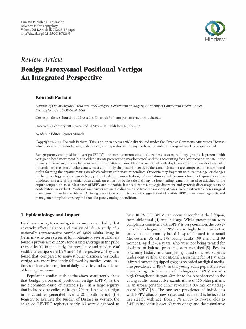
Utricle
Saccule
Cochlea
Superior semicircular canalPosterior
Posterior semicircular canal
Horizontal semicircular canal
Inferior
Superior
Anterior
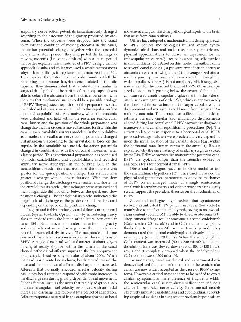
Figure 1: Schematic illustration of the inner ear composed ofthe cochlear and vestibular end organs. Structural relationship ofthe semicircular canals relative to one another and the utricle isdemonstrated.
is not different between the three semicircular canals [16]. Inabout 25% of recurrences, contralateral ear has been reportedto be involved, leading to the suggestion that some systemicfactors might facilitate otoconia detachment [16].
Despite the fact that BPPV is a relatively common condi-tion, because the severity and nature of presenting complaintscan vary, BPPV has a low recognition rate in the primarycare setting [17].The failure to correctly diagnose BPPV likelyaccounts for the poor referral patterns and low proportionof patients who receive appropriate symptom relief from theavailable treatments.
2. Applied Anatomy
The inner ear contributes to two senses: balance and hearing.It is the exclusive end organ for hearing which is served bythe cochlea. The vestibular component of the inner ear isan important end organ for balance which in coordinationwith other inputs (i.e., proprioceptive and visual) to the brainsubserves the sense of balance. The peripheral vestibularsystem consists of five distinct elements on each side: threesemicircular canals and two otolithic organs, the saccule andthe utricle (Figure 1).There are excellent reviews of peripheralvestibular anatomy and function available elsewhere [18, 19].A brief overview is offered here to facilitate an appreciationof the pathophysiology of BPPV.
The inner ear membranous structures and epithelium areencased in the otic capsule which is one of the densest bonesin the body.The otic capsule, located in the petrous portion ofthe temporal bone, unlike other bones in the body, has verylittle turnover, in part because of factors such as osteopontinin inner ear fluids that suppress bone remodeling [20]. Thebony canal houses a membranous duct with the space inbetween being filled by perilymph. The fluid compositionof perilymph is similar to that of cerebrospinal fluid. Themembranous duct is filled with endolymph. Secretion ofendolymph is localized to the stria vascularis in the cochleaand the dark cells in the vestibule. The ampulla of the semi-circular canal secrete a potassium-rich, positively polarizedfluid, the secretion of which is dependent on basolateral Na+,
Advances in Otolaryngology 3
K(+)-ATPase, and Na-K-Cl cotransporter [21]. The ampullarests at one end of the bony/membranous canal and housesthe crista ampullaris. The crista ampullaris includes sensorytransducers (the hair cells) and the supporting structures,as well as a gelatinous fibrillar matrix, referred to as thecupula, in which stereocilia of the hair cells are embedded.The specific gravity of the cupula is similar to that of theendolymph. With rotation of the head, the endolymph fillingthe membranous ducts is displaced resulting in pushing orpulling of the cupula thus deflecting the stereocilia. Depend-ing on the direction of the deflection and the ear in question(toward or away from the kinocilium), the hair cells can bestimulated or suppressed.The resulting signal is carried to thebrain by the vestibular branch of the cranial nerve VIII whichconsists of bipolar afferents whose peripheral endings includelarge calyxes for faithful sensorineural synaptic transmissionfrom the hair cells.
The three canals lie nearly perpendicular to each otherand can code three planes (roll, pitch, and yaw) along the 𝑥-,𝑦-, and 𝑧-axes [22]. Pitch plane corresponds to laying down,sitting up from supine position, or looking up- or downward,while movement along the roll plane corresponds to rotatingthe head toward the right or left in supine position [23].Movement along the yaw plane is rotating head to the leftor right in the sitting position. Therefore, the semicircularcanals are best suited for detecting angular accelerationduring rotational head movements. The kinocilium faces theutricle in the horizontal semicircular canal and faces awayfrom the utricle for the posterior and superior semicircularcanals.Deflection toward the kinocilium (utricopedal) resultsin excitation and away from the kinocilium (utricofugal)produces inhibition. The kinocilia of the crista ampullarisin the posterior and superior (anterior or vertical) semicir-cular ducts are oriented to depolarize the hair cells whenendolymph moves in the utricofugal direction. Head turn toone direction in a given plane results in movement of theendolymph in the opposite direction. Each semicircular canalworks in concert with the corresponding canal located on theother side of the head. They are both oriented in the sameplane; however, hair cells would be expected to convey theopposite signal to the brain due to the opposite direction ofendolymph/cupula displacement on head rotation.
The otolithic organs are best suited for detecting linearacceleration. They consist of the utricle and the saccule.Because of their orientation, which is orthogonal to oneanother, the utricle is more sensitive to linear accelerationand head movements in the horizontal plane, whereas thesaccule is more sensitive to linear acceleration and headmovements in the vertical plane. The saccule is in continuitywith the cochlea throughductus reunions, thus has some low-frequency sound sensitivity. The utricle is more proximateto the semicircular canals which are in continuity with thevestibule, but appears to have some sound sensitivity as well[24]. Macula of the utricle and saccule house the sensoryreceptors, the hair cells. The sensory epithelium projects haircells into an otoconial membrane (a viscous gel layer) onwhich the otoconia rest. The crystals get displaced duringlinear acceleration, which in turn deflects the ciliary bundlesof the hair cells and thus alters the vestibular signal sent to
the brain. Otoconia are calcite crystals composed of calciumcarbonate [25]. They have a specific weight of 2.95 gramsper cubic cm and are typically hexagonal in shape and 3 to30 𝜇m long (see below). Because of their mass, they permitsensitivity to gravitational forces.
Saccular function can be measured using vestibularevoked myogenic potentials (VEMP) [26, 27]. VEMP canhelp identify lesions along the pathway that connects thesaccule via the inferior vestibular nerve and the descendingvestibulospinal pathways to primarily the ipsilateral ster-nocleidomastoid muscle. The stimulus is a series of tonebursts and the evoked potentials are averaged to generatea waveform. Subjectively, dizziness with sensation of fallingis typically associated with saccular dysfunction and VEMPabnormalities [28]. A variant of the cervical VEMP is a myo-genic response that has been evoked around the eye (ocularVEMP) [29, 30]. In ocularVEMP, tone bursts delivered to oneear evoke myogenic potentials in the contralateral inferioroblique muscles with information being conveyed throughafferent fibers travelling in the superior vestibular nerve.Clinically, applications of ocular VEMP are limited. However,in one study, abnormal function of the utricle was implied byabnormal ocular VEMPs recorded in BPPV patients [28].
3. Formation and Structure of Otoconia
Wang and colleagues were able to identify the principalorganic component of the otoconia and partially sequencedand cloned themajor protein component ofmurine otoconia,otoconin 90 [31]. Because of its similarity to secretoryphospholipase A2 (sPLA2), this gene was referred to asPLA2-like (PLA2L) and enabled the identification of humanotoconin 90. The rigid structure of sPLA2 is conveyed by sixor seven disulfide bonds and is conserved in the otoconiaand is essential for optimal interaction of the molecule withthe mineral phase [32]. The molecular weight of otoconin90 ranges between 90 and 100 kD, about half of which isaccounted for by posttranslational modifications, consist-ing predominantly of sulfated glycosaminoglycans. Using ahyaluronidase-gold labeling technique, the localization ofglucuronic acid-containing glycosaminoglycans in the gerbilutricle was examined by Tachibana and Morioka [33]. Theyobserved that otoconia and the gelatinous layer of the otoco-nial membrane were strongly labeled by hyaluronidase-goldand secretory granules in supporting cells suggesting that theorganic matrix of otoconia is secreted from these cells. Thehyaluronidase labeling is lost as otoconia degenerate and areabsorbed into dark cells.
Another protein, otolin, was described as the principalscaffold protein which was initially characterized in the zebrafish [34, 35]. Otolin is a short chain collagen with a highlyinteractive C1q globular domain. Subsequent work in mousedocumented the mammalian ortholog of otolin [32, 36, 37].Zhao and colleagues used gene targeting and protein analysistechniques to demonstrate that otoconin 90 is essential forformation of the organic matrix of otoconia by specificallyrecruiting other matrix components, which include otolin[38]. They demonstrated that this matrix controlled otoconiagrowth andmorphology by embedding the crystallites during
4 Advances in Otolaryngology
seeding and growth. During otoconia development, theorganic matrix forms prior to calcium carbonate (CaCO
3)
deposition and provides optimal calcification efficiency. Yangand colleagues showed that matrix components are recruitedto form the crystal matrix and sequester Ca(2+) for spa-tial specific formation of otoconia [36]. Specifically, theydemonstrated that otoconin 90 binds otolin. In wild typemice otoconin 90 leads to an enrichment of Ca(2+) in theluminal matrices of the utricle and saccule, whereas absenceof otoconin 90 in the null mice leads to significantly reducedmatrix-Ca(2+). Both otoconin 90 and otolin were noted toincrease the propensity of extracellular matrix to calcify incell culture, but together they had a synergistic effect oncalcification.
Andrade and colleagues used immunogold TEM to local-izematrix proteins inmice [39].Theymade several key obser-vations. First, they demonstrated a high density of otoconin90 in the inner core of otoconia, where they are arrangedin oval patterns implying that otoconin 90 is attached toa scaffold consisting of the hexagonal fibrillar meshwork,characteristic of otolin. Second, the level of mineralizationwas much higher in the outer cortex where mineralized fiberbundles are arranged parallel to the surface. Based on thisfinding and observation from decalcification experiments,they concluded that otolin matrix fibrils serve as scaffold toguide mineralization mediated by otoconin 90. Third, theyshowed that individual crystallites assemble into iso-orientedcolumns and that the columns are arranged in parallel lamel-lae which convert into mineralized blocks for hierarchicalassembly into the complex otoconial mosaic. Fourth, theydemonstrated that in young mice the fibrils interconnectingotoconia consisted of the short chain collagen otolin. Fifth,they also observed that in old mice the superficial layer ofmouse otoconia demineralized thus producing weakeningor loss of anchoring of the organic fibrils interconnectingotoconia.
Using energy dispersive X-ray microanalysis and powderX-ray diffraction, otoconia have been described as calcite-based nanocomposites consisting of a relatively uniformouter shape with a cylindrical bulbous body (belly) and threerhombohedral, terminal planes at both ends which are part ofits branches [40]. Degenerative changes can range from mildstructural alteration such as fissures and surface rougheningof the less dense belly area to fractures and disintegrationleading to loss of otoconia [40]. The final component ofthe disintegrating otoconia that leads to fragment formationoccurs in the belly [40]. These degenerative changes tendedto increase with age. With age, the superficial layer of mouseotoconia becomes demineralized resulting in weakening orloss of anchoring of the fibrils interconnecting otoconia [39].Consequently, otoconia can detach from each other and bereleased into the endolymphatic space by minor mechanicaldisturbances. The mechanisms leading to degeneration andeventual fragmentation of otoconia remain unknown.
Other mechanisms that could contribute to otoconiafragmentation include a change in endolymph such as changein pH, specifically acidic range, or calcium concentration.For example, EDTA exposure causes an anisotropic solu-bility of human otoconia, affecting the belly region [41].
Aminoglycosides such as gentamicin can also induce mor-phological changes in the structure of otoconia leading toeventual fragmentation and dissolution [41].
In summary, these results demonstrate that otoconia arecomposed of organic and inorganic components.The organicmatrix is primarily composed of otoconin 90, around anotolin scaffold. The combination of these two componentsattracts and facilitates mineralization of calcium carbonatearound the inorganic matrix to form calcite crystals. Theotoconia are held anchored together with otolin-based fibrils.The structure of otoconia and the fibrils that hold themtogether is affected by the aging process and other diseaseprocesses, thus creating conditions under which otoconia canfragment and separate from the otoconial membrane.
4. Mechanisms of Disease
BPPV is believed to arise from displacement of particulatematter, likely fragments of otoconia from the utricle, intothe semicircular canals. This idea was initially put forth bySchuknecht [42, 43] based on intricate insight in vestibularstructure and function and histopathologic observationsof basophilic staining masses of granular or homogeneousmaterial found attached to the cupula of the posterior semi-circular canal on the affected side. In a subsequent report,Schuknecht and Ruby reported finding copular deposits in37% of temporal bone specimens and that 58% of these werelocated in the posterior canal [44]. Although themechanismsleading to BPPV symptoms were a matter of long-standingdebate [45], today, there is little doubt about the role ofparticulate matter. The involvement of particulate matterwithin the posterior semicircular canal has been establishedintraoperatively in patients with BPPV [46, 47]. Welling andcolleagues prospectively examined the posterior semicircularcanal of patients with and without a clinical history ofBPPV for the presence of particulate matter. No particleswere observed intraoperatively in any of the 73 patientsundergoing labyrinthine surgery (vestibular schwannomaexcision or labyrinthectomy) without a history of BPPV [48].Particulate matter was observed in only 8 of 26 patients withintractable BPPV at the time of the posterior semicircularcanal occlusion procedure. Similarly, Beyea and colleaguesreported 20% incidence of free floating particles in the poste-rior semicircular canal of patients undergoing transmastoidposterior canal plugging for intractable BPPV [49]. However,particulate matter has also been reported in non-BPPVpatients who underwent posterior canal fenestration carriedout in patients undergoing acoustic tumor removal via atranslabyrinthine approach [50]. Particles were identifiedin the membranous labyrinth in nine out of ten patients.However, only one of these patients described positionalvertigo preoperatively. These observations suggest that merepresence of particulate matter is not sufficient and that thereare other conditions that can influence the expression ofdisease symptoms.
Suzuki and colleagues created an in vitro experimentalmodel by placing the posterior semicircular canal isolatedfrom the frog in Ringer’s solution. Saccular otoconia wereused to stimulate the cupula [51]. Posterior semicircular canal
Advances in Otolaryngology 5
ampullary nerve action potentials instantaneously changedaccording to the direction of the gravity produced by oto-conia. When the otoconia were dropped into the canalto mimic the condition of moving otoconia in the canal,the action potentials changed together with the otoconialflow after a latent period. They interpreted the findings asmoving otoconia (i.e., canalolithiasis) with a latent periodthat better explain clinical features of BPPV. Using a similarapproach Otsuka and colleagues used a whole membranouslabyrinth of bullfrogs to replicate the human vestibule [52].They exposed the posterior semicircular canals but left theremaining membranous labyrinth encapsulated in the oticcapsule. They demonstrated that a vibratory stimulus (asurgical drill applied to the surface of the bony capsule) wasable to detach the otoconia from the utricle, consistent withthe view that mechanical insult could be a possible etiologyof BPPV.They adjusted the position of the preparation so thatthe dislodged otoconia were attached to the cupular surfaceto model cupulolithiasis. Alternatively, when the otoconiawere dislodged and held within the posterior semicircularcanal lumen and the position of the whole preparation waschanged so that the otoconiamoved back and forthwithin thecanal lumen, canalolithiasis was modeled. In the cupulolithi-asis model, the vestibular nerve action potentials changedinstantaneously according to the gravitational force on thecupula. In the canalolithiasis model, the action potentialschanged in combination with the otoconial movement aftera latent period. This experimental preparation has been usedto model canalolithiasis and cupulolithiasis and recordedampullary nerve discharges in the bullfrog [53]. In thecanalolithiasis model, the acceleration of the otoconia wasgreater for the quick positional change. This resulted in agreater discharge with a longer duration. With the slowpositional change, the discharges were smaller and shorter. Inthe cupulolithiasis model, the discharges were sustained andtheir magnitude did not differ between the quick and slowpositional changes. The canalolithiasis model influenced themagnitude of discharge of the posterior semicircular canaldepending on the speed of the positional change.
Rajguru and Rabbitt induced canalolithiasis in an animalmodel (oyster toadfish, Opsanus tau) by introducing heavyglass microbeads into the lumen of the lateral semicircularcanal [54]. Bead movement under the action of gravityand canal afferent nerve discharge near the ampulla wererecorded extracellularly in vivo. The magnitude and timecourse of the afferent responses explained the symptoms ofBPPV. A single glass bead with a diameter of about 20 𝜇mmoving at nearly 80 𝜇m/s within the lumen of the canalelicited pathological afferent inputs to the brain equivalentto an angular head velocity stimulus of about 100∘/s. Whenthe head was oriented nose-down, beads moved toward thenose and the lateral canal afferent discharge rate increased.Afferents that normally encoded angular velocity duringoscillatory head rotations responded with tonic increases inthe discharge rate during gravity-dependent beadmovement.Other afferents, such as the units that rapidly adapt to a stepincrease in angular head velocity, responded with an initialincrease in discharge rate followed by a period of adaptation.Afferent responses occurred in the complete absence of head
movement and quantified the pathological inputs to the brainthat arise from canalolithiasis.
Others have adopted a mathematical modeling approachto BPPV. Squires and colleagues utilized known hydro-dynamic calculations and make reasonable geometric andphysical approximations to derive an expression for thetranscupular pressure Δ𝑃
𝑐exerted by a settling solid particle
in canalolithiasis [55]. Based on this model, the authors cameto several conclusions: (1) a pressure amplification occurs asotoconia enter a narrowing duct; (2) an average-sized otoco-nium requires approximately 5 seconds to settle through thewide ampulla, where Δ𝑃
𝑐is not amplified, which suggests a
mechanism for the observed latency of BPPV; (3) an average-sized otoconium beginning below the center of the cupulacan cause a volumetric cupular displacement on the order of30 pL, with nystagmus of order 2∘/s, which is approximatelythe threshold for sensation; and (4) larger cupular volumedisplacement and nystagmus could result from larger and/ormultiple otoconia. This group also utilized their model toestimate dynamic cupular and endolymph displacementselicited during horizontal canal BPPV provocative diagnosticmaneuvers and canalith repositioning procedures [56]. Theactivation latencies in response to a horizontal canal BPPVprovocative diagnostic test were predicted to vary dependingupon the initial location of the canalith debris (e.g., withinthe horizontal canal lumen versus in the ampulla). Resultsexplained why the onset latency of ocular nystagmus evokedby theDix-Hallpike provocativemaneuver for posterior canalBPPV are typically longer than the latencies evoked byanalogous tests for horizontal canal BPPV.
Obrist and colleagues used an in vitro model to testthe canalolithiasis hypothesis [57]. They carefully scaled thephysical and geometrical parameters to study the mechanicsof BPPV on an enlarged model of a single semicircularcanal with laser vibrometry and video particle tracking. Earlyresults support the prevalent theories on the mechanisms ofBPPV.
Zucca and colleagues hypothesized that spontaneousrecovery in untreated BPPV patient (usually in 2–6 weeks) ismainly due to the fact that endolymph, owing to its low cal-cium content (20microM), is able to dissolve otoconia [58].They immersed frog saccular otoconia in normal endolymph(Ca2+ content 20microM) and in Ca2+-rich endolymphaticfluids (up to 500microM) over a 3-week period. Theydemonstrated that normal endolymph can dissolve otoconiavery rapidly (in about 20 hours). When the endolymphaticCa2+ content was increased (50 to 200microM), otoconiadissolution time was slowed down (about 100 to 130 hours,resp.) and it completely stopped when the endolymphaticCa2+ content was of 500microM.
To summarize, based on clinical and experimental evi-dence, displaced fragments of otoconia into the semicircularcanals are now widely accepted as the cause of BPPV symp-toms. However, a critical mass appears to be needed to evokeclinical symptoms, as mere presence of fragments withinthe semicircular canal is not always sufficient to induce achange in vestibular nerve activity. Experimental modelseffectively simulate canalolithiasis and cupulolithiasis provid-ing empirical evidence in support of prevalent hypothesis on
6 Advances in Otolaryngology
pathophysiology of BPPV. Concentration of calcium in theendolymphplays an important role in the rate of resorption ofotoconia and may influence the duration of a BPPV episode.
5. Diagnosis of Cupulolithiasis andCanalolithiasis and Identifying theAffected Canal
Historical recognition of BPPV and use of provocativepositioning techniques in its diagnosis had been a point ofdiscussion. Lanska and Remler [45], based on an excellentreview of the extant medical literature, noted that Baranyin 1921 was the first to describe BPPV in detail. They alsocredited Dix and Hallpike [59] as being the first to clearlydescribe both the currently used provocative positioningtechnique and the essential clinical manifestations of BPPVelicited by that technique.TheDix-Hallpikemaneuver is nowa standard component of the evaluation of the dizzy patient.When performing this procedure, the examiner stands to oneside of the patient and rotates the patient’s head 45 degreesto that side to align the ipsilateral posterior semicircularcanal with the sagittal plane of the body. Next, the examinermoves the patient, whose eyes are open, from the seatedto the supine test-ear-down position and then extends thepatient’s neck slightly so that the chin is pointed slightlyupward. The latency, duration, and direction of nystagmus,if present, and the latency and duration of vertigo, if present,are noted. Each position should be maintained at least 30–45 seconds or until nystagmus resolves. If positive, rotarynystagmus toward the test ear is commonly observed, whichis induced by the movement of free-floating debris withinthe affected canal (away from the cupula, inducing deflectionof the cupula away from the vestibule). If no nystagmus isobserved, or if observed once it resolves, the patient is seatedupright and once again eyes are monitored for nystagmuswhich is once again documented as above. The procedure isrepeated for the opposite ear. More recently, head shakingduring the examhas been proposed to increase the diagnosticyield of the Dix-Hallpike exam [60]. Specifically, patientssuspected of having posterior canal BPPV, but with negativeDix-Hallpike, are recommended to undergo head shakingDix-Hallpike.This subgroupof patients is suspected of havinga milder form of posterior canal BPPV (also see below for“subjective BPPV”). The side-lying test which also stimulatesthe posterior semicircular canal may be more suitable forolder patients because the head and neck are fully supportedby the examination table [7]. In this test, after seating thepatient on the examination table, the head is turned 45∘ awayfrom the involved ear then the patient lies on the side of theinvolved ear [61].
Lansk and Remler point out that “despite their impor-tant contributions, neither Barany nor Dix and Hallpikeunderstood the pathophysiology of BPPV nor did theyappreciate that the positioning techniques they used actu-ally demonstrated pathology in the semicircular canalsrather than the utricle.” They go on to attribute the mod-ern understanding of the pathophysiology of BPPV toSchuknecht’s proposal that the dysfunction resulted from
the gravity-dependent movement of loose or fixed densematerial within the posterior semicircular canal (“cupu-lolithiasis”) [42]. They add that although Schuknecht’s for-mulations were not consistent with all clinical features of thedisease, they led to the modern “canalolithiasis theory” andhighly effective canalith repositioning or liberatory maneu-vers for BPPV. Hall and colleagues brought clarity to under-standing the mechanisms underlying posterior canal BPPVby emphasizing the importance of fatigability of symptoms[62]. Hall and colleagues noted that, under the influence ofgravity, a density differential between the endolymph and thecupula will cause displacement of the cupula when changesin head position occur. They explained that BPPV can bedivided into two types based on the presence or absenceof fatigability. Specifically, nonfatigable nystagmus was theresult of particles being fixed on the cupula (cupulolithiasis),whereas fatigable nystagmus was due to free floating particleswithin the body of posterior semicircular canal. This lattercondition was subsequently referred to as canalolithiasis[63].
Theoretically, if particulate matter can be located in thelong arm of the semicircular canals or attached to the cupula,distally, it could also be present in the short arm or attachedto the cupula, proximally. Indeed a histopathologic studyreported that canaliths have been found to be located in theshort arm of the semicircular canals (proximate to the utricle)[64]. Clinical correlate of this entity has yet to be defined.
Posterior canal BPPV is the most common. In a studyof 614 BPPV patients over an 11-year period, the posteriorsemicircular canal was affected in 543 cases (88.4%), thehorizontal in 39 (6.4%), and the superior canal in 32 (5.2%)[16].
BPPV of the posterior canal, the most inferior canal,is believed to be much more frequent because gravitationalforces settle the particles in this canal, particularly whenthe patient is in the lateral supine position [65]. In contrast,the superior semicircular canal is the highest point of thelabyrinth, thus it is rarely involved in BPPV. To settle particlesinto the superior canal, very marked hyperextension of thehead is required [65].
Several authors have characterized horizontal BPPV [66,67]. Horizontal BPPV is triggered when the head of thesupine patient is turned from side to side and is characterizedby a bidirectional horizontal nystagmus. As with posteriorBPPV there are also two subtypes of horizontal BPPVdepending on the location of the otoconia fragments. Incanalolithiasis, fragments of otoconia move freely in thehorizontal semicircular canal inducing stimulatory utricu-lopetal endolymph flow, whereas in cupulolithiasisotoconiaare attached to the cupula or are trapped in the proximalsegment of the canal near to the cupula. In canalolithiasis,vertigo is triggered by circular acceleration in the plane ofthe horizontal semicircular canal while, in cupulolithiasis,vertigo is triggered when the head changes position, butnot by circular acceleration. In canalolithiasis, nystagmus isgeotropic (toward the undermost ear) and more intense afterrotation of the head toward the affected side. In cupulolithi-asis, the nystagmus during cupulolithiasis of the horizontalsemicircular canal is apogeotropic (away from the undermost
Advances in Otolaryngology 7
ear) and more intense after rotation of the head towardthe unaffected ear. It is recommended that, for patientspresenting with symptoms of BPPV, if the Dix-Hallpike testis negative, a supine roll test be performed to assess for lateralcanal BPPV [68].
Clinically identifying the affected side in horizontal canalBPPV can be challenging. The problem is that during diag-nostic maneuver (e.g., the supine head roll test, also knownas the Pagnini-McClure maneuver), one horizontal canalcannot be isolated from the other, as both head positionsstimulate the horizontal canals resulting in nystagmus witheither ear down. In addition, horizontal canal BPPV causesdirection-changing nystagmus. As noted above, horizontalBPPV-related nystagmus can be geotropic or apogeotropic,but the direction of the nystagmus relative to the ground isthe same in both positions of the supine head roll test. Ewald’ssecond law provides guidance on determining the affectedside. Ewald’s three laws were generated after experimentsin pigeons whose semicircular canals were stimulated usingpneumatic devices [69]. They state that a stimulation of thesemicircular canal causes a movement of the eyes in theplane of the stimulated canal. In the horizontal semicircularcanals, an ampullopetal endolymph movement causes agreater stimulation than an ampullofugal one. In the verticalsemicircular canals, the reverse is true. Ewald’s second lawhasprofound clinical implications. The directional asymmetryimplies that, in persons who have lost inner ear functionon one side, the remaining side should produce highervelocity nystagmus when the head is being rotated towardsthe remaining intact ear (ampullopetal), but less when thehead is being rotated towards the side of vestibular loss. Aronand Bance provided validation of Ewald’s second law in a rarecase combining horizontal canal BPPV with a dead ear onthe contralateral side, therefore arising from a known side[70]. They presented the case of an 87-year-old male withhistory of invasive left ear cholesteatoma which had fistulizedinto the horizontal canal. He underwent successful resectionof disease with postoperative caloric testing showing absentresponses on the left side and a “dead” ear. Ten years later,the patient presentedwith chronic dysequilibriumaggravatedby head movements. Dix-Hallpike test was negative, but asupine head roll test demonstrated bilateral apogeotropic,horizontal nystagmus, which was more pronounced with thehead turned to the left. Videonystagmography demonstratedthe increased slow phase velocity of the nystagmus whenthe head was turned to the left (i.e., away from the affectedear). This case corroborates Ewald’s law for lateralization ofapogeotropic horizontal canal BPPV.
Patients with lateral canal BPPV may also exhibit amild “spontaneous” horizontal nystagmus while in the sittingposition but because presentation of nystagmus is stronglyinfluenced by head position and movements, this type ofnystagmus may be more accurately referred to as pseu-dospontaneous nystagmus [71]. It beats toward the healthyside when the lateral canal BPPV is geotropic and towardthe affected side when it is apogeotropic and when absent[71, 72]; it is sometimes possible to evoke it with a mildshaking of the head [71]. Patientswith apogeotropic showpre-dominantly contralesional head-shaking nystagmus, whereas
patients with geotropic type do not show any directionalpreponderance [72].
The “bow and lean test” has been promoted as a usefulmethod to identify the affected side in horizontal canal BPPV[73]. In a prospective study, for diagnostic purposes, subjectswere divided into two groups: standard head roll test versusthe bow and lean test. The latter group had better remissionrates for both canalolithiasis and cupulolithiasis of the lateralcanal.
Initial presenting history can be an important aid inidentification of the involved canal. There is a relationshipbetween the position that initially provoked vertigo and theaffected semicircular canal in patients with BPPV. Historytaking regarding the side of provoking position at theonset of vertigo helps predict the side affected by BPPV inboth posterior canal BPPV and geotropic horizontal canalBPPV [23]. Although the type of BPPV cannot be predictedby the initial position provoking vertigo, the side of theBPPV is strongly correlated to the head-turning side in thesupine position that initially provide vertigo in this group ofpatients.
When apogeotropic horizontal canal BPPV is suspected,further detailed examinations using additional localizationmethods, including supine head roll test and pitch-planetest, are typically needed. The waking-up position was themost common situation that initially provoked vertigo inall subtypes of BPPV, including geotropic and apogeotropichorizontal canal BPPV [23].
Until recently diagnosing of superior (anterior) semicir-cular canal BPPVwas controversial. Because of its anatomicallocation, the superior semicircular canal is rarely affectedin BPPV. In a retrospective study where over a 1-yearperiod 4,320 patients consulting for otoneurological diseasewere investigated by otological, videonystagmography, andneurological examination, BPPV was diagnosed in 1,430patients (33%) [65]. Among these the posterior semicircularcanal was involved in 1,325 patients (92.6%), the horizontalsemicircular canal in 85 patients (5.9%), the posterior semi-circular canal and ipsilateral superior semicircular canal in19 patients (0.01%), and the superior semicircular canal onlyin one patient (0.0007%). In the 20 patients with superiorsemicircular canal BPPV, the Dix-Hallpike test induced apo-geotropic horizontal torsional nystagmus beating towards theuppermost ear in the lateral supine position with reversal onstanding. That is nystagmus beating towards the uppermostear on theDix-Hallpike test is consistentwithBPPV involvingthe superior semicircular canal of the uppermost ear. Theauthor suggests that the torsional component of nystagmusand not just the vertical component must be taken intoaccount to facilitate the diagnosis.
Multiple canals can be involved at presentation, notuncommonly after head trauma [74]. Involvement ofmultiplecanals can give rise to atypical presentation and challengingmanagement with repositioning maneuvers [74, 75]. It hasbeen argued that the most significant factor in diagnosisof the type of BPPV is observation of the characteristicsof the provoked nystagmus during the diagnostic positionalmaneuvers, although the combination of posterior-anteriorcanal involvement remains particularly challenging [76].
8 Advances in Otolaryngology
Special considerations are warranted in the geriatric pop-ulation, because geriatric patients with BPPV usually reportdizziness or imbalance and do not always describe a rotatorycrisis. Batuecas-Caletrio and colleagues have argued that theDix-Hallpike and supine roll tests should be performed inolder patients with dizziness, despite the fact that they do notcomplain of spinning sensation with positional changes [12].
Finally, a number of studies have reported that the rightear is predominantly affected [10, 12, 16]. It has been suggestedthat prolonged lying may facilitate the deposition of otoconiaon the cupula or contribute to their loosening from the utricle(see below) and that this mechanism might also explain whythe laterality of BPPV often corresponds to the preferred sideof lying during sleep [77].
6. Treatment
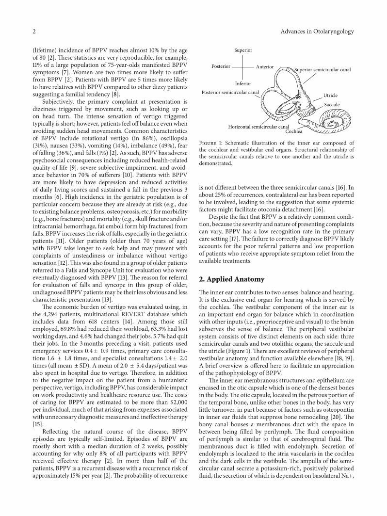
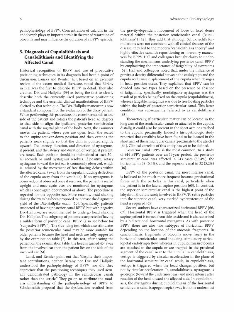
Mechanical repositioning: historically, as the proposedmech-anism involving cupulolithiasis and canalolithiasis becamemore widely accepted, the idea of using head positioningmaneuvers to relieve symptoms of BPPV evolved as analternative to conservative management (i.e., avoidance ofprovocative head positions and changes) and surgical proce-dures. Brandt and Daroff used a physical therapy approachinvolving repeated maneuvers to loosen and disperse thedegenerated otolithic material from the cupula [78]. Theseefforts were followed by liberatory maneuver of Semont [79]and subsequently canalith repositioning procedure of Eply(Figure 2) [80, 81]. Given that the dominant view at thattime was that BPPV arose from the involvement of theposterior semicircular canal, these repositioning procedureswere mainly directed at providing relief of posterior canalBPPV.However, several varieties of BPPV can be experienceddepending on the semicircular canal involved, whether or notcupula is involved, single or multiple canal involvement, andsidedness of the affected structures. In fact, the effectivenessof the repositioning maneuvers varies depending on correctidentification of the affected canal.
To assess the effectiveness of the Epley maneuver intreatment of posterior semicircular canal BPPV, Hilton andPinder, in a Cochrane review, compared Epley maneuverversus placebo Epley maneuver versus untreated controls[82]. They evaluated the frequency and severity of attacks ofvertigo, proportion of patients improved by each interven-tion, and conversion of a “positive” Dix-Hallpike test to a“negative” Dix-Hallpike test. Fifteen randomized trials wereidentified but twelve studies were excluded because of a highrisk of bias, leaving three trials in the review. Trials weremainly excluded because of inadequate concealment duringrandomization, or failure to blind outcome assessors. Thestudies included in the review addressed the efficacy of theEpley maneuver against a sham maneuver or control group.They concluded that the Epley maneuver is a safe effectivetreatment for posterior canal BPPV. A more recent review83 arrived at the same conclusion, noting that based on asystematic review of all relevant randomized controlled trials(Level I evidence) there is strong evidence of efficacy of theEpley maneuver in treatment of posterior canal BPPV, withno serious adverse effects.
Poste
rior s
emic
ircul
ar ca
nal
cana
liths
Figure 2: Illustration of the Epley maneuver for the treatment ofleft posterior canal BPPV. From a seated position (top), the patient’shead is turned 45∘ to the left (2nd from top).The patient is laid downwith the head rotated to the left and neck is extended (3rd from top).Next, the head is turned 90∘ to the right (4th from top) followedby rolling the body to the right such that patient is lying on theright shoulder looking toward the floor (2nd from bottom). Finally,the patient is returned to the seated position with head remainingrotated to the right (bottom). Each position is held for at least 30–60seconds or until nystagmus or vertigo has resolved. Frenzel lensescan facilitate visualization of nystagmus. Adapted fromMitka [151].
There are currently two practice guidelines that areavailable for diagnosis and management of BPPV, one bythe American Academy of Neurology [83] and another byAmericanAcademyofOtolaryngology-Head&Neck Surgery[68]. They both offer thorough reviews of the availableevidence and validate the effectiveness of the Epleymaneuverin management of posterior semicircular canal BPPV.
Amore recent Cochrane review compared amodificationof Epley maneuver to the standard Epley maneuver [84].Nine studies included post-Epley postural restrictions. Suchrestrictions could include using a neck brace, headmovementrestrictions (avoiding bending the head up and down orbending over), and instructions to sleeping upright. Thereview concluded that there was a statistically significantadvantage of post-Epley postural restrictions. Specifically, itwas found that in the experimental group 88.7% versus 78.2%in the control group converted from a positive to negativeDix-Hallpike test.
The sameCochrane review found that there is insufficientevidence to support the routine application of mastoidoscillation during the Epley maneuver. Mastoid oscillationinvolves applying vibration to the mastoid bone behind theear during the maneuver (an “augmented” Epley).
In a prospective study in geriatric BPPV patients, bothclinical and functional aspects of body balance, including
Advances in Otolaryngology 9
postural instability, improved after treatment with the mod-ified Epley maneuver [85]. However, in the older patients,the effectiveness of therapeutic repositioning maneuver (seebelow) is lower than those under 70 and the recurrences aremore frequent [5].
At present, the best available treatment of the horizontalcanal BPPV is the roll (Barbecue or Lempert) maneuver [67,86–89]. The procedure is effective in managing lateral canalBPPV in a lower proportion of patients than Epley maneuveris in control of posterior canal BPPV (<75% versus 88%,resp.). The Gufoni maneuver is another technique that hasbeen reported as effective in treating horizontal canal BPPV.In a double-blind randomized controlled trial, this maneuverproved effective for both geotropic and apogeotropic formswith 84% relief of vertigo after 24 hrs [90].
Options inmanagement of the cupulolithiasis of the hori-zontal semicircular canal are very limitedwhere the challengeis to detach the otoconia fragments from the cupula. It hasbeen suggested that repetitive somersaults may be beneficial.In somersaults, acceleration in the forward-facing directionmay induce an intracanalicular force strong enough to detachotoconia debris from the cupula and the ongoing rollingmay move loose otoconia debris back into the utricle [91].Yamanaka and colleagues described the head-tilt hoppingexercise in treatment of cupulolithiasis associated with BPPVof the horizontal semicircular canal, which is characterizedby apogeotropic direction-changing nystagmus [92]. Thistreatment is designed to release otoconial debris adherent tothe cupula as the patients hopped while tilting their headslaterally. The treatment required several sessions daily over a4-week period. Nystagmus of 33.3% patients with intractablelateral canal BPPV disappeared immediately after the firsttraining session and after 1 and 4 weeks of the training, 56%and 70% of patients that had experienced improvements,respectively. It is important to note, however, given thenatural history of BPPV, that absence of untreated controlgroup significantly weakened the potential for generalizationof this study.
In a recent study of 965 BPPV patients, canalith repo-sitioning procedures (Epley and Barbeque maneuvers) pro-vided long-lasting noninvasive treatment for BPPV with 85%effectiveness after the first procedure [89]. Fourteen percentof patients had recurrences, with this group of patientshaving characteristics of being older or having history of headtrauma or vestibular neuropathy. The authors suggested thatgeriatric patients, because of a significantly higher recurrencerate, should have additional education to minimize potentialmorbidity of their falls.
In general, The Epley maneuver is effective in treatmentof superior semicircular canal in 94.1% of cases after onetreatment and 97.5% after multiple treatments [65]. However,in another report, therapeutic maneuvers were found to bemore effective in resolving posterior canal or horizontal canalBPPV than superior canal BPPV [16]. Specific maneuvers forrelief of superior canal BPPV have been described [93, 94].For example, a sequence of maneuvers consisting of headpositioning beginning supine with head hanging 30 degreesdependent with respect to the body, then supine with headinclined 30 degrees forward, and ending sitting with head 30
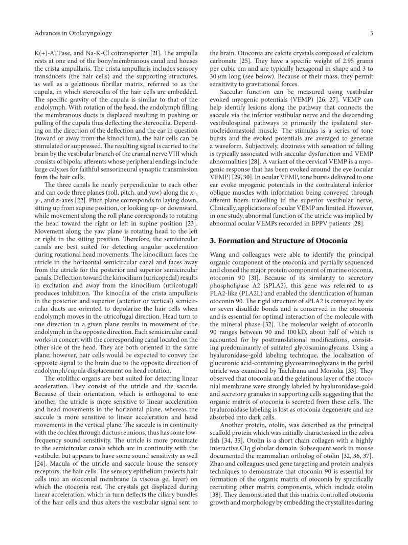
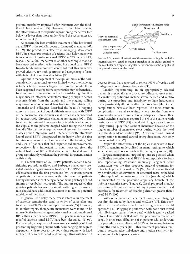
Cochlear nerve
Nerve to superior semicircular canal
Nerve to horizontalsemicircular canal
Nerve to posterior semicircular canal(singular nerve)
Nerve to saccule
Nerve to utricle
Facial nerve
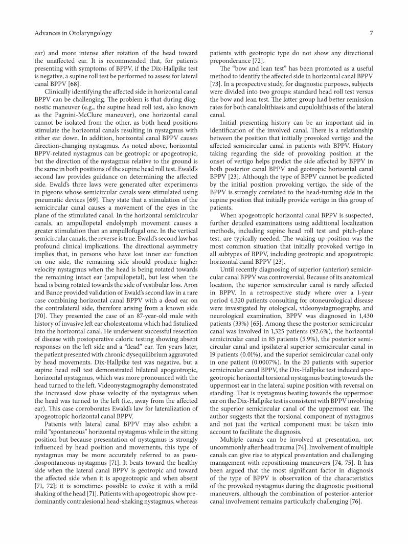
Figure 3: Schematic illustration of the nerves travelling through theinternal auditory canal, including branches of the eighth cranial tothe vestibular end organs. Singular nerve innervates the ampulla ofthe posterior semicircular canal.
degrees forward are reported to relieve 100% of vertigo andnystagmus in one retrospective series [93].
Canalith repositioning, in an appropriately selectedpatient, is a generally safe procedure. Minor adverse eventsof canalith repositioning include severe nausea and vertigoduring the procedure and instability or light-headednessfor approximately 48 hours after the procedure [89]. Othercomplications have also been reported. The most commoncomplication is canal switching, where otoliths from onesemicircular canal are unintentionally displaced into another.Canal switching has been reported in 6% of the patients withposterior canal BPPV [95]. Canal switching appears to morelikely during Epley than Semont maneuver because of thehigher number of maneuver steps during which the headis in the dependent position [96]. A very rare and unusualcomplication is internal carotid artery dissection, with onlyone reported case [97].
Despite the effectiveness of the Epley maneuver to treatBPPV, it remains underutilized in many settings to whichsufferers initially present, such as the emergency room [98].
Surgical management: surgical options are pursued whendebilitating posterior canal BPPV is unresponsive to bed-side repositioning. Posterior ampullary (singular) nervetransaction was the first proposed surgical treatment forintractable posterior canal BPPV [99]. Gacek was motivatedby Schuknecht’s observations of otoconial mass embeddedin the cupula of the posterior canal crista (see above) whichis innervated by the posterior ampullary branch of theinferior vestibular nerve (Figure 3). Gacek proposed singularneurectomy through a tympanotomy approach under localanesthesia for treatment of disabling chronic (greater than 1year) BPPV [100].
Posterior semicircular canal occlusion (canal plugging)was first described by Parnes and McClure [47]. This oper-ation can be effectively performed using a transmastoidapproach [49]. Plugging is performed with bone dust mixedwith fibrinogen sealant (bone pate) which is gently packedinto a fenestration drilled into the posterior semicircularcanal. In one series, all but one of 44 patients who underwentthis procedure were relieved of BPPV on follow-up between6 months and 12 years [101]. This treatment produces tem-porary postoperative imbalance and motion sensitivity forseveral weeks, but spares hearing.
10 Advances in Otolaryngology
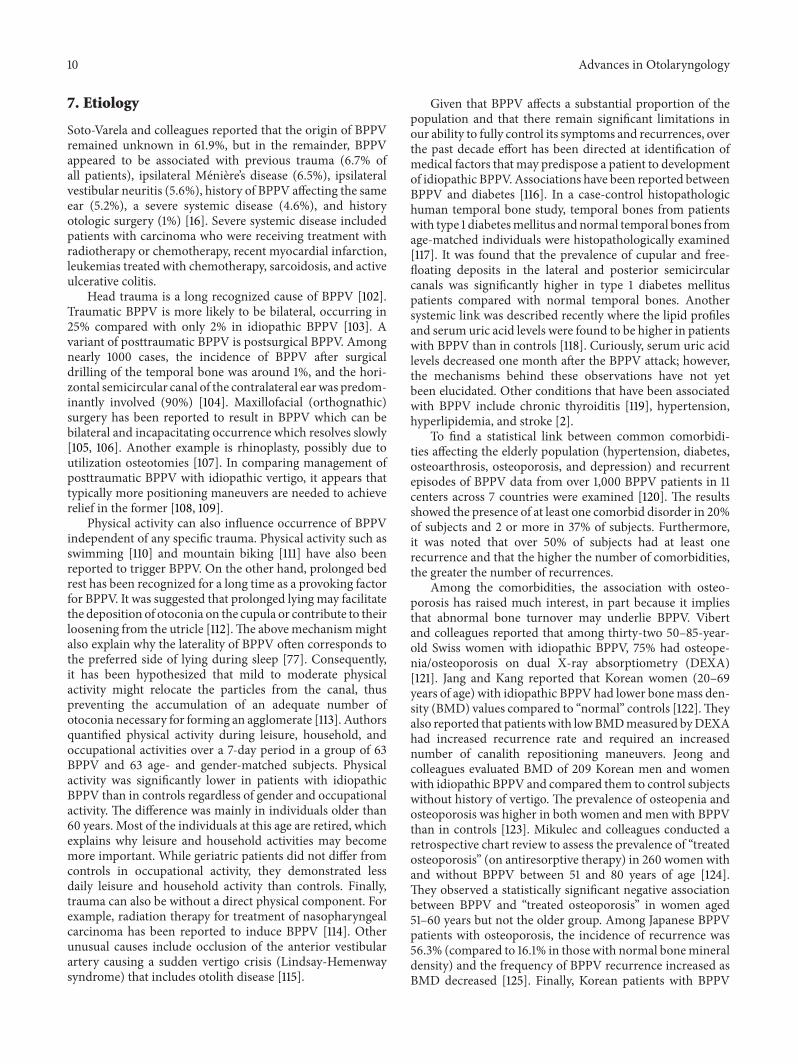
7. Etiology
Soto-Varela and colleagues reported that the origin of BPPVremained unknown in 61.9%, but in the remainder, BPPVappeared to be associated with previous trauma (6.7% ofall patients), ipsilateral Meniere’s disease (6.5%), ipsilateralvestibular neuritis (5.6%), history of BPPV affecting the sameear (5.2%), a severe systemic disease (4.6%), and historyotologic surgery (1%) [16]. Severe systemic disease includedpatients with carcinoma who were receiving treatment withradiotherapy or chemotherapy, recent myocardial infarction,leukemias treated with chemotherapy, sarcoidosis, and activeulcerative colitis.
Head trauma is a long recognized cause of BPPV [102].Traumatic BPPV is more likely to be bilateral, occurring in25% compared with only 2% in idiopathic BPPV [103]. Avariant of posttraumatic BPPV is postsurgical BPPV. Amongnearly 1000 cases, the incidence of BPPV after surgicaldrilling of the temporal bone was around 1%, and the hori-zontal semicircular canal of the contralateral earwas predom-inantly involved (90%) [104]. Maxillofacial (orthognathic)surgery has been reported to result in BPPV which can bebilateral and incapacitating occurrence which resolves slowly[105, 106]. Another example is rhinoplasty, possibly due toutilization osteotomies [107]. In comparing management ofposttraumatic BPPV with idiopathic vertigo, it appears thattypically more positioning maneuvers are needed to achieverelief in the former [108, 109].
Physical activity can also influence occurrence of BPPVindependent of any specific trauma. Physical activity such asswimming [110] and mountain biking [111] have also beenreported to trigger BPPV. On the other hand, prolonged bedrest has been recognized for a long time as a provoking factorfor BPPV. It was suggested that prolonged lying may facilitatethe deposition of otoconia on the cupula or contribute to theirloosening from the utricle [112].The abovemechanismmightalso explain why the laterality of BPPV often corresponds tothe preferred side of lying during sleep [77]. Consequently,it has been hypothesized that mild to moderate physicalactivity might relocate the particles from the canal, thuspreventing the accumulation of an adequate number ofotoconia necessary for forming an agglomerate [113]. Authorsquantified physical activity during leisure, household, andoccupational activities over a 7-day period in a group of 63BPPV and 63 age- and gender-matched subjects. Physicalactivity was significantly lower in patients with idiopathicBPPV than in controls regardless of gender and occupationalactivity. The difference was mainly in individuals older than60 years. Most of the individuals at this age are retired, whichexplains why leisure and household activities may becomemore important. While geriatric patients did not differ fromcontrols in occupational activity, they demonstrated lessdaily leisure and household activity than controls. Finally,trauma can also be without a direct physical component. Forexample, radiation therapy for treatment of nasopharyngealcarcinoma has been reported to induce BPPV [114]. Otherunusual causes include occlusion of the anterior vestibularartery causing a sudden vertigo crisis (Lindsay-Hemenwaysyndrome) that includes otolith disease [115].
Given that BPPV affects a substantial proportion of thepopulation and that there remain significant limitations inour ability to fully control its symptoms and recurrences, overthe past decade effort has been directed at identification ofmedical factors that may predispose a patient to developmentof idiopathic BPPV. Associations have been reported betweenBPPV and diabetes [116]. In a case-control histopathologichuman temporal bone study, temporal bones from patientswith type 1 diabetesmellitus andnormal temporal bones fromage-matched individuals were histopathologically examined[117]. It was found that the prevalence of cupular and free-floating deposits in the lateral and posterior semicircularcanals was significantly higher in type 1 diabetes mellituspatients compared with normal temporal bones. Anothersystemic link was described recently where the lipid profilesand serum uric acid levels were found to be higher in patientswith BPPV than in controls [118]. Curiously, serum uric acidlevels decreased one month after the BPPV attack; however,the mechanisms behind these observations have not yetbeen elucidated. Other conditions that have been associatedwith BPPV include chronic thyroiditis [119], hypertension,hyperlipidemia, and stroke [2].
To find a statistical link between common comorbidi-ties affecting the elderly population (hypertension, diabetes,osteoarthrosis, osteoporosis, and depression) and recurrentepisodes of BPPV data from over 1,000 BPPV patients in 11centers across 7 countries were examined [120]. The resultsshowed the presence of at least one comorbid disorder in 20%of subjects and 2 or more in 37% of subjects. Furthermore,it was noted that over 50% of subjects had at least onerecurrence and that the higher the number of comorbidities,the greater the number of recurrences.
Among the comorbidities, the association with osteo-porosis has raised much interest, in part because it impliesthat abnormal bone turnover may underlie BPPV. Vibertand colleagues reported that among thirty-two 50–85-year-old Swiss women with idiopathic BPPV, 75% had osteope-nia/osteoporosis on dual X-ray absorptiometry (DEXA)[121]. Jang and Kang reported that Korean women (20–69years of age) with idiopathic BPPV had lower bonemass den-sity (BMD) values compared to “normal” controls [122].Theyalso reported that patientswith lowBMDmeasured byDEXAhad increased recurrence rate and required an increasednumber of canalith repositioning maneuvers. Jeong andcolleagues evaluated BMD of 209 Korean men and womenwith idiopathic BPPV and compared them to control subjectswithout history of vertigo. The prevalence of osteopenia andosteoporosis was higher in both women and men with BPPVthan in controls [123]. Mikulec and colleagues conducted aretrospective chart review to assess the prevalence of “treatedosteoporosis” (on antiresorptive therapy) in 260 women withand without BPPV between 51 and 80 years of age [124].They observed a statistically significant negative associationbetween BPPV and “treated osteoporosis” in women aged51–60 years but not the older group. Among Japanese BPPVpatients with osteoporosis, the incidence of recurrence was56.3% (compared to 16.1% in those with normal bonemineraldensity) and the frequency of BPPV recurrence increased asBMD decreased [125]. Finally, Korean patients with BPPV
Advances in Otolaryngology 11
show a higher prevalence of vitamin D deficiency [126]and a recent pilot study showed that in 4 Austrian patientswith BPPV recurrence, vitamin D levels were lower thanthose without recurrence and recurrences were relieved withvitamin D supplementation [127].
Although these studies have investigated the associationbetween BPPV and low bone mineral density, the cooc-currence of two morbidities is not by itself supportive ofcausation. Low bone mineral density (e.g., in premenopausalwomen) might reflect low peak bone mass based on geneticpredisposition, environment, and lifestyle factors [128] andosteopenia/osteoporosis are prevalent disorders of aging.Furthermore, a limitation of asserting an association basedon DEXA scan findings is that any physiological alterationsin bone turnover take about one year to be detectable byDEXA scan [129]. Since osteoporosis is a slowly changingdisease process there is an obvious incongruence betweenDEXA scan findings and BPPV episodes, which tend topresent abruptly and typically are self-limited (days to weeksin duration). An alternative approach is to examine biochem-ical markers of bone turnover, whose levels change rapidly(within 1–3 months) and are more temporally relevant to thenatural history of BPPV. Parham and colleagues used thesemarkers to demonstrate that idiopathic BPPV patients tendto have higher levels of biochemicalmarkers of bone turnoverand that these levels are influenced by vitamin D level [130].
8. Differential Diagnosis
Although BPPV is one of the most common causes ofvertigo, it is not the only disorder associated with recur-rent vertigo. Meniere’s disease, migraines, vertebrobasilarinsufficiency, and panic disorder are also characterized byrecurrent episodes. Caution in diagnosis BPPV is warrantedas other conditions can present with BPPV-like symptoms.For example, central vestibular disorders can give rise topositional nystagmus, which can be mistaken for BPPV. Suchlesions can arise from posterior fossa such as from smallcerebellar strokes [131]. In inner ear pathologies which maycause vestibulopathies, such as perilymph fistulas, nystagmusmay be enhanced by Dix-Hallpike testing. Furthermore, asspontaneous nystagmus associated with a perilymph fistulaimproves, nystagmus toward the affected ear in the downwardposition may misdirect the examiner from the initial patho-logical trigger of the symptoms. Other vestibulopathies thatcause horizontal nystagmus may also be enhanced by Dix-Hallpike testing. Welgampola and colleagues cite anotherexample where spontaneous horizontal nystagmus fromacute peripheral vestibulopathies may be enhanced by Dix-Hallpike testing [132]. Specifically, spontaneous nystagmusfroma right-sided vestibular neuritis will be left beating in theleft lateral and right lateral positions. In contrast, nystagmusfrom left horizontal canal BPPVwill be vigorously left beatingin the left lateral position and will beat less vigorously tothe right in the right lateral position. Low-velocity sustainedhorizontal, vertical, or torsional nystagmus can be foundin patients with clinically definite vestibular migraine [133].BPPVoccursmore frequently in patients withmigraines thanthosewithout [134].One study reported thatmigraineswere 3
timesmore likely inBPPVpatients than in general populationand a family history of migraine is present in nearly 60%of BPPV patients [135]. Prevalence of migraine in patientsthat have been diagnosed with BPPV of childhood is higherthan in the general population, which led to the proposal thatchildhood BPPV is a precursor of migraine [136].
BPPV can occur secondary to various other condi-tions including viral neurolabyrinthitis, Meniere’s disease,and vertebrobasilar ischemia [137]. In Meniere’s disease,it has been suggested that hydropic distension or rupturedamages the otolithic apparatus, leading to the release ofotoconia debris which migrate to the semicircular canalswhere they may result in BPPV [138]. Balatsouras andcolleagues reported that the BPPV associated with Meniere’sdisease differs from idiopathic BPPV in regard to severalepidemiological and clinical features, may follow a differentcourse, and responds less effectively to treatment [139]. Ina retrospective/epidemiological study of 345 BPPV patients,they identified 8.4% incidence of Meniere’s disease. Thepatients with BPPV associated with Meniere’s disease tendedto be greater than 90% female, have had a longer durationof symptoms, commonly had involvement of the horizontalsemicircular canal (25%), had a greater incidence of canalparesis on videonystagmography, needed more therapeuticsessions to relieve their BPPV, and had a higher rate ofrecurrence.
Lee and colleagues reported that BPPV may occur sec-ondary to inner ear diseases, including idiopathic suddensensorineural hearing loss (51%), Meniere’s disease 28.9%)and unilateral vestibulopathy such as acute vestibular neu-ronitis and herpes zoster oticus (20.2%) [140]. They fur-ther noted that Meniere’s associated BPPV most commonlyinvolved the lateral canal. The durations of treatment foridiopathic BPPV and BPPV secondary to inner ear diseaseswere 2.28 and 4.87 days, respectively. The mean duration oftreatment was 6.28 days for idiopathic sudden sensorineuralhearing loss with BPPV, 5.07 days for BPPV with unilat-eral vestibulopathy, and 2.28 days for BPPV with Meniere’sdisease. Of note, BPPV in idiopathic sudden sensorineuralhearing loss patients portends a poor prognosis [141].
There appears to be a variant condition where recurrentvertigo associated with fast head movements and historyconsistentwithBPPV is associatedwith absence of nystagmuson the Dix-Hallpike maneuver [139, 142–145]. This variantis known as “subjective BPPV” [139] or “sitting-up or Type2 BPPV” [145]. It is characterized by vertigo and/or nauseain the absence of nystagmus during the Dix-Hallpike or rolltest. Alvarenga and colleagues suggested that the patientscould have small amounts of calcium carbonate particlesstuck to the cupula or floating in the affected semicircularcanal which may not be enough to cause nystagmus, butsufficient enough to induce nausea and/or vertigo [144].Others suggest that this condition may be related to chroniccanalolithiasis within the short arm of a posterior canal [145].An alternate explanation was put forward when Johkura andcolleagues noted that nystagmus may be present but may betoo subtle to detect without additional instrumentation [146].They investigated 200 geriatric patients with dizziness withan infrared camera and video-oculography and found a faint
12 Advances in Otolaryngology
positional apogeotropic horizontal nystagmus, compatiblewith horizontal semicircular canal BPPV in nearly 50% ofthe patients. This nystagmus was not detectable with Frenzelglasses. Gans has offered a fourth explanation based ona change in the calcium metabolism and the consequentnonabsorption of free otoliths [147], which would increasetheir quantity in the semicircular canals and enable thetriggering of vertigo upon head movement. Alvarenga andcolleagues suggest that the affected labyrinth can be identifiedbased onwhich side induced nausea or vertigo on positioningtesting [144].
Regardless of etiology, BPPV arising from other innerear diseases [140, 148] and subjective BPPV [149] can betreated using the same approach as to typical BPPV, althoughsuccess ratemay be slightly lower. Finally, intracranial tumorscan cause positional vertigo which should be differentiatedfromBPPV based on history and neurotological examinationfindings [150]. The differentiating features include symptomsduration >35 days, sensorineural hearing loss, nonfatigua-bility positional nystagmus, variable duration and latencyof nystagmus on positional changes, nonresponsiveness tocanalith repositioning, and possible rollover phenomenon.This latter entity has been referred to as malignant paroxys-mal positional vertigo [150].
9. Summary and Future Directions
BPPV is believed to be a focal disorder of the inner ear withmanifestations which affect the well-being and functionalcapacity of the sufferers. It occurs more frequently in womenthan men (2 to 1 ratio) and its prevalence increases with age.BPPV arises from displacement of otoconia fragments intothe semicircular; however, a critical mass is needed to evokeclinical symptoms, as mere presence of fragments within thesemicircular canal is not always sufficient to induce a changein vestibular nerve activity. Because of the anatomy of theinner ear, the posterior semicircular canal is more commonlyaffected, but lateral and superior canals can also be involved.In the majority ofcases, diagnostic maneuvers, such as theDix-Hallpike andhead roll test, can effectively lead to identifi-cation of the involved canal, although diagnosing the affectedside in lateral canal BPPV can be challenging. Otoconia arecomposed of organic and inorganic components.The organicmatrix is primarily composed of otoconin 90, around anotolin scaffold. The combination of these two componentsattracts and facilitates mineralization of calcium carbonatearound the inorganic matrix to form calcite crystals. Theotoconia are held anchored together with otolin-based fibrils.The structure of otoconia and the fibrils that hold themtogether is affected by the aging process and other diseaseprocess.
The natural history of BPPV is one of a self-limiteddisease in which symptoms resolve as otoconia fragmentsdissolve into endolymph. The rate of dissolution of thefragments is dependent on calcium concentration within theendolymph, such that low concentrations lead to faster reso-lution. Canalith repositioning maneuvers effectively managethe majority of cases. Evidence supports treatment of theposterior canal with the Epley maneuver, which also appears
to be effective inmanagement of the superior canal BPPV.TheLempert or Gufoni maneuvers can effectively manage lateralcanal BPPV, assuming the affected side has been correctlyidentified.
Themajority of BPPV cases are idiopathic; however, otherinner ear diseases (e.g., labyrinthitis, Meniere’s disease) andhead trauma (e.g., accidental or intraoperative/iatrogenic)can lead to BPPV. Idiopathic BPPVmay be closely associatedwith systemic diseases such as diabetes and osteoporosis.A predisposition to developing BPPV is supported by thehigh rate of idiopathic disease, recurrences months afterresolution, recurrences that can be in contralateral ear, anda familial tendency for the occurrence of BPPV.
Further investigation of systemic causes of idiopathicBPPVmay introduce amajor shift in the clinicalmanagementparadigm for BPPV. Currently, the dominant clinical percep-tion of idiopathic BPPV is that of a purely otologic disorderand, therefore, management is focused on canalith reposi-tioning only. With identification of the clinical variables thatcontribute to idiopathic BPPV or increase vulnerability todevelopment of BPPV, clinical management of BPPV willlikely evolve beyond treatment of acute episodeswith canalithrepositioning to global management aimed at preventing thedisease and its recurrences.
Conflict of Interests
The author declares that there is no conflict of interestsregarding the publication of this paper.
References
[1] H. K. Neuhauser, “Epidemiology of vertigo,”Current Opinion inNeurology, vol. 20, no. 1, pp. 40–46, 2007.
[2] M. Von Brevern, A. Radtke, F. Lezius et al., “Epidemiologyof benign paroxysmal positional vertigo: a population basedstudy,” Journal of Neurology, Neurosurgery and Psychiatry, vol.78, no. 7, pp. 710–715, 2007.
[3] S. Agus, H. Benecke, C. Thum, and M. Strupp, “Clinical anddemographic features of vertigo: findings from the REVERTregistry,” Frontiers in Neurology, vol. 4, article 48, 2013.
[4] N. Saka, T. Imai, T. Seo et al., “Analysis of benign paroxysmalpositional nystagmus in children,” International Journal ofPediatric Otorhinolaryngology, vol. 77, no. 2, pp. 233–236, 2013.
[5] M. A. Kerrigan,M. F. Costigan, K. J. Blatt,M. A.Mathiason, andM. E. Domroese, “Prevalence of benign paroxysmal positionalvertigo in the young adult population,” PM and R, vol. 5, no. 9,pp. 778–785, 2013.
[6] J. S. Oghalai, S. Manolidis, J. L. Barth, M. G. Stewart, and H. A.Jenkins, “Unrecognized benign paroxysmal positional vertigoin elderly patients,” Otolaryngology—Head and Neck Surgery,vol. 122, no. 5, pp. 630–634, 2000.
[7] L. Kollen, K. Frandin, M. Moller, M. Olsen, and C. Moller,“Benign paroxysmal positional vertigo is a common cause ofdizziness and unsteadiness in a large population of 75-year-olds,” Aging—Clinical and Experimental Research, vol. 24, no. 4,pp. 317–323, 2012.
[8] M. Gizzi, S. Ayyagari, and V. Khattar, “The familial incidence ofbenign paroxysmal positional vertigo,”Acta Oto-Laryngologica,vol. 118, no. 6, pp. 774–777, 1998.
Advances in Otolaryngology 13
[9] J. A. Lopez-Escamez, M. J. Gamiz, A. Fernandez-Perez, and M.Gomez-Finana, “Long-termoutcome andhealth-related qualityof life in benign paroxysmal positional vertigo,” EuropeanArchives of Oto-Rhino-Laryngology, vol. 262, no. 6, pp. 507–511,2005.
[10] M.VonBrevern, F. Lezius, K. Tiel-Wilck, A. Radtke, andT. Lem-pert, “Benign paroxysmal positional vertigo: current status ofmedical management,”Otolaryngology: Head and Neck Surgery,vol. 130, no. 3, pp. 381–382, 2004.
[11] F. F. Gananca, J. M. Gazzola, C. F. Gananca, H. H. Caovilla,M. M. Gananca, and O. L. M. Cruz, “Elderly falls associatedwith benign paroxysmal positional vertigo,” Brazilian Journal ofOtorhinolaryngology, vol. 76, no. 1, pp. 113–120, 2010.
[12] A. Batuecas-Caletrio, G. Trinidad-Ruiz, C. Zschaeck et al.,“Benign paroxysmal positional vertigo in the elderly,” Geron-tology, vol. 59, no. 5, pp. 408–412, 2013.
[13] J. Lawson, I. Johnson, D. E. Bamiou, and J. L. Newton, “Benignparoxysmal positional vertigo: clinical characteristics of dizzypatients referred to a Falls and Syncope Unit,” QJM—MonthlyJournal of the Association of Physicians, vol. 98, no. 5, pp. 357–364, 2005.
[14] H. Benecke, S. Agus, D. Kuessner, G. Goodall, and M. Strupp,“The burden and impact of vertigo: findings from the REVERTpatient registry,” Frontiers in Neurology, vol. 4, article 136, 2013.
[15] J. C. Li, C. J. Li, J. Epley, and L. Weinberg, “Cost-effectivemanagement of benign positional vertigo using canalith reposi-tioning,” Otolaryngology—Head and Neck Surgery, vol. 122, no.3, pp. 334–339, 2000.
[16] A. Soto-Varela, S. Santos-Perez, M. Rossi-Izquierdo, and I.Sanchez-Sellero, “Are the three canals equally susceptible tobenign paroxysmal positional vertigo?” Audiology & Neuro-Otology, vol. 18, no. 5, pp. 327–334, 2013.
[17] E. E. Hansson, N. O. Mansson, and A. Hakansson, “Benignparoxysmal positional vertigo among elderly patients in pri-mary health care,”Gerontology, vol. 51, no. 6, pp. 386–389, 2005.
[18] R. D. Rabbitt, E. R. Damiano, and J. W. Grant, Biomechanics ofthe Semicircular Canals andOtolith Organs, Springer, NewYork,NY, USA, 2004.
[19] A. Lysakowski and J. M. Goldberg, “Morphophysiology of thevestibular periphery,” inThe Vestibular System, S. M. Highstein,R. R. Fay, and A. N. Popper, Eds., pp. 57–152, Springer, NewYork, NY, USA, 2004.
[20] C. A. Lopez, E. S. Olson, J. C. Adams, K. Mou, D. T. Denhardt,and R. L. Davis, “Osteopontin expression detected in adultcochleae and inner ear fluids,”Hearing Research, vol. 85, no. 1-2,pp. 210–222, 1995.
[21] E. Ferrary, C. Bernard, O. Oudar, O. Sterkers, and C. Amiel,“Secretion of endolymph by the isolated frog semicircularcanal,”ActaOto-Laryngologica, vol. 112, no. 2, pp. 294–298, 1992.
[22] R. H. I. Blanks, I. S. Curthoys, and C. H. Markham, “Planarrelationships of the semicircular canals in man,” Acta Oto-Laryngologica, vol. 80, no. 3-4, pp. 185–196, 1975.
[23] D. B. Shim, K. M. Ko, J. H. Kim, W. S. Lee, and M. H. Song,“Can the affected semicircular canal be predicted by the initialprovoking position in benign paroxysmal positional vertigo?”Laryngoscope, vol. 123, no. 9, pp. 2259–2263, 2013.
[24] T. Murofushi, I. S. Curthoys, A. N. Topple, J. G. Colebatch, andG. M. Halmagyi, “Responses of guinea pig primary vestibularneurons to clicks,” Experimental Brain Research, vol. 103, no. 1,pp. 174–178, 1995.
[25] M. D. Ross and D. R. Peacor, “The nature and crystal growthof otoconia in the rat,” The Annals of Otology, Rhinology andLaryngology, vol. 84, no. 1, part 1, pp. 22–36, 1975.
[26] J. G. Colebatch and G. M. Halmagyi, “Vestibular evokedpotentials in human neck muscles before and after unilateralvestibular deafferentation,” Neurology, vol. 42, no. 8, pp. 1635–1636, 1992.
[27] T. Seo, A. Miyamoto, M. Node, and M. Sakagami, “Vestibularevoked myogenic potentials of undiagnosed dizziness,” AurisNasus Larynx, vol. 35, no. 1, pp. 27–30, 2008.
[28] T. Seo, N. Saka, S. Ohta, and M. Sakagami, “Detection ofutricular dysfunction using ocular vestibular evoked myogenicpotential in patients with benign paroxysmal positional ver-tigo,” Neuroscience Letters, vol. 550, pp. 12–16, 2013.
[29] N. P. M. Todd, S. M. Rosengren, S. T. Aw, and J. G. Colebatch,“Ocular vestibular evoked myogenic potentials (OVEMPs)produced by air- and bone-conducted sound,” Clinical Neuro-physiology, vol. 118, no. 2, pp. 381–390, 2007.
[30] N. P. M. Todd, S. M. Rosengren, and J. G. Colebatch, “Ashort latency vestibular evoked potential (VsEP) produced bybone-conducted acoustic stimulation,” Journal of the AcousticalSociety of America, vol. 114, no. 6, pp. 3264–3272, 2003.
[31] Y.Wang, P. E. Kowalski, I.Thalmann, D.M.Ornitz, D. L.Mager,and R. Thalmann, “Otoconin-90, the mammalian otoconialmatrix protein, contains two domains of homology to secretoryphospholipase A2,” Proceedings of the National Academy ofSciences of the United States of America, vol. 95, no. 26, pp.15345–15350, 1998.
[32] W. Lu, D. Zhou, J. J. Freeman, I. Thalmann, D. M. Ornitz,and R. Thalmann, “In vitro effects of recombinant otoconin 90upon calcite crystal growth. Significance of tertiary structure,”Hearing Research, vol. 268, no. 1-2, pp. 172–183, 2010.
[33] M. Tachibana and H. Morioka, “Glucuronic acid-containingglycosaminoglycans occur in otoconia: cytochemical evidenceby hyaluronidase-gold labeling,” Hearing Research, vol. 62, no.1, pp. 11–15, 1992.
[34] E. Murayama, P. Herbomel, A. Kawakami, H. Takeda, and H.Nagasawa, “Otolith matrix proteins OMP-1 and Otolin-1 arenecessary for normal otolith growth and their correct anchoringonto the sensory maculae,” Mechanisms of Development, vol.122, no. 6, pp. 791–803, 2005.
[35] E. Murayama, Y. Takagi, T. Ohira, J. G. Davis, M. I. Greene, andH. Nagasawa, “Fish otolith contains a unique structural protein,otolin-1,” European Journal of Biochemistry, vol. 269, no. 2, pp.688–696, 2002.
[36] H. Yang, X. Zhao, Y. Xu, L. Wang, Q. He, and Y. W. Lundberg,“Matrix recruitment and calcium sequestration for spatialspecificOtoconia development,” PLoSONE, vol. 6, no. 5, ArticleID e20498, 2011.
[37] R. Thalmann, I. Thalmann, and W. Lu, “Significance of tertiaryconformation of otoconial matrix proteins—clinical implica-tions,”Acta Oto-Laryngologica, vol. 131, no. 4, pp. 382–390, 2011.
[38] X. Zhao, S. M. Jones, E. N. Yamoah, and Y. W. Lundberg,“Otoconin-90 deletion leads to imbalance but normal hearing:a comparison with other otoconia mutants,” Neuroscience, vol.153, no. 1, pp. 289–299, 2008.
[39] L. R. Andrade, U. Lins, M. Farina, B. Kachar, and R.Thalmann,“Immunogold TEM of otoconin 90 and otolin—relevanceto mineralization of otoconia, and pathogenesis of benignpositional vertigo,” Hearing Research, vol. 292, no. 1-2, pp. 14–25, 2012.
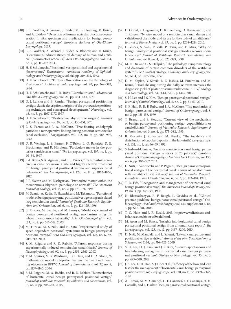
14 Advances in Otolaryngology
[40] L. E. Walther, A. Wenzel, J. Buder, M. B. Bloching, R. Kniep,and A. Blodow, “Detection of human utricular otoconia degen-eration in vital specimen and implications for benign parox-ysmal positional vertigo,” European Archives of Oto-Rhino-Laryngology, 2013.
[41] L. E. Walther, A. Wenzel, J. Buder, A. Blodow, and R. Kniep,“Gentamicin-induced structural damage of human and artifi-cial (biomimetic) otoconia,” Acta Oto-Laryngologica, vol. 134,no. 2, pp. 111–117, 2014.
[42] H. F. Schuknecht, “Positional vertigo: clinical and experimentalobservations,” Transactions—American Academy of Ophthal-mology and Otolaryngology, vol. 66, pp. 319–332, 1962.
[43] H. F. Schuknecht, “Further Observations on the Pathology ofPresbycusis,” Archives of otolaryngology, vol. 80, pp. 369–382,1964.
[44] H. F. Schuknecht and R. R. Ruby, “Cupulolithiasis,” Advances inOto-Rhino-Laryngology, vol. 20, pp. 434–443, 1973.
[45] D. J. Lanska and B. Remler, “Benign paroxysmal positioningvertigo: classic descriptions, origins of the provocative position-ing technique, and conceptual developments,” Neurology, vol.48, no. 5, pp. 1167–1177, 1997.
[46] H. F. Schuknecht, “Destructive labyrinthine surgery,” Archivesof Otolaryngology, vol. 97, no. 2, pp. 150–151, 1973.
[47] L. S. Parnes and J. A. McClure, “Free-floating endolymphparticles: a new operative finding during posterior semicircularcanal occlusion,” Laryngoscope, vol. 102, no. 9, pp. 988–992,1992.
[48] D. B. Welling, L. S. Parnes, B. O’Brien, L. O. Bakaletz, D. E.Brackmann, and R. Hinojosa, “Particulate matter in the pos-terior semicircular canal,” The Laryngoscope, vol. 107, no. 1, pp.90–94, 1997.
[49] J. A. Beyea, S. K. Agrawal, and L. S. Parnes, “Transmastoid semi-circular canal occlusion: a safe and highly effective treatmentfor benign paroxysmal positional vertigo and superior canaldehiscence,” The Laryngoscope, vol. 122, no. 8, pp. 1862–1866,2012.
[50] J. F. Kveton and M. Kashgarian, “Particulate matter within themembranous labyrinth: pathologic or normal?” The AmericanJournal of Otology, vol. 15, no. 2, pp. 173–176, 1994.
[51] M. Suzuki, A. Kadir, N. Hayashi, andM. Takamoto, “Functionalmodel of benign paroxysmal positional vertigo using an isolatedfrog semicircular canal,” Journal of Vestibular Research: Equilib-rium and Orientation, vol. 6, no. 2, pp. 121–125, 1996.
[52] K. Otsuka, M. Suzuki, and M. Furuya, “Model experiment ofbenign paroxysmal positional vertigo mechanism using thewhole membranous labyrinth,” Acta Oto-Laryngologica, vol.123, no. 4, pp. 515–518, 2003.
[53] M. Furuya, M. Suzuki, and H. Sato, “Experimental study ofspeed-dependent positional nystagmus in benign paroxysmalpositional vertigo,” Acta Oto-Laryngologica, vol. 123, no. 6, pp.709–712, 2003.
[54] S. M. Rajguru and R. D. Rabbitt, “Afferent responses duringexperimentally induced semicircular canalithiasis,” Journal ofNeurophysiology, vol. 97, no. 3, pp. 2355–2363, 2007.
[55] T. M. Squires, M. S. Weidman, T. C. Hain, and H. A. Stone, “Amathematical model for top-shelf vertigo: the role of sediment-ing otoconia in BPPV,” Journal of Biomechanics, vol. 37, no. 8,pp. 1137–1146, 2004.
[56] S. M. Rajguru, M. A. Ifediba, and R. D. Rabbitt, “Biomechanicsof horizontal canal benign paroxysmal positional vertigo,”Journal of Vestibular Research: Equilibrium andOrientation, vol.15, no. 4, pp. 203–214, 2005.
[57] D. Obrist, S. Hegemann, D. Kronenberg, O. Hauselmann, andT. Rosgen, “In vitro model of a semicircular canal: design andvalidation of themodel and its use for the study of canalithiasis,”Journal of Biomechanics, vol. 43, no. 6, pp. 1208–1214, 2010.
[58] G. Zucca, S. Valli, P. Valli, P. Perin, and E. Mira, “Why dobenign paroxysmal positional vertigo episodes recover spon-taneously?” Journal of Vestibular Research: Equilibrium andOrientation, vol. 8, no. 4, pp. 325–329, 1998.
[59] M. R. Dix and C. S. Hallpike, “The pathology, symptomatologyand diagnosis of certain common disorders of the vestibularsystem,”TheAnnals of Otology, Rhinology, and Laryngology, vol.61, no. 4, pp. 987–1016, 1952.
[60] D. M. Kaplan, Y. Slovik, B. Z. Joshua, M. Puterman, and M.Kraus, “Head shaking during dix-hallpike exam increases thediagnostic yield of posterior semicircular canal BPPV,” Otologyand Neurotology, vol. 34, 1444, no. 8, p. 1447, 2013.
[61] S. H. Lee and J. S. Kim, “Benign paroxysmal positional vertigo,”Journal of Clinical Neurology, vol. 6, no. 2, pp. 51–63, 2010.
[62] S. F. Hall, R. R. F. Ruby, and J. A. McClure, “The mechanics ofbenign paroxysmal vertigo,” Journal of Otolaryngology, vol. 8,no. 2, pp. 151–158, 1979.
[63] T. Brandt and S. Steddin, “Current view of the mechanismof benign paroxysmal positioning vertigo: cupulolithiasis orcanalolithiasis?” Journal of Vestibular Research: Equilibrium &Orientation, vol. 3, no. 4, pp. 373–382, 1993.
[64] B. Moriarty, J. Rutka, and M. Hawke, “The incidence anddistribution of cupular deposits in the labyrinth,” Laryngoscope,vol. 102, no. 1, pp. 56–59, 1992.
[65] S. Imbaud-Genieys, “Anterior semicircular canal benign parox-ysmal positional vertigo: a series of 20 patients,” EuropeanAnnals ofOtorhinolaryngology,Head andNeckDiseases, vol. 130,no. 6, pp. 303–307, 2013.
[66] D.Nuti, P. Vannucchi, and P. Pagnini, “Benign paroxysmal posi-tional vertigo of the horizontal canal: a form of canalolithiasiswith variable clinical features,” Journal of Vestibular Research:Equilibrium and Orientation, vol. 6, no. 3, pp. 173–184, 1996.
[67] T. D. Fife, “Recognition and management of horizontal canalbenign positional vertigo,”TheAmerican Journal of Otology, vol.19, no. 3, pp. 345–351, 1998.
[68] N. Bhattacharyya, R. F. Baugh, L. Orvidas et al., “Clinicalpractice guideline: benign paroxysmal positional vertigo,” Oto-laryngology: Head and Neck Surgery, vol. 139, supplement 4, no.5, pp. S47–S81, 2008.
[69] T. C. Hain and J. R. Ewald, 2013, http://www.dizziness-and-balance.com/history/Ewald.html.
[70] M. Aron and M. Bance, “Insights into horizontal canal benignparoxysmal positional vertigo from a human case report,” TheLaryngoscope, vol. 123, no. 12, pp. 3197–3200, 2013.
[71] D. Nuti, M. Mandala, and L. Salerni, “Lateral canal paroxysmalpositional vertigo revisited,”Annals of the New York Academy ofSciences, vol. 1164, pp. 316–323, 2009.
[72] S. U. Lee, H. J. Kim, and J. S. Kim, “Pseudo-spontaneous andhead-shaking nystagmus in horizontal canal benign paroxys-mal positional vertigo,” Otology & Neurotology, vol. 35, no. 3,pp. 495–500, 2014.
[73] J. B. Lee, D.H.Han, S. J. Choi et al., “Efficacy of the bow and leantest for the management of horizontal canal benign paroxysmalpositional vertigo,” Laryngoscope, vol. 120, no. 11, pp. 2339–2346,2010.
[74] A. Tomaz, M. M. Gananca, C. F. Gananca, F. F. Gananca, H. H.Caovilla, and L. Harker, “Benign paroxysmal positional vertigo:
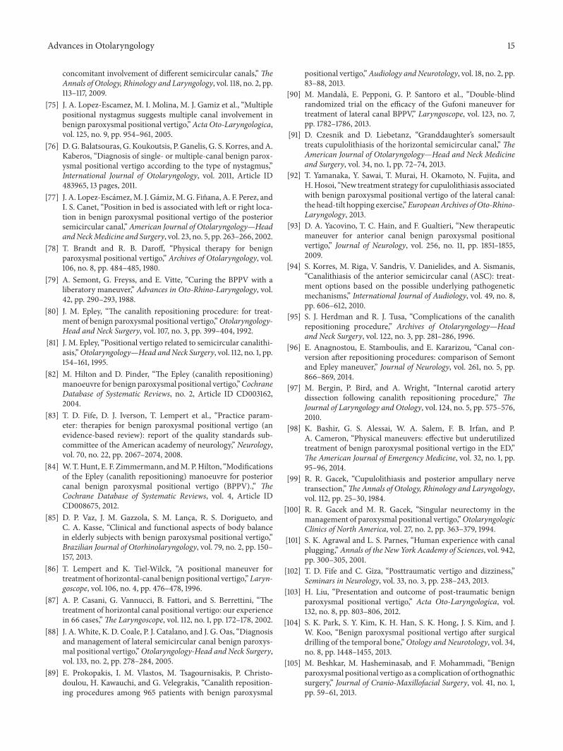
Advances in Otolaryngology 15
concomitant involvement of different semicircular canals,”TheAnnals of Otology, Rhinology and Laryngology, vol. 118, no. 2, pp.113–117, 2009.
[75] J. A. Lopez-Escamez, M. I. Molina, M. J. Gamiz et al., “Multiplepositional nystagmus suggests multiple canal involvement inbenign paroxysmal positional vertigo,”Acta Oto-Laryngologica,vol. 125, no. 9, pp. 954–961, 2005.
[76] D.G. Balatsouras, G. Koukoutsis, P. Ganelis, G. S. Korres, andA.Kaberos, “Diagnosis of single- or multiple-canal benign parox-ysmal positional vertigo according to the type of nystagmus,”International Journal of Otolaryngology, vol. 2011, Article ID483965, 13 pages, 2011.
[77] J. A. Lopez-Escamez,M. J. Gamiz,M. G. Finana, A. F. Perez, andI. S. Canet, “Position in bed is associated with left or right loca-tion in benign paroxysmal positional vertigo of the posteriorsemicircular canal,”American Journal of Otolaryngology—HeadandNeckMedicine and Surgery, vol. 23, no. 5, pp. 263–266, 2002.
[78] T. Brandt and R. B. Daroff, “Physical therapy for benignparoxysmal positional vertigo,” Archives of Otolaryngology, vol.106, no. 8, pp. 484–485, 1980.
[79] A. Semont, G. Freyss, and E. Vitte, “Curing the BPPV with aliberatory maneuver,” Advances in Oto-Rhino-Laryngology, vol.42, pp. 290–293, 1988.
[80] J. M. Epley, “The canalith repositioning procedure: for treat-ment of benign paroxysmal positional vertigo,”Otolaryngology-Head and Neck Surgery, vol. 107, no. 3, pp. 399–404, 1992.
[81] J. M. Epley, “Positional vertigo related to semicircular canalithi-asis,”Otolaryngology—Head andNeck Surgery, vol. 112, no. 1, pp.154–161, 1995.
[82] M. Hilton and D. Pinder, “The Epley (canalith repositioning)manoeuvre for benign paroxysmal positional vertigo,”CochraneDatabase of Systematic Reviews, no. 2, Article ID CD003162,2004.
[83] T. D. Fife, D. J. Iverson, T. Lempert et al., “Practice param-eter: therapies for benign paroxysmal positional vertigo (anevidence-based review): report of the quality standards sub-committee of the American academy of neurology,” Neurology,vol. 70, no. 22, pp. 2067–2074, 2008.
[84] W.T.Hunt, E. F. Zimmermann, andM.P.Hilton, “Modificationsof the Epley (canalith repositioning) manoeuvre for posteriorcanal benign paroxysmal positional vertigo (BPPV).,” TheCochrane Database of Systematic Reviews, vol. 4, Article IDCD008675, 2012.
[85] D. P. Vaz, J. M. Gazzola, S. M. Lanca, R. S. Dorigueto, andC. A. Kasse, “Clinical and functional aspects of body balancein elderly subjects with benign paroxysmal positional vertigo,”Brazilian Journal of Otorhinolaryngology, vol. 79, no. 2, pp. 150–157, 2013.
[86] T. Lempert and K. Tiel-Wilck, “A positional maneuver fortreatment of horizontal-canal benign positional vertigo,” Laryn-goscope, vol. 106, no. 4, pp. 476–478, 1996.
[87] A. P. Casani, G. Vannucci, B. Fattori, and S. Berrettini, “Thetreatment of horizontal canal positional vertigo: our experiencein 66 cases,”The Laryngoscope, vol. 112, no. 1, pp. 172–178, 2002.
[88] J. A.White, K. D. Coale, P. J. Catalano, and J. G. Oas, “Diagnosisand management of lateral semicircular canal benign paroxys-mal positional vertigo,”Otolaryngology-Head and Neck Surgery,vol. 133, no. 2, pp. 278–284, 2005.
[89] E. Prokopakis, I. M. Vlastos, M. Tsagournisakis, P. Christo-doulou, H. Kawauchi, and G. Velegrakis, “Canalith reposition-ing procedures among 965 patients with benign paroxysmal
positional vertigo,”Audiology and Neurotology, vol. 18, no. 2, pp.83–88, 2013.
[90] M. Mandala, E. Pepponi, G. P. Santoro et al., “Double-blindrandomized trial on the efficacy of the Gufoni maneuver fortreatment of lateral canal BPPV,” Laryngoscope, vol. 123, no. 7,pp. 1782–1786, 2013.
[91] D. Czesnik and D. Liebetanz, “Granddaughter’s somersaulttreats cupulolithiasis of the horizontal semicircular canal,” TheAmerican Journal of Otolaryngology—Head and Neck Medicineand Surgery, vol. 34, no. 1, pp. 72–74, 2013.
[92] T. Yamanaka, Y. Sawai, T. Murai, H. Okamoto, N. Fujita, andH.Hosoi, “New treatment strategy for cupulolithiasis associatedwith benign paroxysmal positional vertigo of the lateral canal:the head-tilt hopping exercise,”EuropeanArchives ofOto-Rhino-Laryngology, 2013.
[93] D. A. Yacovino, T. C. Hain, and F. Gualtieri, “New therapeuticmaneuver for anterior canal benign paroxysmal positionalvertigo,” Journal of Neurology, vol. 256, no. 11, pp. 1851–1855,2009.
[94] S. Korres, M. Riga, V. Sandris, V. Danielides, and A. Sismanis,“Canalithiasis of the anterior semicircular canal (ASC): treat-ment options based on the possible underlying pathogeneticmechanisms,” International Journal of Audiology, vol. 49, no. 8,pp. 606–612, 2010.
[95] S. J. Herdman and R. J. Tusa, “Complications of the canalithrepositioning procedure,” Archives of Otolaryngology—Headand Neck Surgery, vol. 122, no. 3, pp. 281–286, 1996.
[96] E. Anagnostou, E. Stamboulis, and E. Kararizou, “Canal con-version after repositioning procedures: comparison of Semontand Epley maneuver,” Journal of Neurology, vol. 261, no. 5, pp.866–869, 2014.
[97] M. Bergin, P. Bird, and A. Wright, “Internal carotid arterydissection following canalith repositioning procedure,” TheJournal of Laryngology and Otology, vol. 124, no. 5, pp. 575–576,2010.
[98] K. Bashir, G. S. Alessai, W. A. Salem, F. B. Irfan, and P.A. Cameron, “Physical maneuvers: effective but underutilizedtreatment of benign paroxysmal positional vertigo in the ED,”The American Journal of Emergency Medicine, vol. 32, no. 1, pp.95–96, 2014.
[99] R. R. Gacek, “Cupulolithiasis and posterior ampullary nervetransection,”TheAnnals of Otology, Rhinology and Laryngology,vol. 112, pp. 25–30, 1984.
[100] R. R. Gacek and M. R. Gacek, “Singular neurectomy in themanagement of paroxysmal positional vertigo,”OtolaryngologicClinics of North America, vol. 27, no. 2, pp. 363–379, 1994.
[101] S. K. Agrawal and L. S. Parnes, “Human experience with canalplugging,”Annals of the New York Academy of Sciences, vol. 942,pp. 300–305, 2001.
[102] T. D. Fife and C. Giza, “Posttraumatic vertigo and dizziness,”Seminars in Neurology, vol. 33, no. 3, pp. 238–243, 2013.
[103] H. Liu, “Presentation and outcome of post-traumatic benignparoxysmal positional vertigo,” Acta Oto-Laryngologica, vol.132, no. 8, pp. 803–806, 2012.
[104] S. K. Park, S. Y. Kim, K. H. Han, S. K. Hong, J. S. Kim, and J.W. Koo, “Benign paroxysmal positional vertigo after surgicaldrilling of the temporal bone,”Otology and Neurotology, vol. 34,no. 8, pp. 1448–1455, 2013.
[105] M. Beshkar, M. Hasheminasab, and F. Mohammadi, “Benignparoxysmal positional vertigo as a complication of orthognathicsurgery,” Journal of Cranio-Maxillofacial Surgery, vol. 41, no. 1,pp. 59–61, 2013.
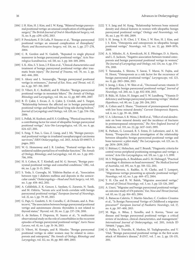
16 Advances in Otolaryngology
[106] J. H. Kim, H. J. Kim, and J. W. Kang, “Bilateral benign paroxys-mal positional vertigo: an unusual complication of orthognathicsurgery,”TheBritish Journal of Oral &Maxillofacial Surgery, vol.51, no. 8, pp. e291–e292, 2013.
[107] P. Persichetti, F. Di Lella, P. Simone et al., “Benign paroxysmalpositional vertigo: an unusual complication of rhinoplasty,”Plastic and Reconstructive Surgery, vol. 114, no. 1, pp. 277–278,2004.
[108] C. R. Gordon and N. Gadoth, “Repeated vs single physicalmaneuver in benign paroxysmal positional vertigo,” Acta Neu-rologica Scandinavica, vol. 110, no. 3, pp. 166–169, 2004.
[109] S. K. Ahn, S. Y. Jeon, J. P. Kim et al., “Clinical characteristics andtreatment of benign paroxysmal positional vertigo after trau-matic brain injury,” The Journal of Trauma, vol. 70, no. 2, pp.442–446, 2011.
[110] S. Aksoy and L. Sennaroglu, “Benign paroxysmal positionalvertigo in swimmers.,” Journal of Ear, Nose, and Throat, vol. 17,no. 6, pp. 307–310, 2007.
[111] D. Vibert, R. C. Redfield, and R. Hausler, “Benign paroxysmalpositional vertigo in mountain bikers,” The Annals of Otology,Rhinology and Laryngology, vol. 116, no. 12, pp. 887–890, 2007.
[112] B. O. Cakir, I. Ercan, Z. A. Cakir, S. Civelek, and S. Turgut,“Relationship between the affected ear in benign paroxysmalpositional vertigo and habitual head-lying side during bedrest,”Journal of Laryngology and Otology, vol. 120, no. 7, pp. 534–536,2006.
[113] L. Pollak,M.Kushnir, andH. S.Goldberg, “Physical inactivity asa contributing factor for onset of idiopathic benign paroxysmalpositional vertigo,” Acta Oto-Laryngologica, vol. 131, no. 6, pp.624–627, 2011.
[114] S. Feng, Y. Fan, L. Guo, Z. Liang, and J. Mi, “Benign paroxys-mal positional vertigo in irradiated nasopharyngeal carcinomasurvivors,” ISRN Otolaryngology, vol. 2013, Article ID 698575, 5pages, 2013.
[115] W. G. Hemenway and J. R. Lindsay, “Postural vertigo due tounilateral sudden partial loss of vestibular function,”TheAnnalsof Otology, Rhinology, and Laryngology, vol. 65, no. 3, pp. 692–706, 1956.
[116] H. S. Cohen, K. T. Kimball, and M. G. Stewart, “Benign paro-xysmal positional vertigo and comorbid conditions,” ORL, vol.66, no. 1, pp. 11–15, 2004.
[117] S. Yoda, S. Cureoglu, M. Yildirim-Baylan et al., “Associationbetween type 1 diabetes mellitus and deposits in the semicir-cular canals,”Otolaryngology—Head and Neck Surgery, vol. 145,no. 3, pp. 458–462, 2011.
[118] A. Celikbilek, Z. K. Gencer, L. Saydam, G. Zararsiz, N. Tanik,and M. Ozkiris, “Serum uric acid levels correlate with benignparoxysmal positional vertigo,” European Journal of Neurology,vol. 21, no. 1, pp. 79–85, 2013.
[119] G. Papi, G. Guidetti, S. M. Corsello, C. di Donato, and A. Pon-tecorvi, “The association between benign paroxysmal positionalvertigo and autoimmune chronic thyroiditis is not related tothyroid status,”Thyroid, vol. 20, no. 2, pp. 237–238, 2010.
[120] A. de Stefano, F. Dispenza, H. Suarez et al., “A multicenterobservational study on the role of comorbidities in the recurrentepisodes of benign paroxysmal positional vertigo,” Auris NasusLarynx, vol. 41, no. 1, pp. 31–36, 2013.
[121] D. Vibert, M. Kompis, and R. Hausler, “Benign paroxysmalpositional vertigo in older women may be related to osteo-porosis and osteopenia,” The Annals of Otology, Rhinology andLaryngology, vol. 112, no. 10, pp. 885–889, 2003.
[122] Y. S. Jang and M. Kang, “Relationship between bone mineraldensity and clinical features in women with idiopathic benignparoxysmal positional vertigo,” Otology and Neurotology, vol.30, no. 1, pp. 95–100, 2009.
[123] S. H. Jeong, S. H. Choi, J. Y. Kim, J. W. Koo, H. J. Kim, andJ. S. Kim, “Osteopenia and osteoporosis in idiopathic benignpositional vertigo,” Neurology, vol. 72, no. 12, pp. 1069–1076,2009.
[124] A. A. Mikulec, K. A. Kowalczyk, M. E. Pfitzinger, D. A. Harris,and L. E. Jackson, “Negative association between treated osteo-porosis and benign paroxysmal positional vertigo in women,”The Journal of Laryngology and Otology, vol. 124, no. 4, pp. 374–376, 2010.
[125] T. Yamanaka, S. Shirota, Y. Sawai, T. Murai, N. Fujita, andH. Hosoi, “Osteoporosis as a risk factor for the recurrence ofbenign paroxysmal positional vertigo,” Laryngoscope, vol. 123,no. 11, pp. 2813–2816, 2013.
[126] S. Jeong, J. Kim, J. W. Shin et al., “Decreased serum vitamin Din idiopathic benign paroxysmal positional vertigo,” Journal ofNeurology, vol. 260, no. 3, pp. 832–838, 2013.
[127] B. Buki, M. Ecker, H. Junger, and Y. W. Lundberg, “Vitamin Ddeficiency and benign paroxysmal positioning vertigo,”MedicalHypotheses, vol. 80, no. 2, pp. 201–204, 2013.
[128] A. Cohen and E. Shane, “Treatment of premenopausal womenwith low bone mineral density,” Current Osteoporosis Reports,vol. 6, no. 1, pp. 39–46, 2008.
[129] U. A. Liberman, S. R.Weiss, J. Broll et al., “Effect of oral alendro-nate on bone mineral density and the incidence of fracturesin postmenopausal osteoporosis,” The New England Journal ofMedicine, vol. 333, no. 22, pp. 1437–1443, 1995.
[130] K. Parham, G. Leonard, R. S. Feinn, D. Lafreniere, and A. M.Kenny, “Prospective clinical investigation of the relationshipbetween idiopathic benign paroxysmal positional vertigo andbone turnover: a pilot study,”The Laryngoscope, vol. 123, no. 11,pp. 2834–2839, 2013.
[131] U. Buttner, C. Helmchen, and T. Brandt, “Diagnostic criteria forcentral versus peripheral positioning nystagmus and vertigo: areview,” Acta Oto-Laryngologica, vol. 119, no. 1, pp. 1–5, 1999.
[132] M. S.Welgampola, A. Bradshaw, andG.M.Halmagyl, “Practicalneurology 4: dizziness on headmovement,”TheMedical Journalof Australia, vol. 195, no. 9, pp. 518–522, 2011.
[133] M. von Brevern, A. Radtke, A. H. Clarke, and T. Lempert,“Migrainous vertigo presenting as episodic positional vertigo,”Neurology, vol. 62, no. 3, pp. 469–472, 2004.
[134] Y. H. Cha and R. W. Baloh, “Migraine associated vertigo,”Journal of Clinical Neurology, vol. 3, no. 3, pp. 121–126, 2007.
[135] A. Uneri, “Migraine and benign paroxysmal positional vertigo:an outcome study of 476 patients,” Ear, Nose andThroat Journal,vol. 83, no. 12, pp. 814–815, 2004.
[136] A. Batuecas-Caletrıo, V. Martın-Sanchez, C. Cordero-Civantoset al., “Is Benign Paroxysmal Vertigo of Childhood a migraineprecursor?” European Journal of Paediatric Neurology, vol. 17,no. 4, pp. 397–400, 2013.
[137] M. Riga, A. Bibas, J. Xenellis, and S. Korres, “Inner eardisease and benign paroxysmal positional vertigo: a criticalreview of incidence, clinical characteristics, and management,”International Journal of Otolaryngology, vol. 2011, Article ID709469, 7 pages, 2011.
[138] G. Psillas, S. Triaridis, K. Markou, M. Tsalighopoulos, and V.Vital, “Benign paroxysmal positional vertigo in the first acuteattack of Meniere's disease,” B-ENT, vol. 7, no. 2, pp. 131–135,2011.
Advances in Otolaryngology 17
[139] D. G. Balatsouras, P. Ganelis, A. Aspris, N. C. Economou, A.Moukos, and G. Koukoutsis, “Benign paroxysmal positionalvertigo associated with Meniere’s disease: epidemiological,pathophysiologic, clinical, and therapeutic aspects,”The Annalsof Otology, Rhinology and Laryngology, vol. 121, no. 10, pp. 682–688, 2012.
[140] N. Lee, J. Ban, K. Lee, and S. M. Kim, “Benign paroxysmal posi-tional vertigo secondary to inner ear disease,” Otolaryngology:Head and Neck Surgery, vol. 143, no. 3, pp. 413–417, 2010.
[141] N. H. Lee and J. H. Ban, “Is BPPV a prognostic factor in idio-pathic sudden sensory hearing loss?” Clinical and ExperimentalOtorhinolaryngology, vol. 3, no. 4, pp. 199–202, 2010.
[142] G. Tirelli, E. D’Orlando, V. Giacomarra, and M. Russolo,“Benign positional vertigo without detectable nystagmus,” TheLaryngoscope, vol. 111, no. 6, pp. 1053–1056, 2001.
[143] D. S. Haynes, J. R. Resser, R. F. Labadie et al., “Treatment ofbenign positional vertigo using the Semont maneuver: efficacyin patients presenting without nystagmus,” Laryngoscope, vol.112, no. 5, pp. 796–801, 2002.
[144] G. A. Alvarenga, M. A. Barbosa, and C. C. Porto, “BenignParoxysmal Positional Vertigo without nystagmus: diagnosisand treatment,” Brazilian Journal of Otorhinolaryngology, vol.77, no. 6, pp. 799–804, 2011.
[145] B. Buki, L. Simon, S. Garab, Y. W. Lundberg, H. Junger, andD. Straumann, “Sitting-up vertigo and trunk retropulsion inpatients with benign positional vertigo but without positionalnystagmus,” Journal of Neurology, Neurosurgery and Psychiatry,vol. 82, no. 1, pp. 98–104, 2011.
[146] K. Johkura, T. Momoo, and Y. Kuroiwa, “Positional nystagmusin patients with chronic dizziness,” Journal of Neurology, Neuro-surgery and Psychiatry, vol. 79, no. 12, pp. 1324–1326, 2008.
[147] R. Gans, “Benign paroxysmal positional vertigo: a commondizziness sensation,” 2014, http://www.audiologyonline.com/articles/article detail.
[148] C. F. Gananca, H. H. Caovilla, J. M. Gazzola, M. M. Gananca,and F. F. Gananca, “Epley’s maneuver in benign paroxysmalpositional vertigo associated with Meniere’s disease,” BrazilianJournal of Otorhinolaryngology, vol. 73, no. 4, pp. 506–512, 2007.
[149] D. G. Balatsouras and S. G. Korres, “Subjective benign paroxys-mal positional vertigo,”Otolaryngology: Head andNeck Surgery,vol. 146, no. 1, pp. 98–103, 2012.
[150] A. L. DeStefano and F. Dispenza, “Malignant paroxysmal posi-tional vertigo,” in Textbook of Vertigo: Diagnosis and Manage-ment, F. Dispenza and A. DeStefano, Eds., pp. 145–150, JaypeeBrothers Medical Publishers, New Delhi, India, 2013.
[151] M.Mitka, “Practice parameter: simplemaneuver is best therapyfor common form of vertigo,” Journal of the American MedicalAssociation, vol. 300, no. 2, pp. 157–158, 2008.

Submit your manuscripts athttp://www.hindawi.com
Stem CellsInternational
Hindawi Publishing Corporationhttp://www.hindawi.com Volume 2014
Hindawi Publishing Corporationhttp://www.hindawi.com Volume 2014
MEDIATORSINFLAMMATION
of
Hindawi Publishing Corporationhttp://www.hindawi.com Volume 2014
Behavioural Neurology
EndocrinologyInternational Journal of
Hindawi Publishing Corporationhttp://www.hindawi.com Volume 2014
Hindawi Publishing Corporationhttp://www.hindawi.com Volume 2014
Disease Markers
Hindawi Publishing Corporationhttp://www.hindawi.com Volume 2014
BioMed Research International
OncologyJournal of
Hindawi Publishing Corporationhttp://www.hindawi.com Volume 2014
Hindawi Publishing Corporationhttp://www.hindawi.com Volume 2014
Oxidative Medicine and Cellular Longevity
Hindawi Publishing Corporationhttp://www.hindawi.com Volume 2014
PPAR Research
The Scientific World JournalHindawi Publishing Corporation http://www.hindawi.com Volume 2014
Immunology ResearchHindawi Publishing Corporationhttp://www.hindawi.com Volume 2014
Journal of
ObesityJournal of
Hindawi Publishing Corporationhttp://www.hindawi.com Volume 2014
Hindawi Publishing Corporationhttp://www.hindawi.com Volume 2014
Computational and Mathematical Methods in Medicine
OphthalmologyJournal of
Hindawi Publishing Corporationhttp://www.hindawi.com Volume 2014
Diabetes ResearchJournal of
Hindawi Publishing Corporationhttp://www.hindawi.com Volume 2014
Hindawi Publishing Corporationhttp://www.hindawi.com Volume 2014
Research and TreatmentAIDS
Hindawi Publishing Corporationhttp://www.hindawi.com Volume 2014
Gastroenterology Research and Practice
Hindawi Publishing Corporationhttp://www.hindawi.com Volume 2014
Parkinson’s Disease
Evidence-Based Complementary and Alternative Medicine
Volume 2014Hindawi Publishing Corporationhttp://www.hindawi.com