Embed Size (px)
Citation preview
Case ReportRetrograde Intramedullary Nail Fixation for DerotationalFemoral Osteotomy for Recurrent Femoropatellar Instability
Maximiliano Barahona ,1 Alvaro Zamorano,1 Cristian Barrientos,1 Mauricio Guzmán,2
Yoshiro Sato,1 and Jaime Hinzpeter1
1Orthopedic Department at Hospital Clinico Universidad de Chile, 999 Santos Dumont Street, Independencia,Santiago 8380456, Chile2Radiology Department at Hospital Clinico Universidad de Chile, 999 Santos Dumont Street, Independencia, Santiago 8380456, Chile
Correspondence should be addressed to Maximiliano Barahona; [email protected]
Received 22 May 2019; Accepted 31 August 2019; Published 11 September 2019
Academic Editor: Dimitrios S. Karataglis
Copyright © 2019 Maximiliano Barahona et al. This is an open access article distributed under the Creative Commons AttributionLicense, which permits unrestricted use, distribution, and reproduction in any medium, provided the original work isproperly cited.
Axial alignment of the femur and tibia is often misdiagnosed in patients with patellofemoral stability problems. Femoral torsion iscritical for patellofemoral biomechanics, so it must be evaluated in every patient before the plan of surgery is decided. This casedescribes a femoral derotational osteotomy due to excessive internal torsion of the femur fixed with a retrograde femoral nail.This type of fixation provides a biomechanical advantage compared to plates. At the two-year follow-up, the patient achievedexcellent results, reaching a functional score of 91 points on the Lysholm scale. Derotational femoral osteotomy should beconsidered in patellofemoral pathology, and a retrograde femoral nail is a valid fixation method for this surgery.
1. Introduction
Patellofemoral instability is a multifactorial pathology, inwhich three main elements participate: lower limb malalign-ment, trochlear dysplasia, and the medial patellofemoral lig-ament (MPFL). Malalignment is a factor in a large percentageof patients who consult for this pathology, and the spectrumis broad. An increase in the distance between the intercondy-lar groove and the tibial tuberosity is just the tip of the ice-berg; on the other end is the miserable malalignmentsyndrome, a rare condition that includes severe internaltorsion of the femur [1, 2].
Femoral torsion is defined as the angle between the fem-oral neck axis and the condyle axis. The pathological cutoffvalue to perform a derotational osteotomy is a matter of cur-rent debate; one of the first values reported was thosedescribed by Cordier and Katthagen, who defined a normalrange between 5° and 25° assuming a normal distribution ofthe data [3]. However, a range of two standard deviations
from the mean sample does not imply a relation to a clinicalproblem like patella instability or pain.
Femoral torsion is a risk factor for patella instability [4].A cadaveric study performed by Kaiser et al. [5] shows thata 10° increase of femoral torsion amplifies the lateral vectorforce to the patella, but a reconstruction of the MPFL alonecan restore it; however, if the femoral torsion is nowincreased to 20°, the reconstruction of the MPFL alone isinsufficient to decrease the lateral vector. This finding addsto the ever-growing evidence that supports the need to per-form derotational osteotomies in patients with increasedfemoral torsion [6, 7].
Also, trochlea dysplasia may be a result of decreased con-tact between the femur and the patella due to an increasedfemoral torsion, emulating the proposed model in hip dys-plasia [8]. So, when the trochlea angle is flat, femoral torsionmust be evaluated and corrected if it is too high.
The knee surgeon must study the entire alignment inpatella instability, including femoral and tibial torsion [9].
HindawiCase Reports in OrthopedicsVolume 2019, Article ID 1893042, 7 pageshttps://doi.org/10.1155/2019/1893042
This case report is aimed at describing a femoral derotationalosteotomy fixed by a retrograde femoral nail for recurrentpatellar instability.
2. Case
The patient was a 17-year-old woman who complains ofrecurrent patellar instability in her left knee (5-7 episodesper year in the last three years) and in her right knee (two epi-sodes). Physical examination revealed a full range of motionin both knees. The left knee had mild effusion and a positiveapprehension test. On the right knee, no effusion or appre-hension was noticed, but a pain in the medial retinaculumwas found. The hip internal rotation was asymmetric, beinggreater in the left hip, so femoral malrotation was suspected.Tibial torsion, assessed by clinical thigh-foot angle, was nor-mal. The preoperative patient-reported score was 19 on theLysholm scale [10] and level 1 on the Tegner scale [11].
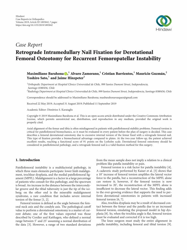
All patients with recurrent patellar instability are studiedwith full-length standing anteroposterior (AP) radiography,computed tomography (CT) of the lower extremities, andknee magnetic resonance imaging (MRI) in our institution.CT measurements are summarised in Table 1, and anincreased femoral internal rotation was the main finding(Figure 1). Femoral torsion was measured according to thetechnique described by Jarrett et al. [12], with an expectedvalue of 15°. Radiography shows mild symmetric valgusalignment in the coronal plane, a femoral-tibial mechanicalangle of 1° of valgus, and a mechanical distal lateral femoralangle (mDLFA) of 86° on the left knee. Knee MRI shows abilateral lesion of the MPFL and chondral type 2 damage inthe lateral facet of the left patella. The trochlea was flat onboth knees, which means that it corresponds to a type Baccording to Dejour’s classification [13].
According to the classification of patellar instability byFrosch and Schmeling [14], the right extremity was a type2: instability without malalignment as femoral torsion wasbelow 25°. The authors use the upper limit to indicate a fem-oral derotational osteotomy according to the findings of Kai-ser et al. [5]; therefore, a medial patellofemoral ligament(MPFL) reconstruction alone was planned. The left extremitywas a type 3e (instability with femoral malrotation); there-fore, a femoral derotational osteotomy was planned.
Our planning was estimating the perimeter of the femurat the level of the osteotomy. For the estimation of the perim-eter, in an axial section of the CT scan at the level of thedesired osteotomy, we measure the radius in the anteropos-terior (2,98 cm) direction and in the midlateral direction(2,66 cm). With these two measurements, we obtained anaverage radius of 2,82 cm. Then, the perimeter calculation isbased on the formula 2 ∗ π ∗ radius; therefore, the perimeterwas 17,7 cm. The perimeter accounted for 360°, so for 17° ofcorrection, a rotation of 0.8 cm should be made.
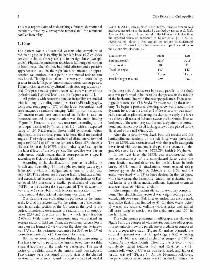
Both knees were operated on during the same surgery.The first step was to perform the femoral osteotomy; for this,a lateral approach in the thigh was performed. The lateralcortex of the distal third of the femoral shaft was exposed.Two clamps were positioned on both sides of the desiredlocation for the osteotomy, and the bone was marked parallel
to the long axis. A transverse bone cut, parallel to the shaftaxis, was performed in between the clamps and in the middleof the horizontal line with fluoroscopy guidance. Then, a ret-rograde femoral nail (T2, Stryker®) was used to fix the osteot-omy. To begin, a proximal blocking screw was placed in thedynamic hole, then the distal end of the osteotomy was exter-nally rotated, as planned, using the clamps to apply the forceto achieve a distance of 0.8 cm between the horizontal lines atboth ends of the osteotomy cut. Immediately after the derota-tional maneuver, two distal locking screws were placed in thedistal end of the nail (Figure 2).
After the osteotomy was fixed, both the gracilis and thesemitendinosus tendons of the left knee were harvested.The left MPFL was reconstructed with the gracilis autograft;it was fixed with two anchors in the patellar side and a biode-gradable screw in the femur (BIORCI®, Smith & Nephew).
In the right knee, the MPFL was reconstructed withthe semitendinosus of the contralateral knee using thesame fixation method described for the left knee. In bothknees, MPFL femoral attachments were selected usingfluoroscopy as described by Schöttle et al. [15], and thegrafts were fixed with 45° of knee flexion. In the left knee,while harvesting the hamstring tendon, an accidental par-tial lesion of the distal medial collateral ligament occurredand was repaired with an anchor.
After surgery, the patient did not present any complica-tions. The rehabilitation protocol was weight bearing as tol-erated, with two canes. Full knee extension was encouraged,and active flexion was limited to 60° for three weeks. After10 weeks, she resumed walking without canes and gainedfull knee range of motion on the right knee and 100° inthe left knee.
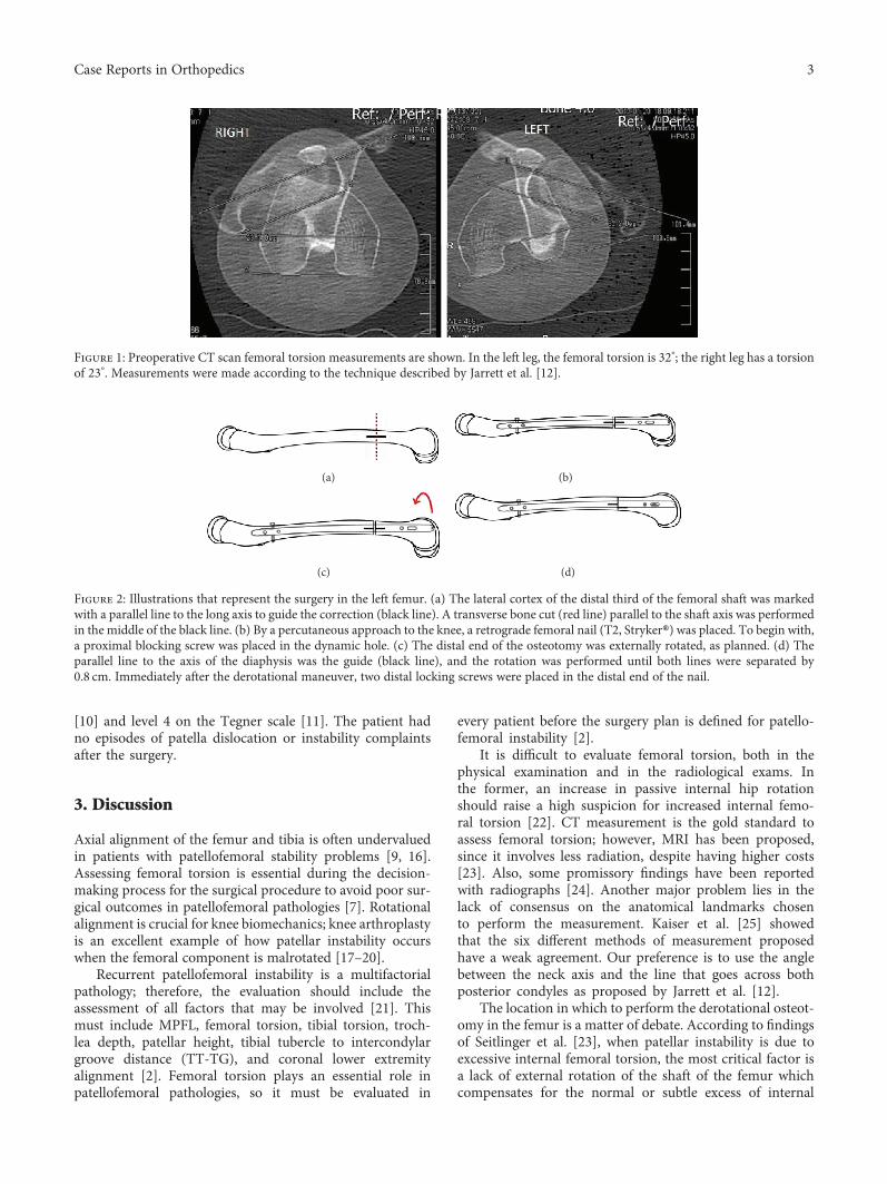
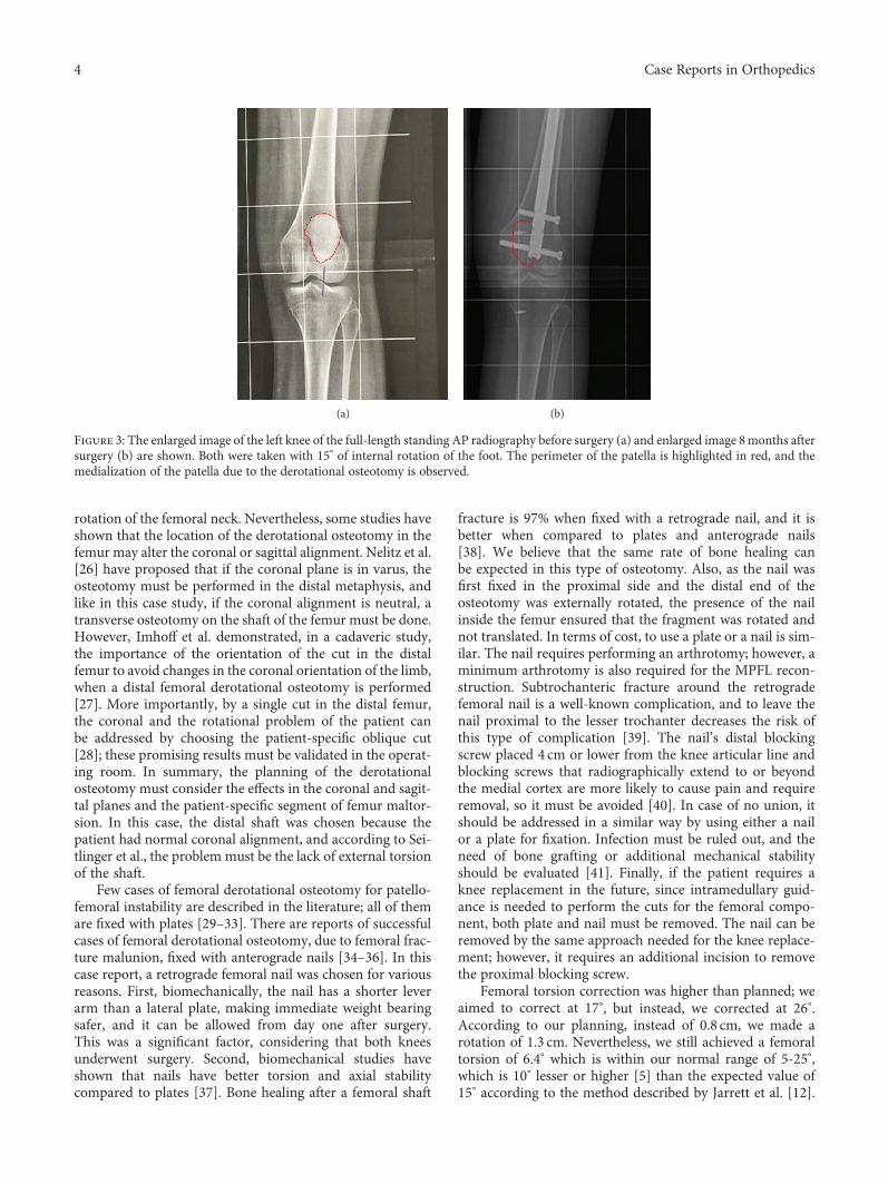
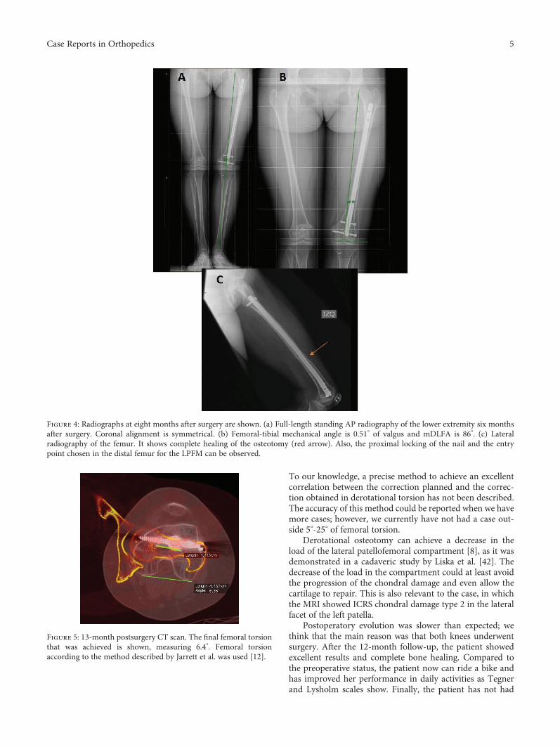
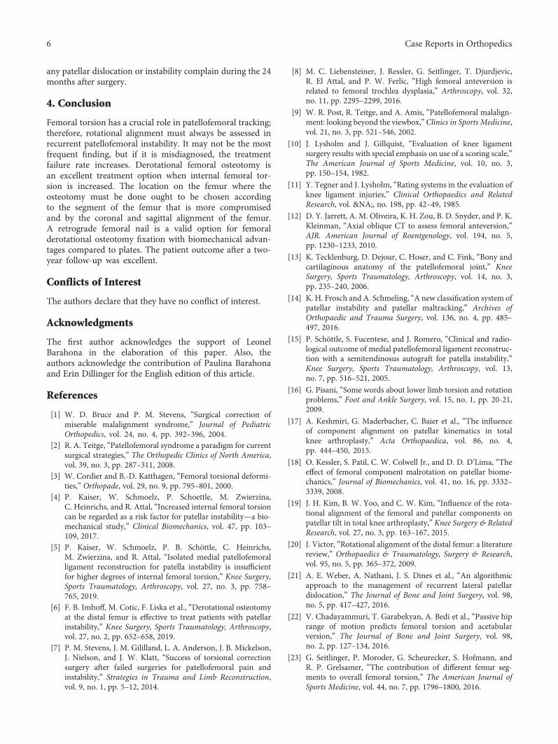
The eight-month postsurgery radiographs are shown inFigure 3 and are compared with the preoperative radiographs.It is remarkable how the patella looks medialised comparedto the preoperative study (Figure 3), and as planned, thecoronal alignment was not changed (Figure 4(a)) achievinga femoral-tibial mechanical angle after surgery of 0.51° ofvalgus. At the eight-month follow-up, the osteotomy wascompletely healed (Figures 4(b) and 4(c)). At the 18-month follow-up, a CT scan was performed, and femoraltorsion was 6.4° (Figure 5). At the 24-month follow-up,the patient-reported outcome was 91 on the Lysholm scale
Table 1: All CT measurements are shown. Femoral torsion wasmeasured according to the method described by Jarrett et al. [12].A femoral torsion of 32° was found in the left side, 17° higher thanthe expected value, so according to Kaiser et al. [5], a MPFLreconstruction alone is not enough to restore patellofemoralkinematics. The trochlea in both knees was type B according tothe Dejour classification [13].
Measurement Right Left
Femoral torsion 23.3° 32.2°
Tibial torsion 18° 21°
Trochlea angle 143° 147°
TT-TG 13mm 14mm
Patellar height (Caton) 0.98 0.87
2 Case Reports in Orthopedics
[10] and level 4 on the Tegner scale [11]. The patient hadno episodes of patella dislocation or instability complaintsafter the surgery.
3. Discussion
Axial alignment of the femur and tibia is often undervaluedin patients with patellofemoral stability problems [9, 16].Assessing femoral torsion is essential during the decision-making process for the surgical procedure to avoid poor sur-gical outcomes in patellofemoral pathologies [7]. Rotationalalignment is crucial for knee biomechanics; knee arthroplastyis an excellent example of how patellar instability occurswhen the femoral component is malrotated [17–20].
Recurrent patellofemoral instability is a multifactorialpathology; therefore, the evaluation should include theassessment of all factors that may be involved [21]. Thismust include MPFL, femoral torsion, tibial torsion, troch-lea depth, patellar height, tibial tubercle to intercondylargroove distance (TT-TG), and coronal lower extremityalignment [2]. Femoral torsion plays an essential role inpatellofemoral pathologies, so it must be evaluated in
every patient before the surgery plan is defined for patello-femoral instability [2].
It is difficult to evaluate femoral torsion, both in thephysical examination and in the radiological exams. Inthe former, an increase in passive internal hip rotationshould raise a high suspicion for increased internal femo-ral torsion [22]. CT measurement is the gold standard toassess femoral torsion; however, MRI has been proposed,since it involves less radiation, despite having higher costs[23]. Also, some promissory findings have been reportedwith radiographs [24]. Another major problem lies in thelack of consensus on the anatomical landmarks chosento perform the measurement. Kaiser et al. [25] showedthat the six different methods of measurement proposedhave a weak agreement. Our preference is to use the anglebetween the neck axis and the line that goes across bothposterior condyles as proposed by Jarrett et al. [12].
The location in which to perform the derotational osteot-omy in the femur is a matter of debate. According to findingsof Seitlinger et al. [23], when patellar instability is due toexcessive internal femoral torsion, the most critical factor isa lack of external rotation of the shaft of the femur whichcompensates for the normal or subtle excess of internal
Figure 1: Preoperative CT scan femoral torsion measurements are shown. In the left leg, the femoral torsion is 32°; the right leg has a torsionof 23°. Measurements were made according to the technique described by Jarrett et al. [12].
(a) (b)
(c) (d)
Figure 2: Illustrations that represent the surgery in the left femur. (a) The lateral cortex of the distal third of the femoral shaft was markedwith a parallel line to the long axis to guide the correction (black line). A transverse bone cut (red line) parallel to the shaft axis was performedin the middle of the black line. (b) By a percutaneous approach to the knee, a retrograde femoral nail (T2, Stryker®) was placed. To begin with,a proximal blocking screw was placed in the dynamic hole. (c) The distal end of the osteotomy was externally rotated, as planned. (d) Theparallel line to the axis of the diaphysis was the guide (black line), and the rotation was performed until both lines were separated by0.8 cm. Immediately after the derotational maneuver, two distal locking screws were placed in the distal end of the nail.
3Case Reports in Orthopedics
rotation of the femoral neck. Nevertheless, some studies haveshown that the location of the derotational osteotomy in thefemur may alter the coronal or sagittal alignment. Nelitz et al.[26] have proposed that if the coronal plane is in varus, theosteotomy must be performed in the distal metaphysis, andlike in this case study, if the coronal alignment is neutral, atransverse osteotomy on the shaft of the femur must be done.However, Imhoff et al. demonstrated, in a cadaveric study,the importance of the orientation of the cut in the distalfemur to avoid changes in the coronal orientation of the limb,when a distal femoral derotational osteotomy is performed[27]. More importantly, by a single cut in the distal femur,the coronal and the rotational problem of the patient canbe addressed by choosing the patient-specific oblique cut[28]; these promising results must be validated in the operat-ing room. In summary, the planning of the derotationalosteotomy must consider the effects in the coronal and sagit-tal planes and the patient-specific segment of femur maltor-sion. In this case, the distal shaft was chosen because thepatient had normal coronal alignment, and according to Sei-tlinger et al., the problem must be the lack of external torsionof the shaft.
Few cases of femoral derotational osteotomy for patello-femoral instability are described in the literature; all of themare fixed with plates [29–33]. There are reports of successfulcases of femoral derotational osteotomy, due to femoral frac-ture malunion, fixed with anterograde nails [34–36]. In thiscase report, a retrograde femoral nail was chosen for variousreasons. First, biomechanically, the nail has a shorter leverarm than a lateral plate, making immediate weight bearingsafer, and it can be allowed from day one after surgery.This was a significant factor, considering that both kneesunderwent surgery. Second, biomechanical studies haveshown that nails have better torsion and axial stabilitycompared to plates [37]. Bone healing after a femoral shaft
fracture is 97% when fixed with a retrograde nail, and it isbetter when compared to plates and anterograde nails[38]. We believe that the same rate of bone healing canbe expected in this type of osteotomy. Also, as the nail wasfirst fixed in the proximal side and the distal end of theosteotomy was externally rotated, the presence of the nailinside the femur ensured that the fragment was rotated andnot translated. In terms of cost, to use a plate or a nail is sim-ilar. The nail requires performing an arthrotomy; however, aminimum arthrotomy is also required for the MPFL recon-struction. Subtrochanteric fracture around the retrogradefemoral nail is a well-known complication, and to leave thenail proximal to the lesser trochanter decreases the risk ofthis type of complication [39]. The nail’s distal blockingscrew placed 4 cm or lower from the knee articular line andblocking screws that radiographically extend to or beyondthe medial cortex are more likely to cause pain and requireremoval, so it must be avoided [40]. In case of no union, itshould be addressed in a similar way by using either a nailor a plate for fixation. Infection must be ruled out, and theneed of bone grafting or additional mechanical stabilityshould be evaluated [41]. Finally, if the patient requires aknee replacement in the future, since intramedullary guid-ance is needed to perform the cuts for the femoral compo-nent, both plate and nail must be removed. The nail can beremoved by the same approach needed for the knee replace-ment; however, it requires an additional incision to removethe proximal blocking screw.
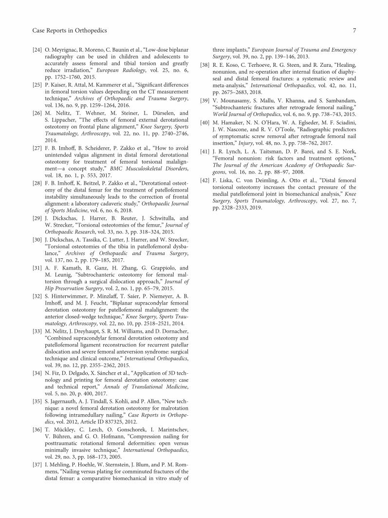
Femoral torsion correction was higher than planned; weaimed to correct at 17°, but instead, we corrected at 26°.According to our planning, instead of 0.8 cm, we made arotation of 1.3 cm. Nevertheless, we still achieved a femoraltorsion of 6.4° which is within our normal range of 5-25°,which is 10° lesser or higher [5] than the expected value of15° according to the method described by Jarrett et al. [12].
(a) (b)
Figure 3: The enlarged image of the left knee of the full-length standing AP radiography before surgery (a) and enlarged image 8months aftersurgery (b) are shown. Both were taken with 15° of internal rotation of the foot. The perimeter of the patella is highlighted in red, and themedialization of the patella due to the derotational osteotomy is observed.
4 Case Reports in Orthopedics
To our knowledge, a precise method to achieve an excellentcorrelation between the correction planned and the correc-tion obtained in derotational torsion has not been described.The accuracy of this method could be reported when we havemore cases; however, we currently have not had a case out-side 5°-25° of femoral torsion.
Derotational osteotomy can achieve a decrease in theload of the lateral patellofemoral compartment [8], as it wasdemonstrated in a cadaveric study by Liska et al. [42]. Thedecrease of the load in the compartment could at least avoidthe progression of the chondral damage and even allow thecartilage to repair. This is also relevant to the case, in whichthe MRI showed ICRS chondral damage type 2 in the lateralfacet of the left patella.
Postoperatory evolution was slower than expected; wethink that the main reason was that both knees underwentsurgery. After the 12-month follow-up, the patient showedexcellent results and complete bone healing. Compared tothe preoperative status, the patient now can ride a bike andhas improved her performance in daily activities as Tegnerand Lysholm scales show. Finally, the patient has not had
Figure 5: 13-month postsurgery CT scan. The final femoral torsionthat was achieved is shown, measuring 6.4°. Femoral torsionaccording to the method described by Jarrett et al. was used [12].
Figure 4: Radiographs at eight months after surgery are shown. (a) Full-length standing AP radiography of the lower extremity six monthsafter surgery. Coronal alignment is symmetrical. (b) Femoral-tibial mechanical angle is 0.51° of valgus and mDLFA is 86°. (c) Lateralradiography of the femur. It shows complete healing of the osteotomy (red arrow). Also, the proximal locking of the nail and the entrypoint chosen in the distal femur for the LPFM can be observed.
5Case Reports in Orthopedics
any patellar dislocation or instability complain during the 24months after surgery.
4. Conclusion
Femoral torsion has a crucial role in patellofemoral tracking;therefore, rotational alignment must always be assessed inrecurrent patellofemoral instability. It may not be the mostfrequent finding, but if it is misdiagnosed, the treatmentfailure rate increases. Derotational femoral osteotomy isan excellent treatment option when internal femoral tor-sion is increased. The location on the femur where theosteotomy must be done ought to be chosen accordingto the segment of the femur that is more compromisedand by the coronal and sagittal alignment of the femur.A retrograde femoral nail is a valid option for femoralderotational osteotomy fixation with biomechanical advan-tages compared to plates. The patient outcome after a two-year follow-up was excellent.
Conflicts of Interest
The authors declare that they have no conflict of interest.
Acknowledgments
The first author acknowledges the support of LeonelBarahona in the elaboration of this paper. Also, theauthors acknowledge the contribution of Paulina Barahonaand Erin Dillinger for the English edition of this article.
References
[1] W. D. Bruce and P. M. Stevens, “Surgical correction ofmiserable malalignment syndrome,” Journal of PediatricOrthopedics, vol. 24, no. 4, pp. 392–396, 2004.
[2] R. A. Teitge, “Patellofemoral syndrome a paradigm for currentsurgical strategies,” The Orthopedic Clinics of North America,vol. 39, no. 3, pp. 287–311, 2008.
[3] W. Cordier and B.-D. Katthagen, “Femoral torsional deformi-ties,” Orthopade, vol. 29, no. 9, pp. 795–801, 2000.
[4] P. Kaiser, W. Schmoelz, P. Schoettle, M. Zwierzina,C. Heinrichs, and R. Attal, “Increased internal femoral torsioncan be regarded as a risk factor for patellar instability—a bio-mechanical study,” Clinical Biomechanics, vol. 47, pp. 103–109, 2017.
[5] P. Kaiser, W. Schmoelz, P. B. Schöttle, C. Heinrichs,M. Zwierzina, and R. Attal, “Isolated medial patellofemoralligament reconstruction for patella instability is insufficientfor higher degrees of internal femoral torsion,” Knee Surgery,Sports Traumatology, Arthroscopy, vol. 27, no. 3, pp. 758–765, 2019.
[6] F. B. Imhoff, M. Cotic, F. Liska et al., “Derotational osteotomyat the distal femur is effective to treat patients with patellarinstability,” Knee Surgery, Sports Traumatology, Arthroscopy,vol. 27, no. 2, pp. 652–658, 2019.
[7] P. M. Stevens, J. M. Gililland, L. A. Anderson, J. B. Mickelson,J. Nielson, and J. W. Klatt, “Success of torsional correctionsurgery after failed surgeries for patellofemoral pain andinstability,” Strategies in Trauma and Limb Reconstruction,vol. 9, no. 1, pp. 5–12, 2014.
[8] M. C. Liebensteiner, J. Ressler, G. Seitlinger, T. Djurdjevic,R. El Attal, and P. W. Ferlic, “High femoral anteversion isrelated to femoral trochlea dysplasia,” Arthroscopy, vol. 32,no. 11, pp. 2295–2299, 2016.
[9] W. R. Post, R. Teitge, and A. Amis, “Patellofemoral malalign-ment: looking beyond the viewbox,” Clinics in Sports Medicine,vol. 21, no. 3, pp. 521–546, 2002.
[10] J. Lysholm and J. Gillquist, “Evaluation of knee ligamentsurgery results with special emphasis on use of a scoring scale,”The American Journal of Sports Medicine, vol. 10, no. 3,pp. 150–154, 1982.
[11] Y. Tegner and J. Lysholm, “Rating systems in the evaluation ofknee ligament injuries,” Clinical Orthopaedics and RelatedResearch, vol. &NA;, no. 198, pp. 42–49, 1985.
[12] D. Y. Jarrett, A. M. Oliveira, K. H. Zou, B. D. Snyder, and P. K.Kleinman, “Axial oblique CT to assess femoral anteversion,”AJR. American Journal of Roentgenology, vol. 194, no. 5,pp. 1230–1233, 2010.
[13] K. Tecklenburg, D. Dejour, C. Hoser, and C. Fink, “Bony andcartilaginous anatomy of the patellofemoral joint,” KneeSurgery, Sports Traumatology, Arthroscopy, vol. 14, no. 3,pp. 235–240, 2006.
[14] K. H. Frosch and A. Schmeling, “A new classification system ofpatellar instability and patellar maltracking,” Archives ofOrthopaedic and Trauma Surgery, vol. 136, no. 4, pp. 485–497, 2016.
[15] P. Schöttle, S. Fucentese, and J. Romero, “Clinical and radio-logical outcome of medial patellofemoral ligament reconstruc-tion with a semitendinosus autograft for patella instability,”Knee Surgery, Sports Traumatology, Arthroscopy, vol. 13,no. 7, pp. 516–521, 2005.
[16] G. Pisani, “Some words about lower limb torsion and rotationproblems,” Foot and Ankle Surgery, vol. 15, no. 1, pp. 20-21,2009.
[17] A. Keshmiri, G. Maderbacher, C. Baier et al., “The influenceof component alignment on patellar kinematics in totalknee arthroplasty,” Acta Orthopaedica, vol. 86, no. 4,pp. 444–450, 2015.
[18] O. Kessler, S. Patil, C. W. Colwell Jr., and D. D. D’Lima, “Theeffect of femoral component malrotation on patellar biome-chanics,” Journal of Biomechanics, vol. 41, no. 16, pp. 3332–3339, 2008.
[19] J. H. Kim, B. W. Yoo, and C. W. Kim, “Influence of the rota-tional alignment of the femoral and patellar components onpatellar tilt in total knee arthroplasty,” Knee Surgery & RelatedResearch, vol. 27, no. 3, pp. 163–167, 2015.
[20] J. Victor, “Rotational alignment of the distal femur: a literaturereview,” Orthopaedics & Traumatology, Surgery & Research,vol. 95, no. 5, pp. 365–372, 2009.
[21] A. E. Weber, A. Nathani, J. S. Dines et al., “An algorithmicapproach to the management of recurrent lateral patellardislocation,” The Journal of Bone and Joint Surgery, vol. 98,no. 5, pp. 417–427, 2016.
[22] V. Chadayammuri, T. Garabekyan, A. Bedi et al., “Passive hiprange of motion predicts femoral torsion and acetabularversion,” The Journal of Bone and Joint Surgery, vol. 98,no. 2, pp. 127–134, 2016.
[23] G. Seitlinger, P. Moroder, G. Scheurecker, S. Hofmann, andR. P. Grelsamer, “The contribution of different femur seg-ments to overall femoral torsion,” The American Journal ofSports Medicine, vol. 44, no. 7, pp. 1796–1800, 2016.
6 Case Reports in Orthopedics
[24] O. Meyrignac, R. Moreno, C. Baunin et al., “Low-dose biplanarradiography can be used in children and adolescents toaccurately assess femoral and tibial torsion and greatlyreduce irradiation,” European Radiology, vol. 25, no. 6,pp. 1752–1760, 2015.
[25] P. Kaiser, R. Attal, M. Kammerer et al., “Significant differencesin femoral torsion values depending on the CT measurementtechnique,” Archives of Orthopaedic and Trauma Surgery,vol. 136, no. 9, pp. 1259–1264, 2016.
[26] M. Nelitz, T. Wehner, M. Steiner, L. Dürselen, andS. Lippacher, “The effects of femoral external derotationalosteotomy on frontal plane alignment,” Knee Surgery, SportsTraumatology, Arthroscopy, vol. 22, no. 11, pp. 2740–2746,2014.
[27] F. B. Imhoff, B. Scheiderer, P. Zakko et al., “How to avoidunintended valgus alignment in distal femoral derotationalosteotomy for treatment of femoral torsional malalign-ment—a concept study,” BMC Musculoskeletal Disorders,vol. 18, no. 1, p. 553, 2017.
[28] F. B. Imhoff, K. Beitzel, P. Zakko et al., “Derotational osteot-omy of the distal femur for the treatment of patellofemoralinstability simultaneously leads to the correction of frontalalignment: a laboratory cadaveric study,” Orthopaedic Journalof Sports Medicine, vol. 6, no. 6, 2018.
[29] J. Dickschas, J. Harrer, B. Reuter, J. Schwitulla, andW. Strecker, “Torsional osteotomies of the femur,” Journal ofOrthopaedic Research, vol. 33, no. 3, pp. 318–324, 2015.
[30] J. Dickschas, A. Tassika, C. Lutter, J. Harrer, and W. Strecker,“Torsional osteotomies of the tibia in patellofemoral dysba-lance,” Archives of Orthopaedic and Trauma Surgery,vol. 137, no. 2, pp. 179–185, 2017.
[31] A. F. Kamath, R. Ganz, H. Zhang, G. Grappiolo, andM. Leunig, “Subtrochanteric osteotomy for femoral mal-torsion through a surgical dislocation approach,” Journal ofHip Preservation Surgery, vol. 2, no. 1, pp. 65–79, 2015.
[32] S. Hinterwimmer, P. Minzlaff, T. Saier, P. Niemeyer, A. B.Imhoff, and M. J. Feucht, “Biplanar supracondylar femoralderotation osteotomy for patellofemoral malalignment: theanterior closed-wedge technique,” Knee Surgery, Sports Trau-matology, Arthroscopy, vol. 22, no. 10, pp. 2518–2521, 2014.
[33] M. Nelitz, J. Dreyhaupt, S. R. M. Williams, and D. Dornacher,“Combined supracondylar femoral derotation osteotomy andpatellofemoral ligament reconstruction for recurrent patellardislocation and severe femoral anteversion syndrome: surgicaltechnique and clinical outcome,” International Orthopaedics,vol. 39, no. 12, pp. 2355–2362, 2015.
[34] N. Fiz, D. Delgado, X. Sánchez et al., “Application of 3D tech-nology and printing for femoral derotation osteotomy: caseand technical report,” Annals of Translational Medicine,vol. 5, no. 20, p. 400, 2017.
[35] S. Jagernauth, A. J. Tindall, S. Kohli, and P. Allen, “New tech-nique: a novel femoral derotation osteotomy for malrotationfollowing intramedullary nailing,” Case Reports in Orthope-dics, vol. 2012, Article ID 837325, 2012.
[36] T. Mückley, C. Lerch, O. Gonschorek, I. Marintschev,V. Bühren, and G. O. Hofmann, “Compression nailing forposttraumatic rotational femoral deformities: open versusminimally invasive technique,” International Orthopaedics,vol. 29, no. 3, pp. 168–173, 2005.
[37] I. Mehling, P. Hoehle, W. Sternstein, J. Blum, and P. M. Rom-mens, “Nailing versus plating for comminuted fractures of thedistal femur: a comparative biomechanical in vitro study of
three implants,” European Journal of Trauma and EmergencySurgery, vol. 39, no. 2, pp. 139–146, 2013.
[38] R. E. Koso, C. Terhoeve, R. G. Steen, and R. Zura, “Healing,nonunion, and re-operation after internal fixation of diaphy-seal and distal femoral fractures: a systematic review andmeta-analysis,” International Orthopaedics, vol. 42, no. 11,pp. 2675–2683, 2018.
[39] V. Mounasamy, S. Mallu, V. Khanna, and S. Sambandam,“Subtrochanteric fractures after retrograde femoral nailing,”World Journal of Orthopedics, vol. 6, no. 9, pp. 738–743, 2015.
[40] M. Hamaker, N. N. O’Hara, W. A. Eglseder, M. F. Sciadini,J. W. Nascone, and R. V. O’Toole, “Radiographic predictorsof symptomatic screw removal after retrograde femoral nailinsertion,” Injury, vol. 48, no. 3, pp. 758–762, 2017.
[41] J. R. Lynch, L. A. Taitsman, D. P. Barei, and S. E. Nork,“Femoral nonunion: risk factors and treatment options,”The Journal of the American Academy of Orthopaedic Sur-geons, vol. 16, no. 2, pp. 88–97, 2008.
[42] F. Liska, C. von Deimling, A. Otto et al., “Distal femoraltorsional osteotomy increases the contact pressure of themedial patellofemoral joint in biomechanical analysis,” KneeSurgery, Sports Traumatology, Arthroscopy, vol. 27, no. 7,pp. 2328–2333, 2019.
7Case Reports in Orthopedics

Stem Cells International
Hindawiwww.hindawi.com Volume 2018
Hindawiwww.hindawi.com Volume 2018
MEDIATORSINFLAMMATION
of
EndocrinologyInternational Journal of
Hindawiwww.hindawi.com Volume 2018
Hindawiwww.hindawi.com Volume 2018
Disease Markers
Hindawiwww.hindawi.com Volume 2018
BioMed Research International
OncologyJournal of
Hindawiwww.hindawi.com Volume 2013
Hindawiwww.hindawi.com Volume 2018
Oxidative Medicine and Cellular Longevity
Hindawiwww.hindawi.com Volume 2018
PPAR Research
Hindawi Publishing Corporation http://www.hindawi.com Volume 2013Hindawiwww.hindawi.com
The Scientific World Journal
Volume 2018
Immunology ResearchHindawiwww.hindawi.com Volume 2018
Journal of
ObesityJournal of
Hindawiwww.hindawi.com Volume 2018
Hindawiwww.hindawi.com Volume 2018
Computational and Mathematical Methods in Medicine
Hindawiwww.hindawi.com Volume 2018
Behavioural Neurology
OphthalmologyJournal of
Hindawiwww.hindawi.com Volume 2018
Diabetes ResearchJournal of
Hindawiwww.hindawi.com Volume 2018
Hindawiwww.hindawi.com Volume 2018
Research and TreatmentAIDS
Hindawiwww.hindawi.com Volume 2018
Gastroenterology Research and Practice
Hindawiwww.hindawi.com Volume 2018
Parkinson’s Disease
Evidence-Based Complementary andAlternative Medicine
Volume 2018Hindawiwww.hindawi.com
Submit your manuscripts atwww.hindawi.com