Embed Size (px)
Citation preview
Guideline Only/Not a Substitute for Clinical Judgment 1
JOINT TRAUMA SYSTEM CLINICAL PRACTICE GUIDELINE (JTS CPG)
Resuscitative Endovascular Balloon Occlusion of the Aorta (REBOA) for Hemorrhagic Shock (CPG ID:38) Reviews the range of accepted management approaches to profound shock and post-traumatic cardiac arrest and establishes indications for considering REBOA as a hemorrhage control adjunct.
Contributors
CDR Jacob Glaser, MC, USN Capt Kyle Stigall, USAF, MC Col Jeremy Cannon, USAF, MC CDR Shane Jensen, MC, USN Jonathan J Morrison, MBChB, PhD Sandy Snyder, RN Maj Rachel Russo, USAF, MC
Lt Col Justin Manley, USAF, MC COL Tyson Becker, MC, USA Col Joseph Dubose, USAF, MC Col Todd Rasmussen, USAF, MC LTC John C Graybill, MC, USA COL Jennifer Gurney, MC, USA Col Stacy Shackelford, USAF, MC
First Publication Date: 16 Jun 2014 Publication Date: 31 Mar 2020 Supersedes: 06 Jul 2017
JTS CPGs are developed and peer reviewed by subject matter experts serving on the Defense Committees on Trauma: the Committee on Tactical Combat Casualty Care; the Committee of Surgical Combat Casualty Care; and the Committee of En Route Combat Casualty Care. Special thanks goes to these individuals who donate their time and share their experience to aid the JTS mission of publishing standardized clinical practice guidelines which improve patient care and save lives.
TABLE OF CONTENTS
Purpose .......................................................................................................................................................................... 3 Background .................................................................................................................................................................... 3 Current Recommendations ........................................................................................................................................... 4 REBOA in Traumatic Arrest & Profound Shock .............................................................................................................. 5
Initial Management ................................................................................................................................................... 5 Resuscitative Thoracotomy ....................................................................................................................................... 6 Trans-abdominal Aortic Occlusion ............................................................................................................................ 6 REBOA ....................................................................................................................................................................... 6
Aeromedical Evacuation Considerations ....................................................................................................................... 8 Training .......................................................................................................................................................................... 8 REBOA Use by Non-surgical Resuscitation Teams ......................................................................................................... 9 REBOA Pitfalls ................................................................................................................................................................ 9 Future Considerations ................................................................................................................................................. 10 Performance Improvement (PI) Monitoring ................................................................................................................ 11
Population of Interest ............................................................................................................................................. 11 Intent (Expected Outcomes) ................................................................................................................................... 11 Performance/Adherence Metrics ........................................................................................................................... 11 Audit Filter ............................................................................................................................................................... 11 Data Source ............................................................................................................................................................. 12 PI Data Capture and Reporting ................................................................................................................................ 12 System Reporting & Frequency ............................................................................................................................... 12
Resuscitative Endovascular Balloon Occlusion of the Aorta (REBOA) for Hemorrhagic Shock CPG ID: 38
Guideline Only/Not a Substitute for Clinical Judgment 2
Responsibilities ........................................................................................................................................................ 12 References ................................................................................................................................................................... 12 Appendix A: Traumatic Arrest Algorithm .................................................................................................................... 17 Appendix B: Algorithm for Use of REBOA for Profound Shock ................................................................................... 18 Appendix C: Aortic Zones ............................................................................................................................................. 19 Appendix D: Equipment and Supplies or REBOA ......................................................................................................... 20 Appendix E: REBOA Steps using 7 French ER-REBOA .................................................................................................. 21 Appendix F: ER-REBOA Procedure Checklist ................................................................................................................ 25 Appendix G: ER-REBOA Quick Reference Guide .......................................................................................................... 26 Appendix H: Aortic Occlusion Procedure Notes .......................................................................................................... 27 Appendix I: Additional Information Regarding Off-Label Uses In CPGs ....................................................................... 28
Resuscitative Endovascular Balloon Occlusion of the Aorta (REBOA) for Hemorrhagic Shock CPG ID: 38
Guideline Only/Not a Substitute for Clinical Judgment 3
PURPOSE
This CPG reviews the range of accepted management approaches for Resuscitative Endovascular Balloon Occlusion of the Aorta (REBOA) as a hemorrhage control adjunct in traumatic shock and post-traumatic cardiac arrest in combat casualties. Updated guidelines exist for the use of REBOA in the civilian clinical setting. The use of REBOA in the military setting is less well defined. Prior CPGs relied heavily on expert opinion and consensus from military thought leaders. Civilian guidelines currently apply to well-resourced civilian centers with expertise in trauma care. Recommendations for use in the military setting must consider the unique challenges of the deployed environment. Mission parameters, tactical situation, casualty’s physical location and evacuation capability also determine the capabilities available for combat casualty care. Mechanisms and patterns of injury, and the availability and experience level of surgical resources and resuscitation teams all influence the care rendered on the field. The optimal management is best determined by the clinician at the bedside. This document does not address the use of REBOA for indications other than trauma and traumatic hemorrhage.
BACKGROUND
Hemorrhage continues to be a leading cause of preventable death on the battlefield. It can be broadly categorized as compressible or non-compressible depending on its location. Non-Compressible Torso Hemorrhage (NCTH) arises from trauma to the torso vessels, pulmonary parenchyma, solid abdominal organs, or the bony pelvis.1 Because NCTH is not amenable to control by direct pressure or extremity tourniquet application, it is particularly lethal.2
Resuscitative Aortic Occlusion (RAO) affords distal hemorrhage control while increasing cardiac afterload and thereby maintaining coronary and cerebral perfusion pressure until direct hemostasis can be achieved.3 RAO has traditionally required a left thoracotomy or laparotomy for aortic exposure.4-7 Resuscitative thoracotomy has a high mortality rate, due largely to the nature of the injuries leading to arrest.8-10 Nonetheless, data from combat theaters indicate that there is a reasonable probability of long-term survival and recovery following RAO in appropriately selected casualties as described in the JTS Emergent Resuscitative Thoracotomy (ERT) CPG.11-13
There is no high grade evidence defining the specific indications for REBOA, nor that REBOA improves survival or outcomes as compared to ERT.14 There is literature demonstrating both a survival benefit with REBOA 15-16 as well as data suggesting that REBOA may actually worsen mortality.17-18 The advent of the wireless ER-REBOA and a better understanding of REBOA indications has led to recent studies demonstrating the non-inferiority of REBOA. In patients that do not require CPR, REBOA has now shown a survival benefit.19-23 In the highest quality prospective analysis available, REBOA improved survival beyond the emergency department and to hospital discharge compared to ERT when applied prior to traumatic cardiac arrest in patients with hemorrhagic shock.23
REBOA is an alternative form of RAO for patients at risk of imminent cardiovascular collapse. It is performed through a femoral artery approach without the need for thoracotomy. REBOA is best applied prior to cardiovascular collapse when the site of hemorrhage is below the diaphragm and no open thoracic intervention is otherwise indicated.23
ERT allows management of thoracic injuries and manual cardiac compression and thus remains the procedure of choice for patients with significant thoracic or cardiac injury. REBOA has been used in
Resuscitative Endovascular Balloon Occlusion of the Aorta (REBOA) for Hemorrhagic Shock CPG ID: 38
Guideline Only/Not a Substitute for Clinical Judgment 4
combination with open thoracotomy and/or sternotomy as a resuscitative bridge to open surgical control of hemorrhage to treat thoracic great vessel injury.24
CURRENT RECOMMENDATIONS
For the purpose of this CPG, REBOA remains contraindicated in the setting of major thoracic hemorrhage or pericardial tamponade.
ERT may improve cardiac index as well as coronary and cerebral perfusion pressure compared to closed chest compression.26 However, when closed chest compressions are combined with REBOA, cardiopulmonary resuscitation is more effective allowing for higher EtCO2 and cardiac compression fraction compared to open cardiac massage and aortic cross clamping.
RAO poses a significant risk of life-threatening and limb-threatening complications. RAO is a time-critical intervention that should never be undertaken without expedient access to definitive hemorrhage control.1–19,7-28,14,17,20
The major rate limiting step with REBOA is accurate and expedient common femoral artery (CFA) access. Ultrasound guided access is the preferred method for CFA access however, up to 50% of cases require open exposure. Smaller access sheaths are associated with improved outcomes.29-32
Initial animal experiments demonstrated the potential merits of REBOA with occlusion times of up to 90 minutes.33-34 However, long occlusion times resulted in nonsurvivable metabolic derangements and organ damage. These side effects were significantly lessened with occlusion times less than 30 minutes.35
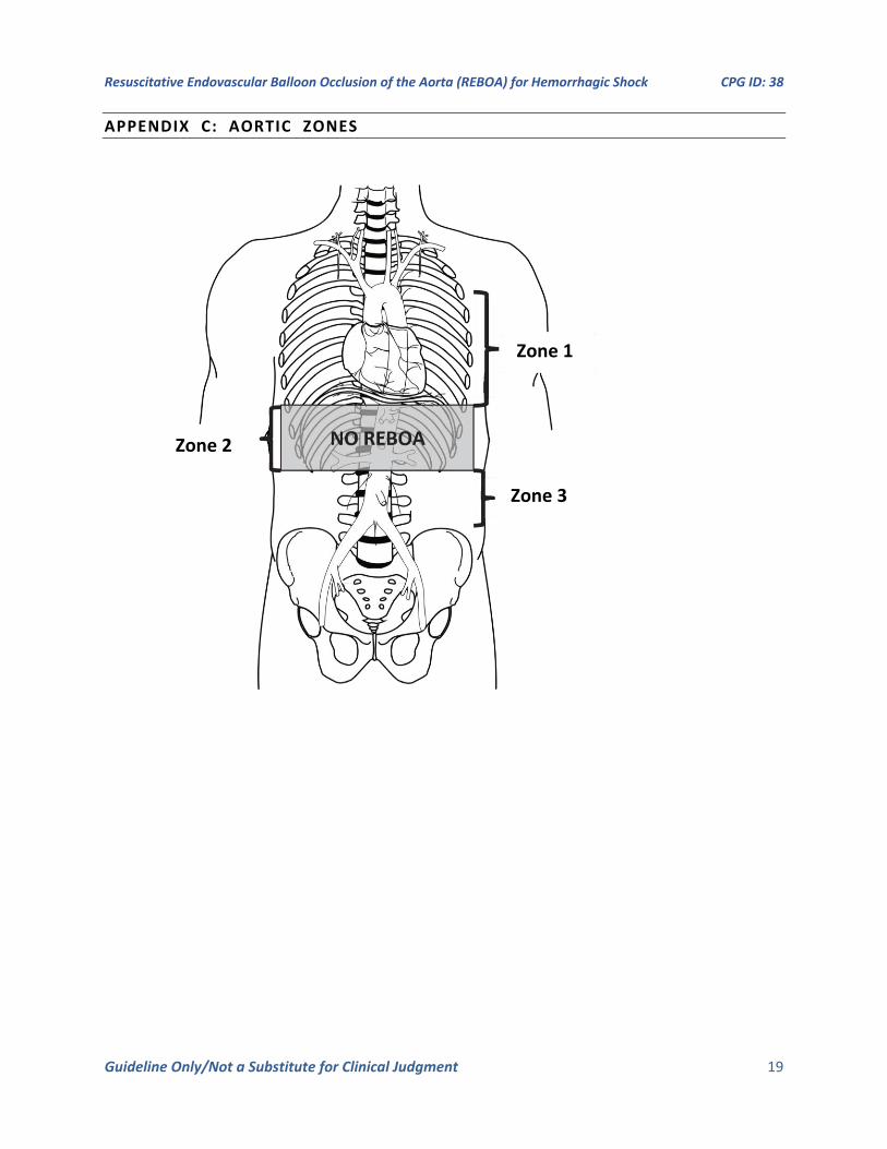
Outcomes for Zone 1 REBOA are optimized if occlusion times are between 15-30 minutes (See Appendix C for an illustration of the Zones of the Aorta). Occlusion times over 30 minutes are associated with higher ischemic complications and higher mortality.17
Outcomes for Zone 3 REBOA are optimized if occlusion times are 30-60 minutes, though survival following longer occlusion times has been reported.36
The American Association for the Surgery of Trauma (AAST) prospective Aortic Occlusion for Resuscitation in Trauma and Acute Care Surgery (AORTA) database contains cases with occlusion times exceeding the above recommendations, but this registry does not track the use of occlusion techniques such as intermittent and/or partial REBOA.41 Partial and intermittent balloon techniques may reduce distal ischemia and extend tolerable occlusion times.37-40 There is currently insufficient data to guide any consensus on this practice.42,36
With increasing REBOA availability and provider experience, REBOA has successfully been utilized in multiple austere military locations.43-47 In austere resuscitations, REBOA has been shown to improve the ability to triage multiple casualties, allow for blood product conservation, and assist in creating a ‘bloodless’ environment for damage control surgery.45,47-48
Properly trained nurses are responsible for assisting in equipment availability and setup, accurate documentation and recording of catheter insertion distance in addition to safe and accurate patient handoff during transfer/transport.
The implementation of this technique must be determined at each site based on training, experience, local resources, and evacuation timelines.
Resuscitative Endovascular Balloon Occlusion of the Aorta (REBOA) for Hemorrhagic Shock CPG ID: 38
Guideline Only/Not a Substitute for Clinical Judgment 5
Documentation of Aortic Occlusion via open thoracotomy or REBOA will be done using the Aortic Occlusion (AO) Procedure Note that is found in Appendix H of this CPG
REBOA IN TRAUMATIC ARREST & PROFOUND SHOCK
Indications for the use of REBOA are summarized below. These indications mirror the indications for resuscitative thoracotomy with the exception that shock or arrest secondary to penetrating chest trauma is a relative contraindication to REBOA (See the JTS Emergency Resuscitative Thoracotomy, 18 Jul 2018 CPG 13 As stated above, there is no high grade evidence defining the specific indications for REBOA. Data is, at best, mixed when comparing survival and outcomes of REBOA versus ERT.14,23 However, there is at least one major trial that demonstrates that REBOA improves survival beyond the emergency department as well as to hospital discharge when compared to ERT for patients who are in hemorrhagic shock, but are pre-cardiac arrest.23 For this reason, early recognition of hemorrhagic shock is vital when identifying patients who may benefit from REBOA.
INITIAL MANAGEMENT
Initial management priorities for patients with traumatic arrest or impending arrest include early control of hemorrhage and hemostatic resuscitation as described in the JTS Damage Control Resuscitation CPG. 50 The initial focus in patients presenting in profound hemorrhagic shock, to include loss of pulses, is to determine the best resuscitative strategy, and whether resuscitation is appropriate or futile in a moribund patient. The following must be rapidly determined:
Mechanism and pattern of injury
Presence of a pulse
Duration of cardiac arrest
Presence or absence of an organized, narrow complex cardiac rhythm and/or organized cardiac activity by ultrasound.
Resources available
Number of concurrent casualties
Patients exsanguinating from abdominal, pelvic, or junctional lower extremity bleeding may be candidates for REBOA. Such patients are identified by penetrating mechanism of injury to abdomen or pelvis, blast or blunt mechanism with positive FAST or suspected pelvic fracture, or massive proximal lower extremity trauma with signs of impending cardiovascular collapse.
Exsanguinating hemorrhage in the chest must be ruled out prior to placing REBOA—this can be done with chest tube placement, thoracostomy, x-ray, or thoracic ultrasound. In cases of major chest hemorrhage, occlusion of the aorta may increase thoracic bleeding and is thus best addressed via thoracotomy or sternotomy.
A decision algorithm for Resuscitative Aortic Occlusion (RAO) is found in Appendix A. If RAO is performed, concurrent hemostatic resuscitation and closed chest cardiac massage should continue while the procedure is performed.51 If RAO is not performed, resuscitative efforts should cease unless there is a compelling reason to consider a non-traumatic arrest.
Resuscitative Endovascular Balloon Occlusion of the Aorta (REBOA) for Hemorrhagic Shock CPG ID: 38
Guideline Only/Not a Substitute for Clinical Judgment 6
RESUSCITATIVE THORACOTOMY
The gold standard for aortic occlusion in traumatic arrest remains a left anterolateral thoracotomy (See JTS Emergent Resuscitative Thoracotomy CPG.)
TRANS- ABDOMINAL AORTIC OCCLUSION
The aorta can also be occluded trans-abdominally at any point along its length. It can be occluded with either application of a clamp or compression with a retractor or manually. Alternatively, balloon occlusion can be considered (below) as this can decrease instruments in the upper abdomen, depending on where the focus of bleeding is located. In obese patients with a large volume of hemoperitoneum or other intra-abdominal pathology, a trans-thoracic approach or a balloon approach to the aorta may be preferable. As with all other forms of RAO, restoration of aortic perfusion should be carefully coordinated with the rest of the team to minimize the effects of reperfusion and blood volume shifts.
REBOA
REBOA can be considered in 6 sequential steps:
1. Arterial access and positioning of sheath
2. Positioning of the balloon
3. Inflation of the balloon
4. Operative/procedural control of bleeding
5. Deflation of the balloon
6. Sheath removal
REBOA can be performed preemptively in patients with high-risk injury patterns and unstable physiologic parameters as described above. In this way, REBOA can be proactive rather than reactive in appropriate patients. The indications for REBOA are summarized in Appendix A for traumatic arrest and Appendix B in cases of profound shock. A schematic of the aortic anatomy is presented in Appendix C.52 If proximal aortic occlusion is required, this is termed Zone 1, whereas distal aortic occlusion is termed Zone 3. Zone 1 REBOA deployment will be used in most patients presenting with hemorrhagic shock, and may be used in all patients with traumatic arrest, regardless of injury pattern, due to the benefits on a patient’s mean arterial pressure (MAP).53
In clinical situations where REBOA is being considered, pre-emptive placement of an arterial line in the common femoral artery (CFA) is recommended. CFA access has consistently been identified as the rate limiting step to REBOA deployment.30 Obtaining early CFA access in the form of an arterial line can greatly decrease REBOA placement time as a preplaced common femoral arterial line can quickly be re-wired to a REBOA introducer in the event of patient deterioration.
Preclinical research has shown a Zone 1 aortic occlusion time of 60 minutes or more results in significant metabolic derangement and organ damage that may offset any gain obtained by early hemorrhage control. 30 minute occlusion times had significantly improved outcomes without evidence of severe physiologic costs. Based on this data, Zone I REBOA should be deployed for no greater than 30 minutes. Zone III REBOA historically has been considered acceptable for up to 4-6 hours.54-55 However, recent analysis in preclinical models have led to the revised recommendation to target Zone III balloon occlusion times of less than 30, and no greater than 60 minutes.54-56,36
Resuscitative Endovascular Balloon Occlusion of the Aorta (REBOA) for Hemorrhagic Shock CPG ID: 38
Guideline Only/Not a Substitute for Clinical Judgment 7
After placing a REBOA, careful management of the femoral sheath is imperative. The majority of complications associated with REBOA use are related to the sheath and access site complications. Reported femoral access complications include arterial disruption, dissection, pseudoaneurysms, hematoma, thromboembolic phenomenon, and extremity ischemia. These complications have resulted in limb loss.27,28 Due to the risk of sheath dislodgement or vessel wall damage excessive patient movement should be avoided. Patients with indwelling sheaths should be positioned supine or reverse Trendelenburg only. If the patient must be moved or turned, they should be kept in a flat position and log-rolled.
The provider, or assistant, should promptly document placement time, pre-/post-placement blood pressure and MAP, and REBOA insertion distance. Please use the Aortic Occlusion (AO) Procedure Note that is found in Appendix H of this CPG for specific REBOA documentation. It is also available on the JTS Website at https://jts.amedd.army.mil/index.cfm/documents/forms_after_action under CPG Forms. Balloon volume and inflation time should be noted at the insertion site for reference by all providers caring for the patient. The provider is responsible for prevention of catheter migration, particularly during patient transport. A provider who is knowledgeable about the management of REBOA should attend to the patient while awaiting definitive surgical repair, to include during transport. The trained provider is responsible for ensuring a safe and competent hand off.
While the sheath is in place, hourly neurovascular assessments of the bilateral lower extremities should be completed. These assessments should continue for 24 hours after removal of the sheath to allow for early identification and intervention of access site complications. In addition, follow-on duplex imaging (24-48 hours post REBOA) of the access site allows for early identification and treatment of any access site complications. This may be performed either at the Role 3 facility or as soon as possible after arrival at a Role 4 facility, depending on resources available to perform and interpret the ultrasound.
Once definitive hemorrhage control has been obtained, the REBOA sheath should be removed and 30 minutes of direct pressure applied to the CFA access site. If arterial pressure monitoring is still required an alternate line site should be considered. An angiogram through the sheath to document distal limb perfusion is best practice, though not always available. If a large sheath size is used, a patient is coagulopathic, or there is technical difficulty in sheath removal a cut down and arterial repair, patch or graft may be required. This may be best accomplished in the Role 3 environment with access to specialists and/or surgical backup.21
The balloon should be deflated once specific vascular control or definitive hemorrhage control has been obtained. Communication with the assistant holding the apparatus securing the catheter and the anesthesia team is critical before consideration of deflating the balloon. When deflating the balloon turn the three-way stopcock and withdraw saline slowly and deflate the balloon slowly as this step can be anticipated to result in a significant decrease in afterload and hypotension and may result in cardiac collapse. Additional resuscitation may be needed while the balloon is slowly deflated. After balloon deflation, the team should anticipate hemodynamic changes related to reperfusion, washout of metabolic byproducts, and acidosis. As such, intermittent balloon inflation and deflation may be necessary during ongoing resuscitation until hemodynamic stability is restored.
Even in an austere environment, protocols for use and follow on care should be planned and discussed prior to implementation. Team training and awareness of pitfalls are critical to ensure the best possible outcomes.
Resuscitative Endovascular Balloon Occlusion of the Aorta (REBOA) for Hemorrhagic Shock CPG ID: 38
Guideline Only/Not a Substitute for Clinical Judgment 8
If definitive hemorrhage is not obtained, leaving the sheath in place without aortic occlusion may be a valid option. By leaving the sheath in place, the REBOA can easily be reinserted and aortic occlusion can quickly be obtained if rebleeding occurs or hemorrhage continues.38 In general, and situation/resource dependent, the sheath should be left in during any active or ongoing resuscitation. The sheath should not be removed immediately prior to transport, and is best removed where vascular complications can be treated and managed.
A sheath MUST NEVER be left in for transfer to a host nation facility.
AEROMEDICAL EVACUATION CONSIDERATIONS
Patients who receive REBOA at a Role 2 and need to be evacuated to a higher level of care should have hemorrhage control addressed and balloon deflated prior to transfer. Under no circumstance should a Zone 1 REBOA remain inflated during transport. In rare situations when a short-distance rotary-wing evacuation to higher level of care is possible, a Zone 3 REBOA inserted at Role 2 may remain inflated during transport however this requires exceptional communication and planning to avoid undue risk of ischemic injury.
If rotary-wing transport is available, a medical provider trained in hemodynamic monitoring and manipulation of the occlusion balloon should accompany the casualty at all times. If a REBOA sheath is in place in a trauma patient, re-placement/re-inflation of the balloon during transport is an option for trained providers in the event of sudden profound hypotension. Simultaneous blood transfusion is needed and balloon inflation time should not exceed 15 min in Zone I.
The essential equipment for REBOA is provided in Appendix D while the appropriate technical steps and considerations are summarized in Appendix E.
TRAINING
Prior to using REBOA, providers should have a thorough knowledge of the device, its indications, use and potential complications. Organized, curriculum-based REBOA training courses such as the American College of Surgeon’s Basic Endovascular Skills for Trauma (BEST) course or the ‘Resuscitation Adjuncts: Prehospital Transfusion & REBOA’ (RAPToR) Course are available. Successful completion of a REBOA training course, including a didactic and hands on skills component, is recommended prior utilization of the device. Skills training can be achieved through high-fidelity simulation, perfused cadaver or live tissue training. Critical skills include access to the CFA with ultrasound and cut down, sheath placement and positioning, and REBOA operation and removal. Anatomically correct models are critical for accurate training of CFA access skills, and thus perfused cadavers are recommended to meet this requirement.57-59
Ultimately, the decision to perform REBOA on patients at high risk for hemorrhagic death will depend on the specific injury pattern, individual provider experience, team training, and local resources.
Resuscitative Endovascular Balloon Occlusion of the Aorta (REBOA) for Hemorrhagic Shock CPG ID: 38
Guideline Only/Not a Substitute for Clinical Judgment 9
REBOA USE BY NON-SURGICAL RESUSCITATION TEAMS
Advanced resuscitation teams may be utilized in austere environments as a bridge to surgical hemorrhage control. Data on the effectiveness of this approach are lacking. REBOA in this setting may be considered in the rare circumstance that all of the following conditions are met:
1. The casualty would otherwise die in 15-30 minutes without REBOA (NCTH, refractory hemorrhagic shock)
2. A physician experienced in REBOA therapy is present
3. Blood product resuscitation, preferably whole blood, is available but failing to resuscitate the patient
4. Time to definitive hemorrhage control is short (ideally <15 min Zone 1, <30 min Zone 3).
The narrow therapeutic window of aortic occlusion is the major limitation of REBOA. Techniques for lengthening aortic occlusion time are being investigated such as partial REBOA (pREBOA), intermittent REBOA (iREBOA), regional hypothermia, and pharmacologic interventions to decrease ischemia or enhance ischemia resistance. Multiple descriptions of pREBOA and iREBOA techniques in animal models have been described. One protocol recommended by the Committee on Tactical Combat Casualty Care (CoTCCC) describes iREBOA as an initial 15min of occlusion time, followed by balloon deflation and reassessment of the patient’s systolic blood pressure (SBP). If SBP > 80mmHg, the balloon should remain deflated. If SBP drops to < 80mmHg the balloon should be re-inflated. If the SBP dropped below 80mmHg in less than 3 minutes, balloon occlusion will be maintained for up to 30 min as resuscitation continues. If decompensation occurs after 3 minutes, the balloon should re-inflated and deflated again after 10 minutes for reassessment. This cycle continues for a total occlusion time up to 120 min or until the patient’s blood pressure remains stable above 80mmHg. iREBOA has come under criticism due to multiple limitations of the initial study and lack of other supportive studies.37-38,42,60
REBOA PITFALLS
Making the decision to perform REBOA too late. Mortality is high after loss of pulses has occurred, as it is with ERT.
Difficulty locating the common femoral artery in the groin. The clinician must be very familiar with open, percutaneous, and ultrasound guided femoral access techniques. Early CFA access is recommended even if REBOA not utilized.
Insertion of the REBOA too low, below the femoral artery bifurcation. The catheter should be placed in the common femoral artery, just below the inguinal ligament. Insertion into the superficial femoral artery is associated with an increased risk of thrombosis and limb loss.
Unrecognized proximal femoral or iliac artery transection preventing endovascular access on the side of the injury. This may occur with penetrating pelvic trauma or severe pelvic fracture—check bilateral femoral pulses and access the side with a stronger pulse if there is a difference. Do not hesitate to switch to the opposite groin or convert to thoracotomy.
Resuscitative Endovascular Balloon Occlusion of the Aorta (REBOA) for Hemorrhagic Shock CPG ID: 38
Guideline Only/Not a Substitute for Clinical Judgment 10
Failure to address chest pathology. Always evaluate the chest by X-ray, ultrasound, or bilateral chest tube placement to identify and treat significant hemothorax or pneumothorax. Convert to thoracotomy to address massive hemothorax.
Catheter or guidewire does not pass freely. This could indicate injury to the vessel. Do not inflate balloon. Consider accessing the opposite groin or convert to thoracotomy.
Over-inflating the balloon. The ER REBOA balloon capacity is 24 ml. Zone 1 may require as little as 8 ml and Zone 3 as little as 2 ml to achieve occlusion. Over-inflation may rupture the balloon or injure the aorta.
Leaving the balloon inflated too long. Only 30 minutes of Zone 1 occlusion is advised, and the shorter the better. Achieve rapid control of bleeding sites with temporizing measures such as clamping to allow the earliest reperfusion; most suturing, ligating, solid organ removal, and vascular shunting may be done after balloon deflation. Death secondary to ischemic injury has been reported with longer occlusion times.
Failure to work with a heightened level of urgency once REBOA is placed. Some patients may regain “stability,” however balloon occlusion is just like a cross clamp, with the same complications of visceral and spinal ischemia. Every effort should be made to restore perfusion as soon as possible to limit ischemia.
Failure to adequately secure the REBOA catheter after balloon inflation, resulting in migration of the balloon. The catheter position must be maintained during and after inflation to avoid distal migration until aortic pressure and pulsatility are restored.
Deflating the balloon too quickly before adequate volume resuscitation. Ensure that the anesthesia team is prepared for reperfusion prior to balloon deflation.
Premature removal of the arterial sheath. The sheath should remain in place if the patient is coagulopathic, may have ongoing bleeding in the abdomen or pelvis, or is being transported within theater to a higher level of care.
Injury to the arterial access point. After removal of the sheath, monitor the instrumented leg closely for re-bleeding and thrombus/intimal injury. Decreased lower extremity perfusion may require further angiography, thrombectomy, or direct arterial repair.
Committing multiple resources to a futile resuscitation. Anticipate massive transfusion, personnel required, surgical supplies, diversion of resources from more salvageable casualties, etc.
FUTURE CONSIDERATIONS
A retrospective capability gap analysis of the UK Joint Theatre Trauma Registry suggested that as many as one in five severely injured casualties have wounds that may be amenable to treatment with REBOA.61 The development of the 7 Fr ER REBOA catheter facilitates insertion of the device and may lead to more widespread use of this approach in the austere environment. Training non-physician caregivers to place REBOAs in the prehospital settings is being investigated.62-63 Partial REBOA, intermittent REBOA, regional hypothermia, and pharmacologic adjuncts continue to undergo validation as a means of prolonging aortic occlusion time.60,39,64 Ongoing research seeks to identify modifications to the REBOA technique that may be required when it is combined with other resuscitation modalities such as tranexamic acid. Researchers are also striving to clarify patient selection, evaluating the impact of REBOA on thoracic injury, and traumatic brain injury.65 All of these advances should refine the
Resuscitative Endovascular Balloon Occlusion of the Aorta (REBOA) for Hemorrhagic Shock CPG ID: 38
Guideline Only/Not a Substitute for Clinical Judgment 11
optimal use of this resuscitation adjunct. Longitudinal data in the civilian and military setting will assist in defining the ideal clinical situation in which REBOA can be of maximal benefit.
PERFORMANCE IMPROVEMENT (PI) MONITORING
POPULATION OF INTEREST
1. All trauma patients with traumatic arrest (SBP 0, HR 0, or absent pulse) or profound shock (SBP < 90).
2. Patients with abdominal, pelvic, or junctional lower extremity injury with body region AIS ≥ 3.
3. Patients who receive REBOA.
INTENT (EXPECTED OUTCOMES)
1. If performed, REBOA was performed for the indication of hemorrhagic shock associated with abdominal, pelvic, or junctional lower extremity bleeding, or other indication is clearly documented.
2. If REBOA performed, the patient was assessed for thoracic hemorrhage (extended focused assessment with sonography in trauma [EFAST] or chest x-ray [CXR] results documented or bilateral chest tubes placed).
3. Blood pressure pre- and post- REBOA and balloon times (inflation and deflation) are documented in REBOA procedure note.
4. Lower extremity pulses are documented after balloon deflation.
5. All patient who undergo REBOA have a subsequent hemorrhage control procedure or documentation that the procedure was not needed.
PERFORMANCE/ADHERENCE METRICS
1. Number and percentage of patients who had REBOA performed for hemorrhagic shock associated with abdominal, pelvic, or junctional lower extremity bleeding.
2. Number and percentage of patients who had REBOA performed who were assessed for thoracic hemorrhage (E-FAST or CXR results documented or bilateral chest tubes placed).
3. Number and percentage of patients who had REBOA performed with complete REBOA procedure note to include documented blood pressure pre and post REBOA and documented balloon times (inflation and deflation).
4. Number and percentage of patients who had REBOA performed with lower extremity pulses documented after balloon deflation.
5. Number and percentage of patients who had REBOA performed with subsequent hemorrhage control procedure or documentation that procedure not needed.
6. Survival of REBOA versus Resuscitative Thoracotomy.
AUDIT FILTER
All patients who receive REBOA, incomplete or absent REBOA procedure note
Resuscitative Endovascular Balloon Occlusion of the Aorta (REBOA) for Hemorrhagic Shock CPG ID: 38
Guideline Only/Not a Substitute for Clinical Judgment 12
DATA SOURCE
Patient Record
Department of Defense Trauma Registry (DoDTR)
PI DATA CAPTURE AND REPORTING
Number of REBOA interventions, performance, and adherence measures will be reported quarterly by JTS PI Branch Chief to the JTS Chief.
JTS will identify REBOA patients in the trauma registry and facilitate capture of complete medical records.
SYSTEM REPORTING & FREQUENCY
The above constitutes the minimum criteria for PI monitoring of this CPG. System reporting will be performed annually; additional PI monitoring and system reporting may be performed as needed.
The system review and data analysis will be performed by the JTS Chief, JTS Program Manager, and the JTS PI Branch.
RESPONSIBILITIES
It is the responsibility of the JTS PI Branch Chief to ensure system-level compliance with this CPG. It is the trauma team leader’s responsibility to ensure familiarity, appropriate compliance and PI monitoring at the local level with this CPG.
REFERENCES
1. Morrison JJ, Rasmussen TE. Noncompressible torso hemorrhage: a review with contemporary definitions and management strategies. Surg Clin North Am 2012;92(4):843–58, vii.
2. Stannard A, Morrison JJ, Scott DJ, et al. The epidemiology of noncompressible torso hemorrhage in the wars in Iraq and Afghanistan. J Trauma Acute Care Surg 2013;74(3):830–4.
3. Mattox KL, Feliciano DV. Role of external cardiac compression in truncal trauma. J Trauma 1982;22(11):934–6.
4. Mattox KL, Wall MJ, Tsai P. Trauma thoracotomy: principles and techniques. In: Mattox KL, Moore EE, Feliciano DV, editors. Trauma. New York: McGraw Hill Medical; 2013. p. 461–7.
5. Ledgerwood AM, Kazmers M, Lucas CE. The role of thoracic aortic occlusion for massive hemoperitoneum. J Trauma 1976;16(08):610–5.
6. Burlew CC, Moore EE, Moore F a, et al. Western Trauma Association critical decisions in trauma: resuscitative thoracotomy. J Trauma Acute Care Surg 2012;73(6):1359–63.
7. Working Group Ad Hoc Subcommittee on Outcomes, American College of Surgeons-Committee on Trauma. Practice Management Guidelines for Emergency Department Thoracotomy. J Am Coll Surg 2001;193(3):303–9.
8. Seamon MJ, Fisher CA, Gaughan JP, Kulp H, Dempsey DT, Goldberg AJ. Emergency department thoracotomy: survival of the least expected. World J Surg 2008;32(4):604–12.
Resuscitative Endovascular Balloon Occlusion of the Aorta (REBOA) for Hemorrhagic Shock CPG ID: 38
Guideline Only/Not a Substitute for Clinical Judgment 13
9. Branney SW, Moore EE, Feldhaus KM, Wolfe RE. Critical analysis of two decades of experience with postinjury emergency department thoracotomy in a regional trauma center. J Trauma 1998;45(1):85–7.
10. Passos EM, Engels PT, Doyle JD, et al. Societal costs of inappropriate emergency department thoracotomy. J Am Coll Surg 2012;214(1):18–25.
11. Edens JW, Beekley AC, Chung KK, et al. Longterm outcomes after combat casualty emergency department thoracotomy. J Am Coll Surg 2009;209(2):188–97.
12. Mitchell TA, Waldrep KB, Sams VG, et al. An 8-year review of Operation Enduring Freedom and Operation Iraqi Freedom resuscitative thoracotomies. Mil Med 2015;180(3):S33-S36.
13. Joint Trauma System, Emergent Resuscitative Thoracotomy Clinical Practice Guideline, 18 Jul 2018; https://jts.amedd.army.mil/assets/docs/cpgs/JTS_Clinical_Practice_Guidelines_(CPGs)/Emergent_Resuscitative_Thoracotomy_ERT_18_Jul_2018_ID20.pdf Accessed Mar 2020.
14. Joseph B, Zeeshan M, Sakran JV, et al. Nationwide analysis of resuscitative endovascular balloon occlusion of the aorta in civilian trauma. JAMA Surg 2019;154:500.
15. Moore L, Brenner M, Kozare RA, et al. Implementation of resuscitative endovascular balloon occlusion of the aorta as an alternative to resuscitative thoracotomy for noncompressible truncal hemorrhage. J Trauma Acute Care Surg 2014;79:523-532.
16. Abe T, Uchida M, Nagata I, et al. Resuscitative endovascular balloon occlusion of the aorta versus aortic cross clamping among patients with critical trauma: a nationwide cohort study in Japan. Crit Care 2016;20:400-410.
17. Inoue J, Shiraishi A, Yoshiyuki A, Haruta K, Matsui H, Otomo Y. Resuscitative endovascular balloon occlusion of the aorta might be dangerous in patients with severe torso trauma: A propensity score analysis. J Trauma Acute Care Surg 2016;80:559-567.
18. Norii T, Crandall C, Terasaka Y. Survival of severe blunt trauma patients treated with resuscitative endovascular balloon occlusion of the aorta compared with propensity score/adjusted untreated patients. J Trauma Acute Care Surg. 2015;78:721-728.
19. Perina DG, Kang CS, Bulger EM, et al. Authors' Response to Letter to the Editor by Allen et al regarding Joint statement from the American College of Surgeons Committee on Trauma (ACS COT) and the American College of Emergency Physicians (ACEP) regarding the clinical use of Resuscitative Endovascular Balloon Occlusion of the Aorta (REBOA) by Brenner et al. Trauma Surg Acute Care Open. 2018 Mar 6;3(1):e000172.
20. Manzano-Nunez R, Orlas CP, Herrera-Escobar JP, et al. A meta-analysis of the incidence of complications associated with groin access after the use of resuscitative endovascular balloon occlusion of the aorta in trauma patients. J Trauma Acute Care Surg. 2018 Sep;85(3):626-634.
21. Taylor JR, Harvin JA, Martin C, Holcomb JB, Moore LJ. Vascular complications from resuscitative endovascular balloon occlusion of the aorta: life over limb? J Trauma Acute Care Surg. 2017;83(1 Suppl1):S120–S123.
22. DuBose JJ1, Scalea TM, Brenner M, Skiada D, et al. The AAST prospective Aortic Occlusion for Resuscitation in Trauma and Acute Care Surgery (AORTA) registry: Data on contemporary utilization and outcomes of aortic occlusion and resuscitative balloon occlusion of the aorta (REBOA). J Trauma Acute Care Surg. 2016 Sep;81(3):409-19
23. Brenner M, Inaba K, Aiolfi A, DuBose J, et al. Resuscitative Endovascular Balloon Occlusion of the Aorta and Resuscitative Thoracotomy in select patients with hemorrhagic shock: early results from the American Association for the Surgery of Trauma's Aortic Occlusion in Resuscitation for Trauma and Acute Care Surgery Registry. J Am Coll Surg. 2018 May; 226(5):730-740.
Resuscitative Endovascular Balloon Occlusion of the Aorta (REBOA) for Hemorrhagic Shock CPG ID: 38
Guideline Only/Not a Substitute for Clinical Judgment 14
24. Ordoñez CA, Parra MW, Manzano-Nunez R, et al. Intraoperative combination of resuscitative endovascular balloon occlusion of the aorta and a median sternotomy in hemodynamically unstable patients with penetrating chest. J Trauma Acute Care Surg. May 2018; 84(5):752–757,
25. Boczar ME, Howard MA, Rivers EP, et al. A technique revisited: Hemodynamic comparison of closed- and open-chest cardiac massage during human cardiopulmonary resuscitation. Critical Care Medicine. 23(3):498-503, March 1995. trauma: is this feasible? J Trauma Acute Care Surg 2018;84:752–7.
26. Doucet J, Coimbra R. REBOA: is it ready for prime time? J Vasc Bras 2017;16:1–3. Norii T, Crandall C, Terasaka Y. Survival of severe blunt trauma patients treated with resuscitative endovascular balloon occlusion of the aorta compared with propensity score-adjusted untreated patients. J Trauma Acute Care Surg 2015;78:721–8.
27. Davidson AJ, Russo RM, Reva VA, et al. The pitfalls of REBOA: risk factors and mitigation strategies. J Trauma Acute Care Surg 2017;84:192–202.
28. Ribeiro Junior MAF, Feng CYD, Nguyen ATM, et al. The complications associated with resuscitative endovascular balloon occlusion of the aorta (REBOA). World J Emerg Surg 2018;13:20.
29. Matsumura Y, Matsumoto J, Kondo H, et al. Early arterial access for REBOA is related to survival outcome in trauma. J Trauma Acute Care Surg 2018;1.
30. Romagnoli A, Teeter W, Pasley J, et al. Time to aortic occlusion: it’s all about access. J Trauma Acute Care Surg 2017;83:1161–4.
31. Brenner M, Moore L, Teeter W, et al. Exclusive clinical experience with a lower profile device for resuscitative endovascular balloon occlusion of the aorta (REBOA). Am J Surg 2019;217:1126–9.
32. Matsumura Y, Matsumoto J, Kondo H, et al. Fewer REBOA complications with smaller devices and partial occlusion: evidence from a multicentre registry in Japan. Emerg Med J 2017;34:793–9.
33. Markov NP, Percival TJ, Morrison JJ, et al. Physiologic tolerance of descending thoracic aortic balloon occlusion in a swine model of hemorrhagic shock. Surgery 2013;153(6):848–56.
34. Brenner ML, Moore LJ, Dubose JJ, et al. A clinical series of resuscitative endovascular balloon occlusion of the aorta for hemorrhage control and resuscitation. J Trauma Acute Care Surg 2013;75(3):506–11.
35. Kauvar DS, Dubick MA, Martin MJ. Large animal models of proximal aortic balloon occlusion in traumatic hemorrhage: review and identification of knowledge gaps relevant to expanded use. J Surg Res 2019;236:247–58.
36. Bulger EM, Perina DG, Zaffer Q,et al. Clinical use of resuscitative endovascular balloon occlusion of the aorta (REBOA) in civilian trauma systems in the USA, 2019: a joint statement from the American College of Surgeons Committee on Trauma, the American College of Emergency Physicians, the National Association of Emergency Medical Services Physicians and the National Association of Emergency Medical Technicians. Trauma Surg Acute Care Open 2019;4:e000376. doi:10.1136/tsaco-2019-000376
37. Kuckelman JP, Barron M, Moe D, et al. Extending the golden hour for zone 1 resuscitative endovascular balloon occlusion of the aorta: improved survival and reperfusion injury with intermittent versus continuous resuscitative endovascular balloon occlusion of the aorta in a porcine severe truncal hemorrhage model. J Trauma Acute Care Surg. 2018;85(2):318–326.
38. Butler FK Jr, Holcomb JB, Shackelford S, et al. Advanced resuscitative care in tactical combat casualty care: TCCC Guidelines Change 18-01:14 October 2018. J Spec Oper Med. Winter 2018;18(4):37-55.
Resuscitative Endovascular Balloon Occlusion of the Aorta (REBOA) for Hemorrhagic Shock CPG ID: 38
Guideline Only/Not a Substitute for Clinical Judgment 15
39. Johnson MA, Tibbits EM, Hoareau GL, et al. Endovascular perfusion augmentation for critical care: partial aortic occlusion for treatment of severe ischemia-reperfusion shock. Shock. 2019 May;51(5):659-666.
40. Johnson MA, Williams TK, Ferencz SE, et al. The effect of resuscitative endovascular balloon occlusion of the aorta, partial aortic occlusion and aggressive blood transfusion on traumatic brain injury in a swine multiple injuries model. J Trauma Acute Care Surg. 2017 Jul; 83(1):61-70.
41. Brenner M, Bishoy Z, Coimbra R, AAST Multi-Institutional Trials Committee, et al. Right into the danger zone: complications of Resuscitative Endovascular Balloon Occlusion of the Aorta (REBOA) at zone 1 and 3 from the AAST aortic occlusion for resuscitation in trauma and acute care surgery (AORTA) trial. Presented at the 2019 AAST meeting.
42. Williams T, Neff L, Johnson, MA. Letter to the Editor: Intermittent REBOA Translational Science Papers, Journal of Trauma and Acute Care Surgery: Aug 28, 2019 - Volume Publish Ahead of Print - Issue – p doi: 10.1097/TA.0000000000002496
43. Fisher AD, Teeter WA, Cordova CB, et al. The Role I Resuscitation Team and Resuscitative Endovascular Balloon Occlusion of the Aorta. J Spec Oper Med. Summer 2017;17(2):65-73.
44. Glaser J, Teeter W, Fernandez N. Resuscitative Endovascular Balloon Occlusion of the Aorta (REBOA) as an adjunct to damage control surgery for combat trauma. Journal of Endovascular Resuscitation and Trauma Management, [S.l.], v. 1, n. 1, p. 58-62, Aug. 2017.
45. Manley JD, Mitchell BJ, Dubose JJ, et al. A modern case series of Resuscitative Endovascular Balloon Occlusion of the Aorta (REBOA) in an out-of-hospital, combat casualty care setting. J Spec Oper Med 2017; 17 (1), 1-8.
46. Reva VA, Hörer TM, Makhnovskiy AI. Field and en route resuscitative endovascular occlusion of the aorta: A feasible military reality? J Trauma Acute Care Surg. 2017 Jul;83(1 Suppl 1):S170-S176.
47. Northern DM, Manley JD, Lyon R, et al. Recent advances in austere combat surgery: Use of aortic balloon occlusion as well as blood challenges by special operations medical forces in recent combat operations. J Trauma Acute Care Surg. 2018 Jul;85(1S Suppl 2):S98-S103
48. Lyon RF, Northern DM. REBOA by a non-surgeon as an adjunct during MASCAL. Am J Emerg Med. 2018 Jun;36(6):1121.e5-1121.e6. doi: 10.1016/j.ajem.2018.02.013. Epub 2018 Feb 13.
49. Biffl WL, Fox CJ, Moore EE. The role of REBOA in the control of exsanguinating torso hemorrhage: J Trauma Acute Care Surg 2015;78(5):1054–8.
50. Joint Trauma System, Damage Control Resuscitation CPG, 12 Jul 2019; https://jts.amedd.army.mil/assets/docs/cpgs/JTS_Clinical_Practice_Guidelines_(CPGs)/Damage_Control_Resuscitation_12_Jul_2019_ID18.pdf Accessed Mar 2020.
51. Teeter WA, Bradley MJ, Romagnoli A, et al. Treatment effect or effective treatment? Cardiac compression fraction and end-tidal carbon dioxide are higher in patients with resuscitative endovascular balloon occlusion of the aorta compared with resuscitative thoracotomy and open-chest cardiac massage. Am Surg. 2018 Oct 1;84(10):1691-1695.
52. Stannard A, Eliason JL, Rasmussen TE. Resuscitative endovascular balloon occlusion of the aorta (REBOA) as an adjunct for hemorrhagic shock. J Trauma 2011;71(6):1869–72.
53. Tibbits EM, Hoareau GL, Simon MA, et al. Location is everything: the hemodynamic effects of REBOA in zone 1 versus zone 3 of the aorta. J Trauma Acute Care Surg. 2018;85(1):101–107.
54. Reva VA, Matsumura Y, Hörer T, et al. Resuscitative endovascular balloon occlusion of the aorta: what is the optimum occlusion time in an ovine model of hemorrhagic shock? Eur J Trauma Emerg Surg. 2018 Aug;44(4):511-518.
Resuscitative Endovascular Balloon Occlusion of the Aorta (REBOA) for Hemorrhagic Shock CPG ID: 38
Guideline Only/Not a Substitute for Clinical Judgment 16
55. Bekdache O, Paradis T, Shen YBH, et al. Resuscitative endovascular balloon occlusion of the aorta (REBOA): indications: advantages and challenges of implementation in traumatic non-compressible torso hemorrhage. Trauma Surgery & Acute Care Open 2019;4:e000262.
56. Kauvar DS, Dubick MA, Martin MJ. Large animal models of proximal aortic balloon occlusion in traumatic hemorrhage: review and identification of knowledge gaps relevant to expanded use. J Surg Res 2019;236:247–58.
57. Villamaria CY, Eliason JL, Napolitano LM, et al. Endovascular Skills for Trauma and Resuscitative Surgery (ESTARS) course: curriculum development, content validation, and program assessment. J Trauma Acute Care Surg 2014;76(4):929–35; discussion 935–6.
58. Brenner M, Hoehn M, Pasley J, et al. Basic endovascular skills for trauma course: bridging the gap between endovascular techniques and the acute care surgeon. J Trauma Acute Care Surg 2014;77(2):286–91.
59. Brenner M, Hoehn M, Stein DM, et al. Central pressurized cadaver model (CPCM) for resuscitative endovascular balloon occlusion of the aorta (REBOA) training and device testing. J Trauma Acute Care Surg 2015;78(1):197–200.
60. Glaser JJ, Fisher AD, Shackelford SA, Butler F, Rasmussen TE. A contemporary report on U.S. military guidelines for the use of whole blood and resuscitative endovascular balloon occlusion of the aorta. J Trauma Acute Care Surg. 2019 Jul;87
61. Morrison JJ, Ross JD, Rasmussen TE, et al. Resuscitative endovascular balloon occlusion of the aorta: a gap analysis of severely injured UK combat casualties. Shock 2014;41(5):388–93.
62. Pasley JD, Teeter WA, Gamble WB, et al. Bringing Resuscitative Endovascular Balloon Occlusion of the Aorta (REBOA) Closer to the Point of Injury. J Spec Oper Med. 2018 Spring;18(1):33-36.
63. Saito N, Matsumoto H, Yagi T, et al. Evaluation of the safety and feasibility of resuscitative endovascular balloon occlusion of the aorta. J Trauma Acute Care Surg 2015;78(5):897–903; discussion 904.
64. Simon MA, Tibbits EM, Hoareau GL, et al. Lower extremity cooling reduces ischemia-reperfusion injury following Zone 3 REBOA in a porcine hemorrhage model. J Trauma Acute Care Surg. 2018 Sep;85(3):512-518.
65. Williams AM, Bhatti UF, Dennahy IS, Graham NJ, Nikolian VC, Chtraklin K, Chang P, Zhou J, Biesterveld BE, Eliason J, Alam HB. Traumatic brain injury may worsen clinical outcomes after prolonged partial resuscitative endovascular balloon occlusion of the aorta in severe hemorrhagic shock model. Journal of Trauma and Acute Care Surgery. 2019 Mar 1;86(3):415-23.
Resuscitative Endovascular Balloon Occlusion of the Aorta (REBOA) for Hemorrhagic Shock CPG ID: 38
Guideline Only/Not a Substitute for Clinical Judgment 17
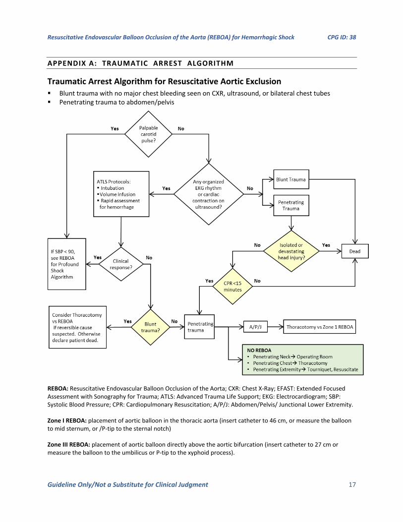
APPENDIX A: TRAUMATIC ARREST ALGORITHM
Traumatic Arrest Algorithm for Resuscitative Aortic Exclusion Blunt trauma with no major chest bleeding seen on CXR, ultrasound, or bilateral chest tubes
Penetrating trauma to abdomen/pelvis
REBOA: Resuscitative Endovascular Balloon Occlusion of the Aorta; CXR: Chest X-Ray; EFAST: Extended Focused Assessment with Sonography for Trauma; ATLS: Advanced Trauma Life Support; EKG: Electrocardiogram; SBP: Systolic Blood Pressure; CPR: Cardiopulmonary Resuscitation; A/P/J: Abdomen/Pelvis/ Junctional Lower Extremity.
Zone I REBOA: placement of aortic balloon in the thoracic aorta (insert catheter to 46 cm, or measure the balloon to mid sternum, or /P-tip to the sternal notch)
Zone III REBOA: placement of aortic balloon directly above the aortic bifurcation (insert catheter to 27 cm or measure the balloon to the umbilicus or P-tip to the xyphoid process).
Resuscitative Endovascular Balloon Occlusion of the Aorta (REBOA) for Hemorrhagic Shock CPG ID: 38
Guideline Only/Not a Substitute for Clinical Judgment 18
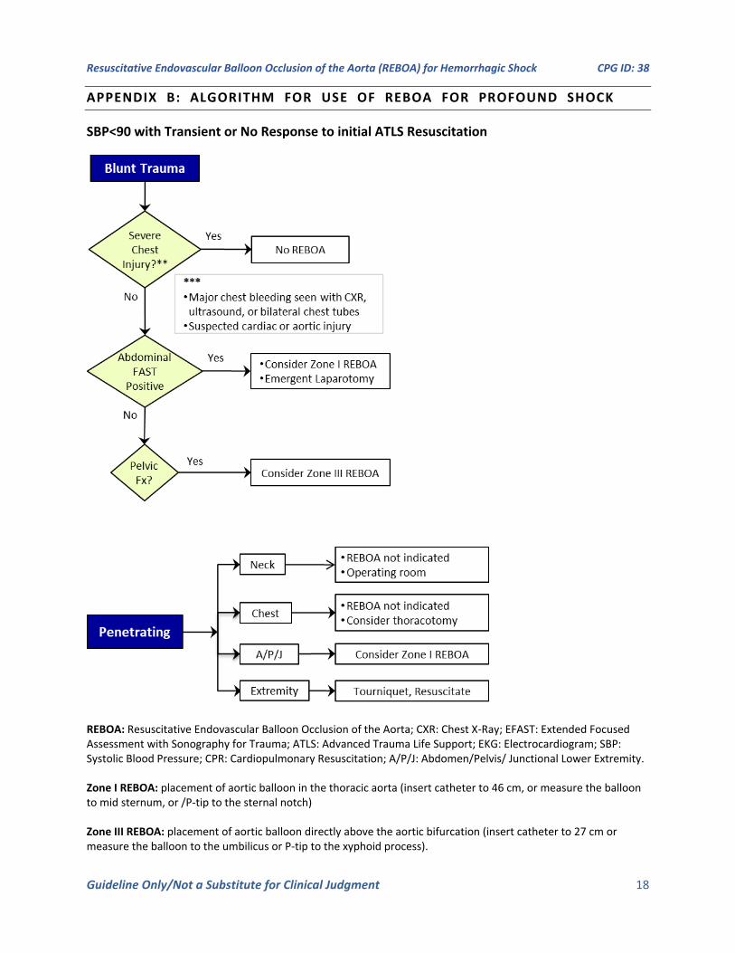
APPENDIX B: ALGORITHM FOR USE OF REBOA FOR PROFOUND SHOCK
SBP<90 with Transient or No Response to initial ATLS Resuscitation
REBOA: Resuscitative Endovascular Balloon Occlusion of the Aorta; CXR: Chest X-Ray; EFAST: Extended Focused Assessment with Sonography for Trauma; ATLS: Advanced Trauma Life Support; EKG: Electrocardiogram; SBP: Systolic Blood Pressure; CPR: Cardiopulmonary Resuscitation; A/P/J: Abdomen/Pelvis/ Junctional Lower Extremity.
Zone I REBOA: placement of aortic balloon in the thoracic aorta (insert catheter to 46 cm, or measure the balloon to mid sternum, or /P-tip to the sternal notch)
Zone III REBOA: placement of aortic balloon directly above the aortic bifurcation (insert catheter to 27 cm or measure the balloon to the umbilicus or P-tip to the xyphoid process).
Resuscitative Endovascular Balloon Occlusion of the Aorta (REBOA) for Hemorrhagic Shock CPG ID: 38
Guideline Only/Not a Substitute for Clinical Judgment 19
APPENDIX C: AORTIC ZONES
Zone 2
Zone 1
Zone 3
Resuscitative Endovascular Balloon Occlusion of the Aorta (REBOA) for Hemorrhagic Shock CPG ID: 38
Guideline Only/Not a Substitute for Clinical Judgment 20
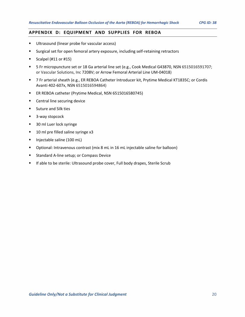
APPENDIX D: EQUIPMENT AND SUPPLIES FOR REBOA
Ultrasound (linear probe for vascular access)
Surgical set for open femoral artery exposure, including self-retaining retractors
Scalpel (#11 or #15)
5 Fr micropuncture set or 18 Ga arterial line set (e.g., Cook Medical G43870, NSN 6515016591707; or Vascular Solutions, Inc 7208V; or Arrow Femoral Arterial Line UM-04018)
7 Fr arterial sheath (e.g., ER REBOA Catheter Introducer kit, Prytime Medical KT1835C; or Cordis Avanti 402-607x, NSN 6515016594864)
ER REBOA catheter (Prytime Medical, NSN 6515016580745)
Central line securing device
Suture and Silk ties
3-way stopcock
30 ml Luer lock syringe
10 ml pre filled saline syringe x3
Injectable saline (100 mL)
Optional: Intravenous contrast (mix 8 mL in 16 mL injectable saline for balloon)
Standard A-line setup; or Compass Device
If able to be sterile: Ultrasound probe cover, Full body drapes, Sterile Scrub
Resuscitative Endovascular Balloon Occlusion of the Aorta (REBOA) for Hemorrhagic Shock CPG ID: 38
Guideline Only/Not a Substitute for Clinical Judgment 21
APPENDIX E: REBOA STEPS USING 7 FRENCH ER-REBOA
The procedure may reviewed online using the following links:
Part 1: https://www.youtube.com/watch?v=-U7MkU3eA7E
Part 2: https://www.youtube.com/watch?v=DZ5LCEt7PBk
STEP 1: Arterial Access and Positioning of the Sheath
Establishing Arterial Access:
Access to the arterial circulation for REBOA for trauma should be obtained through the common femoral artery using one of three techniques: percutaneous, open exposure (e.g., cut down), or exchange over a guide wire from an existing common femoral arterial line.
Ultrasound is used to identify the common femoral artery above the branch of the profunda and the needle visualized passing into the common femoral artery (linear array transducer preferred). Ultrasound guided access improves first pass access and decreases complications.1 Once identified, the artery should be entered at a 45-degree angle with the needle, using either a 5 Fr micropuncture kit or 18 gauge femoral arterial line kit. After the wire has been passed into the artery, the needle is removed and a small incision made at the interface of the wire and skin and the catheter is passed over the wire.
Using landmarks, the location of the inguinal ligament is identified between the Anterior Superior Iliac Spine (ASIS) and pubic symphysis (NOT the inguinal crease). The common femoral artery is then accessed 2 cm below the inguinal ligament.
Selection and Positioning of Initial Sheath:
If REBOA is indicated, the arterial access catheter must be upsized to a 7 Fr sheath. This maneuver is accomplished by placing a 0.035 guide wire greater than twice the length of the existing arterial catheter through its inner lumen allowing the catheter to be removed over the wire while maintaining arterial access. After a larger opening is created at the wire/skin interface, the 7 Fr working sheath with its internal dilator in position can be inserted over the wire. When urgently needed, a 7 Fr sheath may be placed as the initial step by placing the 7F sheath over the 0.035 guide wire though this can increase risk of access site damage.
The sheath’s internal dilator must be firmly held in place to allow a smooth reverse taper from the wire to the diameter of the sheath to avoid arterial intimal injury. Once the dilator and sheath have been advanced over the wire through the skin into the artery, the dilator and wire are removed, leaving the sheath in place. It is important that the operator assure that the stopcock is in the “off” position to reduce bleeding.
Resuscitative Endovascular Balloon Occlusion of the Aorta (REBOA) for Hemorrhagic Shock CPG ID: 38
Guideline Only/Not a Substitute for Clinical Judgment 22
STEP 2: Selection and Positioning of the Balloon
Selection of a Balloon:
The ER REBOA (Prytime Medical, New Braunfels, TX) is the only REBOA device covered by this CPG, as that it is the product chosen for use by the DoD. This is wire-free and fluoroscopy free and smaller caliber than previously used balloons, allowing fewer steps for insertion and a smaller introducer sheath (7 Fr). It also has arterial pressure monitoring capability.
Balloon Preparation:
Attach 30cc syringe to the ER-REBOA balloon port. The syringe will be filled with 24cc of 1/3 contrast 2/3 saline, or all saline if contrast not available. Negative pressure should be applied to the balloon to remove any air, then locked in place with the plunger at the 30cc mark on the syringe.
The a-line should be flushed with saline. The balloon will now pass easily into the peel-away sheath.
If using the pressure monitoring capabilities, the pressure sensor and tubing should be attached to the catheter’s arterial stopcock and flushed with saline using standard arterial line setup and transducer connected to a monitor. Once the catheter is inserted, continuous care must be taken to prevent inadvertent emboli (air, thrombus, etc…) as well as keeping the a-line patent.
Balloon Positioning:
For Zone I occlusion, the catheter should be inserted 46 cm (or measured with the balloon from the midsternum, or the P-tip from the sternal notch to the femoral access catheter). For Zone III occlusion, the catheter should be inserted 28 cm (or the balloon measured at the umbilicus or the P-tip measured from the xiphoid process to the femoral access catheter). Distances are noted on the catheter shaft.
The peel away sheath is advanced over the P-tip and balloon to protect these as they enter the 7F sheath. The peel away sheath is advanced into the end of the 7F sheath approximately 5mm or until it hits a “stop.” The REBOA catheter is then advanced 10cm into the sheath. The peel away sheath can then be slid back onto the catheter hub or removed, if full advancement is necessary. The catheter should be advanced to the predetermined depth. Plain film x-ray, ultrasound, or fluoroscopy can confirm correct positioning of the catheter and adjustments can be made if necessary, prior to inflation. There are two radio-opaque markers on the catheter to designate the location of the balloon. In cases of arrest there is no role for position confirmation and this can be done at a later time when the patient is stable.
STEP 3: Inflation of the Balloon, Securing of the Apparatus, and Monitoring
Inflation of the Balloon:
A 30 ml syringe should be used. Fill syringe to 24cc with 1/3 iodinated contrast and 2/3 saline, or all saline if contrast not available.2 Balloon should be inflated until the blood pressure is augmented and contralateral femoral pulse is stopped, approximately 8 ml for Zone I or 2 ml for Zone III.
Do not over-inflate the balloon—balloon capacity is 24 ml—over-inflation can rupture the balloon or damage the aorta. Balloon inflation can be guided by fluoroscopy, hemodynamic response, and/or loss of the contralateral pulse. When fluoroscopy is available, inflate the balloon until the outer edges of the balloon change from convex to parallel as the balloon takes on the contour of the aortic wall. When inflation appears adequate to gain aortic wall apposition and/or central blood pressure is augmented, the three-way stopcock on the shaft of the balloon should be locked to maintain inflation and occlusion while other maneuvers are undertaken. Confirmatory X-ray may be used for radiographic confirmation of location. If no imaging is available in the austere environment, definitive confirmation of the balloon positioning should be accomplished directly with “hands-on” at the time of

Resuscitative Endovascular Balloon Occlusion of the Aorta (REBOA) for Hemorrhagic Shock CPG ID: 38
Guideline Only/Not a Substitute for Clinical Judgment 23
laparotomy. If the balloon is found to be malpositioned (e.g., Zone 2) the balloon can be deflated and catheter positioned to Zone I or III and the balloon re-inflated.
Securing the Inflated Balloon and Sheath:
As the central aortic pressure improves, there is a risk of the catheter moving caudally. To prevent catheter migration, HOLD the catheter in place or secure the catheter to the sheath, and sheath to the patient with a central line attachment device. For added monitoring and security, assign an assistant the task of holding the apparatus until balloon deflation is desired.
Managing the patient pre-op:
A trained assistant should monitor and communicate the “big three” factors imperative to maintenance of successful REBOA: mean arterial pressure (MAP), maintenance of catheter position, and maintenance of occlusion (balloon inflation).
MAP: Immediately upon balloon inflation, and successful arterial occlusion, the MAP increases. In order to prevent negative effects of increased circulating volume leading to hypertension, the clinician should consider partial aortic occlusion if the MAP exceeds 100. The arterial waveform should be monitored for changes including over-dampening (flattened waveform) or under-dampening (hyper-dynamic waveform). Measures should be taken to ensure that the transducer, pressure tubing, and lines are problem-free. The pressure monitoring system should include dedicated pressure tubing, fully primed and air-free, not of excessive length, and with minimal use of stopcocks. Be sure all connections are tight, but not over-tightened.
Catheter position: The clinician should frequently check the measured distance of the catheter at the sheath to ensure that the catheter is not migrating. Notify the physician if catheter migration has occurred.
Maintenance of occlusion: Distal pulses should be monitored frequently. If pulses are present, and partial-REBOA is not intended, then balloon occlusion is not achieved and must be corrected. Notify the physician to add 0.5mm saline to the balloon and recheck MAP and distal pulses for evidence of complete occlusion.
STEP 4: Operative/Procedural Control of Bleeding
Control of bleeding below the diaphragm must occur very quickly, with a goal to keep the total aortic occlusion time less than 30 minutes. It is therefore important to start with damage control maneuvers to control bleeding such as clamping of the splenic or renal hilum, Pringle maneuver, clamping of any injured blood vessel, packing, or obtaining proximal and distal control of an injured blood vessel. At times, definitive control of bleeding such as solid organ removal, ligation of clamped vessels, or vascular shunt placement, may be deferred until after the REBOA has been deflated.
In patients with pelvic fractures, interventional radiology embolization may be considered when available, after intra-abdominal hemorrhage has been ruled out or controlled and the REBOA has been positioned in Zone 3.
STEP 5: Deflation of the Balloon
The balloon should be deflated once hemorrhage control has been obtained. Communicating with the assistant securing the catheter and the anesthesia team is critical before deflating the balloon. When deflating the balloon turn the three-way stopcock and withdraw saline slowly as this step can be anticipated to result in significant hypotension and may result in cardiac collapse. Further resuscitation may be necessary while deflating the balloon. While one person focuses on slowly deflating the balloon, another should hold the catheter and sheath in the position to avoid unintentional migration should the need to rapidly re-inflate the balloon arise. After balloon deflation, the team should anticipate hemodynamic changes related to reperfusion, washout of metabolic byproducts, and acidosis. As such, intermittent balloon inflation and deflation may be necessary during ongoing resuscitation until hemodynamic stability is restored.
Resuscitative Endovascular Balloon Occlusion of the Aorta (REBOA) for Hemorrhagic Shock CPG ID: 38
Guideline Only/Not a Substitute for Clinical Judgment 24
STEP 6: Removal of the Balloon and Sheath
After REBOA is no longer required, the balloon may be deflated. It is imperative to have close communication with the anesthesia team to anticipate transient hypotension when the balloon is deflated. Once definitive hemorrhage control has been obtained, the REBOA sheath should be removed and 30 minutes of direct pressure applied to the CFA access site.
An angiogram through the sheath to document distal limb perfusion is best practice, though not always available. An aortogram may be best accomplished in the Role 3 environment with access to specialists and/or surgical backup.
The sheath should not be removed immediately prior to transport and is best removed where vascular complications can be treated and managed. If the anticipated patient transport time is less than 4 hours, the sheath may remain in place in patients with a high risk of rebleeding/continued bleeding. If patient transport time exceeds 4 hours the sheath should be removed at least 30 minutes prior to transport to allow for sufficient hemostasis at the CFA puncture site. These patients should be monitored closely en route for signs of access site complications. While the sheath is in place and up to 24hrs after removal, the patient should undergo bilateral lower extremity neurovascular checks every 1 hour. Providers should have a low threshold to involve vascular surgery or obtain a lower extremity arteriogram is any abnormalities arise.
Again, the sheath must NEVER be left in place for transfer to a host nation hospital.
Open vascular repair may be needed if a large sheath size is used, the patient is coagulopathic, or there is technical difficulty in sheath removal. If open surgical repair of the arterial access site is necessary, the femoral artery proximal and distal to the sheath entry site should be exposed to allow control. Proximally, this may require dissection for 2 cm to 3 cm underneath the inguinal ligament as an assistant uses a narrow handheld retractor (e.g., short Wylie renal vein retractor) to lift the inguinal ligament off of the femoral sheath. Exposure distal to the sheath entry site often requires identification and control of both the superficial and profunda femoris arteries. Once proximal and distal exposure and control with vessel loops or vascular clamps have been accomplished, the sheath may be removed. Consideration should be made for passage of embolectomy catheters distally to remove any potential clot and assure back bleeding. The resulting arteriotomy, especially the intima, should be closely examined and tailored with Potts scissors if necessary to allow primary transverse closure. Closure of the arteriotomy should be performed transversely using 5-0 or 6-0 permanent monofilament suture in either an interrupted or running fashion with care to capture all layers of the arterial wall with passage of the needle. Before closing the last suture, forward bleeding and back bleeding of the arterial segments should be allowed followed by flushing of the surface with heparinized saline. Restoration of flow through the arterial segment should be confirmed using manual palpation for pulses distally and use of continuous wave Doppler of both the artery and more distal extremity. If there is any question of flow, it is recommended to perform an angiogram and appropriate intervention if any abnormalities are noted. Closure of the soft tissues above the femoral artery is accomplished in layers using absorbable suture in the soft tissues.
References
1. Marquis-Gravel G1, Tremblay-Gravel M1, Lévesque J1, Généreux P1,2,3, Schampaert E1, Palisaitis D1, Doucet M1, Charron T1, Terriault P1, Tessier P1. Ultrasound guidance versus anatomical landmark approach for femoral artery access in coronary angiography: A randomized controlled trial and a meta-analysis. J Interv Cardiol. 2018 Aug;31(4):496-503. doi: 10.1111/joic.12492. Epub 2018 Jan 25.
2. American College of Surgeons Basic Endovascular Skills for Trauma (BEST) Course https://www.facs.org/quality-programs/trauma/education/best
Resuscitative Endovascular Balloon Occlusion of the Aorta (REBOA) for Hemorrhagic Shock CPG ID: 38
Guideline Only/Not a Substitute for Clinical Judgment 25
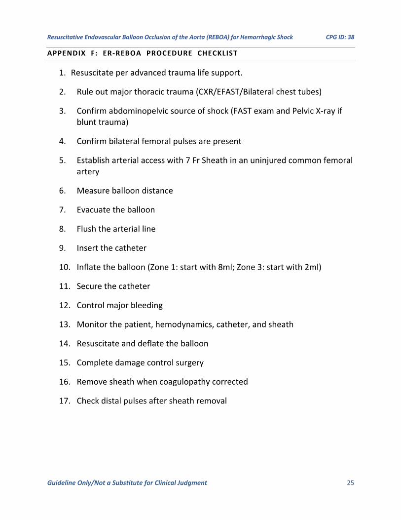
APPENDIX F: ER-REBOA PROCEDURE CHECKLIST
1. Resuscitate per advanced trauma life support.
2. Rule out major thoracic trauma (CXR/EFAST/Bilateral chest tubes)
3. Confirm abdominopelvic source of shock (FAST exam and Pelvic X-ray if blunt trauma)
4. Confirm bilateral femoral pulses are present
5. Establish arterial access with 7 Fr Sheath in an uninjured common femoral artery
6. Measure balloon distance
7. Evacuate the balloon
8. Flush the arterial line
9. Insert the catheter
10. Inflate the balloon (Zone 1: start with 8ml; Zone 3: start with 2ml)
11. Secure the catheter
12. Control major bleeding
13. Monitor the patient, hemodynamics, catheter, and sheath
14. Resuscitate and deflate the balloon
15. Complete damage control surgery
16. Remove sheath when coagulopathy corrected
17. Check distal pulses after sheath removal
Resuscitative Endovascular Balloon Occlusion of the Aorta (REBOA) for Hemorrhagic Shock CPG ID: 38
Guideline Only/Not a Substitute for Clinical Judgment 26
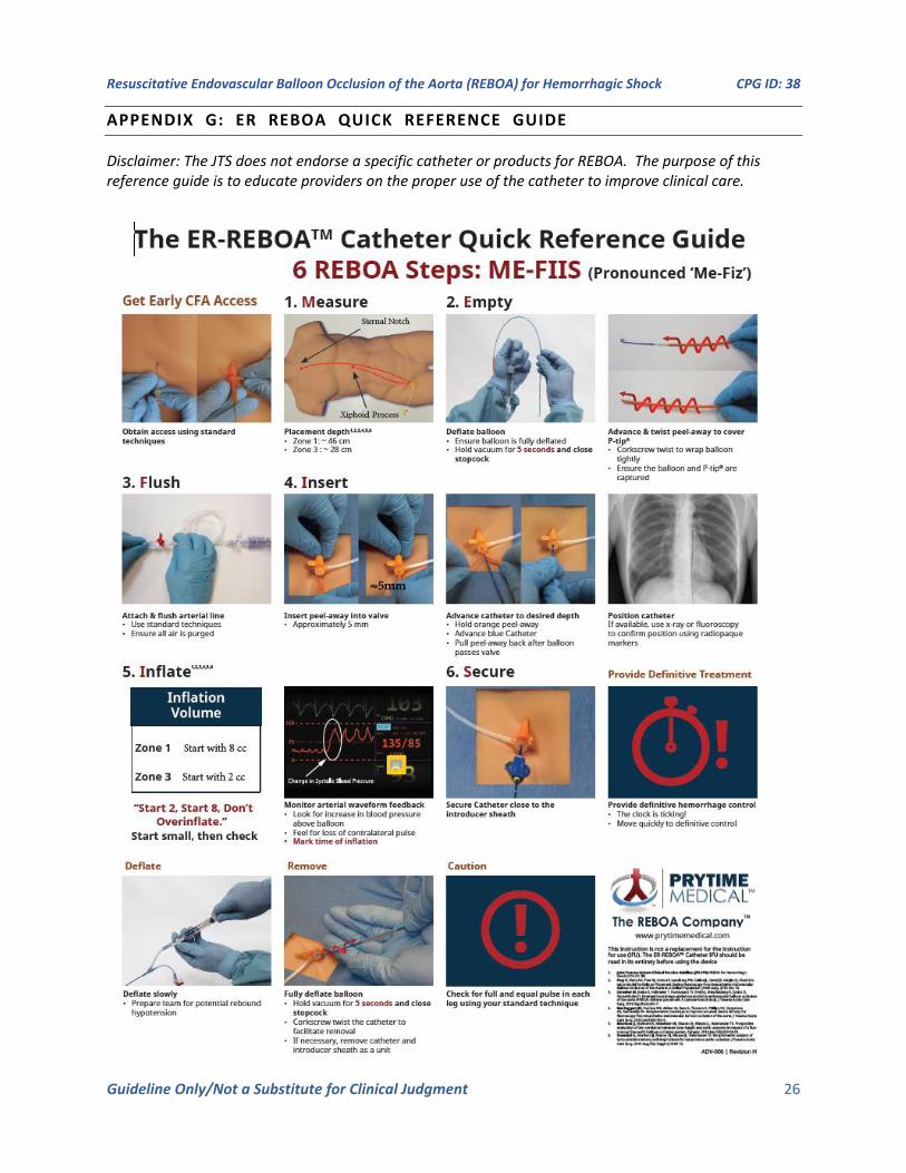
APPENDIX G: ER REBOA QUICK REFERENCE GUIDE
Disclaimer: The JTS does not endorse a specific catheter or products for REBOA. The purpose of this reference guide is to educate providers on the proper use of the catheter to improve clinical care.
Resuscitative Endovascular Balloon Occlusion of the Aorta (REBOA) for Hemorrhagic Shock CPG ID: 38
Guideline Only/Not a Substitute for Clinical Judgment 27
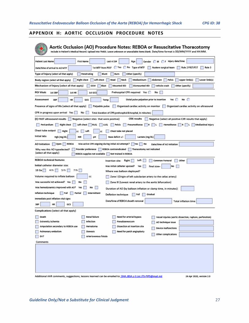
APPENDIX H: AORTIC OCCLUSION PROCEDURE NOTES
Resuscitative Endovascular Balloon Occlusion of the Aorta (REBOA) for Hemorrhagic Shock CPG ID: 38
Guideline Only/Not a Substitute for Clinical Judgment 28
APPENDIX I: ADDITIONAL INFORMATION REGARDING OFF-LABEL USES IN CPGS
PURPOSE
The purpose of this Appendix is to ensure an understanding of DoD policy and practice regarding inclusion in CPGs of “off-label” uses of U.S. Food and Drug Administration (FDA)–approved products. This applies to off-label uses with patients who are armed forces members.
BACKGROUND
Unapproved (i.e. “off-label”) uses of FDA-approved products are extremely common in American medicine and are usually not subject to any special regulations. However, under Federal law, in some circumstances, unapproved uses of approved drugs are subject to FDA regulations governing “investigational new drugs.” These circumstances include such uses as part of clinical trials, and in the military context, command required, unapproved uses. Some command requested unapproved uses may also be subject to special regulations.
ADDITIONAL INFORMATION REGARDING OFF-LABEL USES IN CPGS
The inclusion in CPGs of off-label uses is not a clinical trial, nor is it a command request or requirement. Further, it does not imply that the Military Health System requires that use by DoD health care practitioners or considers it to be the “standard of care.” Rather, the inclusion in CPGs of off-label uses is to inform the clinical judgment of the responsible health care practitioner by providing information regarding potential risks and benefits of treatment alternatives. The decision is for the clinical judgment of the responsible health care practitioner within the practitioner-patient relationship.
ADDITIONAL PROCEDURES
Balanced Discussion
Consistent with this purpose, CPG discussions of off-label uses specifically state that they are uses not approved by the FDA. Further, such discussions are balanced in the presentation of appropriate clinical study data, including any such data that suggest caution in the use of the product and specifically including any FDA-issued warnings.
Quality Assurance Monitoring
With respect to such off-label uses, DoD procedure is to maintain a regular system of quality assurance monitoring of outcomes and known potential adverse events. For this reason, the importance of accurate clinical records is underscored.
Information to Patients
Good clinical practice includes the provision of appropriate information to patients. Each CPG discussing an unusual off-label use will address the issue of information to patients. When practicable, consideration will be given to including in an appendix an appropriate information sheet for distribution to patients, whether before or after use of the product. Information to patients should address in plain language: a) that the use is not approved by the FDA; b) the reasons why a DoD health care practitioner would decide to use the product for this purpose; and c) the potential risks associated with such use.