Embed Size (px)
Citation preview

RESEARCH Open Access
Feasibility study of volumetric modulated arctherapy for the treatment of retroperitonealsarcomasCarmen Llacer-Moscardo1*, François Quenet2, David Azria1, Pascal Fenoglietto1
Abstract
Background: Radiotherapy for retroperitoneal sarcomas remains controversial and a technical challengeconsidering the threshold of contiguous critical organs tolerance. We performed consecutive RapidArc dosimetricplans in preoperative or postoperative setting.
Methods: A dosimetric study was carried out from six preoperative (group A) and four postoperative (group B)CT-scans, performed in 7 patients.Prescribed dose was 45 and 50 Gy for groups A and B, respectively. The planning target volume (PTV) was definedas the clinical target volume (CTV) plus 5 mm. The CTV encompassed the gross tumor volume (GTV) plus 10 mmor the tumoral bed. The dosimetric plans were optimized on a RapidArc Eclipse console using the progressiveresolution algorithm, PRO version 8.8. Normalization method allowed the coverage of 99% of the PTV by 95% ofthe dose.
Results: Mean PTV were 2318.5 ± 2223.9 cc [range 348-6198 cc] and 698.3 ± 216.6 cc [range 463 -933 cc] forgroups A and B, respectively. Plans were optimized for single arcs in group B and for single or two arcs in group A.The contralateral kidney volume receiving 5 Gy (V5Gy) was 21.5 ± 23.3% [range 0-55%] and 3.1 ± 2.6% [range 0-7.3%] for groups A and B, respectively. The mean dose received by 1% of the kidney (D1%) was 5.6 ± 2.4 Gy [range3.6 -7.6 Gy] for group A and 5.4 ± 0.7 Gy [range 4.3-6 Gy] for group B. The volume of small bowel excluding thePTV (small bowel-PTV) that received 40 Gy and 30 Gy (V40Gy and V30Gy) in group A were 7.5 ± 4.4% [range 5.4-14.1%] and 18.5 ± 7.1% [range 10-30.4%], respectively.In group B, small bowel-PTV V40Gy and V30Gy were 4.7 ± 3.3% [range 3.3-8%] and 21.6 ± 7.5% [range 9.4-30%]respectively. In a second step, we treated two patients in the postoperative group. Treatment time delivery withone arc was 74 seconds. No severe acute toxicity was observed.
Conclusion: RapidArc technology for retroperitoneal sarcomas showed acceptable dosimetric results inpreoperative or postoperative clinical situation. From the first treated patients, acute tolerability was good toexcellent.
BackgroundRetroperitoneal sarcoma is a rare and very heteroge-neous disease representing about 10-15% of all soft tis-sue sarcomas. Surgery is the main treatment, butmicroscopic or gross residual disease may remain afterthe procedure, compromising local control and survival[1-4]. Since local progression rather than metastatic
dissemination is the main cause of death, the role ofradiotherapy in association to surgery has been investi-gated. There are no randomized trials comparing post-operative to preoperative radiotherapy and theappropriate strategy is not well defined today.Based on the results of phase III randomized trials for
limb soft tissue sarcoma, postoperative RT has beenadopted by some teams in retroperitoneal sarcomas.Nevertheless, this approach raises the problem of thetumor underdosing due to the nearby critical organs atrisk (OAR), with the consequence to increase the risk of
* Correspondence: [email protected] of Radiation Oncology, CRLC Val D’Aurelle Paul-Lamarque,Montpellier, FranceFull list of author information is available at the end of the article
Llacer-Moscardo et al. Radiation Oncology 2010, 5:83http://www.ro-journal.com/content/5/1/83
© 2010 Llacer-Moscardo et al; licensee BioMed Central Ltd. This is an Open Access article distributed under the terms of the CreativeCommons Attribution License (http://creativecommons.org/licenses/by/2.0), which permits unrestricted use, distribution, andreproduction in any medium, provided the original work is properly cited.

local recurrence. This concern was confirmed by severalauthors who reported a high local relapse rate inside theradiotherapy field with considerable toxicity, dissuadingpostoperative radiotherapy [4-6].The single randomized trial about adjuvant radiother-
apy in resectable retroperitoneal sarcomas [7,8] com-pared a standard external beam radiotherapy (EBRT)delivering 50-55 Gy to an experimental therapy thatassociated a single dose (20 Gy) of intraoperative radio-therapy (IORT) using electrons with a low dose post-operative EBRT (35-40 Gy). With a median follow-up of8 years, the number of locoregional recurrence was sig-nificantly reduced in the experimental arm, as well asthe enteral toxicity.Preoperative radiotherapy has some theoretical advan-
tages in the management of retroperitoneal sarcomas,such as the reduction of tumor seeding during surgeryand the shift of radiosensitive viscera outside the treat-ment field [9]. Prospective trials showed the feasibility ofpreoperative radiotherapy in this context [10-12].Regarding IMRT, it is now well established that this
technique usually provides high conformity and offersimproved OAR sparing when compared to 3 D confor-mational radiotherapy. IMRT use has already beeninvestigated for the treatment of retroperitoneal sarco-mas [13-15]. Although large fields may be required forthose tumors, more particularly in preoperative setting,this does not preclude the employment of IMRT [14],but the dose inhomogeneity within the target canincrease considerably, especially in the vicinity of kid-neys. To improve dose homogeneity throughout theplanning tumor volume (PTV), multiplying fields maybe necessary, having the effect to increase the treatmenttime per fraction [16]. Some authors investigated thefeasibility of diminishing the size of fields to only irradi-ate specifically the portion of the clinical tumor volume(CTV) at the higher risk of relapse [13].In this context, the purpose of this study was to assess
dosimetric aspects using RapidArc technology for thetreatment of retroperitoneal sarcoma. The feasibility ofvolumetric arc therapy was evaluated in several dosi-metric plans obtained before or after surgery. We usedtwo different dose levels (45 and 50 Gy) adapted to theclinical situation, in order to protect normal tissuesincluding small bowel, contralateral kidney and spinalcord and achieve an excellent coverage of the whole tar-get volume. In addition, we investigated the opportunityto deliver complex radiotherapy treatments in a shorttreatment time. Finally, we directly implemented thesephysical data into the clinic.
MethodsThis dosimetric study was carried out from ten CT-scans performed in a series of seven consecutive patients
with resectable retroperitoneal sarcoma. Patients under-went either a single preoperative or postoperative CT-scan or both exams, providing six preoperative (groupA) and four postoperative cases (group B). The dosi-metric analysis was performed using RapidArctechnology.
Radiotherapy treatment planningPatients underwent CT scan-based virtual simulation(GE lightspeed RT16 Milwaukee, USA). Patients wereplaced in supine position with the arms above the head,using a special support (Sinmed, The Netherlands) andknees were placed with a knee support (Sinmed, TheNetherlands). Intravenous contrast was not used consid-ering that renal function of those patients could bealtered. 4DCT Scanner was performed to include tumormotion during breathing with 2.5 mm thick slices at2.5 mm intervals. Tumor (GTV) or tumor bed weremanually contoured on the CT images. The isocenterwas set in the middle of the GTV if preoperatively orthe tumoral bed if postoperatory, using our virtual simu-lation console (Advantagesim, GE Milwaukee, USA). Inthe case of preoperative radiotherapy (Group A), theCTV included the tumor and a margin around obtainedby a three-dimensional 10 mm expansion, except poster-iorly in regards of the vertebral body or bone, where themargin was adapted to sculpt these structures. In thepostoperative planning (Group B), the CTV was definedtogether by the surgeon and the radiation oncologist toinclude the tumor bed and all the areas at risk. Toaccount for set-up inaccuracies, a PTV was defined by athree-dimensional 5 mm expansion of CTV in all direc-tions, except close to the spinal cord where it wasreduced if necessary. The PTV margin was chosen after4DCT scanner evaluation.Kidneys or contralateral kidney were completely con-
toured. A planning organ at risk volume (PRV) of 3 cmwas added to the contralateral kidney for two reasons:first, because of the potential internal movement of thisstructure and second, to be able to define a constraintlimiting the dose delivered around the kidney. Smallbowel and spinal cord were contoured from 2 cm aboveto 2 cm below the extension of the tumor or the tumorbed corresponding to the portion of the irradiatedorgan. Liver was contoured as a whole organ when itwas close to the target volume.The dose prescribed to the PTV was 50 and 45 Gy in
25 fractions for Groups A and B, respectively.Dose constraints to the OAR were based on the avail-
able IMRT studies (Table 1). The maximal dose (Dmax)allowed for the small bowel was the prescribed dose.Dose received by 50% and 30% of the small bowel (SBD50, SB D30) should not exceed 30 Gy and 40 Gy,respectively. The maximal dose allowed to contralateral
Llacer-Moscardo et al. Radiation Oncology 2010, 5:83http://www.ro-journal.com/content/5/1/83
Page 2 of 10

kidney was 12 Gy, but we systematically tried to mini-mize global dose to the whole volume. Liver couldreceive 20 Gy to the whole volume and 40 Gy to 30% ofthe volume. The maximal tolerated dose to the spinalcord was 45 Gy.The RapidArc plan optimization was generated by the
progressive resolution optimizer (PRO) algorithm of theEclipse workstation (Varian Medical System, Palo Alto,USA) in a version 8.8 allowing multiple arcs. Single ordouble gantry rotation was used depending on the thick-ness of the volume. Each arc had systematically ancounter-clockwise rotation of 358° from 179° to 181°and opposite if two arcs. The beams shared the sameisocenter with different collimator rotation to increasethe modulation capacities of the algorithm.Plan acceptance criteria required that at least 95% of
the dose covers 99% of the PTV volume.
Evaluation toolsDose Volume Histograms (DVH) were generated toevaluate the three different plans. For PTV, the para-meters D1% and D99% were used as surrogate markersfor maximum and minimum doses. Mean dose (Dmean)was also reported.The degree of conformity of the plans was defined as
the ratio between the volume receiving at least 95% ofthe prescribed dose and the volume of the PTV (CI95%).The homogeneity index (HI) was expressed by D5% -
D95% (difference between the dose covering 5% and 95%of the PTV). For all patients DVH for OAR (bowel,bowel excluding PTV, kidneys and spinal cord) werecalculated and reported. A set of Vx values and Dmean
was therefore reported. The number of Monitor Units(MU) per fraction required for each plan and the treat-ment delivery time (from start to the end of the
irradiation), dimension of the fields and collimator angleare reported in Table 2.Following the results of the study, the two last conse-
cutive patients of group B were treated by receiving45 Gy.
Quality assurance for treated patientsWe conducted a quality control of the dosimetric plansregarding the 2 patients treated in this study. It con-sisted in a comparison between the previous dose calcu-lated by the planning system and the actual measureddose delivered by the linac. Two different methods wereused. The first one consisted of calculating the plan in acylindrical phantom of 20 cm diameter and then mea-suring the dose at the central point of this phantom byan ionisation chamber of 0.125 cc (PTW, Freiburg, Ger-many). The second method used an amorphous siliconportal imager (AS1000 Varian Medical System, Plo Alto,US) as a detection matrix with a resolution of 0.39 mm/pixel at the machine isocenter. The dose collected wascompared to a previous distribution on water using theGlaAs algorithm and the Epiqa software (Epidos, Brtai-slavia, Slovakia)[17].
ResultsTechnical data are summarized in Table 2. Our caseswere characterized by very large target volumes invol-ving wide fields until 36 cm of length. This resulted in alow number of MU delivered (380.7 and 332.3 forGroups A and B, respectively) due to a high output fac-tor of the machine. Postoperative plans were optimizedfor one arc, and some preoperative plans, specially thosewith the largest PTV, required 2 arcs. Even in thosecases, the number of MU was not significantlyincreased.
Table 1 Literature dose constrains for IMRT
Author n° cases preop postop dose to PTV (Gy) dose constraints (Gy)
contralateral kidney small bowel spinal cord liver
Tzeng [26] 16 16 0 45 ± 12,5 < 23 < 45 < 45 < 33
54 to <20 cc
Bossi [13] 18 18 0 50 < 10 to 50% V55Gy < 50% < 48 V50Gy < 33%
< 50 V30Gy < 100%
Koshy [14] 11 9 2 45-50,4 12 to 100% < 45
15 to 50% D75% 48 V40Gy < 50%
D50% 50 V30Gy < 100%
Present study 10 6 4 45-50 < 12 < 45-50 < 45
V40Gy <30% V40Gy <30%
V30Gy <50% V30Gy <40%
Llacer-Moscardo et al. Radiation Oncology 2010, 5:83http://www.ro-journal.com/content/5/1/83
Page 3 of 10

For the treated patients, the treatment time was 74 sec-onds using one arc. Quality control analysis showedacceptable results with a difference between the calcu-lated and measured doses of 1.2% and 1.7% in thecylindrical phantom. Percentage of points meeting thecriteria of 3%-3 mm for the gamma index was 98.3%and 95.7% for both patients.Figure 1 and 2 shows examples of dose distribution
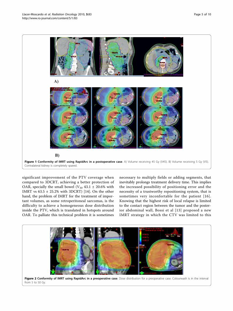
for the preoperative and postoperative cases. Dosimetricdata for PTV and OAR are recorded in table 3 andDVH results are shown in figures 3 and 4. All planswere normalized aiming to obtain V95% > 99% for thePTV. When we evaluated GTV (preoperative cases)-CTV (postoperative cases) DVH in Figure 3, we couldobserve that for all cases the dose distribution washomogeneous. Nevertheless, homogeneity (representedby D5%-D95%) inside the PTV could reach 12 and 18%for the two largest volumes (6198 and 4085 cc) of thepreoperative group.Concerning the OAR, the dose constraints initially
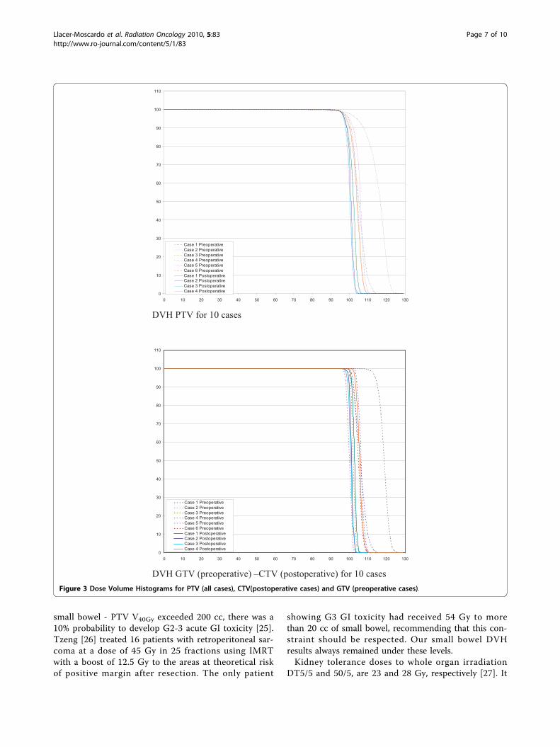
required (Table 1) were largely respected. With regardsto the bowel and bowel-PTV we presented the DVHresults for all cases, showing the important variability ofbowel volume from one case to another. V40Gy rangedfrom 66.6 cc to 962.8 cc for group A and from 18.7 ccto 695.3 cc for group B. Mean small bowel D1% was 53± 2.9 Gy, with a Dmax of 59 Gy in the portion includedin the PTV for the largest tumor. The volume of smallbowel-PTV receiving the prescribed dose was alwaysbelow 3 cc.Dose constraints were largely respected for the kidney
and the spinal cord.
Early clinical practiceTreated patients were 29 and 47 years old respectively,and were diagnosed with a liposarcoma at the histologi-cal examination. They did not present any comorbidityfactors. The treatment strategy was approved by a pluri-disciplinar committee. PTV volumes were 933 and 463cc, respectively. They underwent surgery combined toIORT at a single dose of 15 Gy delivered by an 80 mmdiameter collimator, and then received postoperativeradiotherapy at a dose of 45 Gy in 25 fractions.Acute toxicity was evaluated according to the Com-
mon Toxicology Criteria grading system (CTC V.03).Both patients showed G1 nausea-vomiting. Pain andneuropathy was G0 and no patient presented any skinreactions or weight loss.
DiscussionIMRT for retroperitoneal sarcoma has already been stu-died and implemented to clinical practice by someteams. Dose constraints criteria of those series areshown in Table 1. On the one hand, IMRT has proved aTa
ble
2Te
chnical
dataforRap
idArc
Case
Preo
perative1
Preo
perative2
Preo
perative3
Preo
perative4
Preo
perative5
Preo
perative6
Postop
erative1
Postop
erative2
Postop
erative3
Postop
erative4
Dose[Gy]
5050
5050
5050
4545
4545
VolumePTV[cc]
6198
384
2535
348
4085
361
895
463
502
933
Leng
htPTV[cm]
2414
229,3
319,41
2921
1624
Num
berof
arcs
21
21
21
11
11
MU
177+
190
337
233+
217
433
158+
132
372
295
322
374
338
Energy
[MV]
1818
66
1818
1818
1818
Collim
ator
Ang
le30/330
3045/315
4535/335
45345
3045
30
Xjaw
[cm]
23/23,4
12,7
22,4/23
12,2
26,4/27,5
11,8
1515
17,9
23,9
Yjaw
[cm]
33,4/33,4
14,7
23,5/24,4
1236,5/36,5
11,8
3022,5
18,5
29,5
Llacer-Moscardo et al. Radiation Oncology 2010, 5:83http://www.ro-journal.com/content/5/1/83
Page 4 of 10
significant improvement of the PTV coverage whencompared to 3DCRT, achieving a better protection ofOAR, specially the small bowel (V30 43.1 ± 20.6% withIMRT vs 63.5 ± 25.2% with 3DCRT) [14]. On the otherhand, the problem of IMRT for the treatment of impor-tant volumes, as some retroperitoneal sarcomas, is thedifficulty to achieve a homogeneous dose distributioninside the PTV, which is translated in hotspots aroundOAR. To palliate this technical problem it is sometimes
necessary to multiply fields or adding segments, thatinevitably prolongs treatment delivery time. This impliesthe increased possibility of positioning error and thenecessity of a trustworthy repositioning system, that issometimes very inconfortable for the patient [16].Knowing that the highest risk of local relapse is limitedto the contact region between the tumor and the poster-ior abdominal wall, Bossi et al [13] proposed a newIMRT strategy in which the CTV was limited to this
A)
B) Figure 1 Conformity of IMRT using RapidArc in a postoperative case. A) Volume receiving 45 Gy (V45). B) Volume receiving 5 Gy (V5).Contralateral kidney is completely spared.
Figure 2 Conformity of IMRT using RapidArc in a preoperative case. Dose distribution for a preoperative case. Colourwash is in the intervalfrom 5 to 50 Gy.
Llacer-Moscardo et al. Radiation Oncology 2010, 5:83http://www.ro-journal.com/content/5/1/83
Page 5 of 10

area, reducing the volume of the target in an attempt todecrease toxicity. IMRT plans were compared to3DCRT and showed a significant better sparing withIMRT of the contralateral kidney. No significant advan-tage for small bowel was observed with IMRT in theirstudy where they defined the CTV as a part of thewhole GTV. Additionally, the presence of the tumorshifted small bowel outside of the PTV.Many authors reported for other tumor sites dosi-
metric plans at least similar for RapidArc when com-pared to IMRT with a static gantry position [18-23].RapidArc was implemented since 2008 in our institutionin a daily practice for several localisations. Therefore wedecided to evaluate this innovative technique for thetreatment of retroperitoneal sarcomas.We found in the frame of our dosimetric study better
DVH results than those expected at the initial planningtime taking into account that we studied very largevolumes (Table 3). Our choice regarding the normaliza-tion method was specific for this localisation. We initi-ally decided to cover 99% of the PTV by 95% of the
prescribed dose. This resulted in a better dose coveragein the edge of the volume, but compromised homogene-ity, particularly for the largest preoperative case, wherewe obtained a maximal dose of 124% inside the PTV.This hotspot wouldn’t have been observed if we hadcovered 95% of the volume by 95% of the dose. Never-theless, we may wonder whether the presence of thesehotspots inside the PTV is really problematic knowingthat this lesion will be removed.Regarding the organs at risk, small bowel DVH
showed that V30Gy and V40Gy results were better thaninitially required for both groups. Hotspots in the smallbowel were systematically in the portion included in thePTV for the biggest case. The portion of bowel - PTVirradiated above the prescribed dose was always verylimited (< 3cc).To allow reproducible correlation between the volume
of small bowel receiving a dose range and toxicity, DVHdata were expressed in cc. Some authors showed that aV30Gy > 450 cc was correlated to a significant higheracute gastro-intestinal (GI) toxicity [24] and that when
Table 3 Dosimetric results for PTV and OAR
Preoperative Postoperative
MEAN SD MAX MIN MEAN SD MAX MIN
PTV
Volume [cc] 2318,5 2223,9 6198,0 348,0 698,3 216,6 933,0 463,0
D1% [%] 111,2 6,7 124,0 103,4 105,3 2,4 109,1 103,1
D95% [%] 99,3 2,3 103,9 96,8 97,5 0,4 98,2 97,3
D5% [Gy] 109,4 6,2 121,8 102,5 104,5 2,0 107,6 102,6
V107% [%] 25,6 30,9 90,0 0,0 2,1 3,6 8,3 0,0
V95% [%] 99,0 0,0 99,0 99,0 99,0 0,0 99,0 99,0
D5%-D95% [%] 10,1 4,1 17,9 5,7 6,9 1,7 9,4 5,3
CI 95% 1,1 0,1 1,2 1,1 1,2 0,0 1,3 1,2
Spinal Cord
D1% [Gy] 28,1 12,6 40,0 1,9 32,6 4,4 39,2 28,1
Dmax [Gy] 32,0 13,6 44,0 3,1 35,0 4,8 41,0 30,0
Kidney
Volume [cc] 149,9 60,1 173,8 105,2 171,6 31,6 209,6 139,0
V5 Gy [%] 21,5 23,3 55,0 0,0 3,1 2,6 7,3 0,0
Dmean [Gy] 3,5 1,9 5,2 1,4 2,9 0,5 3,8 2,5
D1% [Gy] 5,6 2,4 7,6 3,6 5,4 0,7 6,0 4,3
Bowel
Volume [cc] 1421,3 729,7 2720,0 628,8 1494,9 533,7 2406,0 1105,0
V30Gy [%] 33,2 12,0 50,9 19,0 30,5 11,5 43,0 11,9
V40Gy [%] 22,4 9,1 35,4 10,6 15,6 9,6 28,9 1,7
D1% [Gy] 53,4 2,9 59,6 50,7 46,9 0,8 47,7 45,7
V Prescription dose [%] 12,2 9,2 28,0 2,8 8,7 7,7 21,0 0,0
Bowel-PTV
Volume [cc] 1183,6 645,3 2392,0 481,4 1343,7 613,2 2205,0 825,2
V30Gy [%] 18,5 7,1 30,5 10,1 21,6 7,5 30,0 9,4
V40Gy [%] 7,5 4,4 14,1 1,3 4,7 3,3 8,0 0,0
V Prescription dose [%] 0,6 0,9 2,2 0,0 0,7 1,3 2,9 0,0
Llacer-Moscardo et al. Radiation Oncology 2010, 5:83http://www.ro-journal.com/content/5/1/83
Page 6 of 10
small bowel - PTV V40Gy exceeded 200 cc, there was a10% probability to develop G2-3 acute GI toxicity [25].Tzeng [26] treated 16 patients with retroperitoneal sar-coma at a dose of 45 Gy in 25 fractions using IMRTwith a boost of 12.5 Gy to the areas at theoretical riskof positive margin after resection. The only patient
showing G3 GI toxicity had received 54 Gy to morethan 20 cc of small bowel, recommending that this con-straint should be respected. Our small bowel DVHresults always remained under these levels.Kidney tolerance doses to whole organ irradiation
DT5/5 and 50/5, are 23 and 28 Gy, respectively [27]. It
0
10
20
30
40
50
60
70
80
90
100
110
0 10 20 30 40 50 60 70 80 90 100 110 120 130
Case 1 PreoperativeCase 2 PreoperativeCase 3 PreoperativeCase 4 PreoperativeCase 5 PreoperativeCase 6 PreoperativeCase 1 PostoperativeCase 2 PostoperativeCase 3 PostoperativeCase 4 Postoperative
DVH PTV for 10 cases
0
10
20
30
40
50
60
70
80
90
100
110
0 10 20 30 40 50 60 70 80 90 100 110 120 130
Case 1 PreoperativeCase 2 PreoperativeCase 3 PreoperativeCase 4 PreoperativeCase 5 PreoperativeCase 6 PreoperativeCase 1 PostoperativeCase 2 PostoperativeCase 3 PostoperativeCase 4 Postoperative
DVH GTV (preoperative) –CTV (postoperative) for 10 cases Figure 3 Dose Volume Histograms for PTV (all cases), CTV(postoperative cases) and GTV (preoperative cases).
Llacer-Moscardo et al. Radiation Oncology 2010, 5:83http://www.ro-journal.com/content/5/1/83
Page 7 of 10

has been reported that in the absence of concomitantchemotherapy or latent nephropathy, doses under 15 Gyare not likely to provoke radiation-induced nephropathy[28]. Another important concept is that kidney consistsof multiple independent functional structures very sensi-tive to radiation. For this reason, despite the problem oftotal dose, there is the problem of quantity of irradiatedvolume even at low doses. May et al [29] showed thatthe percentage of bilateral renal volume receiving atleast 10 Gy and the mean kidney dose were significantpredictors of subsequent G2 renal complications (p =0.017 and p = 0.0095 respectively).In our study respectively mean and maximal doses
received by the contralateral kidney were 3.45 Gy and7.6 Gy for the preoperative and 2.94 Gy and 6 Gy for
the postoperative plans, which is much lower thanaccepted doses. One could be worried about the respira-tion-induced motion of the kidneys making uncertainthe doses received. Some authors studied this phenom-enon showing a maximal movement of kidneys incephalo-caudal direction, with displacements varyingaround 16 ± 8 mm [30,31] justifying the PRV of 3 cmthat we created around this structure to allow respect ofdose constraints. Furthermore, as those patients will bemonorenal in most of the cases, we recommend the pre-scription of a pre-treatment renal scintigraphy to assesthe functionality of the remaining kidney.Concerning the dose for retroperitoneal sarcomas,
limitation of dose prescription was assessed by the toler-ance of the organs at risk. Our results open the question
0
10
20
30
40
50
60
70
80
90
100
0 1 2 3 4 5 6 7 8 9 10
mean preoperativemean postoperative
0
200
400
600
800
1000
1200
1400
1600
0 10 20 30 40 50 60 70
Bowel Mean preoperative
Bowel Mean postoperative
Bowel - PTV Mean preoperative
Bowel - PTV pean postoperative
A B
0
250
500
750
1000
1250
1500
1750
2000
2250
2500
2750
3000
0 5 10 15 20 25 30 35 40 45 50 55 60 65
Case 1 PreoperativeCase 2 PreoperativeCase 3 PreoperativeCase 4 PreoperativeCase 5 PreoperativeCase 6 PreoperativeCase 1 PostoperativeCase 2 PostoperativeCase 3 PostoperativeCase 4 Postoperative
0
250
500
750
1000
1250
1500
1750
2000
2250
2500
2750
0 5 10 15 20 25 30 35 40 45 50 55 60 65
Case 1 PreoperativeCase 2 PreoperativeCase 3 PreoperativeCase 4 PreoperativeCase 5 PreoperativeCase 6 PreoperativeCase 1 PostoperativeCase 2 PostoperativeCase 3 PostoperativeCase 4 Postoperative
C D Figure 4 Dose Volume Histograms (DVH) for OAR. A) mean DVH for contralateral kidney. B) mean DVH for small bowel and small bowel -PTV. C) Small bowel DVH results for all cases. D) Small bowel-PTV DVH results for all cases.
Llacer-Moscardo et al. Radiation Oncology 2010, 5:83http://www.ro-journal.com/content/5/1/83
Page 8 of 10

of dose escalation and will be the object of furtherstudies.Another important point is the reduction achieved in
delivery time, which is a major advantage of RapidArc.Even if static gantry IMRT allows acceptable dose distri-bution, the average fraction time is about 20 minutes[13,20]. Shorter treatment time will reduce the likeli-hood of intrafraction baseline shifts in PTV and organsat risk position. Taking into account that those patientsare painful in most of the cases because of the psoasinvasion and have big difficulties to stay laying on theaccelerator table, RapidArc technology offers a solutionimproving treatment comfort and decreasing the possi-bility of set-up errors.Even if the available evidence from retrospective stu-
dies and prospective non randomized trials strongly sug-gests that conventional preoperative radiation is bettertolerated, we treated using RapidArc technology twopatients of the postoperative group with excellent clini-cal tolerance.
ConclusionsRapidArc for retroperitoneal sarcomas achieved accepta-ble dosimetric results in preoperative or postoperativesetting, even for large volumes. The two first treatedpatients presented a good tolerability. Currently, we arecontinuing to treat patients with this technique offeringa rapid and safe procedure. Longer follow-up is war-ranted to assess long-term toxicity and local control.
Author details1Department of Radiation Oncology, CRLC Val D’Aurelle Paul-Lamarque,Montpellier, France. 2Department of Surgical Oncology, CRLC Val D’AurellePaul-Lamarque, Montpellier, France.
Authors’ contributionsCLLM, PF and FQ designed and coordinated the study. Patient accrual andclinical data collection was done by CLLM and FQ. Data analysis, physicsdata and treatment planning data collection was done by PF and CLLM.CLLM prepared the manuscript. DA and PF revised critically for importantintellectual content. All authors read and approved the final manuscript.
Competing interestsThe authors declare that they have no competing interests.
Received: 9 July 2010 Accepted: 20 September 2010Published: 20 September 2010
References1. Karakousis CP, Velez AF, Gerstenbluth R, Driscoll DL: Resectability and
survival in retroperitoneal sarcomas. Ann Surg Oncol 1996, 3(2):150-8.2. Lewis JJ, Leung D, Woodruff JM, Brennan MF: Retroperitoneal soft-tissue
sarcoma: analysis of 500 patients treated and followed at a singleinstitution. Ann Surg 1998, 228(3):355-65.
3. Raut CP, Pisters PW: Retroperitoneal sarcomas: Combined modalitytreatment approaches. J Surg Oncol 2006, 94(1):81-7.
4. Catton CN, O’Sullivan B, Kotwall C, Cummings B, Hao Y, Fornasier V:Outcome and prognosis in retroperitoneal soft tissue sarcoma. Int J RadOncol Biol Phys 1994, 29(5):1005-10.
5. Gilbeau L, Kantor G, Lagarde P, Thomas L, Kind M, Richaud P, Coindre JM,Bonichon F, Bui BN: Surgical resection and radiotherapy for primaryretroperitoneal soft tissue sarcoma. Radiother and Oncol 2002,65(3):137-43.
6. Stoeckle E, Coindre JM, Bonvalot S, Kantor G, Terrier P, Bonichon F, NguyenBui B, French Federation of Cancer Centers Sarcoma Group: Prognosticfactors in retroperitoneal sarcoma: a multivariate analysis of a series of165 patients of the French Cancer Center Federation Sarcoma Group.Cancer 2001, 92(2):359-68.
7. kinsella TJ, Syndelar WF, Lack E, Glatstein E, Rosenberg SA: Preliminaryresults of a randomized study of adjuvant radiation therapy inresectable adult retroperitoneal soft tissue sarcomas. J Clin Oncol 1988,6(1):18-25.
8. Sindelar WF, Kinsella TJ, Chen PW, DeLaney TF, Tepper JE, Rosenberg SA,Glatstein E: Intraoperative radiotherapy in retroperitoneal sarcomas. Finalresults of a prospective, randomized, clinical trial. Arch Surg 1993,128(4):402-10.
9. Caudle AS, Tepper JE, Calvo BF, Meyers MO, Goyal LK, Cance WG, Kim HJ:Complications associated with neoadjuvant radiotherapy in themultidisciplinary treatment of retroperitoneal sarcomas. Ann Surg Oncol2007, 14(2):577-82.
10. Jones JJ, Catton CN, O’Sullivan B, Couture J, Heisler RL, Kandle RA,Swallow CJ: Initial results of a trial of preoperative external-beamradiation therapy and postoperative brachytherapy for retroperitonealsarcoma. Ann Surg Oncol 2002, 9(4):346-54.
11. Gieschen Hl, Spiro IJ, Suit HD, Ott MJ, Rattner DW, Ancukiewicz M,Willet CG: Long-term results of intraoperative electron beamradiotherapy for primary and recurrent retroperitoneal soft tissuesarcoma. Int J Radiat Oncol Biol Phys 2001, 50(1):127-31.
12. Petersen IA, Haddock MG, Donohue JH, Nagorney DM, Grill JP, Sargent DJ,Gunderson LL: Use of intraoperative electron beam radiotherapy in themanagement of retroperitoneal soft tissue sarcoma. Int J Radiat OncolBiol Phys 2002, 52(2):469-75.
13. Bossi A, De Wever I, Van Limbergen E, Vanstraelen B: Intensity modulatedradiation-therapy for preoperative posterior wall irradiation ofretroperitoneal liposarcomas. Int J Radiat Oncol Biol Phys 2007,67(1):164-70, Erratum in: Int J Radiat Oncol Biol Phys. 2007,68(1):317.Dosage error in article text.
14. Koshy M, Landry JC, Lawson JD, Staley CA, Esiashvili N, Howell R,Ghavidel S, Davis LW: Intensity modulated radiation therapy forretroperitoneal sarcoma: a case for dose escalation and organ at risktoxicity reduction. Sarcoma 2003, 7(3-4):137-48.
15. Musat E, Kantor G, Caron J, Lagarde P, Laharie H, Stoeckle E, Angles J,Gilbeau L, Bui BN: Comparison of intensity-modulated postoperativeradiotherapy with conventional postoperative conformal radiotherapyfor retroperitoneal sarcoma. Cancer Radiother 2004, 8(4):255-61.
16. Hong L, Alektiar K, Chui C, LoSasso T, Hunt M, Spirou S, Yang J, Amols H,Ling C, Fuks Z, Leibel S: IMRT of large fields: whole-abdomen irradiation.Int j Radiat Oncol Biol Phys 2002, 54(1):278-89.
17. Nicolini G, Vanetti E, Clivio A, Fogliata A, Korreman S, Bocanek J, Cozzi L:The GLAas algorithm for portal dosimetry and quality assurance ofRapidArc and intensity modulated rotational therapy. Radiation Oncology2008, 3:24.
18. Cozzi L, Dinshaw KA, Shrivastava SK, Mahantshetty U, Engineer R,Deshpande DD, Jamema SV, Vanetti E, Clivio A, Nicolini G, Foliata A: Atreatment planning study comparing volumetric arc modulation withRapidArc and fixed field IMRT for cervix uteri radiotherapy. RadiotherOncol 2008, 89(2):180-91.
19. Palma D, Vollans E, James K, Nakano S, Moiseenko V, Shaffer R, McKenzie M,Morris J, Otto K: Volumetric modulated arc therapy for delivery ofprostate radiotherapy: comparison with intensity-modulatedradiotherapy and three-dimensional conformal radiotherapy. Int J RadiatOncol Biol Phys 2008, 72(4):996-1001.
20. Alexander AS, Wells D, Berrang T, Parsons C, Mydin A, Shaffer R, Wong F,Sayers D, Otto K: Volumetric Arc Therapy (VMAT) reduces treatment timecompared to conventional IMRT (cIMRT) while mantaining similar planquality in whole pelvic gynecologic radiotherapy. Int J Radiat Oncol BiolPhys 2008, 72(1):S366.
21. Fogliata A, Clivio A, Nicolini G, Vanetti E, Cozzi L: Intensity modulation withphotons for benign intracranial tumours: a planning comparison of
Llacer-Moscardo et al. Radiation Oncology 2010, 5:83http://www.ro-journal.com/content/5/1/83
Page 9 of 10

volumetric single arc, helical arc and fixed gantry techniques. RadiotherOncol 2008, 89(3):254-62.
22. Clivio A, Fogliata A, Francetti-Pellanda A, Nicolini G, Vanetti E,Wyttenbach R, Cozzi L: Volumetric-modulated arc radiotherapy forcarcinomas of the anal canal: A treatment planning comparison withfixed field IMRT. Radiother Oncol 2009, 92(1):118-24.
23. Vanetti E, Clivio A, Nicolini G, Fogliata A, Ghosh-Laskar S, Agarwal JP,Upreti RR, Budrukkar A, Murthy V, Deshpande DD, Shrivastava SK,Dinshaw KA, Cozzi L: Volumetric modulated arc radiotherapy forcarcinomas of the oro-pharynx and larynx: a treatment planningcomparison with fixed field gantry. Radiother Oncol 2009, 92(1):111-7.
24. Devisetty K, Mell LK, Salama JK, Schomas DA, Miller RC, Jani AB, Roeske JC,Aydogan B, Chumura SJ: A multi-institutional acute gastrointestinaltoxicity analysis of anal cancer patients treated with concurrentintensity-modulated radiation therapy (IMRT) and chemotherapy.Radiother Oncol 2009, 93:298-301.
25. Fiorino C, Alongi F, Perna L, Broggi S, Cattaneo GM, Cozzarini C, DiMuzio N, Fazio F, Calandrino R: Dose-volume relationships for acutebowel toxicity in patients treated with pelvic nodal irradiation forprostate cancer. Int J Radiat Oncol Biol Phys 2009, 75(1):29-35.
26. Tzeng CW, Fiveash JB, Popple RA, Arnoletti JP, Russo SM, Urist MM,Bland KI, Heslin MJ: Preoperative radiation therapy with selective doseescalation to the margin at risk for retroperitoneal sarcoma. Cancer 2006,107(2):371-9.
27. Emami B, Lyman J, Brown A, Coia L, Goitein M, Munzenrider JE, Shank B,Solin LJ, Wesson M: Tolerance of normal tissue to therapeutic irradiation.Int J Radiat Oncol Biol Phys 1991, 21(1):109-122.
28. Cassady JR: Clinical radiation nephropathy. Int J Radiat Oncol Biol Phys1995, 31(5):1249-56.
29. May KS, Khushalani NI, Chandrasekhar R, Wilding GE, Lyer RV, Ma WW,Flaherty L, Russo RC, Fakih M, Kuvshinoff BW, Gibbs JF, Javle MM, Yang Gy:Analysis of clinical and dosimetric factors assocated with change inrenal function in patients with gastrointestinal malignances afterchemoradiation to the abdomen. Int J Radiat Oncol Biol Phys 2010,76(4):1193-8.
30. Ahmad NR, Huq MS, Corn BW: Respiration-induced motion of the kidneysin whole abdominal radiotherapy. Radiother Oncol 1997, 42(1):87-90.
31. Bussels B, Goethals L, Feron M, Bielen D, Dymarkowski S, Suetens P,Haustermans K: Respiration-induced movement of the upper abdominalorgans: a pitfall for the tree-dimensional conformal radiation treatmentof pancreatic cancer. Radiother Oncol 2003, 68(1):69-74.
doi:10.1186/1748-717X-5-83Cite this article as: Llacer-Moscardo et al.: Feasibility study of volumetricmodulated arc therapy for the treatment of retroperitoneal sarcomas.Radiation Oncology 2010 5:83.
Submit your next manuscript to BioMed Centraland take full advantage of:
• Convenient online submission
• Thorough peer review
• No space constraints or color figure charges
• Immediate publication on acceptance
• Inclusion in PubMed, CAS, Scopus and Google Scholar
• Research which is freely available for redistribution
Submit your manuscript at www.biomedcentral.com/submit
Llacer-Moscardo et al. Radiation Oncology 2010, 5:83http://www.ro-journal.com/content/5/1/83
Page 10 of 10