Embed Size (px)
Citation preview
Research ArticleChondrogenic Potency Analyses of Donor-MatchedChondrocytes and Mesenchymal Stem Cells Derived from BoneMarrow, Infrapatellar Fat Pad, and Subcutaneous Fat
John Garcia, Claire Mennan, Helen S. McCarthy, Sally Roberts,James B. Richardson, and Karina T. Wright
ISTM, Keele University, Robert Jones and Agnes Hunt Orthopaedic Hospital NHS Foundation Trust, Oswestry,Shropshire SY10 7AG, UK
Correspondence should be addressed to Karina T. Wright; [email protected]
Received 22 July 2016; Accepted 4 September 2016
Academic Editor: Wesley N. Sivak
Copyright © 2016 John Garcia et al. This is an open access article distributed under the Creative Commons Attribution License,which permits unrestricted use, distribution, and reproduction in any medium, provided the original work is properly cited.
Autologous chondrocyte implantation (ACI) is a cell-based therapy that has been used clinically for over 20 years to treat cartilageinjuries more efficiently in order to negate or delay the need for joint replacement surgery. In this time, very little has changedin the ACI procedure, but now many centres are considering or using alternative cell sources for cartilage repair, in particularmesenchymal stem cells (MSCs). In this study, we have tested the chondrogenic potential of donor-matched MSCs derived frombone marrow (BM), infrapatellar fat pad (FP), and subcutaneous fat (SCF), compared to chondrocytes. We have confirmed thatthere is a chondrogenic potency hierarchy ranging across these cell types, with the most potent being chondrocytes, followed byFP-MSCs, BM-MSCs, and lastly SCF-MSCs. We have also examined gene expression and surface marker profiles in a predictivemodel to identify cells with enhanced chondrogenic potential. In doing so, we have shown that Sox-9, Alk-1, and Coll X expressions,as well as immunopositivity for CD49c and CD39, have predictive value for all of the cell types tested in indicating chondrogenicpotency. The findings from this study have significant clinical implications for the refinement and development of novel cell-basedcartilage repair strategies.
1. Introduction
Autologous chondrocyte implantation (ACI) for the treat-ment of focal chondral and/or osteochondral lesions haschanged very little since its inception [1], but there remainsscope for improvement. While we and others have reported asignificant level of improved joint function and a reductionin pain following treatment with ACI [2–4], disadvantagessuch as cost, potential donor-sitemorbidity, and the quality ofrepair tissue formed remain. Althoughwe have shown donor-site morbidity to be minimal [5], there is also the added riskof chondrocyte dedifferentiation during culture expansion[6, 7], the extent of which is likely to impact on the abilityof the chondrocytes to redifferentiate upon implantation intothe defect site.
Mesenchymal stem cells (MSCs) isolated from the bonemarrow (BM-MSCs) have been used in several clinical trials
as an alternative cell source for use in cell therapies totreat cartilage injuries and osteoarthritis [8–10]. The processof acquiring a sample of bone marrow, however, resultsin an additional, painful procedure for the patient. Theinfrapatellar fat pad (FP) is often routinely removed anddisposed of as surgical waste during arthroscopy or open kneesurgery and may provide an accessible alternative source ofMSCs (FP-MSCs) with demonstrable chondrogenic capacityin vitro [11, 12]. Another accessible source of MSCs, althoughstudied to a lesser extent for their chondrogenic propensity,is MSCs derived from subcutaneous fat (SCF-MSCs) [13, 14].The ability to utilise these tissues for the treatment of cartilageinjuries has the potential to improve the way we currentlytreat patients.
An important factor to consider when comparing andcontrasting the properties of different cell types is the “donorimpact” as donor demographics, such as age and gender, are
Hindawi Publishing CorporationStem Cells InternationalVolume 2016, Article ID 6969726, 11 pageshttp://dx.doi.org/10.1155/2016/6969726
2 Stem Cells International
Table 1: Donor demographics.
ID Gender Age (years) PathologyDonor 1 Male 71 OA with extensive joint degenerationDonor 2 Female 67 OA with loss of joint spaceDonor 3 Female 75 Patellofemoral OA and loss of joint space in medial compartmentDonor 4 Female 81 OADonor 5 Male 74 OA with joint stiffness
factors which are known to affect cell proliferation and differ-entiation capacity [15–17].The impact of donor is particularlycritical for autologous treatment regimes and in decidingwhether such a cell-based therapy represents the appropriatetreatment option for an individual patient. Unravelling theimpact of tissue and donor source and developing tools topredict the efficacy of cell-based treatments will likely resultin the refinement of existing treatments and may providevaluable additional information for consideration duringthe decision making process of cost benefit versus clinicalefficacy.
In this study, we have examined 4 different cell types(chondrocytes, BM-MSCs, FP-MSCs, and SCF-MSCs) andtested the chondrogenic potential of each population ofcells. This study compares donor-matched cell types and wasdesigned to establish the impact of tissue source and donoron chondrogenic differentiation capacity and to continue theprocess of establishing a marker panel indicative of chon-drogenic potency and likely clinical success. Such marker(s)could be screened for and used in the selection of a partic-ular cell type and/or subpopulation of cells with enhancedchondrogenic capability prior to treatment. We envisage thattaken together this information could significantly improvethe success of cell-based therapies for cartilage injuries andperhaps even lead to the development of novel individualisedtreatments for cartilage repair.
2. Materials and Methods
2.1. Patients. All samples were obtained after patients hadprovided written informed consent; favourable ethical ap-proval was given by the National Research Ethics Ser-vice (11/NW/0875) and all experiments were performed inaccordance with relevant guidelines and regulations. Donor-matched samples of cartilage, BM, FP, and SCFwere obtainedfrom 5 patients (2 males and 3 females, ages 67–81 years)undergoing total knee replacement (TKR) surgery (Table 1).
2.2. Isolation of Chondrocytes. Macroscopically normal artic-ular cartilage was excised from the femoral condyles ofpatients undergoing TKR. Cartilage tissue was weighed,minced into small pieces with a sterile scalpel, and digestedin collagenase type II (250 IU/mg dry weight, Worthington,New Jersey, USA) for 16 hours at 37∘C.The resulting suspen-sionwas passed through a 40𝜇mcell strainer and centrifuged(350×g for 10 minutes) to produce a cell pellet that wasresuspended in Dulbecco’s Modified Eagle’s Medium/F-12
(DMEM/F-12) with 1% (v/v) penicillin/streptomycin (P/S)and 10% (v/v) foetal calf serum (FCS, all Life Technolo-gies, Paisley, UK), hereafter referred to as complete culturemedium, at a seeding density of 5 × 10³/cm2.
2.3. Isolation of MSCs from Bone Marrow. Bone marrow as-pirates and bone chips were obtained from the tibial plateauof patients undergoing TKR. Bone marrow was first dilutedwith an equal volume of phosphate buffered saline (PBS, LifeTechnologies) then split between two 50mL tubes, layeredonto 10mL of Lymphoprep� (Alere Technologies AS, Oslo,Norway), and centrifuged (900×g for 20 minutes). The buffycoat, containing mononuclear cells, was aspirated and addedto complete culture medium and centrifuged (750×g for10 minutes). The resulting cell pellet was resuspended incomplete culture medium and BM-MSCs were seeded at adensity of 20 million cells per 75 cm2 tissue culture flask.Cells were left to adhere for 24 hours before the media werechanged and the nonadherent cells were removed. Bone chipswere placed in a 175 cm2 culture flask with 30mL of completemedia for 7 days to allow the plastic adherent cells to migrateout of the bone chips.
2.4. Isolation of MSCs from Adipose Tissues. Human FP andSCF tissue samples were obtained from patients and pro-cessed within 2 hours of receipt from the operating theatre.The FP was dissected from the innermost zone, to avoidcontamination with synovium derived cells as describedpreviously [12]. Dissected FP and SCF tissues were washedin PBS, minced, and digested with 1mg/mL collagenase typeI (≥125 digesting units/mg, Sigma-Aldrich, Poole, UK) inserum-free media for 1 h at 37∘C. The resulting cells werestrained through a 40𝜇m nylon cell strainer and centrifuged(350×g for 10min). Cells were then seeded at a density of5 × 10
3/cm2 in complete culture medium. All cultures weremaintained in a humidified incubator at 37∘C and 5% CO
2.
2.5. RNA Extraction and Quantitative Real-Time PolymeraseChain Reaction (qRT-PCR). After trypsinisation at passages3-4, 2 × 10⁵ monolayer cells were centrifuged (500×gfor 5 minutes), frozen in liquid N
2, and stored at −80∘C
briefly prior to extraction. Cells were thawed on ice andmessenger RNA (mRNA) was extracted using an RNeasy�extraction kit (Qiagen, Hilden, Germany) according to themanufacturer’s recommendations. RNA was converted tocDNA using a High-Capacity cDNA Reverse TranscriptaseKit� (Applied Biosystems, Warrington, UK) according to the
Stem Cells International 3
manufacturer’s instructions. qRT-PCR was used to evaluatethe expression of specific genes, indicative of chondrogenicpotency or hypertrophy [18]. RT-qPCR analysis was per-formed on the Quant Studio 3 Real-Time Quantitative PCRSystem (Applied Biosystems) using SYBR green QuantiTectprimer assays for the chondrogenic genes Sox-9, collagentype II (Coll II), aggrecan (ACAN), frizzled-related protein(FRZB), and the following genes indicative of hypertrophy:activin receptor-like kinase 1 (Alk-1) and collagen type X(Coll X). Peptidylprolyl isomerase A (PPIA) and TATA-binding protein (TBP) were used as reference genes (Qiagen).The relative expression of each genewas determined using thecomparative CT method [19].
2.6. Flow Cytometry. Flow cytometry was used to assess theimmunoprofile of chondrocytes andMSCs prior to chondro-genic differentiation. Cells at passages 3-4 were harvested,pelleted, and resuspended in 2%bovine serumalbumin (BSA,Sigma-Aldrich) in PBS. FC receptors were blocked for 1 h at4∘C using 10% (v/v) human IgG (Grifols, Barcelona, Spain)in 2% (v/v) BSA in PBS (immunobuffer). The cells werethen washed with immunobuffer and centrifuged (350×gfor 8minutes). Cells were stained for 30minutes at 4∘Cwith fluorochrome conjugated antibodies against cell surfacemarkers indicative of MSC according to the InternationalSociety for Cellular Therapy (ISCT) [20]. Markers probedfor were CD90-phycoerythrin (PE) (clone 5E10), CD105-allophycocyanin (APC) (clone 266), and CD73-brilliant vio-let 421 (BV421) (clone AD2), CD19-BV421 (clone HIB19),CD34-APC (clone 581), CD45-PE (clone HI30), HLA-DR-APC (clone TU36), and CD14-PerCP-Cy5.5 (clone M𝜙P9).CD markers which have been reported as putative chondro-genic potency markers [21–28] were also probed for; theseincluded CD49c-PE (clone C3 II.1), CD166-BV421 (clone3A6), CD39-APC (clone TU66), CD44-peridinin chloro-phyll protein-cyanine 5.5 (PerCp-Cy5.5) (clone G44-26), andCD271-BV421 (clone C40-1457) (BD Biosciences). Appro-priate isotype-matched IgG controls were used throughout(BD Biosciences). Data from at least 10,000 stained cells ispresented which was analysed using a FACSCanto II flowcytometer (BDBiosciences) and BD FACSDiva v.7.0 software.
2.7. Chondrogenic Differentiation. The chondrogenic poten-tial of all cultured cell populations was assessed at passage4 using an established 3D pellet culture system [12, 29].Briefly, 2 × 10⁵ cells were centrifuged to produce a cell pelletwhich was maintained in DMEM F12, FBS (10% v/v), P/S(1%) ITS (1%, v/v), ascorbic acid (0.1mM) (Sigma-Aldrich),dexamethasone (10 nM), and transforming growth factor 𝛽-1(TGF-𝛽1, PeproTech, London,UK) (10 ng/mL). After 28 days,cell pellets were frozen in liquid nitrogen and stored at −80∘Cprior to use. In total, 𝑛 = 6 pellets per donor were produced,𝑛 = 3 for glycosaminoglycan (GAG)/DNA analysis and 𝑛 = 3for histological analysis.
2.8. GAG/DNA Analysis of Chondrogenic Pellets. Pellets weredigested using papain to release GAGs and DNA. A digestionbuffer consisting of 50mM sodium phosphate (BDH), 2mM
EDTA (Sigma-Aldrich), and 20mMN-acetyl cysteine (BDH)was prepared and the pH adjusted to 6. Papain (Sigma-Aldrich) was added to the digestion buffer to reach a finalconcentration of 125𝜇g/mL. Each chondrogenic pellet wasdigested using 200𝜇L of papain digest solution and placed ina 60∘C oven for 3 hours. The digest suspensions were mixedvigorously every 30 minutes by vortexing the tubes. Sampleswere then centrifuged at 1000×g for 5mins, aliquoted, andstored at −20∘C until further use.
The dimethylmethylene blue (DMMB) assay was usedto quantitate GAGs [30, 31]. Standards were prepared bydissolving chondroitin sulphate (Sigma-Aldrich) frombovinetrachea in PBS to create appropriate serial dilutions. Fiftymicrolitres of sample or standard was added in triplicate toa 96-well plate and 200𝜇L of the DMMB staining solutionwas added to each well. The absorbance was immediatelyread at 𝐴530 nm and 𝐴590 nm. A standard curve was plotted(𝐴530 nm/𝐴590 nm) − (𝐴530 nmblank/𝐴590 nmblank), from whichthe total GAG content in each sample was calculated usingthe equation of the curve.
The PicoGreen� fluorescence assay (Invitrogen) was usedto quantitate the amount of double stranded DNA in solutionand was conducted according to the manufacturer’s instruc-tions. Fluorescence was read on a plate reader configured toexcitation = 480 nm and emission = 520 nm. The normalisa-tion of GAG content in chondrogenic pellets was achieved bydividing the total GAG content of a given pellet by the DNAcontent of that same pellet.
2.9. Histological Analysis of Chondrogenic Pellets. Pellets werecryosectioned (7 𝜇m) onto poly-L-lysine coated slides (CellPath, Newtown, UK) and stained for GAGs with toluidineblue (BDH) metachromatic stain for 30 seconds and thenwashed briefly in tap water. Slides were left to air dry beforemounting in Pertex (Cell Path). Chondrogenic pellets wereassessed following toluidine blue staining using a modifiedversion of the Bern score [32]. In brief, cells pellets wereassessed using the following criteria: uniformity and intensityof toluidine blue staining and distance between cells/amountof matrix produced and cell morphology. Each of these threecategories was scored from 0 to 3.
2.10. Statistical Analysis. TheShapiro-Wilk normality test wasused to assess the distribution of quantitative data. A one-wayANOVAwith Bonferroni’smultiple comparisons test wasused to test for significant differences between cell types withregard to gene expression, immunoprofile, GAG quantita-tion, and histological scores for chondrogenic pellets. Pear-son’s correlation coefficients were determined for gene-geneexpression analyses and chondrogenic assessments (GAGquantitation and histological analyses). Multilevel modellingwas conducted to determinewhether gene expression and cellsurface marker positivity were predictors of chondrogenicoutcome asmeasured by GAG content of the pellet and histo-logical scoring. In these models, cell source, gene expression,and cell surface marker positivity were considered as fixedeffects, while the donor was considered as a random effect.The donor effect was determined using Wald’s tests. Graphs
4 Stem Cells International
Sox-9
0.0
0.5
1.0
1.5
2.0
Expr
essio
n re
lativ
eto
refe
renc
e gen
es
BM FP SCF Ch(a)
Coll II
BM FP SCF Ch0.0
0.5
1.0
1.5
2.0
Expr
essio
n re
lativ
eto
refe
renc
e gen
es
(b)
Aggrecan
BM FP SCF Ch0
20
40
60
80
Expr
essio
n re
lativ
eto
refe
renc
e gen
es
(c)
BM FP SCF Ch
FRZB
0
1
2
3
4
Expr
essio
n re
lativ
eto
refe
renc
e gen
es
(d)
Coll X
BM FP SCF Ch
200400600800
Expr
essio
n re
lativ
eto
refe
renc
e gen
es
02468
10
(e)
Alk-1
BM FP SCF Ch0
1
2
3
Expr
essio
n re
lativ
eto
refe
renc
e gen
es
∗
(f)
Alk-1 Coll X FRZB Aggrecan Coll II
Sox-9
Coll II
Aggrecan
FRZB
Coll X
Significant associations are indicated in bold.p = 0.20r = −0.33
p = 0.32r = 0.26
p = 0.19r = −0.33
p = 0.005r = 0.65
p = 0.33r = 0.25
p = 0.076r = −0.44
p = 0.03r = 0.53
p = 0.07r = 0.45
p = 0.23r = 0.30
p = 0.001r = −0.74
p = 0.026r = 0.54
p = 0.001r = 0.70
p = 0.09r = 0.43
p = 0.28r = 0.28
p = 0.15r = −0.36
(g)
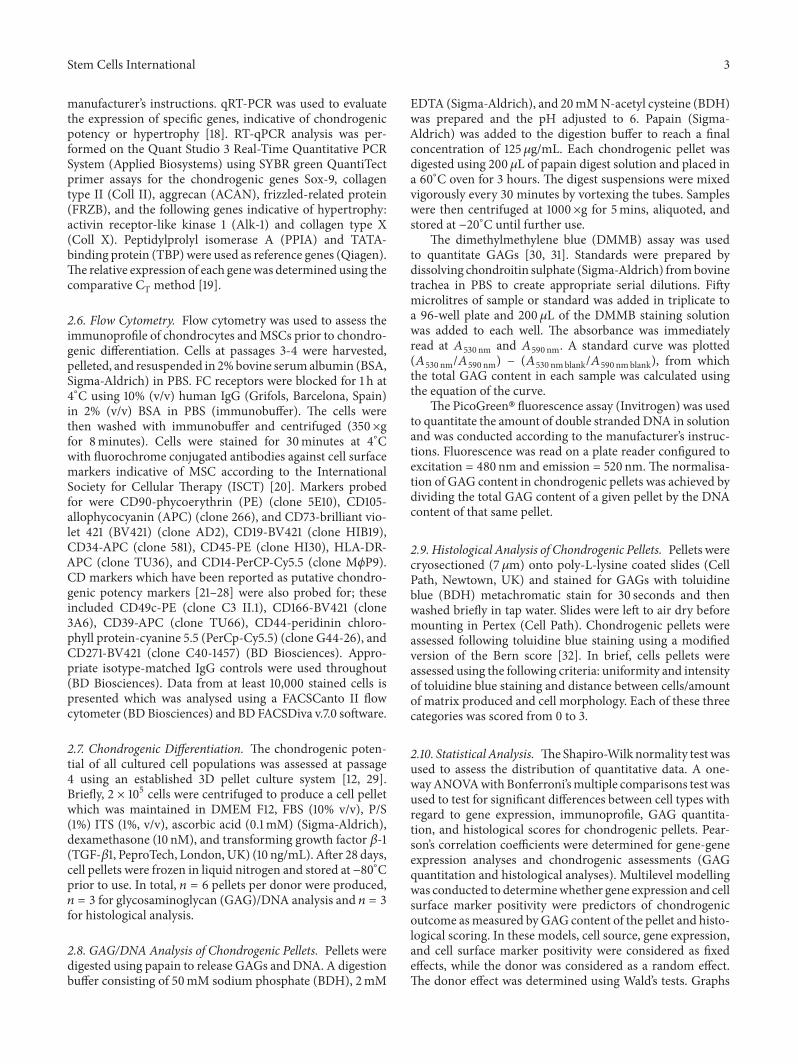
Figure 1: The expression of chondrogenic and hypertrophic genes in monolayer cell populations prior to chondrogenesis. ((a)–(f))Chondrocytes (Ch), bonemarrowMSC (BM), fat padMSC (FP), and subcutaneous fat MSC (SCF). Data shown are themeans ± the standarddeviation of triplicate runs and 5 donors for each cell population. One-way ANOVA and post hoc Bonferroni tests were used to test forsignificant differences in gene expression levels between cell types. (g) Pearson’s correlation analysis matrix comparing genes which may bepredictive of chondrogenic potential; significant correlations are in bold. Gene expression is expressed relative to the reference genes PPIAand TBP.
are shown as means ± standard deviation, with statisticalsignificance considered at ∗𝑝 < 0.05, ∗∗𝑝 < 0.01, and ∗∗∗𝑝 <0.001. All statistical analyses were performed in GraphPadPrism version 6 (GraphPad Software, California, USA) andSPSS version 20 (IBM, New York, USA).
3. Results
3.1. Chondrogenic/Hypertrophic Gene Expression prior toChondrogenic Differentiation. Perhaps not too surprisingly,of the cell types tested in this donor-matched study, thechondrogenic potency genes (Sox-9, Coll II, aggrecan, andFRZB)were consistently expressed at the highest levels in cul-ture expanded chondrocytes. Further, chondrocytes demon-strated the lowest expression profiles for the hypertrophic
genes tested (Alk-1 and Coll X). Of the MSC populationsthat we have examined, BM-MSCs displayed chondrogenicand hypertrophic profiles that most closely resembled thoseof culture expanded chondrocytes. In contrast, the adiposesources of MSCs investigated (FP-MSCs and SCF-MSCs)were least like culture expanded chondrocytes and demon-strated the lowest chondrogenic potency and the highesthypertrophic gene expression profiles. SCF-MSCs expressedAlk-1 at significantly higher levels than chondrocytes andBM-MSCs (𝑝 = 0.044 and 𝑝 = 0.034, resp.) (Figures 1(a)–1(f)).
Gene expression associations for all of the cell typesexamined in this study were tested using Pearson’s correla-tion coefficient analyses and are presented in a correlationmatrix (Figure 1(g)). Significant interactions noted betweenthe chondrogenic potency genes were as follows: aggrecan
Stem Cells International 5
Percentage of positive cells (mean% ± SD)Markers
Chondrocytes BM-MSCs FP-MSCs SCF-MSCsCD73 91.2 (±17.7) 87.1 (±18.0) 99.9 (±0.1) 99.9 (±0.0)
CD90 98.0 (±3.8) 96.4 (±2.5) 99.9 (±0.0) 99.9 (±0.1)
CD105 99.4 (±0.7) 96.7 (±4.1) 98.1 (±4.1) 99.9 (±0.1)
CD34 9.5 (±8.0) 5.1 (±5.4) 74.5 (±15.6) 62.2 (±20.8)
CD45 1.5 (±0.6) 1.3 (±0.4) 2.4 (1.3) 1.6 (±0.6)
CD14 20.4 (±27.1) 14.8 (±12.0) 17.9 (±36.5) 21.4 (±39.3)
CD19 1.3 (±0.8) 2.0 (±1.2) 1.4 (±0.6) 1.6 (±0.5)
HLA-DR 1.4 (±0.6) 1.3 (±0.8) 1.7 (±0.7) 1.3 (±0.6)
(a)
∗
0
50
100
% im
mun
opos
itivi
ty
BM FP SCFCh(b)
∗∗∗
∗∗∗∗∗
BM FP SCFCh0
50
100
% im
mun
opos
itivi
ty
(c)BM FP SCFCh
0
50
100
% im
mun
opos
itivi
ty
(d)
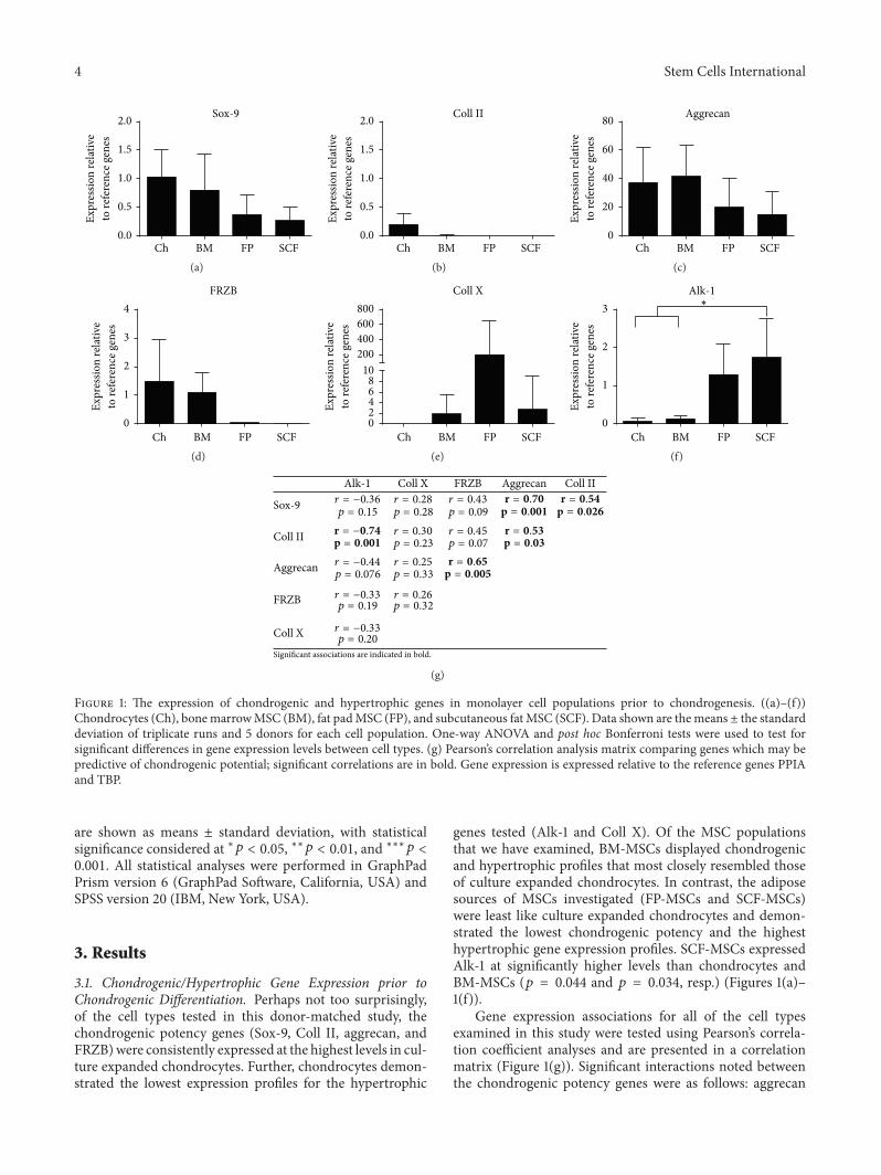
Figure 2: Immunoprofiles for MSC markers and putative chondrogenic potency markers of culture expanded cells prior to chondrogenesis.(a) ISCTMSC immunoprofiles. Immunoprofiles for the putative chondrogenicmarkers CD49c (b), CD166 (c), and CD39 (d). Flow cytometrywas used to detect the percentage of positive cells for each marker on monolayer cell populations of chondrocytes (Ch), bone marrow MSC(BM), fat pad MSC (FP), and subcutaneous fat MSC (SCF) prior to chondrogenesis. Data shown are the means ± the standard deviation of 5donors for each cell population. One-way ANOVA and post hoc Bonferroni tests were used to test for significant differences in the positivityof cell surface markers between cell types.
was positively associated with Sox-9, Coll II, and FRZB; inaddition, Sox-9 was positively associated with Coll II. Therewas also a significant negative association observed betweenColl II and Alk-1 expression.
3.2. MSC/Chondrogenic Immunoprofiling. Flow cytometryanalyses revealed immunopositivity for the MSC markersCD73, CD90, and CD105 for all of the populations of cellsexamined, but to varying levels. FP and SCF derived MSCsadhered to ISCT criteria (i.e., >95% positive); chondrocytesand BM-MSCs also adhered to ISCT criteria for CD90 andCD105 positivity but were <95% positive for CD73. All of thecell populations tested were <2% positive for CD19, CD45,and HLA-DR, in line with ISCT criteria. Some positivitywas recorded in all of the cell populations tested for CD34with high levels (62.2–74.4% positivity) seen in the adiposederived MSCs, which also adheres to ISCT [33]; in addition,>2% of BM-MSCs were also CD34 positive, which does notconform to recommendations by the ISCT [20]. CD14 waspresent on all cell populations, ranging on average between14.8 and 21.4% positivity for each cell type (Figure 2(a)).Differences between cell types for putative chondrogenicpotency marker positivity were noted for CD49c, CD166,and CD39 (Figures 2(b)–2(d)). Chondrocytes showed sig-nificantly greater positivity for CD49c compared to SCF-MSCs (𝑝 = 0.014), whereas the adipose derived MSCs
showed significantly higher positivity for CD166 comparedto chondrocytes or BM-MSCs (𝑝 = 0.0046 and 𝑝 = 0.0002,resp., for FP-MSCs and 𝑝 = 0.021 and 𝑝 = 0.01, resp., forSCF-MSCs). No differences were noted for CD44 or CD271,in that all cell types were >95% positive for CD44 and <5%positive for CD271 (data not shown).
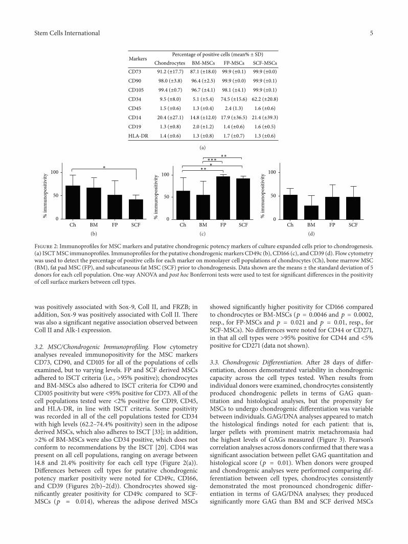
3.3. Chondrogenic Differentiation. After 28 days of differ-entiation, donors demonstrated variability in chondrogeniccapacity across the cell types tested. When results fromindividual donors were examined, chondrocytes consistentlyproduced chondrogenic pellets in terms of GAG quan-titation and histological analyses, but the propensity forMSCs to undergo chondrogenic differentiation was variablebetween individuals. GAG/DNA analyses appeared to matchthe histological findings noted for each patient: that is,larger pellets with prominent matrix metachromasia hadthe highest levels of GAGs measured (Figure 3). Pearson’scorrelation analyses across donors confirmed that there was asignificant association between pellet GAG quantitation andhistological score (𝑝 = 0.01). When donors were groupedand chondrogenic analyses were performed comparing dif-ferentiation between cell types, chondrocytes consistentlydemonstrated the most pronounced chondrogenic differ-entiation in terms of GAG/DNA analyses; they producedsignificantly more GAG than BM and SCF derived MSCs
6 Stem Cells International
Don
or 1
Don
or 2
Don
or 3
Don
or 4
Don
or 5
GAG
/DN
A (𝜇
g/𝜇
g)G
AG/D
NA
(𝜇g/𝜇
g)G
AG/D
NA
(𝜇g/𝜇
g)G
AG/D
NA
(𝜇g/𝜇
g)G
AG/D
NA
(𝜇g/𝜇
g)
0
20
40
60
0
20
40
60
0
20
40
60
0
20
40
60
0
20
40
60
BM FP SCFCh
BM FP SCFCh
BM FP SCFCh
BM FP SCFCh
BM FP SCFCh
(a)
Ch BM SCFFP
(b)
Figure 3: Chondrogenic assessments of pellet cultures between donors. (a) Production of GAG/DNA in pellet cultures from chondrocytes(Ch), bonemarrowMSC (BM), fat padMSC (FP), and subcutaneous fatMSC (SCF). GAGs weremeasured after chondrogenic differentiationusing the DMMB assay and normalised to the DNA content of pellets; each donor is represented in individual graphs. Data shown are themeans ± the standard deviation of triplicate pellets. (b) Chondrogenic pellets from Ch, BM, FP, and SCF showing representative toluidineblue staining for each donor. Scale bars represent 200 𝜇m.
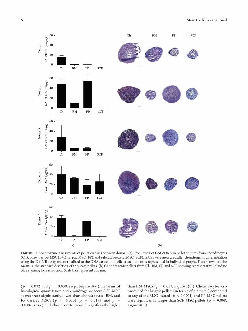
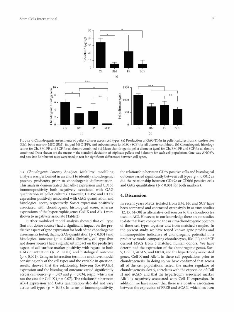
(𝑝 = 0.032 and 𝑝 = 0.030, resp., Figure 4(a)). In terms ofhistological quantitation and chondrogenic score SCF-MSCscores were significantly lower than chondrocytes, BM, andFP derived MSCs (𝑝 < 0.0001, 𝑝 = 0.0195, and 𝑝 =0.0082, resp.) and chondrocytes scored significantly higher
than BM-MSCs (𝑝 = 0.013, Figure 4(b)). Chondrocytes alsoproduced the largest pellets (in terms of diameter) comparedto any of the MSCs tested (𝑝 < 0.0001) and FP-MSC pelletswere significantly larger than SCF-MSC pellets (𝑝 = 0.008,Figure 4(c)).
Stem Cells International 7
∗
∗
GAG
/DN
A (𝜇
g/𝜇
g)
0
10
20
30
40
50
BM FP SCFCh(a)
∗
∗
∗∗∗
∗∗
BM FP SCFCh0
2
4
6
8
10
Chon
drog
enic
hist
olog
y sc
ore
(b)
∗∗
∗∗∗
Pelle
t dia
met
er (𝜇
m)
0
500
1000
1500
BM FP SCFCh(c)
Figure 4: Chondrogenic assessments of pellet cultures across cell types. (a) Production of GAG/DNA in pellet cultures from chondrocytes(Ch), bone marrow MSC (BM), fat pad MSC (FP), and subcutaneous fat MSC (SCF) for all donors combined. (b) Chondrogenic histologyscores for Ch, BM, FP, and SCF for all donors combined. (c) Mean chondrogenic pellet diameter (𝜇m) for Ch, BM, FP, and SCF for all donorscombined. Data shown are the means ± the standard deviation of triplicate pellets and 5 donors for each cell population. One-way ANOVAand post hoc Bonferroni tests were used to test for significant differences between cell types.
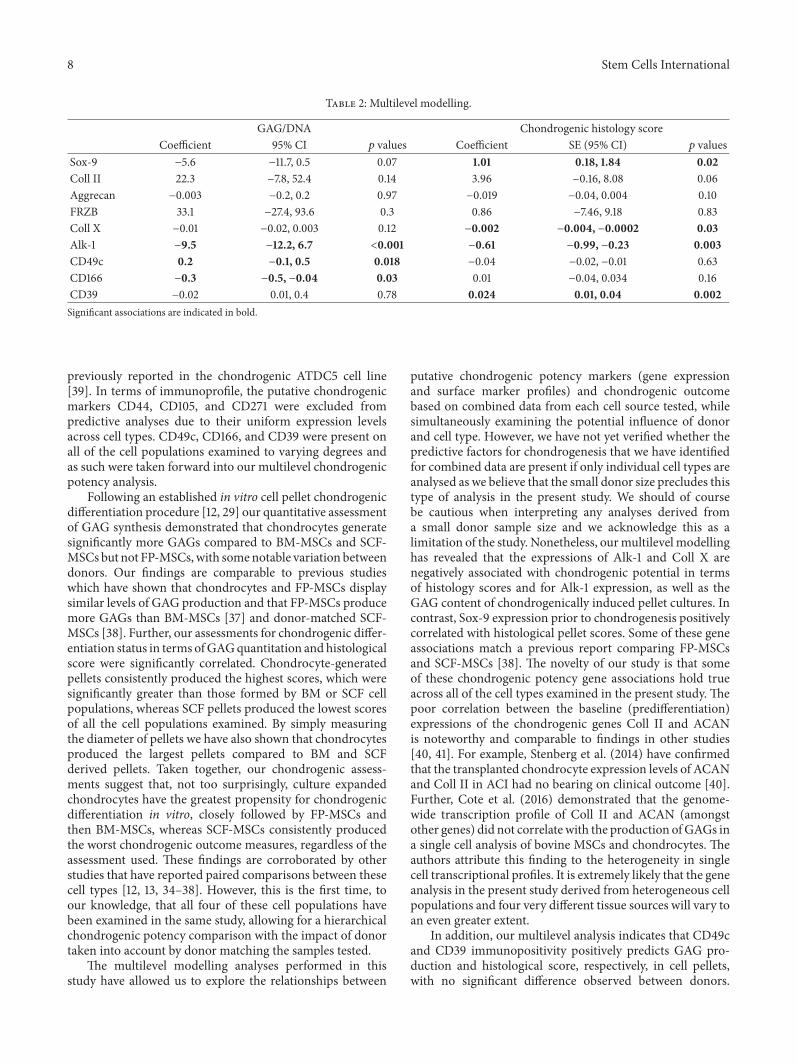
3.4. Chondrogenic Potency Analyses. Multilevel modellinganalysis was performed in an effort to identify chondrogenicpotency predictors prior to chondrogenic differentiation.This analysis demonstrated that Alk-1 expression and CD166immunopositivity both negatively associated with GAGquantitation in pellet cultures. However, CD49c and CD39expression positively associated with GAG quantitation andhistological score, respectively. Sox-9 expression positivelyassociated with chondrogenic histological score, whereasexpressions of the hypertrophic genes Coll X and Alk-1 wereshown to negatively associate (Table 2).
Further multilevel model analysis showed that cell type(but not donor source) had a significant impact on the pre-dictive aspect of gene expression for both of the chondrogenicassessments tested, that is, GAG quantitation (𝑝 < 0.001) andhistological outcome (𝑝 < 0.001). Similarly, cell type (butnot donor source) had a significant impact on the predictiveaspect of cell surface marker positivity with regard to bothGAG quantitation (𝑝 < 0.001) and histological outcome(𝑝 < 0.001). Using an interaction term in a multilevel modelconsisting only of the cell types and the variable in question,results showed that the relationship between Sox-9/Alk-1expression and the histological outcome varied significantlyacross cell source (𝑝 = 0.03 and 𝑝 = 0.034, resp.), which wasnot the case for Coll X (𝑝 = 0.07). The relationship betweenAlk-1 expression and GAG quantitation also did not varyacross cell types (𝑝 = 0.43). In terms of immunopositivity,
the relationship between CD39 positive cells and histologicaloutcome varied significantly between cell types (𝑝 < 0.001) asdid the relationship between CD49c or CD166 positive cellsand GAG quantitation (𝑝 < 0.001 for both markers).
4. Discussion
In recent years MSCs isolated from BM, FP, and SCF havebeen compared and contrasted extensively in in vitro studies[12, 13, 34–38] as alternative cell sources to the chondrocytesused in ACI. However, to our knowledge there are no studiesto date that have compared the in vitro chondrogenic potencyof these cell types together and from matched samples. Inthe present study, we have tested known gene profiles andimmunoprofiles indicative of chondrogenic potential in apredictive model comparing chondrocytes, BM, FP, and SCFderived MSCs from 5 matched human donors. We havedetermined the expression of the chondrogenic genes, Sox-9, Coll II, ACAN, and FRZB, and the hypertrophy associatedgenes, Coll X and Alk-1, in these cell populations prior tochondrogenesis. In doing so, we have confirmed that acrossall of the cell populations tested, the master regulator ofchondrogenesis, Sox-9, correlates with the expression of CollII and ACAN and that the hypertrophy associated markerAlk-1 is negatively associated with Coll II expression. Inaddition, we have shown that there is a positive associationbetween the expression of FRZB and ACAN, which has been
8 Stem Cells International
Table 2: Multilevel modelling.
GAG/DNA Chondrogenic histology scoreCoefficient 95% CI 𝑝 values Coefficient SE (95% CI) 𝑝 values
Sox-9 −5.6 −11.7, 0.5 0.07 1.01 0.18, 1.84 0.02Coll II 22.3 −7.8, 52.4 0.14 3.96 −0.16, 8.08 0.06Aggrecan −0.003 −0.2, 0.2 0.97 −0.019 −0.04, 0.004 0.10FRZB 33.1 −27.4, 93.6 0.3 0.86 −7.46, 9.18 0.83Coll X −0.01 −0.02, 0.003 0.12 −0.002 −0.004, −0.0002 0.03Alk-1 −9.5 −12.2, 6.7 <0.001 −0.61 −0.99, −0.23 0.003CD49c 0.2 −0.1, 0.5 0.018 −0.04 −0.02, −0.01 0.63CD166 −0.3 −0.5, −0.04 0.03 0.01 −0.04, 0.034 0.16CD39 −0.02 0.01, 0.4 0.78 0.024 0.01, 0.04 0.002Significant associations are indicated in bold.
previously reported in the chondrogenic ATDC5 cell line[39]. In terms of immunoprofile, the putative chondrogenicmarkers CD44, CD105, and CD271 were excluded frompredictive analyses due to their uniform expression levelsacross cell types. CD49c, CD166, and CD39 were present onall of the cell populations examined to varying degrees andas such were taken forward into our multilevel chondrogenicpotency analysis.
Following an established in vitro cell pellet chondrogenicdifferentiation procedure [12, 29] our quantitative assessmentof GAG synthesis demonstrated that chondrocytes generatesignificantly more GAGs compared to BM-MSCs and SCF-MSCs but not FP-MSCs,with somenotable variation betweendonors. Our findings are comparable to previous studieswhich have shown that chondrocytes and FP-MSCs displaysimilar levels of GAG production and that FP-MSCs producemore GAGs than BM-MSCs [37] and donor-matched SCF-MSCs [38]. Further, our assessments for chondrogenic differ-entiation status in terms ofGAGquantitation andhistologicalscore were significantly correlated. Chondrocyte-generatedpellets consistently produced the highest scores, which weresignificantly greater than those formed by BM or SCF cellpopulations, whereas SCF pellets produced the lowest scoresof all the cell populations examined. By simply measuringthe diameter of pellets we have also shown that chondrocytesproduced the largest pellets compared to BM and SCFderived pellets. Taken together, our chondrogenic assess-ments suggest that, not too surprisingly, culture expandedchondrocytes have the greatest propensity for chondrogenicdifferentiation in vitro, closely followed by FP-MSCs andthen BM-MSCs, whereas SCF-MSCs consistently producedthe worst chondrogenic outcome measures, regardless of theassessment used. These findings are corroborated by otherstudies that have reported paired comparisons between thesecell types [12, 13, 34–38]. However, this is the first time, toour knowledge, that all four of these cell populations havebeen examined in the same study, allowing for a hierarchicalchondrogenic potency comparison with the impact of donortaken into account by donor matching the samples tested.
The multilevel modelling analyses performed in thisstudy have allowed us to explore the relationships between
putative chondrogenic potency markers (gene expressionand surface marker profiles) and chondrogenic outcomebased on combined data from each cell source tested, whilesimultaneously examining the potential influence of donorand cell type. However, we have not yet verified whether thepredictive factors for chondrogenesis that we have identifiedfor combined data are present if only individual cell types areanalysed as we believe that the small donor size precludes thistype of analysis in the present study. We should of coursebe cautious when interpreting any analyses derived froma small donor sample size and we acknowledge this as alimitation of the study. Nonetheless, ourmultilevel modellinghas revealed that the expressions of Alk-1 and Coll X arenegatively associated with chondrogenic potential in termsof histology scores and for Alk-1 expression, as well as theGAG content of chondrogenically induced pellet cultures. Incontrast, Sox-9 expression prior to chondrogenesis positivelycorrelated with histological pellet scores. Some of these geneassociations match a previous report comparing FP-MSCsand SCF-MSCs [38]. The novelty of our study is that someof these chondrogenic potency gene associations hold trueacross all of the cell types examined in the present study. Thepoor correlation between the baseline (predifferentiation)expressions of the chondrogenic genes Coll II and ACANis noteworthy and comparable to findings in other studies[40, 41]. For example, Stenberg et al. (2014) have confirmedthat the transplanted chondrocyte expression levels of ACANand Coll II in ACI had no bearing on clinical outcome [40].Further, Cote et al. (2016) demonstrated that the genome-wide transcription profile of Coll II and ACAN (amongstother genes) did not correlate with the production ofGAGs ina single cell analysis of bovine MSCs and chondrocytes. Theauthors attribute this finding to the heterogeneity in singlecell transcriptional profiles. It is extremely likely that the geneanalysis in the present study derived from heterogeneous cellpopulations and four very different tissue sources will vary toan even greater extent.
In addition, our multilevel analysis indicates that CD49cand CD39 immunopositivity positively predicts GAG pro-duction and histological score, respectively, in cell pellets,with no significant difference observed between donors.
Stem Cells International 9
Other studies have shown that CD49c positivity on chon-drocytes and CD39 positivity on synovium derived MSCsare associated with increased in vitro chondrogenic potential[21, 27]; however, our results are the first to demonstratethese relationships across matched chondrocytes, BM-MSCs,and adipose derived MSCs. Perhaps surprisingly CD166positivity did not indicate chondrogenic potential, as hasbeen previously shown [24, 26]; in fact immunopositivityfor this marker was negatively associated with chondrogenicassessments. One potential explanation for this findingmightbe that CD166 was expressed at significantly greater levelson SCF-MSCs compared to chondrocytes and BM-MSCsand that in our hands SCF-MSCs have been shown toconsistently demonstrate a poor propensity for chondrogenicdifferentiation. Interestingly, we have demonstrated throughthis multicell type, donor-matched study that the sourceof cells significantly influences both GAG production inpellet culture and also the histological score of the pellet. Incontrast, the donor had no demonstrable impact on eitherof the chondrogenic assessments tested, although as statedpreviously we must be cautious with this finding as ourresults are based on a small cohort of donors. Follow-upstudies should be geared towards understanding the molec-ular mechanisms that account for the differences observedbetween cell populations and in the development of methodsto select cells with enhanced chondrogenic potential.
5. Conclusions
We have demonstrated the chondrogenic predictive valueof high levels of Sox-9 and low levels of collagen type Xor Alk-1 expression as well as immunopositivity for CD49cand CD39 in a combined data analysis of chondrocytes,BM-MSCs, FP-MSCs, and SCF-MSCs. Further individualanalyses on larger donor cohorts will be required to validatethese findings for individual cell types before these predictivefactors could be used as selection criteria prior to thetransplantation or banking of each cell type in the treatmentof cartilage injuries. We have also shown, using donor-matched samples, that cell type significantly influences thechondrogenic potency of the MSC sources examined in thisstudy; we have demonstrated that MSCs sourced from theinfrapatellar fat pad of the knee or bone marrow provide the“next best” alternative to chondrocytes, in terms of in vitrochondrogenic differentiation capacity. Further, our resultshave consistently shown that SCF derived MSCs have thepoorest propensity for chondrogenic differentiation. Thesefindings have important clinical implications, not only for theunderstanding ofMSC chondrogenic differentiation capacity,but also for the development of cell therapy strategies toscreen for and select potent cell types prior to application inthe treatment of cartilage injuries.
Competing Interests
The authors declare that there is no conflict of interestsregarding the publication of this paper.
Acknowledgments
The authors are grateful to the Engineering and PhysicalSciences Research Council Centre for Doctoral Training andArthritis Research UK (Grants 20253, 19429, and 18480) forsupporting this work and to Miss Jade Perry for contributingto some of the chondrogenic data analysis described.
References
[1] M. Brittberg, A. Lindahl, A. Nilsson, C. Ohlsson, O. Isaksson,and L. Peterson, “Treatment of deep cartilage defects in theknee with autologous chondrocyte transplantation,” The NewEngland Journal of Medicine, vol. 331, no. 14, pp. 889–895, 1994.
[2] A.M. Bhosale, J. H. Kuiper,W. E. B. Johnson, P. E. Harrison, andJ. B. Richardson, “Midterm to long-term longitudinal outcomeof autologous chondrocyte implantation in the knee joint: amultilevel analysis,” The American Journal of Sports Medicine,vol. 37, supplement 1, pp. 131S–138S, 2009.
[3] J. B. Richardson, B. Caterson, E. H. Evans, B. A. Ashton, andS. Roberts, “Repair of human articular cartilage after implan-tation of autologous chondrocytes,” Journal of Bone and JointSurgery—Series B, vol. 81, no. 6, pp. 1064–1068, 1999.
[4] D. B. F. Saris, J. Vanlauwe, J. Victor et al., “Treatment of symp-tomatic cartilage defects of the knee: characterized chondrocyteimplantation results in better clinical outcome at 36 months ina randomized trial compared to microfracture,” The AmericanJournal of Sports Medicine, vol. 37, supplement 1, pp. 10S–19S,2009.
[5] H. S. McCarthy, J. B. Richardson, J. C. Parker, and S. Roberts,“Evaluating joint morbidity after chondral harvest for autolo-gous chondrocyte implantation (ACI): a study of ACI-treatedankles and hips with a knee chondral harvest,” Cartilage, vol. 7,no. 1, pp. 7–15, 2016.
[6] H. Holtzer, J. Abbott, J. Lash, and S. Holtzer, “The loss of pheno-typic traits by differentiated cells in vitro, I. dedifferentiation ofcartilage cells,” Proceedings of the National Academy of Sciences,vol. 46, no. 12, pp. 1533–1542, 1960.
[7] M. Schnabel, S. Marlovits, G. Eckhoff et al., “Dedifferentiation-associated changes in morphology and gene expressionin primary human articular chondrocytes in cell culture,”Osteoarthritis and Cartilage, vol. 10, no. 1, pp. 62–70, 2002.
[8] S. Wakitani, T. Okabe, S. Horibe et al., “Safety of autologousbone marrow-derived mesenchymal stem cell transplantationfor cartilage repair in 41 patients with 45 joints followed forup to 11 years and 5 months,” Journal of Tissue Engineering andRegenerative Medicine, vol. 5, no. 2, pp. 146–150, 2011.
[9] I. Akgun, M. C. Unlu, O. A. Erdal et al., “Matrix-induced au-tologous mesenchymal stem cell implantation versus matrix-induced autologous chondrocyte implantation in the treatmentof chondral defects of the knee: a 2-year randomized study,”Archives of Orthopaedic and Trauma Surgery, vol. 135, no. 2, pp.251–263, 2015.
[10] A. Vega, M. A. Martın-Ferrero, F. D. Canto et al., “Treatment ofknee osteoarthritis with allogeneic bone marrow mesenchymalstem cells: a randomized controlled trial,” Transplantation, vol.99, no. 8, pp. 1681–1690, 2015.
[11] J. L. Dragoo, B. Samimi, M. Zhu et al., “Tissue-engineeredcartilage and bone using stem cells from human infrapatellarfat pads,” The Journal of Bone & Joint Surgery—British Volume,vol. 85, no. 5, pp. 740–747, 2003.
10 Stem Cells International
[12] J. Garcia, K. Wright, S. Roberts et al., “Characterisation ofsynovial fluid and infrapatellar fat pad derived mesenchymalstromal cells: the influence of tissue source and inflammatorystimulus,” Scientific Reports, vol. 6, Article ID 24295, 2016.
[13] H. Busser, M. Najar, G. Raicevic et al., “Isolation and charac-terization of human mesenchymal stromal cell subpopulations:comparison of bonemarrow and adipose tissue,” Stem Cells andDevelopment, vol. 24, no. 18, pp. 2142–2157, 2015.
[14] J. Freitag, J. Ford, D. Bates et al., “Adipose derivedmesenchymalstem cell therapy in the treatment of isolated knee chondrallesions: design of a randomised controlled pilot study compar-ing arthroscopic microfracture versus arthroscopic microfrac-ture combined with postoperative mesenchymal stem cellinjections,” BMJ Open, vol. 5, no. 12, Article ID e009332, 2015.
[15] A. E. Aksu, J. P. Rubin, J. R. Dudas, and K. G. Marra, “Roleof gender and anatomical region on induction of osteogenicdifferentiation of human adipose-derived stem cells,” Annals ofPlastic Surgery, vol. 60, no. 3, pp. 306–322, 2008.
[16] K. A. Payne, D. M. Didiano, and C. R. Chu, “Donor sex and ageinfluence the chondrogenic potential of human femoral bonemarrow stem cells,” Osteoarthritis and Cartilage, vol. 18, no. 5,pp. 705–713, 2010.
[17] M. S. Choudhery, M. Badowski, A. Muise, J. Pierce, and D. T.Harris, “Donor age negatively impacts adipose tissue-derivedmesenchymal stem cell expansion and differentiation,” Journalof Translational Medicine, vol. 12, no. 1, article 8, 2014.
[18] F. Dell’Accio, C. De Bari, and F. P. Luyten, “Molecular mark-ers predictive of the capacity of expanded human articularchondrocytes to form stable cartilage in vivo,” Arthritis andRheumatism, vol. 44, no. 7, pp. 1608–1619, 2001.
[19] T. D. Schmittgen and K. J. Livak, “Analyzing real-time PCR databy the comparative CT method,” Nature Protocols, vol. 3, no. 6,pp. 1101–1108, 2008.
[20] M. Dominici, K. Le Blanc, I. Mueller et al., “Minimal crite-ria for defining multipotent mesenchymal stromal cells. TheInternational Society for Cellular Therapy position statement,”Cytotherapy, vol. 8, no. 4, pp. 315–317, 2006.
[21] S. P. Grogan, A. Barbero, J. Diaz-Romero et al., “Identification ofmarkers to characterize and sort human articular chondrocyteswith enhanced in vitro chondrogenic capacity,” Arthritis andRheumatism, vol. 56, no. 2, pp. 586–595, 2007.
[22] M. C. Arufe, A. De la Fuente, I. Fuentes, F. J. de Toro, andF. J. Blanco, “Chondrogenic potential of subpopulations ofcells expressing mesenchymal stem cell markers derived fromhuman synovial membranes,” Journal of Cellular Biochemistry,vol. 111, no. 4, pp. 834–845, 2010.
[23] T. Jiang, W. Liu, X. Lv et al., “Potent in vitro chondrogenesis ofCD105 enriched human adipose-derived stem cells,” Biomateri-als, vol. 31, no. 13, pp. 3564–3571, 2010.
[24] D. Pretzel, S. Linss, S. Rochler et al., “Relative percentageand zonal distribution of mesenchymal progenitor cells inhuman osteoarthritic and normal cartilage,” Arthritis Research&Therapy, vol. 13, no. 2, article R64, 2011.
[25] P. Niemeyer, J. M. Pestka, G. M. Salzmann, N. P. Sudkamp,and H. Schmal, “Influence of cell quality on clinical outcomeafter autologous chondrocyte implantation,” American Journalof Sports Medicine, vol. 40, no. 3, pp. 556–561, 2012.
[26] C. B. Chang, S. A. Han, E. M. Kim, S. Lee, S. C. Seong, and M.C. Lee, “Chondrogenic potentials of human synovium-derivedcells sorted by specific surface markers,” Osteoarthritis andCartilage, vol. 21, no. 1, pp. 190–199, 2013.
[27] F. Gullo and C. De Bari, “Prospective purification of a sub-population of human synovial mesenchymal stem cells withenhanced chondro-osteogenic potency,” Rheumatology, vol. 52,no. 10, pp. 1758–1768, 2013.
[28] Y.Mifune, T.Matsumoto, S.Murasawa et al., “Therapeutic supe-riority for cartilage repair by CD271-positive marrow stromalcell transplantation,” Cell Transplantation, vol. 22, no. 7, pp.1201–1211, 2013.
[29] B. Johnstone, T. M. Hering, A. I. Caplan, V. M. Goldberg, andJ. U. Yoo, “In vitro chondrogenesis of bone marrow-derivedmesenchymal progenitor cells,” Experimental Cell Research, vol.238, no. 1, pp. 265–272, 1998.
[30] R. W. Farndale, C. A. Sayers, and A. J. Barrett, “A direct spec-trophotometric microassay for sulfated glycosaminoglycans incartilage cultures,” Connective Tissue Research, vol. 9, no. 4, pp.247–248, 1982.
[31] R.W. Farndale, D. J. Buttle, and A. J. Barrett, “Improved quanti-tation and discrimination of sulphated glycosaminoglycans byuse of dimethylmethylene blue,” Biochim Biophys Acta, vol. 883,no. 2, pp. 173–177, 1986.
[32] S. P. Grogan, A. Barbero, V. Winkelmann et al., “Visual histo-logical grading system for the evaluation of in vitro-generatedneocartilage,” Tissue Engineering, vol. 12, no. 8, pp. 2141–2149,2006.
[33] P. Bourin, B. A. Bunnell, L. Casteilla et al., “Stromal cellsfrom the adipose tissue-derived stromal vascular fraction andculture expanded adipose tissue-derived stromal/stem cells: ajoint statement of the International Federation for AdiposeTherapeutics and Science (IFATS) and the International Societyfor CellularTherapy (ISCT),”Cytotherapy, vol. 15, no. 6, pp. 641–648, 2013.
[34] C. Karlsson, C. Brantsing, T. Svensson et al., “Differentiationof human mesenchymal stem cells and articular chondro-cytes: analysis of chondrogenic potential and expression pat-tern of differentiation-related transcription factors,” Journal ofOrthopaedic Research, vol. 25, no. 2, pp. 152–163, 2007.
[35] A. English, E. A. Jones, D. Corscadden et al., “A comparativeassessment of cartilage and joint fat pad as a potential source ofcells for autologous therapy development in knee osteoarthri-tis,” Rheumatology, vol. 46, no. 11, pp. 1676–1683, 2007.
[36] Y. Liu, C. T. Buckley, R. Downey, K. J. Mulhall, and D. J. Kelly,“The role of environmental factors in regulating the devel-opment of cartilaginous grafts engineered using osteoarthritichuman infrapatellar fat pad-derived stem cells,”Tissue Engineer-ing Part A, vol. 18, no. 15-16, pp. 1531–1541, 2012.
[37] T. Vinardell, E. J. Sheehy, C. T. Buckley, and D. J. Kelly, “Acomparison of the functionality and in vivo phenotypic stabilityof cartilaginous tissues engineered from different stem cellsources,” Tissue Engineering—Part A, vol. 18, no. 11-12, pp. 1161–1170, 2012.
[38] S. Lopa, A. Colombini, D. Stanco, L. de Girolamo, V. Sansone,and M. Moretti, “Donor-matched mesenchymal stem cellsfrom knee infrapatellar and subcutaneous adipose tissue ofosteoarthritic donors display differential chondrogenic andosteogenic commitment,” European Cells and Materials, vol. 27,pp. 298–311, 2014.
[39] L. Lodewyckx, F. Cailotto, S. Thysen, F. P. Luyten, and R. J.Lories, “Tight regulation of wingless-type signaling in the artic-ular cartilage—subchondral bone biomechanical unit: tran-scriptomics in Frzb-knockout mice,” Arthritis Research andTherapy, vol. 14, article R16, 2012.

Stem Cells International 11
[40] J. Stenberg, T. S. de Windt, J. Synnergren et al., “Clinical out-come 3 years after autologous chondrocyte implantation doesnot correlate with the expression of a predefined gene markerset in chondrocytes prior to implantation but is associatedwith critical signaling pathways,” Orthopaedic Journal of SportsMedicine, vol. 2, no. 9, 2014.
[41] A. J. Cote, C. M. McLeod, M. J. Farrell et al., “Single-celldifferences in matrix gene expression do not predict matrixdeposition,” Nature Communications, vol. 7, p. 10865, 2016.
Submit your manuscripts athttp://www.hindawi.com
Hindawi Publishing Corporationhttp://www.hindawi.com Volume 2014
Anatomy Research International
PeptidesInternational Journal of
Hindawi Publishing Corporationhttp://www.hindawi.com Volume 2014
Hindawi Publishing Corporation http://www.hindawi.com
International Journal of
Volume 2014
Zoology
Hindawi Publishing Corporationhttp://www.hindawi.com Volume 2014
Molecular Biology International
GenomicsInternational Journal of
Hindawi Publishing Corporationhttp://www.hindawi.com Volume 2014
The Scientific World JournalHindawi Publishing Corporation http://www.hindawi.com Volume 2014
Hindawi Publishing Corporationhttp://www.hindawi.com Volume 2014
BioinformaticsAdvances in
Marine BiologyJournal of
Hindawi Publishing Corporationhttp://www.hindawi.com Volume 2014
Hindawi Publishing Corporationhttp://www.hindawi.com Volume 2014
Signal TransductionJournal of
Hindawi Publishing Corporationhttp://www.hindawi.com Volume 2014
BioMed Research International
Evolutionary BiologyInternational Journal of
Hindawi Publishing Corporationhttp://www.hindawi.com Volume 2014
Hindawi Publishing Corporationhttp://www.hindawi.com Volume 2014
Biochemistry Research International
ArchaeaHindawi Publishing Corporationhttp://www.hindawi.com Volume 2014
Hindawi Publishing Corporationhttp://www.hindawi.com Volume 2014
Genetics Research International
Hindawi Publishing Corporationhttp://www.hindawi.com Volume 2014
Advances in
Virolog y
Hindawi Publishing Corporationhttp://www.hindawi.com
Nucleic AcidsJournal of
Volume 2014
Stem CellsInternational
Hindawi Publishing Corporationhttp://www.hindawi.com Volume 2014
Hindawi Publishing Corporationhttp://www.hindawi.com Volume 2014
Enzyme Research
Hindawi Publishing Corporationhttp://www.hindawi.com Volume 2014
International Journal of
Microbiology