Embed Size (px)
Citation preview
Research ArticleAssessment of Hair Aluminum, Lead, and Mercury ina Sample of Autistic Egyptian Children: Environmental RiskFactors of Heavy Metals in Autism
Farida El Baz Mohamed,1 Eman Ahmed Zaky,1 Adel Bassuoni El-Sayed,2
Reham Mohammed Elhossieny,1 Sally Soliman Zahra,1 Waleed Salah Eldin,3
Walaa Yousef Youssef,1 Rania Abdelmgeed Khaled,1 and Azza Mohamed Youssef1
1Pediatrics Department, Faculty of Medicine, Ain Shams University, Cairo, Egypt2National Institute of Standards, Giza, Egypt3Community Medicine Department, Ain Shams University, Cairo, Egypt
Correspondence should be addressed to Sally Soliman Zahra; [email protected]
Received 24 June 2015; Revised 24 August 2015; Accepted 2 September 2015
Academic Editor: Michael E. Behen
Copyright © 2015 Farida El Baz Mohamed et al.This is an open access article distributed under the Creative Commons AttributionLicense, which permits unrestricted use, distribution, and reproduction in anymedium, provided the originalwork is properly cited.
Background and Aims. The etiological factors involved in the etiology of autism remain elusive and controversial, but both geneticand environmental factors have been implicated.The aimof this studywas to assess the levels andpossible environmental risk factorsand sources of exposure to mercury, lead, and aluminum in children with autism spectrum disorder (ASD) as compared to theirmatched controls.Methods. One hundred ASD children were studied in comparison to 100 controls. All participants were subjectedto clinical evaluation andmeasurement of mercury, lead, and aluminum through hair analysis which reflects past exposure.Results.The mean Levels of mercury, lead, and aluminum in hair of the autistic patients were significantly higher than controls. Mercury,lead, and aluminum levels were positively correlated withmaternal fish consumptions, living nearby gasoline stations, and the usageof aluminum pans, respectively. Conclusion. Levels of mercury, lead, and aluminum in the hair of autistic children are higher thancontrols. Environmental exposure to these toxic heavy metals, at key times in development, may play a causal role in autism.
1. Introduction
The autism spectrum describes a range of conditions classifiedas neurodevelopmental disorders in the fifth revision of theAmerican Psychiatric Association’s Diagnostic and StatisticalManual of Mental Disorders 5th Edition (DSM-5). Thesedisorders are characterized by social deficits and communica-tion difficulties, stereotyped or repetitive behaviors and inter-ests, sensory issues, and in some cases cognitive delays [1].
The increase of ASDs prevalence cannot be fullyexplained by advances in diagnostics or sudden genetic shifts.There is a growing consensus among scientists and cliniciansthat ASDs ensue from an interaction between biological vul-nerability factors and environmental or iatrogenic insults [2].
This points to the importance of environmental factorsand raises the possibility of an etiological role for toxic
exposures: either prenatal, postnatal, or in some cumulativepattern that combines the effect of maternal, gestational, andinfant exposures [3].
Some possible sources of heavy metal poisoning includechemical products, fertilizers, industrial paint, buildingmate-rials, fish that is high in mercury, silver dental fillings, andmercury-containing preservatives (thiomersal) in vaccines.Lead may be found in the dirt near roads and can still befound in paint from older houses. Children eating paint chipsor those with pica may develop toxic lead levels [4].
Genetically, children with autism may be less able todetoxify toxic environmental agents, and this inability maypredispose them to suffer neural damage consistent withautistic behavioral traits [4].
Women with chronic metal exposure (who have accumu-lated high tissue levels ofmercury and othermetals)may pass
Hindawi Publishing CorporationBehavioural NeurologyVolume 2015, Article ID 545674, 9 pageshttp://dx.doi.org/10.1155/2015/545674

2 Behavioural Neurology
potentially toxic metals to their fetuses or intoxicate infantsthrough nursing [5].
We conducted the study to examine the possible risk fac-tors and sources of exposure to mercury, lead, and aluminumin children with autistic spectrum disorder and assess thelevels of heavy metals in hair of both autistic and controlgroups.
2. Participants
This case control study included one hundred autistic chil-dren (84 boys and 16 girls); their ages ranged from 2.5 to15 years with mean of 6.2 ± 2.4 years. The children werediagnosed according to the DSM-IV TR (2000) criteriaby pediatric psychiatrists in the Child Psychiatry Clinic,Children’s Hospital, Ain Shams University from December2011 to December 2014. A control group was selected, whichincluded one hundred age- and gender-matched healthy chil-dren.These childrenwere friends and neighbors, unrelated tothe study group.
A written consent from the parents and bioethicalresearch committee approval were taken. Patients wereexcluded from the study if they were suffering from liveror kidney disease, anemia, or current treatment for irondeficiency, progressive neurological disorders, or unstableepilepsy. Also children with mercury dental amalgam, pre-vious use of DMSA or other chelators were excluded.
All of the children admitted to the study received routinechildhood vaccinations.
3. Methods
All children in the current study were subjected to thefollowing: detailed history taking with special emphasis onantenatal or maternal history asking about maternal dietaryhabits (the type and amount of fish consumption by themother during pregnancy especially canned tuna fish and theimported frozen mackerel fish (the cheapest fish in Egypt)),maternal dental work (filling amalgam or removal), and ifRho(D) immune globulin was given during pregnancy.
Developmental history was taken laying stress on alldevelopmental milestones (gross motor, fine motor, sphinc-ter control, language, cognitive development, and socialmilestones). Also, behavioral disorders (history of pica,stereotypic behavior) and dietary history (being breast fedor artificially fed, duration, weaning history, and problemsduring weaning) were noted.
Potential environmental toxic exposures were particu-larly noted such as gasoline station in close proximity to thechild’s home, cooking habits (type of utensils used especiallyaluminum pans), and the age of the patients’ house (type ofpaint and water pipes).
Also, past history ofmajor childhood illnesses and immu-nizations was taken.
Thorough clinical examination of all body systems withspecial emphasis on neurological examination.
All autistic children were subjected to a full clinical childpsychiatric evaluation for diagnosis of autistic spectrum dis-order and exclusion of other psychiatric disorders according
to Diagnostic and Statistical Manual of Mental Disorders 4thEdition, Text Revision (DSM-IV-TR) [6].
The severity of autistic symptomatology was measured bythe Childhood Autism Rating Scale (CARS) [7]. It consists of15 categories, each rated on a four-point scale. The individualis considered nonautistic when his total score falls in therange of 15–29, mildly-to-moderately autistic when his totalscore falls in the range of 30–36, and severely autistic whenhis total score falls in the range of 37–60 [8]. Based on theadministration of multiple assessments, insights into variousaspects of autism are gained.
IQ assessment (intelligent quotient) using Stanford-BinetIntelligence Scale [9]: ranges of IQ are as follows: 20–30denotes severe mental retardation, 31–49 is moderate mentalretardation, 50–70 is mild mental retardation, 71–89 is belowaverage, 90–109 is normal IQ, and 110–125 is above average.
Hair Specimen Collection. We decided to use hair mineralanalysis to evaluate the long term metal exposure. Hairsampling is a noninvasive technique; it is the best indicatorof a given mineral in the body. These samples were collectedfrom cases and control by single cutting from the occipitalregion. The samples were cut to lengths of about 1.5–2 cmusing clean stainless steel scissors. A minimum of 5–10mg ofhair was required for the hair analysis assay. Approximately100 strands of hair (50mg) were used. Adhesive paper wasplaced over the end of the hair strands closest to the scalp;the paper was marked with an arrow indicating the end ofhair closest to the scalp. The samples were placed in a sealedplastic bag [10].
Instrumentation. The measurements of lead and aluminumwere made by Electrothermal Atomic Absorption Spec-trometer Zeenit 700 (Germany) equipped with Zeemanbackground correction and automatic autosampler. Hydridegeneration technique was used for mercury measurements.
3.1. Materials and Reagents. Calibration solutions were pre-pared from Pb, Hg, and Al certified reference materials ofconcentration 1000mg/L maintained at National Institute ofStandards (NIS):
(i) concentrated nitric acid with purity of 69%,(ii) sodium borohydride with purity 98%,(iii) sodium hydroxide with purity of 99%,(iv) ammonium phosphate monobasic 1% which is pre-
pared to be used as a modifier for lead measurementsby electrothermal atomic absorption spectrometer(AAS).
3.1.1. Sample Preparation. Hair samples were washed withpure acetone and three times with ultrapure water and putin a drying furnace at 70∘C over night. After cooling, thesamples were caught in small pieces of 2mm length. About0.15–0.20 g of hair samples was weighed, mixed with 2mLconcentrated nitric acid, and left in a furnace at 90∘C for 24hours. After cooling, the samples were transferred to 25mLmeasuring flask and completed by ultrapure water. Further
Behavioural Neurology 3
dilution was required when the concentrated range exceededthe calibration range. Test values were reported in mg/kg.
3.1.2. Statistical Methodology. The collected data was revised,coded, tabulated, and introduced to Statistical Package forSocial Science (SPSS 15.0.1 for windows; SPSS Inc., Chicago,IL, 2001). Data was presented and suitable analysis was doneaccording to the type of data obtained for each parameter:
(1) Descriptive statistics:mean standard deviation (±SD)and range were used for parametric numerical data,while median was used for nonparametric numericaldata and frequency and percentage of nonnumericaldata.
(2) Analytical statistics: Student’s t-test was used to assessthe statistical significance of the difference betweentwo study group means, Mann-Whitney test (U test)was used to assess the statistical significance of thedifference of a nonparametric variable between twostudy groups, ANOVA test was used to assess thestatistical significance of the difference between morethan two study group means, the Kruskal-Wallis testis was used to assess the statistical significance ofthe difference between more than two study groupnonparametric variables, Chi-square test was usedto examine the relationship between two qualitativevariables, and Fisher’s exact test was used to examinethe relationship between two qualitative variableswhen the expected count is less than 5 in more than20% of cells.
(i) 𝑃 value of 0.05 was considered significant.
4. Results
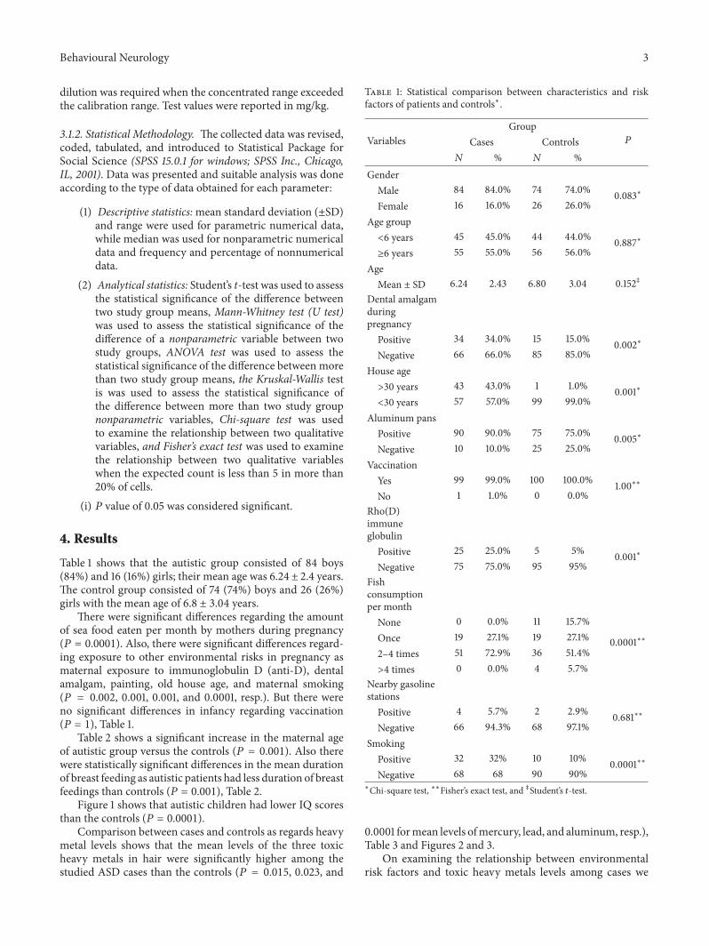
Table 1 shows that the autistic group consisted of 84 boys(84%) and 16 (16%) girls; their mean age was 6.24 ± 2.4 years.The control group consisted of 74 (74%) boys and 26 (26%)girls with the mean age of 6.8 ± 3.04 years.
There were significant differences regarding the amountof sea food eaten per month by mothers during pregnancy(𝑃 = 0.0001). Also, there were significant differences regard-ing exposure to other environmental risks in pregnancy asmaternal exposure to immunoglobulin D (anti-D), dentalamalgam, painting, old house age, and maternal smoking(𝑃 = 0.002, 0.001, 0.001, and 0.0001, resp.). But there wereno significant differences in infancy regarding vaccination(𝑃 = 1), Table 1.
Table 2 shows a significant increase in the maternal ageof autistic group versus the controls (𝑃 = 0.001). Also therewere statistically significant differences in the mean durationof breast feeding as autistic patients had less duration of breastfeedings than controls (𝑃 = 0.001), Table 2.
Figure 1 shows that autistic children had lower IQ scoresthan the controls (𝑃 = 0.0001).
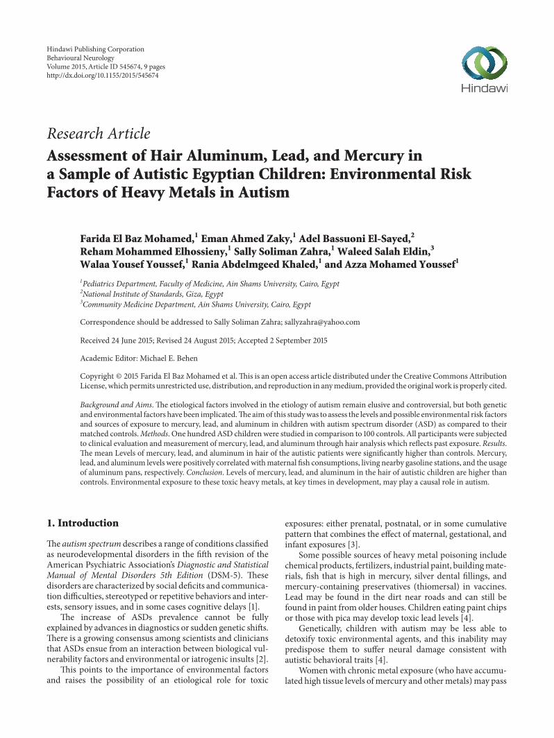
Comparison between cases and controls as regards heavymetal levels shows that the mean levels of the three toxicheavy metals in hair were significantly higher among thestudied ASD cases than the controls (𝑃 = 0.015, 0.023, and
Table 1: Statistical comparison between characteristics and riskfactors of patients and controls∗.
VariablesGroup
𝑃Cases Controls𝑁 % 𝑁 %
GenderMale 84 84.0% 74 74.0% 0.083∗Female 16 16.0% 26 26.0%
Age group<6 years 45 45.0% 44 44.0% 0.887∗≥6 years 55 55.0% 56 56.0%
AgeMean ± SD 6.24 2.43 6.80 3.04 0.152‡
Dental amalgamduringpregnancyPositive 34 34.0% 15 15.0% 0.002∗Negative 66 66.0% 85 85.0%
House age>30 years 43 43.0% 1 1.0% 0.001∗<30 years 57 57.0% 99 99.0%
Aluminum pansPositive 90 90.0% 75 75.0% 0.005∗Negative 10 10.0% 25 25.0%
VaccinationYes 99 99.0% 100 100.0% 1.00∗∗No 1 1.0% 0 0.0%
Rho(D)immuneglobulinPositive 25 25.0% 5 5% 0.001∗Negative 75 75.0% 95 95%
Fishconsumptionper monthNone 0 0.0% 11 15.7%
0.0001∗∗Once 19 27.1% 19 27.1%2–4 times 51 72.9% 36 51.4%>4 times 0 0.0% 4 5.7%
Nearby gasolinestationsPositive 4 5.7% 2 2.9% 0.681∗∗Negative 66 94.3% 68 97.1%
SmokingPositive 32 32% 10 10% 0.0001∗∗Negative 68 68 90 90%
∗Chi-square test, ∗∗Fisher’s exact test, and ‡Student’s 𝑡-test.
0.0001 formean levels ofmercury, lead, and aluminum, resp.),Table 3 and Figures 2 and 3.
On examining the relationship between environmentalrisk factors and toxic heavy metals levels among cases we

4 Behavioural Neurology
Table 2: Comparison between studied groups as regards perinatal data.
GroupPCases Controls
Range Mean ± SD Range Mean ± SDBreast feeding (months) 2.0–24.0 13.5 ± 7.2 2.0–24.0 17.1 ± 4.9 0.001∗
Weaning (months) 2.0–18.0 6.1 ± 3.1 4.0–7.0 5.7 ± 0.9 0.292Age of mother at conception (years) 17.0–40.0 26.5 ± 4.5 19.0–31.0 24.7 ± 3.0 0.005∗∗Student’s 𝑡-test.
Table 3: Comparison between cases and controls as regards heavy metal levels.
GroupPCases Controls
Mean ±SD Median Mean ±SD MedianLead level (mg/kg) 3.31 3.92 2.04 2.06 2.45 1.32 0.015
∗
Mercury level (mg/kg) 0.39 0.37 0.28 0.25 0.16 0.20 0.023∗
Aluminium level (mg/kg) 59.19 37.98 53.00 16.78 17.31 11.11 0.0001∗
∗Mann-Whitney test.
Cases ControlsIQ
0102030405060708090
100
Mea
n IQ
scor
e
Figure 1: Comparison between cases and controls as regards IQscore, 𝑃 = 0.0001, Student’s t-test.
Lead level Mercury level
CasesControls
0
0.5
1
1.5
Mea
n (m
g/kg
)
2
2.5
3
3.5
Figure 2: Mean lead and mercury levels in hair in both groups.
Cases ControlsAluminum level
05
1015202530354045505560657075
Mea
n (m
g/kg
)
Figure 3: Mean aluminum level in hair in both groups.
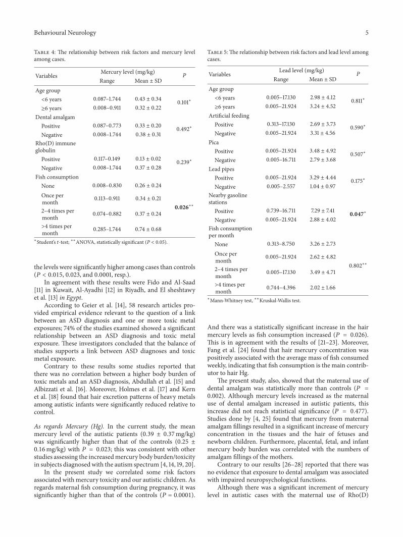
found the following: there was a significant relation betweenfish consumption in the patient and the mercury levels;also the nearby gasoline stations affected the lead levels,while aluminum pans usage increased aluminum level in thestudied autistic patients (𝑃 = 0.026, 0.047, and 0.04, resp.),Tables 4, 5, and 6.
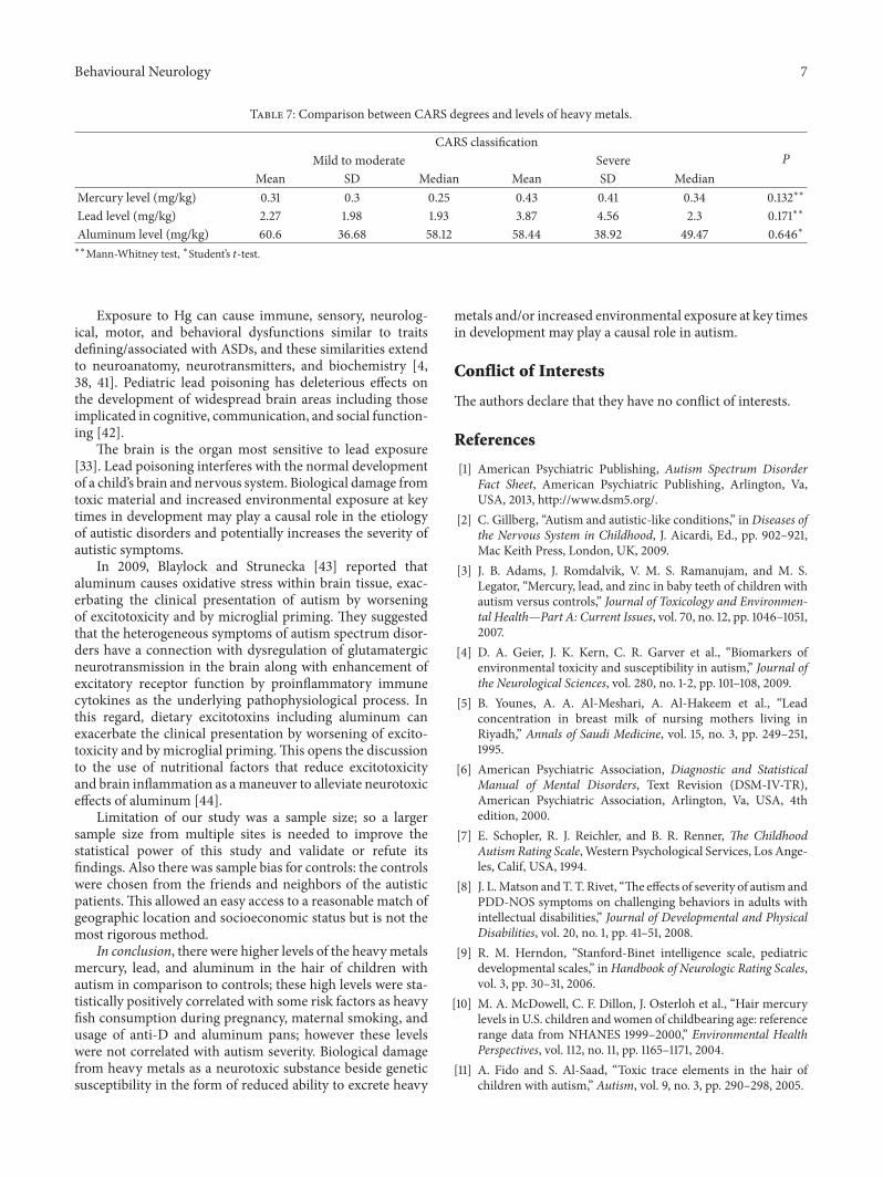
An important finding in the current study was theabsence of significant association between the severity of theautistic manifestations, as measured by the CARS scale, incases and the heavy metals level, Table 7.
5. Discussion
The current study was conducted on 100 autistic children;their ages ranged from 2.5 to 15 years with a mean 6.4 ±2.4. Eighty-four percent of them were males and 16% werefemales, with a male/female ratio of 4 : 1.
On comparing between the levels of lead, mercury, andaluminum in hair of the autistic patients and the controls,
Behavioural Neurology 5
Table 4: The relationship between risk factors and mercury levelamong cases.
Variables Mercury level (mg/kg) PRange Mean ± SD
Age group<6 years 0.087–1.744 0.43 ± 0.34 0.101∗≥6 years 0.008–0.911 0.32 ± 0.22
Dental amalgamPositive 0.087–0.773 0.33 ± 0.20 0.492∗Negative 0.008–1.744 0.38 ± 0.31
Rho(D) immuneglobulin
Positive 0.117–0.149 0.13 ± 0.02 0.239∗Negative 0.008–1.744 0.37 ± 0.28
Fish consumptionNone 0.008–0.830 0.26 ± 0.24
0.026∗∗Once permonth 0.113–0.911 0.34 ± 0.21
2–4 times permonth 0.074–0.882 0.37 ± 0.24
>4 times permonth 0.285–1.744 0.74 ± 0.68
∗Student’s 𝑡-test; ∗∗ANOVA, statistically significant (P < 0.05).
the levels were significantly higher among cases than controls(𝑃 < 0.015, 0.023, and 0.0001, resp.).
In agreement with these results were Fido and Al-Saad[11] in Kuwait, Al-Ayadhi [12] in Riyadh, and El sheshtawyet al. [13] in Egypt.
According to Geier et al. [14], 58 research articles pro-vided empirical evidence relevant to the question of a linkbetween an ASD diagnosis and one or more toxic metalexposures; 74% of the studies examined showed a significantrelationship between an ASD diagnosis and toxic metalexposure. These investigators concluded that the balance ofstudies supports a link between ASD diagnoses and toxicmetal exposure.
Contrary to these results some studies reported thatthere was no correlation between a higher body burden oftoxic metals and an ASD diagnosis, Abdullah et al. [15] andAlbizzati et al. [16]. Moreover, Holmes et al. [17] and Kernet al. [18] found that hair excretion patterns of heavy metalsamong autistic infants were significantly reduced relative tocontrol.
As regards Mercury (Hg). In the current study, the meanmercury level of the autistic patients (0.39 ± 0.37mg/kg)was significantly higher than that of the controls (0.25 ±0.16mg/kg) with 𝑃 = 0.023; this was consistent with otherstudies assessing the increasedmercury body burden/toxicityin subjects diagnosed with the autism spectrum [4, 14, 19, 20].
In the present study we correlated some risk factorsassociated withmercury toxicity and our autistic children. Asregards maternal fish consumption during pregnancy, it wassignificantly higher than that of the controls (𝑃 = 0.0001).
Table 5:The relationship between risk factors and lead level amongcases.
Variables Lead level (mg/kg) PRange Mean ± SD
Age group<6 years 0.005–17.130 2.98 ± 4.12 0.811∗≥6 years 0.005–21.924 3.24 ± 4.52
Artificial feedingPositive 0.313–17.130 2.69 ± 3.73 0.590∗Negative 0.005–21.924 3.31 ± 4.56
PicaPositive 0.005–21.924 3.48 ± 4.92 0.507∗Negative 0.005–16.711 2.79 ± 3.68
Lead pipesPositive 0.005–21.924 3.29 ± 4.44 0.175∗Negative 0.005–2.557 1.04 ± 0.97
Nearby gasolinestationsPositive 0.739–16.711 7.29 ± 7.41 0.047∗Negative 0.005–21.924 2.88 ± 4.02
Fish consumptionper monthNone 0.313–8.750 3.26 ± 2.73
0.802∗∗Once permonth 0.005–21.924 2.62 ± 4.82
2–4 times permonth 0.005–17.130 3.49 ± 4.71
>4 times permonth 0.744–4.396 2.02 ± 1.66
∗Mann-Whitney test, ∗∗Kruskal-Wallis test.
And there was a statistically significant increase in the hairmercury levels as fish consumption increased (𝑃 = 0.026).This is in agreement with the results of [21–23]. Moreover,Fang et al. [24] found that hair mercury concentration waspositively associated with the average mass of fish consumedweekly, indicating that fish consumption is the main contrib-utor to hair Hg.
The present study, also, showed that the maternal use ofdental amalgam was statistically more than controls (𝑃 =0.002). Although mercury levels increased as the maternaluse of dental amalgam increased in autistic patients, thisincrease did not reach statistical significance (𝑃 = 0.477).Studies done by [4, 25] found that mercury from maternalamalgam fillings resulted in a significant increase of mercuryconcentration in the tissues and the hair of fetuses andnewborn children. Furthermore, placental, fetal, and infantmercury body burden was correlated with the numbers ofamalgam fillings of the mothers.
Contrary to our results [26–28] reported that there wasno evidence that exposure to dental amalgam was associatedwith impaired neuropsychological functions.
Although there was a significant increment of mercurylevel in autistic cases with the maternal use of Rho(D)

6 Behavioural Neurology
Table 6: Comparison between selected risk factors and aluminumhair levels among autistic cases.
Aluminum level (mg/kg) PMean ±SD Median
Age group<6 years 63.89 35.51 58.12 0.101∗≥6 years 55.35 39.80 48.91
Fish consumptionYes 49.34 19.68 58.12 0.880∗No 59.50 38.43 52.93
Dental amalgamPositive 55.21 42.73 41.14 0.205∗Negative 61.25 35.46 54.87
House age1 58.06 34.78 52.93 0.931∗∗2 60.05 40.51 53.08
Rho(D) immuneglobulin
Positive 55.30 25.43 54.82 0.994∗Negative 59.40 38.61 52.93
Aluminum pansPositive 61.71 38.46 53.92 0.04∗Negative 36.58 24.78 30.64
∗Student’s 𝑡-test.
immune globulin, this increment was not statistically sig-nificant (𝑃 = 0.239). This finding was supported by [29]that revealed a significant association between total mercuryexposure during the prenatal and early postnatal periodsfrom thimerosal-containing immunoglobulins and the sever-ity of autism. Also, studies done [21, 26] demonstrated thesame results. On the other hand, [30, 31] found that prenatalexposure to thimerosal-containing Rho(D) immune globulindoes not increase the risk of autism.
As regards Lead (Pb). In the current study, themean lead levelin the autistic patients (3.31 ± 3.92mg/kg) was significantlyhigher than that of the controls (2.06 ± 2.45mg/kg) with 𝑃 =0.015. Jiang et al. [32] studied heavy metal concentrations inhair of preschool autistic children and found that hair leadconcentration was significantly elevated.
The current study showed a statistical increase in leadlevels with a presence of nearby gasoline stations (𝑃 < 0.047).This is in agreement with this result, a study done by Naeheret al. [33] who demonstrated that the lead levels of childrenliving nearby gas stations were marginally higher than forchildren living away from gas stations.
As regards Aluminum (Al). In the current study, the meanaluminum level in the autistic patients (59.19±37.98mg/Kg)was significantly higher than that of the controls (16.78 ±17.3198mg/Kg) with 𝑃 = 0.0001. This is in agreement withTomljenovic and Shaw [34], who showed that Al, a highlyneurotoxic metal and the most commonly used vaccine
adjuvant,may be a significant contributing factor to the risingprevalence of ASD in the Western world.
As regards the usage of aluminum pans, it was signif-icantly higher in the studied cases than the controls (𝑃 =0.005). Abu-Taweel et al. [35] documented experimentallythat perinatal oral Al exposure including use of aluminumpans, particularly during pregnancy and lactation period, canaffect the in utero developing fetus of mice. So aluminumexposure has potential and long lasting neurotoxic hazardsand might modify the properties of the dopaminergic systemand thus can change the threshold of that system or otherrelated systems at later ages.
An interesting result in our study was the absence ofstatistically significant relation between the levels of mer-cury, lead, and aluminum and autism severity. This wasnot consistent with [13, 36] that found that, on comparinghair concentration of autistic cases versus controls, elevatedhair concentrations were noted for heavy metals in autisticchildren and correlated with the severity of symptoms. Also,Adams et al. [37] found that the body burden of toxic metalswas significantly related to the variations in the severityof autism. The metals of greatest influence were lead (Pb),mercury (Hg), and aluminum (Al). Geier et al. [38] suggestedthat the impact of toxic metals may be more evident insubjects diagnosed with moderate to severe ASD as opposedto participants diagnosed with a mild ASD.
It may be argued that children with ASD are not the onlychildren exposed to potentially toxic metals; the reason whyautistic patients show greater concentration of potentiallytoxic metals in tissue may be the result of a greater abilityto accumulate toxins, which in turn leads to an alteration ofbiochemical processes. Also, children with autistic spectrumdisorders displayed lower levels of the nutritional elementscalcium, copper, chromium, manganese, magnesium, iron,selenium, and cobalt. Since autistic children display pooreating habits, the low tissue levels may be explained by aninadequate nutritional intake.
Therefore, it is believed that ASD patients have problemswith the chemical pathway that allows them to detoxifymetals to alleviate different cluster of autistic symptoms [39].Evidence shows that autistic children show an increasedbuild-up of toxins which may not arise simply from excessiveexposure but fromamarked inability to process and eliminatetoxins from the body. Such amechanism could lead to a back-up of toxic heavy metals and chemical toxins and increasesfree radical activity in the body [40].
Adams et al. [37] observed that toxic metal excre-tion pathways may significantly vary among study subjectsdiagnosed with moderate to severe ASD as opposed toparticipants diagnosed with a mild ASD. This may be ofa particular importance when examining hair toxic metalconcentrations in young children because previous studieshave suggested that hair toxic concentrations may be relatedto toxic metal excretion rates. Emerging evidence supportsthe theory that some ASDs may result from a combinationof genetic/biochemical susceptibility, specifically a reducedability to excrete mercury (Hg), and exposure to Hg at criticaldevelopmental periods [4, 38, 41].
Behavioural Neurology 7
Table 7: Comparison between CARS degrees and levels of heavy metals.
CARS classificationPMild to moderate Severe
Mean SD Median Mean SD MedianMercury level (mg/kg) 0.31 0.3 0.25 0.43 0.41 0.34 0.132∗∗
Lead level (mg/kg) 2.27 1.98 1.93 3.87 4.56 2.3 0.171∗∗
Aluminum level (mg/kg) 60.6 36.68 58.12 58.44 38.92 49.47 0.646∗∗∗Mann-Whitney test, ∗Student’s 𝑡-test.
Exposure to Hg can cause immune, sensory, neurolog-ical, motor, and behavioral dysfunctions similar to traitsdefining/associated with ASDs, and these similarities extendto neuroanatomy, neurotransmitters, and biochemistry [4,38, 41]. Pediatric lead poisoning has deleterious effects onthe development of widespread brain areas including thoseimplicated in cognitive, communication, and social function-ing [42].
The brain is the organ most sensitive to lead exposure[33]. Lead poisoning interferes with the normal developmentof a child’s brain and nervous system. Biological damage fromtoxic material and increased environmental exposure at keytimes in development may play a causal role in the etiologyof autistic disorders and potentially increases the severity ofautistic symptoms.
In 2009, Blaylock and Strunecka [43] reported thataluminum causes oxidative stress within brain tissue, exac-erbating the clinical presentation of autism by worseningof excitotoxicity and by microglial priming. They suggestedthat the heterogeneous symptoms of autism spectrum disor-ders have a connection with dysregulation of glutamatergicneurotransmission in the brain along with enhancement ofexcitatory receptor function by proinflammatory immunecytokines as the underlying pathophysiological process. Inthis regard, dietary excitotoxins including aluminum canexacerbate the clinical presentation by worsening of excito-toxicity and by microglial priming.This opens the discussionto the use of nutritional factors that reduce excitotoxicityand brain inflammation as amaneuver to alleviate neurotoxiceffects of aluminum [44].
Limitation of our study was a sample size; so a largersample size from multiple sites is needed to improve thestatistical power of this study and validate or refute itsfindings. Also there was sample bias for controls: the controlswere chosen from the friends and neighbors of the autisticpatients.This allowed an easy access to a reasonable match ofgeographic location and socioeconomic status but is not themost rigorous method.
In conclusion, there were higher levels of the heavymetalsmercury, lead, and aluminum in the hair of children withautism in comparison to controls; these high levels were sta-tistically positively correlated with some risk factors as heavyfish consumption during pregnancy, maternal smoking, andusage of anti-D and aluminum pans; however these levelswere not correlated with autism severity. Biological damagefrom heavy metals as a neurotoxic substance beside geneticsusceptibility in the form of reduced ability to excrete heavy
metals and/or increased environmental exposure at key timesin development may play a causal role in autism.
Conflict of Interests
The authors declare that they have no conflict of interests.
References
[1] American Psychiatric Publishing, Autism Spectrum DisorderFact Sheet, American Psychiatric Publishing, Arlington, Va,USA, 2013, http://www.dsm5.org/.
[2] C. Gillberg, “Autism and autistic-like conditions,” inDiseases ofthe Nervous System in Childhood, J. Aicardi, Ed., pp. 902–921,Mac Keith Press, London, UK, 2009.
[3] J. B. Adams, J. Romdalvik, V. M. S. Ramanujam, and M. S.Legator, “Mercury, lead, and zinc in baby teeth of children withautism versus controls,” Journal of Toxicology and Environmen-tal Health—Part A: Current Issues, vol. 70, no. 12, pp. 1046–1051,2007.
[4] D. A. Geier, J. K. Kern, C. R. Garver et al., “Biomarkers ofenvironmental toxicity and susceptibility in autism,” Journal ofthe Neurological Sciences, vol. 280, no. 1-2, pp. 101–108, 2009.
[5] B. Younes, A. A. Al-Meshari, A. Al-Hakeem et al., “Leadconcentration in breast milk of nursing mothers living inRiyadh,” Annals of Saudi Medicine, vol. 15, no. 3, pp. 249–251,1995.
[6] American Psychiatric Association, Diagnostic and StatisticalManual of Mental Disorders, Text Revision (DSM-IV-TR),American Psychiatric Association, Arlington, Va, USA, 4thedition, 2000.
[7] E. Schopler, R. J. Reichler, and B. R. Renner, The ChildhoodAutism Rating Scale, Western Psychological Services, Los Ange-les, Calif, USA, 1994.
[8] J. L.Matson andT. T. Rivet, “The effects of severity of autism andPDD-NOS symptoms on challenging behaviors in adults withintellectual disabilities,” Journal of Developmental and PhysicalDisabilities, vol. 20, no. 1, pp. 41–51, 2008.
[9] R. M. Herndon, “Stanford-Binet intelligence scale, pediatricdevelopmental scales,” inHandbook of Neurologic Rating Scales,vol. 3, pp. 30–31, 2006.
[10] M. A. McDowell, C. F. Dillon, J. Osterloh et al., “Hair mercurylevels inU.S. children andwomen of childbearing age: referencerange data from NHANES 1999–2000,” Environmental HealthPerspectives, vol. 112, no. 11, pp. 1165–1171, 2004.
[11] A. Fido and S. Al-Saad, “Toxic trace elements in the hair ofchildren with autism,” Autism, vol. 9, no. 3, pp. 290–298, 2005.
8 Behavioural Neurology
[12] L. Y. Al-Ayadhi, “Heavy metals and trace elements in hair sam-ples of autistic children in central Saudi Arabia,” Neurosciences,vol. 10, no. 3, pp. 213–218, 2005.
[13] E. Elsheshtawy, S. Tobar, K. Sherra, S. Atallah, and R. Elkasaby,“Study of some biomarkers in hair of children with autism,”Middle East Current Psychiatry, vol. 18, no. 1, pp. 6–10, 2011.
[14] D. A. Geier, J. K. Kern, P. G. King, L. K. Sykes, and M. R.Geier, “Hair toxic metal concentrations and autism spectrumdisorder severity in young children,” International Journal ofEnvironmental Research and Public Health, vol. 9, no. 12, pp.4486–4497, 2012.
[15] M. M. Abdullah, A. R. Ly, W. A. Goldberg et al., “Heavy metalin children’s tooth enamel: related to autism and disruptivebehaviors?” Journal of Autism andDevelopmental Disorders, vol.42, no. 6, pp. 929–936, 2012.
[16] A. Albizzati, L. More, D. Di Candia, M. Saccani, and C. Lenti,“Normal concentrations of heavy metals in autistic spectrumdisorders,”Minerva Pediatrica, vol. 64, no. 1, pp. 27–31, 2012.
[17] A. S. Holmes, M. F. Blaxill, and B. E. Haley, “Reduced levels ofmercury in first baby haircuts of autistic children,” InternationalJournal of Toxicology, vol. 22, no. 4, pp. 277–285, 2003.
[18] J. K. Kern, B. D. Grannemann, M. H. Trivedi, and J. B. Adams,“Sulfhydryl-reactivemetals in autism,” Journal of Toxicology andEnvironmental Health, Part A: Current Issues, vol. 70, no. 8, pp.715–721, 2007.
[19] F. El-baz, R.M. Elhossiny, A. B. Elsayed, and G.M. Gaber, “Hairmercury measurement in Egyptian autistic children,” EgyptianJournal of Medical Human Genetics, vol. 11, no. 2, pp. 135–141,2010.
[20] Y. M. Al-Farsi, M. M. Al-Sharbati, M. I. Waly et al., “Effectof suboptimal breast-feeding on occurrence of autism: a case-control study,” Nutrition, vol. 28, no. 7-8, pp. e27–e32, 2012.
[21] E. Blaurock-Busch, O. R. Amin, and T. Rabah, “Heavy metalsand trace elements in hair and urine of a sample of Arabchildren with autistic spectrum disorder,” Maedica, vol. 6, no.4, pp. 247–257, 2011.
[22] P. Li, X. Feng, P. Liang, H. Man Chan, H. Yan, and L. Chen,“Mercury in the seafood and human exposure in coastal area ofGuangdong Province, South China,” Environmental Toxicologyand Chemistry, vol. 32, no. 3, pp. 541–547, 2012.
[23] J. G. Dorea, R. C. Marques, and C. Isejima, “Neurodevelopmentof Amazonian infants: antenatal and postnatal exposure tomethyl- and ethylmercury,” Journal of Biomedicine and Biotech-nology, vol. 2012, Article ID 132876, 9 pages, 2012.
[24] T. Fang, K. J. Aronson, and L. M. Campbell, “Freshwater fish-consumption relations with total hair mercury and seleniumamong women in Eastern China,” Archives of EnvironmentalContamination and Toxicology, vol. 62, no. 2, pp. 323–332, 2012.
[25] L. Palkovicova, M. Ursinyova, V. Masanova, Z. Yu, and I. Hertz-Picciotto, “Maternal amalgam dental fillings as the source ofmercury exposure in developing fetus and newborn,” Journal ofExposure Science and Environmental Epidemiology, vol. 18, no.3, pp. 326–331, 2008.
[26] J. B. Adams, C. E. Holloway, F. George, and D. Quig, “Analysesof toxic metals and essential minerals in the hair of Arizonachildren with autism and associated conditions, and theirmothers,” Biological Trace Element Research, vol. 110, no. 3, pp.193–209, 2006.
[27] S. S. Yalcin, K. Yurdakok, S. Yalcin, D. Engur-Karasimav, and T.Coskun, “Maternal and environmental determinants of breast-milkmercury concentrations,”Turkish Journal of Pediatrics, vol.52, no. 1, pp. 1–9, 2010.
[28] A. Dursun, The levels of lead, mercury, cadmium in cordblood, breast milk and newborn hair [Ph.D. thesis], HacettepeUniversity, Ankara, Turkey, 2008.
[29] D. A. Geier andM. R. Geier, “A prospective study of thimerosal-containing Rho(D)-immune globulin administration as a riskfactor for autistic disorders,” Journal of Maternal-Fetal andNeonatal Medicine, vol. 20, no. 5, pp. 385–390, 2007.
[30] L. A. Croen, M. Matevia, C. K. Yoshida, and J. K. Grether,“Maternal Rh D status, anti-D immune globulin exposureduring pregnancy, and risk of autism spectrum disorders,”American Journal of Obstetrics and Gynecology, vol. 199, no. 3,pp. 234.e1–234.e6, 2008.
[31] C. S. Price, W. W. Thompson, B. Goodson et al., “Prenatal andinfant exposure to thimerosal from vaccines and immunoglob-ulins and risk of autism,” Pediatrics, vol. 126, no. 4, pp. 656–664,2010.
[32] H.-X. Jiang, L.-S. Chen, J.-G. Zheng, S. Han, N. Tang, andB. R. Smith, “Aluminum-induced effects on Photosystem IIphotochemistry in citrus leaves assessed by the chlorophyll afluorescence transient,”Tree Physiology, vol. 28, no. 12, pp. 1863–1871, 2008.
[33] L. P. Naeher, M. Aguilar-Villalobos, and T. Miller, “Bloodlead survey of children, pregnant women, professional drivers,street workers, and office workers in Trujillo, Peru,” Archives ofEnvironmental Health, vol. 59, no. 7, pp. 359–362, 2004.
[34] L. Tomljenovic and C. A. Shaw, “Aluminum vaccine adjuvants:are they safe?” Current Medicinal Chemistry, vol. 18, no. 17, pp.2630–2637, 2011.
[35] G. M. Abu-Taweel, J. S. Ajarem, and M. Ahmad, “Neurobehav-ioral toxic effects of perinatal oral exposure to aluminum onthe developmental motor reflexes, learning, memory and brainneurotransmitters of mice offspring,” Pharmacology Biochem-istry and Behavior, vol. 101, no. 1, pp. 49–56, 2012.
[36] E. Blaurock-Busch, O. R. Amin, H. H. Dessoki, and T. Rabah,“Toxic metals and essential elements in hair and severity ofsymptoms among children with autism,” Maedica, vol. 7, no. 1,pp. 38–48, 2012.
[37] J. B. Adams, M. Baral, E. Geis et al., “The severity of autismis associated with toxic metal body burden and red blood cellglutathione levels,” Journal of Toxicology, vol. 2009, Article ID532640, 7 pages, 2009.
[38] D. A. Geier, P. G. King, L. K. Sykes, and M. R. Geier, “Acomprehensive review of mercury provoked autism,” IndianJournal of Medical Research, vol. 128, no. 4, pp. 383–411, 2008.
[39] M. D. L. Priya and A. Geetha, “Level of trace elements (copper,zinc, magnesium and selenium) and toxic elements (lead andmercury) in the hair and nail of childrenwith autism,”BiologicalTrace Element Research, vol. 142, no. 2, pp. 148–158, 2011.
[40] J. Mutter, J. Naumann, R. Schneider, H. Walach, and B. Haley,“Mercury and autism: accelerating evidence,” Neuroendocrinol-ogy Letters, vol. 26, no. 5, pp. 439–446, 2005.
[41] D. Austin, “An epidemiological analysis of the ‘autism asmercury poisoning’ hypothesis,” International Journal of Riskand Safety in Medicine, vol. 20, no. 3, pp. 135–142, 2008.
Behavioural Neurology 9
[42] T. I. Lidsky and J. S. Schneider, “Lead neurotoxicity in children:basic mechanisms and clinical correlates,” Brain, vol. 126, part 1,pp. 5–19, 2003.
[43] R. L. Blaylock and A. Strunecka, “Immune-glutamatergic dys-function as a central mechanism of the autism spectrumdisorders,” Current Medicinal Chemistry, vol. 16, no. 2, pp. 157–170, 2009.
[44] S.W. Bihaqi,M. Sharma, A. P. Singh, andM. Tiwari, “Neuropro-tective role of Convolvulus pluricaulis on aluminium inducedneurotoxicity in rat brain,” Journal of Ethnopharmacology, vol.124, no. 3, pp. 409–415, 2009.

Submit your manuscripts athttp://www.hindawi.com
Stem CellsInternational
Hindawi Publishing Corporationhttp://www.hindawi.com Volume 2014
Hindawi Publishing Corporationhttp://www.hindawi.com Volume 2014
MEDIATORSINFLAMMATION
of
Hindawi Publishing Corporationhttp://www.hindawi.com Volume 2014
Behavioural Neurology
EndocrinologyInternational Journal of
Hindawi Publishing Corporationhttp://www.hindawi.com Volume 2014
Hindawi Publishing Corporationhttp://www.hindawi.com Volume 2014
Disease Markers
Hindawi Publishing Corporationhttp://www.hindawi.com Volume 2014
BioMed Research International
OncologyJournal of
Hindawi Publishing Corporationhttp://www.hindawi.com Volume 2014
Hindawi Publishing Corporationhttp://www.hindawi.com Volume 2014
Oxidative Medicine and Cellular Longevity
Hindawi Publishing Corporationhttp://www.hindawi.com Volume 2014
PPAR Research
The Scientific World JournalHindawi Publishing Corporation http://www.hindawi.com Volume 2014
Immunology ResearchHindawi Publishing Corporationhttp://www.hindawi.com Volume 2014
Journal of
ObesityJournal of
Hindawi Publishing Corporationhttp://www.hindawi.com Volume 2014
Hindawi Publishing Corporationhttp://www.hindawi.com Volume 2014
Computational and Mathematical Methods in Medicine
OphthalmologyJournal of
Hindawi Publishing Corporationhttp://www.hindawi.com Volume 2014
Diabetes ResearchJournal of
Hindawi Publishing Corporationhttp://www.hindawi.com Volume 2014
Hindawi Publishing Corporationhttp://www.hindawi.com Volume 2014
Research and TreatmentAIDS
Hindawi Publishing Corporationhttp://www.hindawi.com Volume 2014
Gastroenterology Research and Practice
Hindawi Publishing Corporationhttp://www.hindawi.com Volume 2014
Parkinson’s Disease
Evidence-Based Complementary and Alternative Medicine
Volume 2014Hindawi Publishing Corporationhttp://www.hindawi.com