Embed Size (px)
Citation preview
BioMed Central
Reproductive Biology and Endocrinology
ss
Open AcceResearchMale mice with deleted Wolframin (Wfs1) gene have reduced fertilityKlari Noormets*1, Sulev Kõks2,3, Ants Kavak3, Andres Arend4, Marina Aunapuu4, Aivi Keldrimaa1, Eero Vasar2 and Vallo Tillmann1Address: 1Department of Paediatrics, University of Tartu, 6 Lunini Street, 51014 Tartu, Estonia, 2Department of Physiology, University of Tartu, 19 Ravila Street, 50411 Tartu, Estonia, 3Institute of Veterinary Medicine and Animal Sciences, Estonian University of Life Sciences, 62 Kreutzwaldi Street, 51014 Tartu, Estonia and 4Department of Anatomy, Chair of Histology and Embryology, University of Tartu, 19 Ravila Street, 50411 Tartu, Estonia
Email: Klari Noormets* - [email protected]; Sulev Kõks - [email protected]; Ants Kavak - [email protected]; Andres Arend - [email protected]; Marina Aunapuu - [email protected]; Aivi Keldrimaa - [email protected]; Eero Vasar - [email protected]; Vallo Tillmann - [email protected]
* Corresponding author
AbstractBackground: Wolfram Syndrome (WS) is an autosomal recessive disorder characterised by non-autoimmune diabetes mellitus, optic atrophy, cranial diabetes insipidus and sensorineural deafness.Some reports have described hypogonadism in male WS patients. The aim of our study was to findout whether Wfs1 deficient (Wfs1KO) male mice have reduced fertility and, if so, to examinepossible causes.
Methods: Wfs1KO mice were generated by homologous recombination. Both Wfs1KO and wildtype (wt) male mice were mated with wt female mice. The number of litters and the number ofpups were counted and pregnancy rates calculated. The motility and morphology of the sperm andthe histology of testes were analysed. Serum testosterone and FSH concentrations were alsomeasured.
Results: The pregnancy rate in wt females mated with Wfs1KO males was significantly lower thanin the control group (15% vs. 32%; p < 0.05), but there was no significant difference in litter size.Analysis of male fertility showed that, in the Wfs1KO group, eight males out of 13 had pupswhereas in the control group all 13 males had at least one litter. Sperm motility was not affectedin Wfs1KO mice, but Wfs1KO males had less proximal bent tails (14.4 +/- 1.2% vs. 21.5 +/- 1.3 p< 0.05) and less abnormal sperm heads (22.8 +/- 1.8 vs. 31.5 +/- 3.5, p < 0.05) than wt males. Testeshistology revealed significantly reduced number of spermatogonia (23.9 +/- 4.9 vs. 38.1 +/- 2.8; p <0.05) and Sertoli cells (6.4 +/- 0.5 vs. 9.2 +/- 1.0; p < 0.05) in Wfs1KO mice. Serum testosteroneand FSH concentrations did not differ between the two groups.
Conclusion: The impaired fertility of Wfs1KO male mice is most likely due to changes in spermmorphology and reduced number of spermatogenic cells. The exact mechanism through which theWfs1 gene influences sperm morphology needs to be clarified in further studies.
Published: 10 August 2009
Reproductive Biology and Endocrinology 2009, 7:82 doi:10.1186/1477-7827-7-82
Received: 19 March 2009Accepted: 10 August 2009
This article is available from: http://www.rbej.com/content/7/1/82
© 2009 Noormets et al; licensee BioMed Central Ltd. This is an Open Access article distributed under the terms of the Creative Commons Attribution License (http://creativecommons.org/licenses/by/2.0), which permits unrestricted use, distribution, and reproduction in any medium, provided the original work is properly cited.
Page 1 of 8(page number not for citation purposes)
Reproductive Biology and Endocrinology 2009, 7:82 http://www.rbej.com/content/7/1/82
BackgroundWolfram syndrome (WS), also known as DIDMOAD syn-drome, was first described by Wolfram and Wagener in1938. It is an autosomal recessive disorder usually diag-nosed in childhood when non-autoimmune type I diabe-tes occurs with optic atrophy, cranial diabetes insipidusand sensorineural deafness [1,2]. Other abnormalitiesrelated to this syndrome are dilated renal outflow tracts,multiple neurological abnormalities and various neuro-logical and psychiatric disorders [2-5]. Involvement of thehypothalamus, brain stem (central sleep apnoea), andcerebellum (ataxia) may develop in the third decade orlater [1].
Wolfram syndrome is caused by mutation in the Wfs1gene on chromosome 4p16 [6]. This gene is responsiblefor encoding wolframin, a glycoprotein of the endoplas-mic reticulum, although the function of the wolframinprotein is not fully understood [7-9]. There is growing evi-dence that Wfs1 plays an important role in the pathogen-esis of endoplasmic reticulum (ER) stress and apoptosis[8-10]. Genetic association studies have also indicated therole of Wfs1 in the development of type 2 diabetes [11].
As yet there has been no data regarding the fertility ofpatients with WS. Previous studies have described anteriorpituitary dysfunction [5] and, in male patients, the pres-ence of primary gonadal atrophy and hypergonadotropichypogonadism [2-5]. As far as we know, the role of theWfs1 gene in fertility has not been studied.
Mice lacking the Wfs1 gene (Wfs1KO) were created at theLaboratory of Physiology, University of Tartu [12]. Thisanimal model of WS is useful to study the various organ-systems of WS, including fertility.
The aim of our study was to determine whether the fertil-ity of Wfs1KO male mice is reduced and if so, to explorepossible reasons. Regarding the possible causes ofimpaired fertility, we have focused on sperm morphology.
MethodsAnimalsIn accordance with the European Communities Directive(86/609/EEC), the Estonian National Board of AnimalExperiments granted permission (No. 86, 28.08. 2007)for the animal experiments described in this study. Micewere housed under standard laboratory conditions on a12-hour light/dark cycle (lights on at 07:00 hours) withfree access to food and water.
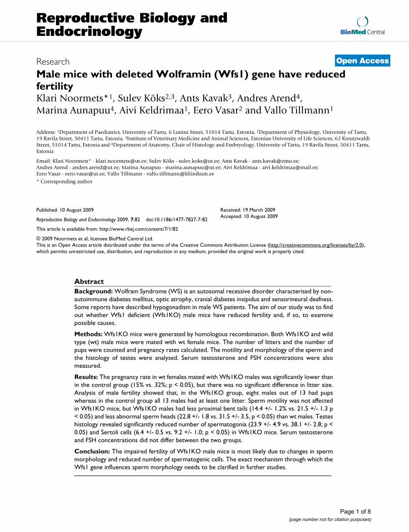
Wfs1 deficient (Wfs1KO) mice were generated by target-ing construct to replace most of the coding region of thewfs1 gene (Figure 1). Briefly, the 8.8 kb BamHI restrictionfragment from the PAC clone 391-J24 (RPCI21 library,MRC UK HGMP Resource Centre, UK) was subcloned
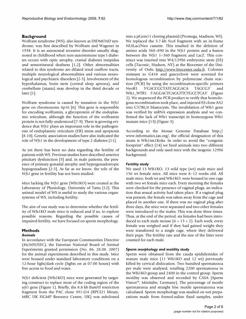
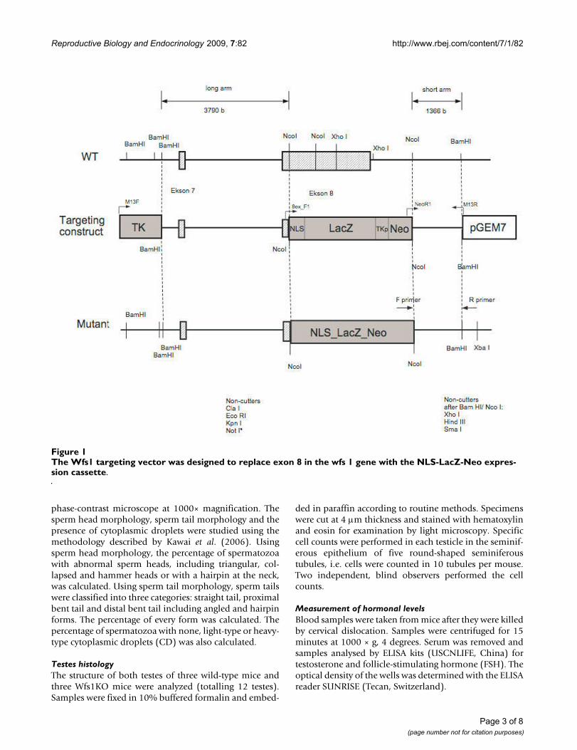
into a pGem11 cloning plasmid (Promega, Madison, WI).We replaced the 3.7-kb NcoI fragment with an in-frameNLSLacZNeo cassette. This resulted in the deletion ofamino acids 360–890 in the Wfs1 protein and a fusionbetween the Wfs1 1–360 fragment and LacZ. This con-struct was inserted into W4/129S6 embryonic stem (ES)cells (Taconic, Hudson, NY) at the Biocenter of the Uni-versity of Oulu http://www.biocenter.oulu.fi. Coloniesresistant to G418 and gancyclovir were screened forhomologous recombination by polymerase chain reac-tion (PCR) by using the recombination-specific primersNeoR1 5'GACCGCTATCAGGACA TAGCG3' andWfs1_WTR1 5'AGGACTCAGGTTCTGCCTCA3' (Figure2). We sequenced the PCR product to verify that homolo-gous recombination took place, and injected ES clone 8A2into C57BL/6 blastocysts. The invalidation of Wfs1 genewas verified by mRNA expression analysis and we con-firmed the lack of Wfs1 transcript in homozygous Wfs1mutant mice [13] (Figure 3).
According to the Mouse Genome Database http://www.informatics.jax.org/, the official designation of thisstrain is Wfs1tm1Koks. In order to avoid the "congenicfootprint" effect [14] we bred animals into two differentbackgrounds and only used mice with the isogenic 129S6background.
Fertility studyWe used 13 Wfs1KO, 13 wild type (wt) male mice and156 wt female mice. All mice were 8–12 weeks old. Allmale mice, both wt and Wfs1KO, were housed in one cagewith two wt female mice each. Every morning the femaleswere checked for the presence of vaginal plugs, an indica-tion that sexual activity had taken place. If a vaginal plugwas present, the female was taken away from the cage andplaced in another one. If there was no vaginal plug afterthree days, the mice were separated and two other femaleswere introduced to the males. This was done three times.Thus, at the end of the period, six females had been intro-duced to each male mouse (6 × 13 × 2). Each week, everyfemale was weighed and if they had gained weight theywere transferred to a single cage, where they deliveredtheir pups. The fertility rate and the size of the litter werecounted for each male.
Sperm morphology and motility studySperm were obtained from the cauda epididymides ofmature male mice (11 Wfs1KO and 12 wt) previouslykilled by cervical dislocation. Two hundred spermatozoaper male were analysed, totalling 2200 spermatozoa inthe Wfs1KO group and 2400 in the control group. Spermmotility was observed and recorded by CASA (SpermVision™, Minitübe, Germany). The percentage of motilespermatozoa and straight line motile spermatozoa wascalculated. Sperm morphology was studied on wet prepa-rations made from formol-saline fixed samples, under
Page 2 of 8(page number not for citation purposes)
Reproductive Biology and Endocrinology 2009, 7:82 http://www.rbej.com/content/7/1/82
phase-contrast microscope at 1000× magnification. Thesperm head morphology, sperm tail morphology and thepresence of cytoplasmic droplets were studied using themethodology described by Kawai et al. (2006). Usingsperm head morphology, the percentage of spermatozoawith abnormal sperm heads, including triangular, col-lapsed and hammer heads or with a hairpin at the neck,was calculated. Using sperm tail morphology, sperm tailswere classified into three categories: straight tail, proximalbent tail and distal bent tail including angled and hairpinforms. The percentage of every form was calculated. Thepercentage of spermatozoa with none, light-type or heavy-type cytoplasmic droplets (CD) was also calculated.
Testes histologyThe structure of both testes of three wild-type mice andthree Wfs1KO mice were analyzed (totalling 12 testes).Samples were fixed in 10% buffered formalin and embed-
ded in paraffin according to routine methods. Specimenswere cut at 4 μm thickness and stained with hematoxylinand eosin for examination by light microscopy. Specificcell counts were performed in each testicle in the seminif-erous epithelium of five round-shaped seminiferoustubules, i.e. cells were counted in 10 tubules per mouse.Two independent, blind observers performed the cellcounts.
Measurement of hormonal levelsBlood samples were taken from mice after they were killedby cervical dislocation. Samples were centrifuged for 15minutes at 1000 × g, 4 degrees. Serum was removed andsamples analysed by ELISA kits (USCNLIFE, China) fortestosterone and follicle-stimulating hormone (FSH). Theoptical density of the wells was determined with the ELISAreader SUNRISE (Tecan, Switzerland).
The Wfs1 targeting vector was designed to replace exon 8 in the wfs 1 gene with the NLS-LacZ-Neo expression cassetteFigure 1The Wfs1 targeting vector was designed to replace exon 8 in the wfs 1 gene with the NLS-LacZ-Neo expres-sion cassette.
Page 3 of 8(page number not for citation purposes)
Reproductive Biology and Endocrinology 2009, 7:82 http://www.rbej.com/content/7/1/82
Statistical analysisAll data was analysed using the statistical software pack-age, SAS version 9.1 (SAS Institute Inc, Cary, North Caro-lina, USA). The Chi-square test or Fisher's Exact Test(when expected values were <5%) was used to comparethe fertility rates, and the Student's t-test was used to com-pare the sperm morphology, litter size, occurrence of thevaginal plugs and concentrations of the hormonesbetween the groups. Mean ± SEM are shown. P values <0.05 were considered statistically significant.
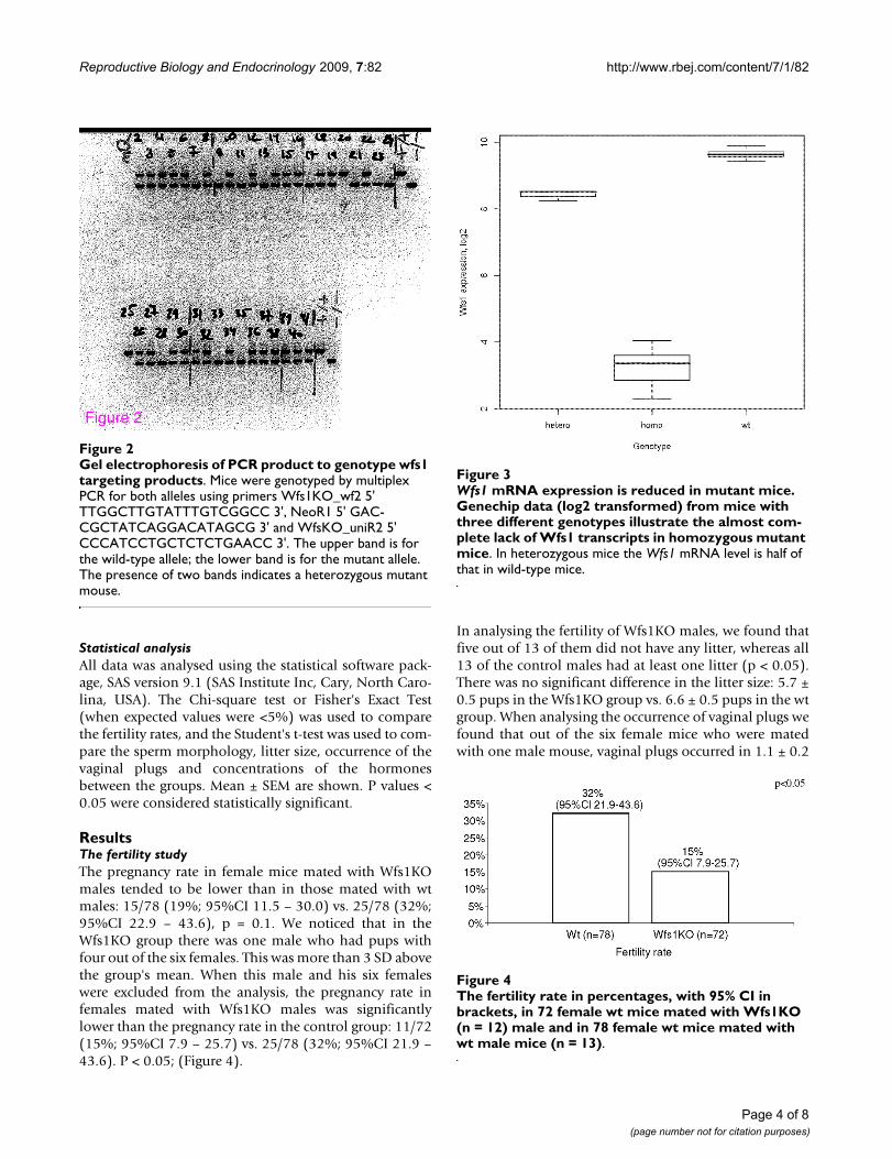
ResultsThe fertility studyThe pregnancy rate in female mice mated with Wfs1KOmales tended to be lower than in those mated with wtmales: 15/78 (19%; 95%CI 11.5 – 30.0) vs. 25/78 (32%;95%CI 22.9 – 43.6), p = 0.1. We noticed that in theWfs1KO group there was one male who had pups withfour out of the six females. This was more than 3 SD abovethe group's mean. When this male and his six femaleswere excluded from the analysis, the pregnancy rate infemales mated with Wfs1KO males was significantlylower than the pregnancy rate in the control group: 11/72(15%; 95%CI 7.9 – 25.7) vs. 25/78 (32%; 95%CI 21.9 –43.6). P < 0.05; (Figure 4).
In analysing the fertility of Wfs1KO males, we found thatfive out of 13 of them did not have any litter, whereas all13 of the control males had at least one litter (p < 0.05).There was no significant difference in the litter size: 5.7 ±0.5 pups in the Wfs1KO group vs. 6.6 ± 0.5 pups in the wtgroup. When analysing the occurrence of vaginal plugs wefound that out of the six female mice who were matedwith one male mouse, vaginal plugs occurred in 1.1 ± 0.2
Gel electrophoresis of PCR product to genotype wfs1 tar-geting productsFigure 2Gel electrophoresis of PCR product to genotype wfs1 targeting products. Mice were genotyped by multiplex PCR for both alleles using primers Wfs1KO_wf2 5' TTGGCTTGTATTTGTCGGCC 3', NeoR1 5' GAC-CGCTATCAGGACATAGCG 3' and WfsKO_uniR2 5' CCCATCCTGCTCTCTGAACC 3'. The upper band is for the wild-type allele; the lower band is for the mutant allele. The presence of two bands indicates a heterozygous mutant mouse.
Figure 2
Wfs1 mRNA expression is reduced in mutant miceFigure 3Wfs1 mRNA expression is reduced in mutant mice. Genechip data (log2 transformed) from mice with three different genotypes illustrate the almost com-plete lack of Wfs1 transcripts in homozygous mutant mice. In heterozygous mice the Wfs1 mRNA level is half of that in wild-type mice.
The fertility rate in percentages, with 95% CI in brackets, in 72 female wt mice mated with Wfs1KO (n = 12) male and in 78 female wt mice mated with wt male mice (n = 13)Figure 4The fertility rate in percentages, with 95% CI in brackets, in 72 female wt mice mated with Wfs1KO (n = 12) male and in 78 female wt mice mated with wt male mice (n = 13).
Page 4 of 8(page number not for citation purposes)
Reproductive Biology and Endocrinology 2009, 7:82 http://www.rbej.com/content/7/1/82
females in the Wfs1KO group, compared to their occur-rence in 2.5 ± 0.4 females out of six in the wt group (p <0.05).
Sperm motility and morphology studySperm motility was not affected in Wfs1KO mice. Surpris-ingly, the mean percentage of motile sperm was evenhigher in the Wfs1KO mice than in the wt mice, whereasno statistical differences were observed in the percentageof straight motility (Table 1).
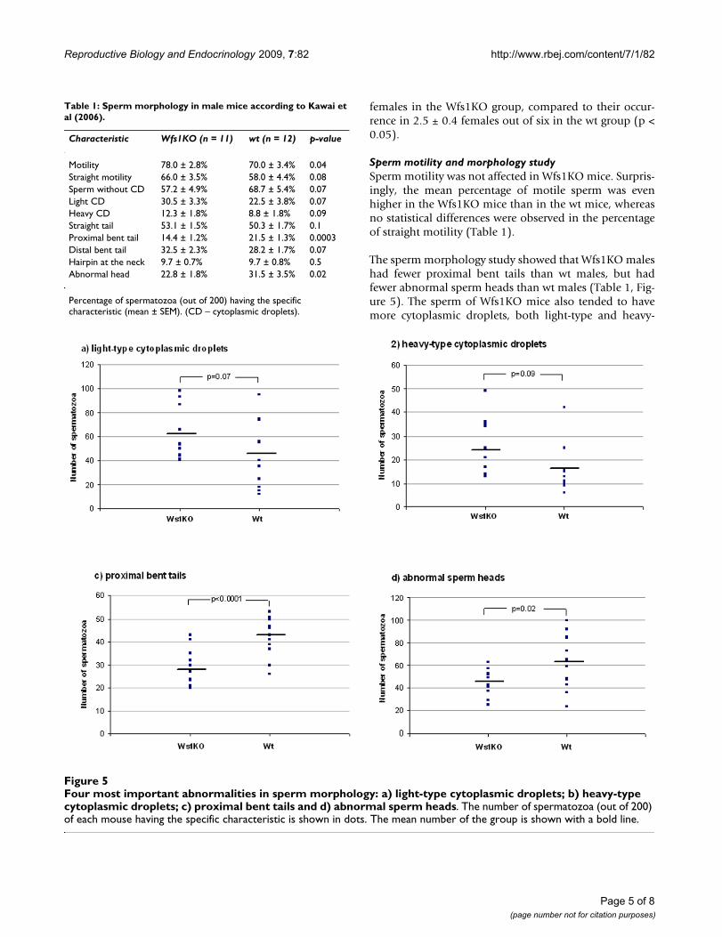
The sperm morphology study showed that Wfs1KO maleshad fewer proximal bent tails than wt males, but hadfewer abnormal sperm heads than wt males (Table 1, Fig-ure 5). The sperm of Wfs1KO mice also tended to havemore cytoplasmic droplets, both light-type and heavy-
Four most important abnormalities in sperm morphology: a) light-type cytoplasmic droplets; b) heavy-type cytoplasmic drop-lets; c) proximal bent tails and d) abnormal sperm headsFigure 5Four most important abnormalities in sperm morphology: a) light-type cytoplasmic droplets; b) heavy-type cytoplasmic droplets; c) proximal bent tails and d) abnormal sperm heads. The number of spermatozoa (out of 200) of each mouse having the specific characteristic is shown in dots. The mean number of the group is shown with a bold line.
Table 1: Sperm morphology in male mice according to Kawai et al (2006).
Characteristic Wfs1KO (n = 11) wt (n = 12) p-value
Motility 78.0 ± 2.8% 70.0 ± 3.4% 0.04Straight motility 66.0 ± 3.5% 58.0 ± 4.4% 0.08Sperm without CD 57.2 ± 4.9% 68.7 ± 5.4% 0.07Light CD 30.5 ± 3.3% 22.5 ± 3.8% 0.07Heavy CD 12.3 ± 1.8% 8.8 ± 1.8% 0.09Straight tail 53.1 ± 1.5% 50.3 ± 1.7% 0.1Proximal bent tail 14.4 ± 1.2% 21.5 ± 1.3% 0.0003Distal bent tail 32.5 ± 2.3% 28.2 ± 1.7% 0.07Hairpin at the neck 9.7 ± 0.7% 9.7 ± 0.8% 0.5Abnormal head 22.8 ± 1.8% 31.5 ± 3.5% 0.02
Percentage of spermatozoa (out of 200) having the specific characteristic (mean ± SEM). (CD – cytoplasmic droplets).
Page 5 of 8(page number not for citation purposes)
Reproductive Biology and Endocrinology 2009, 7:82 http://www.rbej.com/content/7/1/82
type, but the difference was not statistically significant(Table 1, Figure 5).
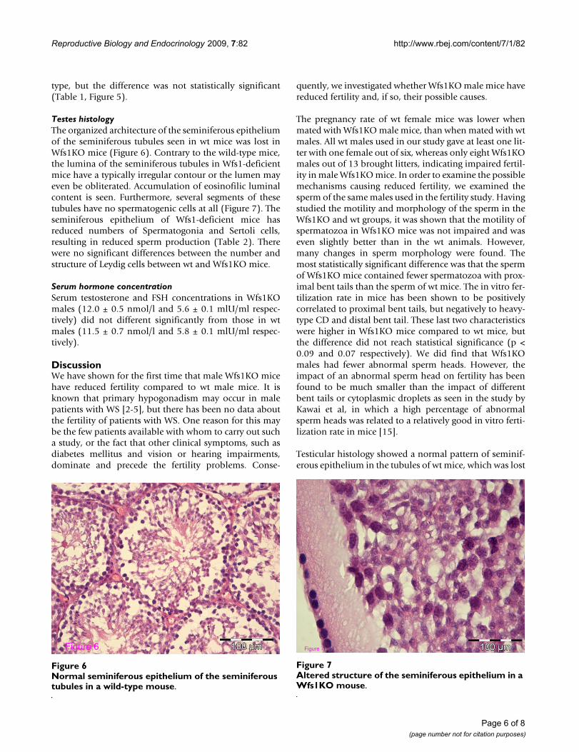
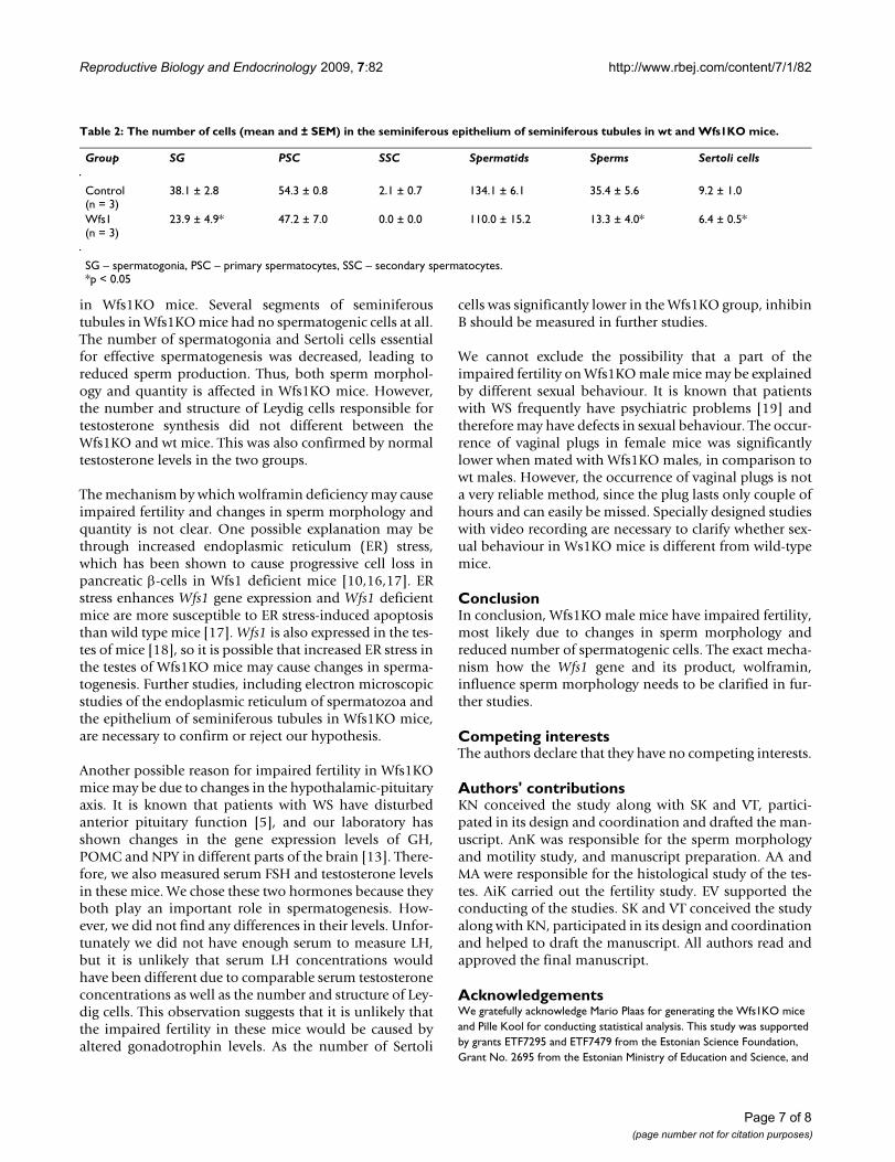
Testes histologyThe organized architecture of the seminiferous epitheliumof the seminiferous tubules seen in wt mice was lost inWfs1KO mice (Figure 6). Contrary to the wild-type mice,the lumina of the seminiferous tubules in Wfs1-deficientmice have a typically irregular contour or the lumen mayeven be obliterated. Accumulation of eosinofilic luminalcontent is seen. Furthermore, several segments of thesetubules have no spermatogenic cells at all (Figure 7). Theseminiferous epithelium of Wfs1-deficient mice hasreduced numbers of Spermatogonia and Sertoli cells,resulting in reduced sperm production (Table 2). Therewere no significant differences between the number andstructure of Leydig cells between wt and Wfs1KO mice.
Serum hormone concentrationSerum testosterone and FSH concentrations in Wfs1KOmales (12.0 ± 0.5 nmol/l and 5.6 ± 0.1 mlU/ml respec-tively) did not different significantly from those in wtmales (11.5 ± 0.7 nmol/l and 5.8 ± 0.1 mlU/ml respec-tively).
DiscussionWe have shown for the first time that male Wfs1KO micehave reduced fertility compared to wt male mice. It isknown that primary hypogonadism may occur in malepatients with WS [2-5], but there has been no data aboutthe fertility of patients with WS. One reason for this maybe the few patients available with whom to carry out sucha study, or the fact that other clinical symptoms, such asdiabetes mellitus and vision or hearing impairments,dominate and precede the fertility problems. Conse-
quently, we investigated whether Wfs1KO male mice havereduced fertility and, if so, their possible causes.
The pregnancy rate of wt female mice was lower whenmated with Wfs1KO male mice, than when mated with wtmales. All wt males used in our study gave at least one lit-ter with one female out of six, whereas only eight Wfs1KOmales out of 13 brought litters, indicating impaired fertil-ity in male Wfs1KO mice. In order to examine the possiblemechanisms causing reduced fertility, we examined thesperm of the same males used in the fertility study. Havingstudied the motility and morphology of the sperm in theWfs1KO and wt groups, it was shown that the motility ofspermatozoa in Wfs1KO mice was not impaired and waseven slightly better than in the wt animals. However,many changes in sperm morphology were found. Themost statistically significant difference was that the spermof Wfs1KO mice contained fewer spermatozoa with prox-imal bent tails than the sperm of wt mice. The in vitro fer-tilization rate in mice has been shown to be positivelycorrelated to proximal bent tails, but negatively to heavy-type CD and distal bent tail. These last two characteristicswere higher in Wfs1KO mice compared to wt mice, butthe difference did not reach statistical significance (p <0.09 and 0.07 respectively). We did find that Wfs1KOmales had fewer abnormal sperm heads. However, theimpact of an abnormal sperm head on fertility has beenfound to be much smaller than the impact of differentbent tails or cytoplasmic droplets as seen in the study byKawai et al, in which a high percentage of abnormalsperm heads was related to a relatively good in vitro ferti-lization rate in mice [15].
Testicular histology showed a normal pattern of seminif-erous epithelium in the tubules of wt mice, which was lost
Altered structure of the seminiferous epithelium in a Wfs1KO mouseFigure 7Altered structure of the seminiferous epithelium in a Wfs1KO mouse.
Figure 7
Normal seminiferous epithelium of the seminiferous tubules in a wild-type mouseFigure 6Normal seminiferous epithelium of the seminiferous tubules in a wild-type mouse.
Figure 6
Page 6 of 8(page number not for citation purposes)
Reproductive Biology and Endocrinology 2009, 7:82 http://www.rbej.com/content/7/1/82
in Wfs1KO mice. Several segments of seminiferoustubules in Wfs1KO mice had no spermatogenic cells at all.The number of spermatogonia and Sertoli cells essentialfor effective spermatogenesis was decreased, leading toreduced sperm production. Thus, both sperm morphol-ogy and quantity is affected in Wfs1KO mice. However,the number and structure of Leydig cells responsible fortestosterone synthesis did not different between theWfs1KO and wt mice. This was also confirmed by normaltestosterone levels in the two groups.
The mechanism by which wolframin deficiency may causeimpaired fertility and changes in sperm morphology andquantity is not clear. One possible explanation may bethrough increased endoplasmic reticulum (ER) stress,which has been shown to cause progressive cell loss inpancreatic β-cells in Wfs1 deficient mice [10,16,17]. ERstress enhances Wfs1 gene expression and Wfs1 deficientmice are more susceptible to ER stress-induced apoptosisthan wild type mice [17]. Wfs1 is also expressed in the tes-tes of mice [18], so it is possible that increased ER stress inthe testes of Wfs1KO mice may cause changes in sperma-togenesis. Further studies, including electron microscopicstudies of the endoplasmic reticulum of spermatozoa andthe epithelium of seminiferous tubules in Wfs1KO mice,are necessary to confirm or reject our hypothesis.
Another possible reason for impaired fertility in Wfs1KOmice may be due to changes in the hypothalamic-pituitaryaxis. It is known that patients with WS have disturbedanterior pituitary function [5], and our laboratory hasshown changes in the gene expression levels of GH,POMC and NPY in different parts of the brain [13]. There-fore, we also measured serum FSH and testosterone levelsin these mice. We chose these two hormones because theyboth play an important role in spermatogenesis. How-ever, we did not find any differences in their levels. Unfor-tunately we did not have enough serum to measure LH,but it is unlikely that serum LH concentrations wouldhave been different due to comparable serum testosteroneconcentrations as well as the number and structure of Ley-dig cells. This observation suggests that it is unlikely thatthe impaired fertility in these mice would be caused byaltered gonadotrophin levels. As the number of Sertoli
cells was significantly lower in the Wfs1KO group, inhibinB should be measured in further studies.
We cannot exclude the possibility that a part of theimpaired fertility on Wfs1KO male mice may be explainedby different sexual behaviour. It is known that patientswith WS frequently have psychiatric problems [19] andtherefore may have defects in sexual behaviour. The occur-rence of vaginal plugs in female mice was significantlylower when mated with Wfs1KO males, in comparison towt males. However, the occurrence of vaginal plugs is nota very reliable method, since the plug lasts only couple ofhours and can easily be missed. Specially designed studieswith video recording are necessary to clarify whether sex-ual behaviour in Ws1KO mice is different from wild-typemice.
ConclusionIn conclusion, Wfs1KO male mice have impaired fertility,most likely due to changes in sperm morphology andreduced number of spermatogenic cells. The exact mecha-nism how the Wfs1 gene and its product, wolframin,influence sperm morphology needs to be clarified in fur-ther studies.
Competing interestsThe authors declare that they have no competing interests.
Authors' contributionsKN conceived the study along with SK and VT, partici-pated in its design and coordination and drafted the man-uscript. AnK was responsible for the sperm morphologyand motility study, and manuscript preparation. AA andMA were responsible for the histological study of the tes-tes. AiK carried out the fertility study. EV supported theconducting of the studies. SK and VT conceived the studyalong with KN, participated in its design and coordinationand helped to draft the manuscript. All authors read andapproved the final manuscript.
AcknowledgementsWe gratefully acknowledge Mario Plaas for generating the Wfs1KO mice and Pille Kool for conducting statistical analysis. This study was supported by grants ETF7295 and ETF7479 from the Estonian Science Foundation, Grant No. 2695 from the Estonian Ministry of Education and Science, and
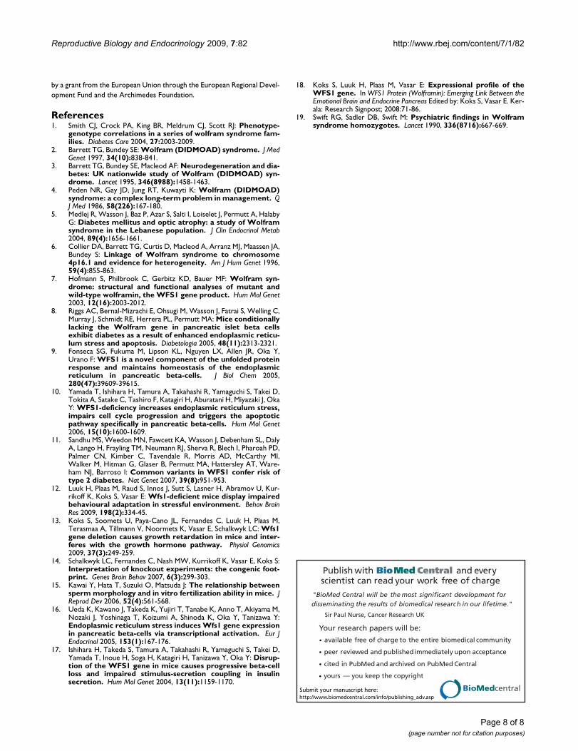
Table 2: The number of cells (mean and ± SEM) in the seminiferous epithelium of seminiferous tubules in wt and Wfs1KO mice.
Group SG PSC SSC Spermatids Sperms Sertoli cells
Control(n = 3)
38.1 ± 2.8 54.3 ± 0.8 2.1 ± 0.7 134.1 ± 6.1 35.4 ± 5.6 9.2 ± 1.0
Wfs1(n = 3)
23.9 ± 4.9* 47.2 ± 7.0 0.0 ± 0.0 110.0 ± 15.2 13.3 ± 4.0* 6.4 ± 0.5*
SG – spermatogonia, PSC – primary spermatocytes, SSC – secondary spermatocytes.*p < 0.05
Page 7 of 8(page number not for citation purposes)
Reproductive Biology and Endocrinology 2009, 7:82 http://www.rbej.com/content/7/1/82
Publish with BioMed Central and every scientist can read your work free of charge
"BioMed Central will be the most significant development for disseminating the results of biomedical research in our lifetime."
Sir Paul Nurse, Cancer Research UK
Your research papers will be:
available free of charge to the entire biomedical community
peer reviewed and published immediately upon acceptance
cited in PubMed and archived on PubMed Central
yours — you keep the copyright
Submit your manuscript here:http://www.biomedcentral.com/info/publishing_adv.asp
BioMedcentral
by a grant from the European Union through the European Regional Devel-opment Fund and the Archimedes Foundation.
References1. Smith CJ, Crock PA, King BR, Meldrum CJ, Scott RJ: Phenotype-
genotype correlations in a series of wolfram syndrome fam-ilies. Diabetes Care 2004, 27:2003-2009.
2. Barrett TG, Bundey SE: Wolfram (DIDMOAD) syndrome. J MedGenet 1997, 34(10):838-841.
3. Barrett TG, Bundey SE, Macleod AF: Neurodegeneration and dia-betes: UK nationwide study of Wolfram (DIDMOAD) syn-drome. Lancet 1995, 346(8988):1458-1463.
4. Peden NR, Gay JD, Jung RT, Kuwayti K: Wolfram (DIDMOAD)syndrome: a complex long-term problem in management. QJ Med 1986, 58(226):167-180.
5. Medlej R, Wasson J, Baz P, Azar S, Salti I, Loiselet J, Permutt A, HalabyG: Diabetes mellitus and optic atrophy: a study of Wolframsyndrome in the Lebanese population. J Clin Endocrinol Metab2004, 89(4):1656-1661.
6. Collier DA, Barrett TG, Curtis D, Macleod A, Arranz MJ, Maassen JA,Bundey S: Linkage of Wolfram syndrome to chromosome4p16.1 and evidence for heterogeneity. Am J Hum Genet 1996,59(4):855-863.
7. Hofmann S, Philbrook C, Gerbitz KD, Bauer MF: Wolfram syn-drome: structural and functional analyses of mutant andwild-type wolframin, the WFS1 gene product. Hum Mol Genet2003, 12(16):2003-2012.
8. Riggs AC, Bernal-Mizrachi E, Ohsugi M, Wasson J, Fatrai S, Welling C,Murray J, Schmidt RE, Herrera PL, Permutt MA: Mice conditionallylacking the Wolfram gene in pancreatic islet beta cellsexhibit diabetes as a result of enhanced endoplasmic reticu-lum stress and apoptosis. Diabetologia 2005, 48(11):2313-2321.
9. Fonseca SG, Fukuma M, Lipson KL, Nguyen LX, Allen JR, Oka Y,Urano F: WFS1 is a novel component of the unfolded proteinresponse and maintains homeostasis of the endoplasmicreticulum in pancreatic beta-cells. J Biol Chem 2005,280(47):39609-39615.
10. Yamada T, Ishihara H, Tamura A, Takahashi R, Yamaguchi S, Takei D,Tokita A, Satake C, Tashiro F, Katagiri H, Aburatani H, Miyazaki J, OkaY: WFS1-deficiency increases endoplasmic reticulum stress,impairs cell cycle progression and triggers the apoptoticpathway specifically in pancreatic beta-cells. Hum Mol Genet2006, 15(10):1600-1609.
11. Sandhu MS, Weedon MN, Fawcett KA, Wasson J, Debenham SL, DalyA, Lango H, Frayling TM, Neumann RJ, Sherva R, Blech I, Pharoah PD,Palmer CN, Kimber C, Tavendale R, Morris AD, McCarthy MI,Walker M, Hitman G, Glaser B, Permutt MA, Hattersley AT, Ware-ham NJ, Barroso I: Common variants in WFS1 confer risk oftype 2 diabetes. Nat Genet 2007, 39(8):951-953.
12. Luuk H, Plaas M, Raud S, Innos J, Sutt S, Lasner H, Abramov U, Kur-rikoff K, Koks S, Vasar E: Wfs1-deficient mice display impairedbehavioural adaptation in stressful environment. Behav BrainRes 2009, 198(2):334-45.
13. Koks S, Soomets U, Paya-Cano JL, Fernandes C, Luuk H, Plaas M,Terasmaa A, Tillmann V, Noormets K, Vasar E, Schalkwyk LC: Wfs1gene deletion causes growth retardation in mice and inter-feres with the growth hormone pathway. Physiol Genomics2009, 37(3):249-259.
14. Schalkwyk LC, Fernandes C, Nash MW, Kurrikoff K, Vasar E, Koks S:Interpretation of knockout experiments: the congenic foot-print. Genes Brain Behav 2007, 6(3):299-303.
15. Kawai Y, Hata T, Suzuki O, Matsuda J: The relationship betweensperm morphology and in vitro fertilization ability in mice. JReprod Dev 2006, 52(4):561-568.
16. Ueda K, Kawano J, Takeda K, Yujiri T, Tanabe K, Anno T, Akiyama M,Nozaki J, Yoshinaga T, Koizumi A, Shinoda K, Oka Y, Tanizawa Y:Endoplasmic reticulum stress induces Wfs1 gene expressionin pancreatic beta-cells via transcriptional activation. Eur JEndocrinol 2005, 153(1):167-176.
17. Ishihara H, Takeda S, Tamura A, Takahashi R, Yamaguchi S, Takei D,Yamada T, Inoue H, Soga H, Katagiri H, Tanizawa Y, Oka Y: Disrup-tion of the WFS1 gene in mice causes progressive beta-cellloss and impaired stimulus-secretion coupling in insulinsecretion. Hum Mol Genet 2004, 13(11):1159-1170.
18. Koks S, Luuk H, Plaas M, Vasar E: Expressional profile of theWFS1 gene. In WFS1 Protein (Wolframin): Emerging Link Between theEmotional Brain and Endocrine Pancreas Edited by: Koks S, Vasar E. Ker-ala: Research Signpost; 2008:71-86.
19. Swift RG, Sadler DB, Swift M: Psychiatric findings in Wolframsyndrome homozygotes. Lancet 1990, 336(8716):667-669.
Page 8 of 8(page number not for citation purposes)