Embed Size (px)
Citation preview
MOLECULAR AND CELLULAR BIOLOGY, Mar. 2004, p. 1930–1943 Vol. 24, No. 50270-7306/04/$08.00�0 DOI: 10.1128/MCB.24.5.1930–1943.2004Copyright © 2004, American Society for Microbiology. All Rights Reserved.
Replication Protein A (RPA) Phosphorylation Prevents RPAAssociation with Replication Centers
Vitaly M. Vassin,1 Marc S. Wold,2 and James A. Borowiec1*Department of Biochemistry and New York University Cancer Institute, New York University School of
Medicine, New York, New York 10016,1 and Department of Biochemistry, University ofIowa College of Medicine, Iowa City, Iowa 522422
Received 11 August 2003/Returned for modification 15 September 2003/Accepted 24 November 2003
Mammalian replication protein A (RPA) undergoes DNA damage-dependent phosphorylation at numeroussites on the N terminus of the RPA2 subunit. To understand the functional significance of RPA phosphory-lation, we expressed RPA2 variants in which the phosphorylation sites were converted to aspartate (RPA2D) oralanine (RPA2A). Although RPA2D was incorporated into RPA heterotrimers and supported simian virus 40DNA replication in vitro, the RPA2D mutant was selectively unable to associate with replication centers in vivo.In cells containing greatly reduced levels of endogenous RPA2, RPA2D again did not localize to replicationsites, indicating that the defect in supporting chromosomal DNA replication is not due to competition with thewild-type protein. Use of phosphospecific antibodies demonstrated that endogenous hyperphosphorylated RPAbehaves similarly to RPA2D. In contrast, under DNA damage or replication stress conditions, RPA2D, likeRPA2A and wild-type RPA2, was competent to associate with DNA damage foci as determined by colocalizationwith �-H2AX. We conclude that RPA2 phosphorylation prevents RPA association with replication centers invivo and potentially serves as a marker for sites of DNA damage.
DNA-damaging stress leads to the inception of a variety ofcellular responses that serve to minimize mutation and preventgenomic instability. In particular, the cell cycle checkpointapparatus is activated to block S phase entry and, in those cellsin the replicative phase, to both inhibit firing of late origins ofDNA replication and avert the collapse of replication forksblocked by damage (3). The DNA repair machinery is mobi-lized in concert to repair lesions and to allow eventual restartof stalled replication forks. One factor that plays essential rolesboth during DNA replication and in the repair- and recombi-nation-mediated recovery from damage is replication proteinA (RPA), the eukaryotic single-stranded (ss) DNA-bindingprotein (27, 52).
RPA is a heterotrimeric protein consisting, in mammaliancells, of �70- (RPA1), 30- (RPA2), and 14 (RPA3)-kDa sub-units. During DNA replication, RPA acts at the fork, stabiliz-ing ssDNA and facilitating nascent strand synthesis by thereplicative DNA polymerases. Under DNA-damaging condi-tions, RPA-ssDNA complexes act to recruit and activate a keycheckpoint mediator consisting of the ATR and ATRIP (ATR-interacting protein) protein-kinase complex (54). At DNAdamage-dependent nuclear foci, RPA interacts with repair andrecombination components to process double-strand DNAbreaks and other lesions (19). RPA activity is regulated byvarious stress conditions. In particular, heat shock (12, 47, 48),exposure to UV radiation (9), and treatment with DNA-alky-lating agents (30) each cause the generation of an RPA spe-cies that is unable to support DNA replication in vitro. In thecase of heat shock, the inhibition of RPA activity is mediated
by a stress-dependent association with the nucleolar proteinnucleolin (12, 47).
In an area with potential regulatory significance, RPA un-dergoes both stress-dependent and -independent phosphory-lation on the extreme N terminus of the RPA2 subunit. A basallevel of RPA modification by cyclin-cdk complexes occurs attwo sites (16, 35). Following stress, such as exposure to ionizing(31) or UV (9) radiation, or treatment with radiomimeticagents, such as camptothecin (CPT) (42), human RPA2 can bephosphorylated at five or more additional sites out of a possi-ble seven by the phosphatidylinositol 3-kinase-related kinases(PIKKs) DNA-PK, ATM, and perhaps ATR (7, 10, 17, 18, 31,33, 35, 46, 53). ATM and ATR are activated in response toDNA damage and replication stress, and they modify variouseffectors that facilitate the damage and cell cycle checkpointresponses (1). DNA-PK is required directly in the repair ofdouble-strand DNA breaks and in V(D)J recombination (15).These data could suggest that the function of stress-dependentmodification of RPA is to repress DNA replication or to pro-mote recovery from DNA damage, but there are as yet nocompelling data for either role. While the results of certainstudies suggest that RPA modification by PIKKs may lead tothe inhibition of DNA replication in vitro and in vivo (9, 37),direct testing of this possibility has not shown any appreciableeffects of RPA phosphorylation on binding to ssDNA or onreplication in vitro using a simian virus 40 (SV40)-based assay(7, 23).
Because previous work has primarily studied the effects ofmammalian RPA phosphorylation using in vitro systems, it ispossible that the modulation of RPA activity by phosphoryla-tion might be observed only in the cellular milieu. Testing thishypothesis, we found that RPA2 phosphorylation mutants thatmimic the hyperphosphorylated form were unable to localizeto replication centers in normal cells. Interestingly, binding of
* Corresponding author. Mailing address: Dept. of Biochemistryand New York University Cancer Institute, New York UniversitySchool of Medicine, New York, NY 10016. Phone: (212) 263-8453.Fax: (212) 263-8166. E-mail: [email protected].
1930
Dow
nloa
ded
from
http
s://j
ourn
als.
asm
.org
/jour
nal/m
cb o
n 12
Feb
ruar
y 20
22 b
y 12
4.22
7.14
.147
.
the hyperphosphorylation mimic to DNA damage foci wasunaffected, as determined by colocalization with the DNAdamage marker �-H2AX. Similar behavior was observed withendogenous hyperphosphorylated RPA. We conclude thatRPA phosphorylation following damage both prevents RPAfrom catalyzing DNA replication and potentially serves as amarker to recruit repair factors to sites of DNA damage.
MATERIALS AND METHODS
Cell lines and stress treatments. U2-OS and HeLa cells were maintained inMcCoy’s 5 M and Dulbecco’s modified Eagle’s media, respectively, supple-mented with 10% fetal bovine serum and 50 �g of gentamicin/ml. When theeffect of stress was examined, the cells were treated with either 1 �M CPT(Sigma) for 1 or 3 h, 2.5 mM hydroxyurea (HU; Sigma) for either 1 or 3 h, or 7�M aphidicolin (Sigma) for 3 h. To inhibit the cellular checkpoint response, cellswere treated with 5 mM caffeine for 30 min prior to stress. Transfection exper-iments were performed using Effectene (Qiagen).
Generation of RPA2 mutant constructs. To generate the myc-RPA2wt andmyc-RPA2D mammalian expression vectors, the human RPA2 genes from plas-mids p11dtRPA and p11dtRPA � 32Asp8 (4, 24) were inserted into the XbaI andBstBI sites of the pEF6/Myc-HisA vector (Invitrogen), resulting in pERPA2wtand pERPA2D. Expression of the His6 tag from pEF6/Myc-HisA was preventedby mutating the ATG codon at position 1863 to a TGA codon. Vectors express-ing the intermediate RPA2 phosphorylation mutants and RPA2A were con-structed by a combination of site-directed mutagenesis of either pERPA2wt orpERPA2D (as appropriate) at positions 23, 29, and 33 and replacement of largersegments of the RPA2 N terminus with synthetic oligonucleotides encodingmutant phosphorylation regions. Detailed cloning procedures are available uponrequest.
Protein purification and in vitro replication assay. The RPARPA2wt andRPARPA2D heterotrimers were expressed in Escherichia coli BL21 transformedwith p11dtRPA and p11dtRPA · 32Asp8, respectively, and purified as describedpreviously (24, 26). The SV40 large tumor (T) antigen used for SV40 DNAreplication reactions was prepared from extracts of Sf9 cells infected with therecombinant baculovirus Ac941SVT (5) and purified using immunoaffinity chro-matography (6). The AS65 fraction lacking RPA was prepared from HeLa cellextracts by ammonium sulfate fractionation according to the method of Wobbeet al. (51). SV40 DNA replication reaction mixtures (50 �l) containing 40 mMcreatine phosphate (diTris salt; pH 7.8); 7 mM MgCl2; 4 mM ATP; 25 �g ofcreatine kinase/ml; 0.4 mM dithiothreitol; 200 �M (each) CTP, GTP, and UTP;100 �M (each) dATP, dGTP, and dCTP; 25 �M [3H]dTTP (�500 cpm/pmol);0.2 �g of the ori-containing plasmid pSV01�EP (50); 200 �g of the AS65fraction; 0 to 700 ng of RPARPA2wt or RPARPA2D; and 750 ng of SV40 T antigenwere incubated at 37°C for 2 h. Replication activity was determined by precipi-tating the high-molecular-weight DNA with trichloroacetic acid and quantitatingthe amount of 3H in the precipitate by scintillation counting. To examine theDpnI resistance of the replication products, replication reaction mixtures con-taining 600 ng of either RPARPA2wt or RPARPA2D and 100 �M [�-32P]dCTP(1,000 cpm/pmol) to label the replication products were incubated at 37°C for2.5 h. Following removal of protein by phenol extraction, the DNA productswere first linearized by digestion with PstI and then either mock treated orincubated with 2.5 U of DpnI to cleave nonreplicated DNA. The digestionproducts were separated by electrophoresis through a 1.1% agarose gel andvisualized both by ethidium bromide staining and by autoradiography.
Immunoprecipitation and immunoblotting. Transfected U2-OS cells werelysed in lysis buffer (50 mM Tris-HCl, pH 7.4, 150 mM NaCl, 1% [vol/vol] NP-40,1 mM phenylmethylsulfonyl fluoride, 0.1 mM Na3VO4, 1 mM NaF, and 1 �geach of aprotinin, leupeptin, and pepstatin per ml). The cell extracts were thenincubated at 4°C for 2 h with 70A anti-RPA1 monoclonal antibody (28) conju-gated to CNBr-activated Sepharose beads (Amersham Biosciences). The immu-noprecipitate was washed five times with lysis buffer and resolved by sodiumdodecyl sulfate-polyacrylamide gel electrophoresis (SDS-PAGE) (13% [wt/vol]polyacrylamide). To test RPA2 phosphorylation and myc-RPA2 expression, cellswere directly lysed in SDS-PAGE sample buffer and proteins were separated bySDS-PAGE. For phosphatase treatment, cells were lysed in � protein phospha-tase buffer (New England Biolabs) containing 1% Triton X-100 and 1 �g each ofaprotinin, leupeptin, and pepstatin per ml. Cell lysates (�20 �g of protein) werethen incubated with 400 U of � protein phosphatase for 30 min at 30°C or mocktreated in the presence of protein phosphatase inhibitors (0.1 mM Na3VO4, 1mM NaF). The Western blots were developed with an anti-RPA2 34A mouse
monoclonal antibody (28) or a rabbit polyclonal anti-pSer4/pSer8-RPA2 anti-body obtained from Bethyl Laboratories, Inc. (Montgomery, Tex.). Proteins weredetected using enhanced chemiluminescence (Amersham Biosciences).
Cell cycle assay. Forty-eight hours posttransfection, cells were incubated with10 �M bromodeoxyuridine (BrdU). After a 30-min incubation, the cells werefixed and processed according to the BrdU Flow Kit manual (BD Pharmingen).Following incubation with rat anti-BrdU (Harlan Sera-Lab) and rabbit anti-myc(Upstate Biotechnology) antibodies, the cells were stained with anti-rat fluores-cein isothiocyanate-conjugated and anti-rabbit phycoerythrin-conjugated sec-ondary antibodies (Jackson ImmunoResearch Laboratories). The DNA wasstained with 7-aminoactinomycin D, and the cells were subjected to fluorescence-activated cell sorting (FACS) analysis.
Immunofluorescence microscopy. Transfected cells were processed by twomethods. To test protein expression and transfection efficiency, the cells werefirst washed with phosphate-buffered saline (PBS), fixed with 4% (wt/vol) form-aldehyde in PBS for 15 min at room temperature, and then extracted with PBScontaining 0.5% (vol/vol) Triton X-100 for 5 min. To study chromatin-boundproteins, the cells were extracted with 0.5% (vol/vol) Triton X-100 for 5 min priorto fixation as described previously (13). When required, cells were incubated inmedia containing 10 �M BrdU for 10 min prior to harvest. For detection ofincorporated BrdU, DNA was denatured with HCl using standard procedures.RPA2 silencing was achieved using a short interfering RNA (siRNA) duplextargeted to the 5�-CCUAGUUUCACAAUCUGUU sequence found in the 3�noncoding region of RPA2 mRNA. Prepared cells were incubated, as required,with rabbit anti-myc (Upstate Biotechnology), mouse anti-RPA1 70A and anti-RPA2 34A (28), rabbit anti-pSer4/pSer8-RPA2 (Bethyl Laboratories), rat anti-BrdU (Harlan Sera-Lab), and mouse anti-�H2AX (Upstate Biotechnology) an-tibodies. Following staining with the appropriate secondary antibodies (JacksonImmunoResearch Laboratories), the cells were examined by epifluorescencemicroscopy using a Zeiss Axiophot microscope. To calculate the relative fre-quency of myc-RPA2-positive cells (see Fig. 6H and 8M), the fraction of cellstransfected with myc-RPA2wt or the myc-tagged RPA mutants was first deter-mined by processing cells without prior Triton X-100 extraction (e.g.,Ftransfection:wt and Ftransfection:D4). Separately, the fraction of cells showing sig-nificant chromatin staining was also determined (e.g., Fchromatin:wt and Fchromatin:
D4). The relative frequency of cells that were positive, for example, for myc-RPA2D4 chromatin staining was calculated using the following formula: relativefrequency (Fchromatin:D4/Ftransfection:D4)/(Fchromatin:wt/Ftransfection:wt) � 100%.Each value determined was the result of three independent experiments.
RESULTS
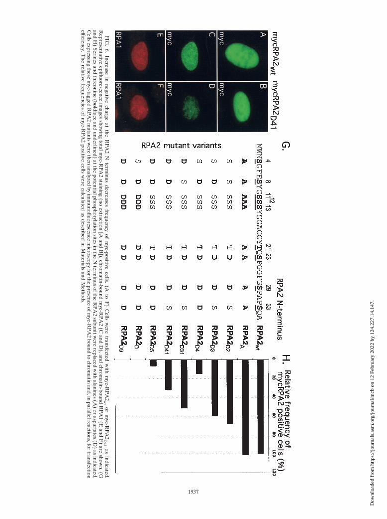
The RPA2D phosphorylation mimic localizes to the nucleusbut is not chromatin bound. To understand the functionalsignificance of RPA phosphorylation, we generated varioushuman RPA2 constructs in which subsets of the nine potentialN-terminal phosphorylation sites were mutated. Previous stud-ies have shown that two of the RPA2 sites (S23 and S29) arephosphorylated in a cell cycle-dependent manner by cyclin-cdk2 complexes (16, 35). At least five of the other seven (S4,S8, S11, S12, S13, T21, and S33) can be phosphorylated inresponse to UV irradiation (53). Ionizing irradiation and treat-ment with the radiomimetic agent CPT cause similar RPAhyperphosphorylation and likely modification of most if not allof these same sites (31, 42). Various data strongly suggest thatthe PIKK members DNA-PK and ATM, and likely ATR, canindependently modify the RPA stress-dependent sites (7, 10,17, 18, 31, 33, 35, 46), although only two (T21 and S33) havecanonical SQ/TQ sequences that are PIKK targets (1). Both ofthe cyclin-cdk2 sites and six of the stress-dependent sites (S8,S11, S12, S13, T21, and S33) were replaced by aspartate tomimic phosphate (generating the RPA2D mutant; see Fig. 6Gfor a schematic showing the construction of this and othermutants). Although an aspartate residue is not the same asphosphoserine or phosphothreonine, the use of aspartate res-idues to imitate phosphate has been shown in many cases tohave identical effects on protein structure and activity (25, 49).
VOL. 24, 2004 REGULATION OF RPA BINDING TO REPLICATION CENTERS 1931
Dow
nloa
ded
from
http
s://j
ourn
als.
asm
.org
/jour
nal/m
cb o
n 12
Feb
ruar
y 20
22 b
y 12
4.22
7.14
.147
.
In the RPA2A mutant, these same eight sites were converted toalanines to prevent phosphorylation (see also Fig. 6G). All ofthe mutants and the wild-type RPA2 control (RPA2wt) con-tained a C-terminal myc tag.
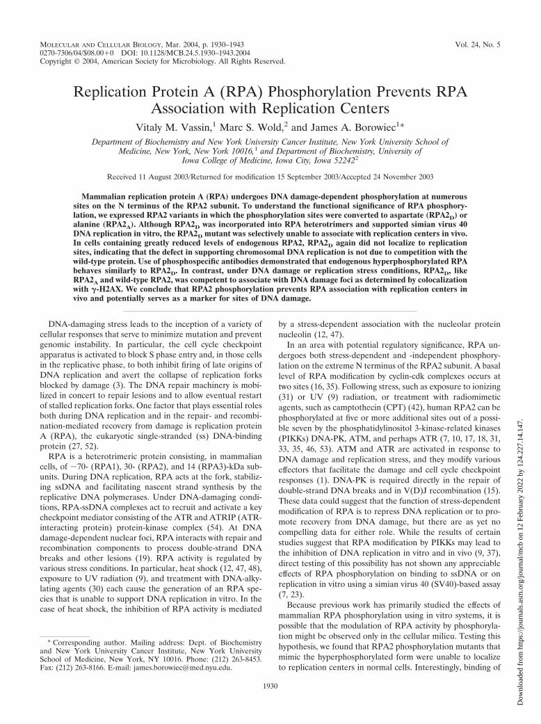
The RPA2wt subunit was expressed in human U2-OS cells.To detect the chromatin-bound fraction of RPA2, transfectedcells were extracted with nonionic detergent prior to formal-dehyde fixation (13). Under such conditions, RPA bound tochromatin in nuclear replication foci can be selectively visual-ized. The transfected RPA2wt subunit nearly completely colo-calized with the endogenous RPA1 and exhibited a punctatedistribution throughout the nucleus, consistent with its recruit-ment to DNA replication centers (Fig. 1A to D). To confirmthis observation, transfected cells were pulse-labeled withBrdU prior to fixation, and the sites of RPA2wt localizationand BrdU incorporation were examined. As expected, theRPA2wt subunit showed nearly complete colocalization withreplicating chromatin (Fig. 1E to H). Taken together, theseresults indicate that the recombinant RPA2wt subunit can func-tionally replace endogenous RPA2 in supporting chromosomalDNA replication.
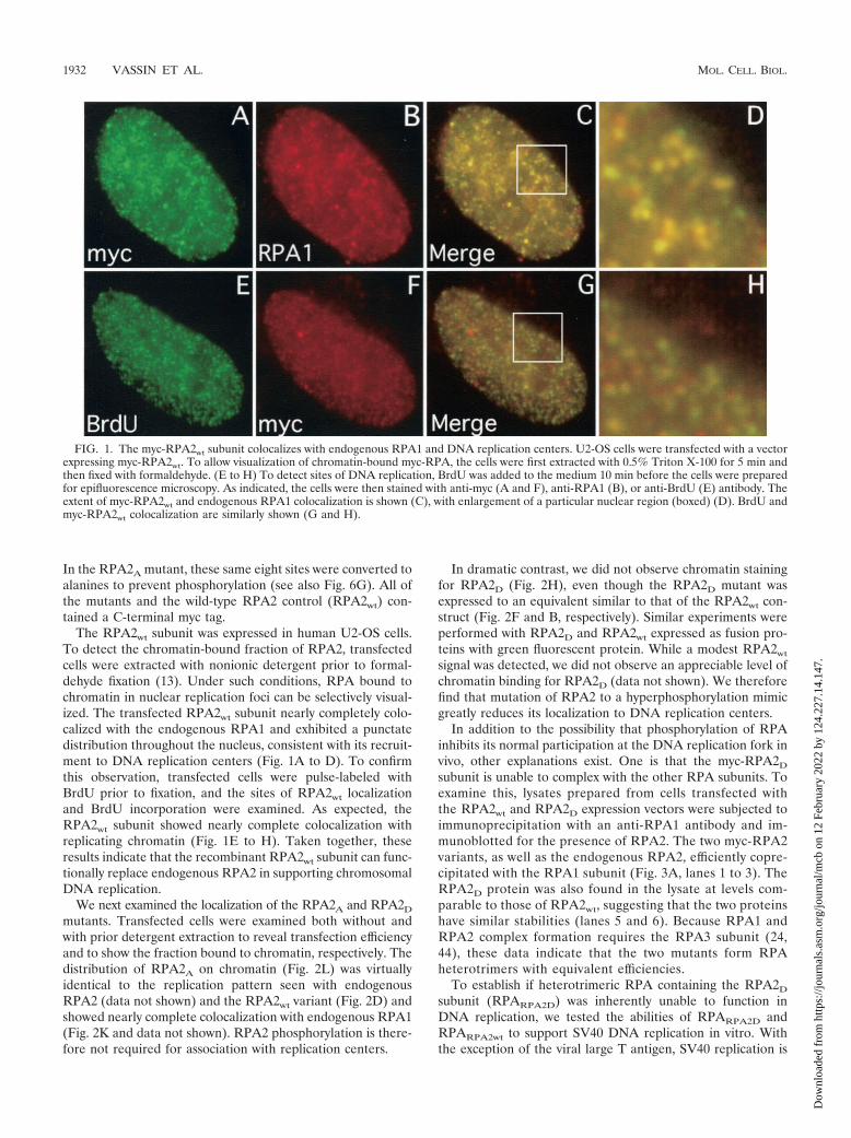
We next examined the localization of the RPA2A and RPA2D
mutants. Transfected cells were examined both without andwith prior detergent extraction to reveal transfection efficiencyand to show the fraction bound to chromatin, respectively. Thedistribution of RPA2A on chromatin (Fig. 2L) was virtuallyidentical to the replication pattern seen with endogenousRPA2 (data not shown) and the RPA2wt variant (Fig. 2D) andshowed nearly complete colocalization with endogenous RPA1(Fig. 2K and data not shown). RPA2 phosphorylation is there-fore not required for association with replication centers.
In dramatic contrast, we did not observe chromatin stainingfor RPA2D (Fig. 2H), even though the RPA2D mutant wasexpressed to an equivalent similar to that of the RPA2wt con-struct (Fig. 2F and B, respectively). Similar experiments wereperformed with RPA2D and RPA2wt expressed as fusion pro-teins with green fluorescent protein. While a modest RPA2wt
signal was detected, we did not observe an appreciable level ofchromatin binding for RPA2D (data not shown). We thereforefind that mutation of RPA2 to a hyperphosphorylation mimicgreatly reduces its localization to DNA replication centers.
In addition to the possibility that phosphorylation of RPAinhibits its normal participation at the DNA replication fork invivo, other explanations exist. One is that the myc-RPA2D
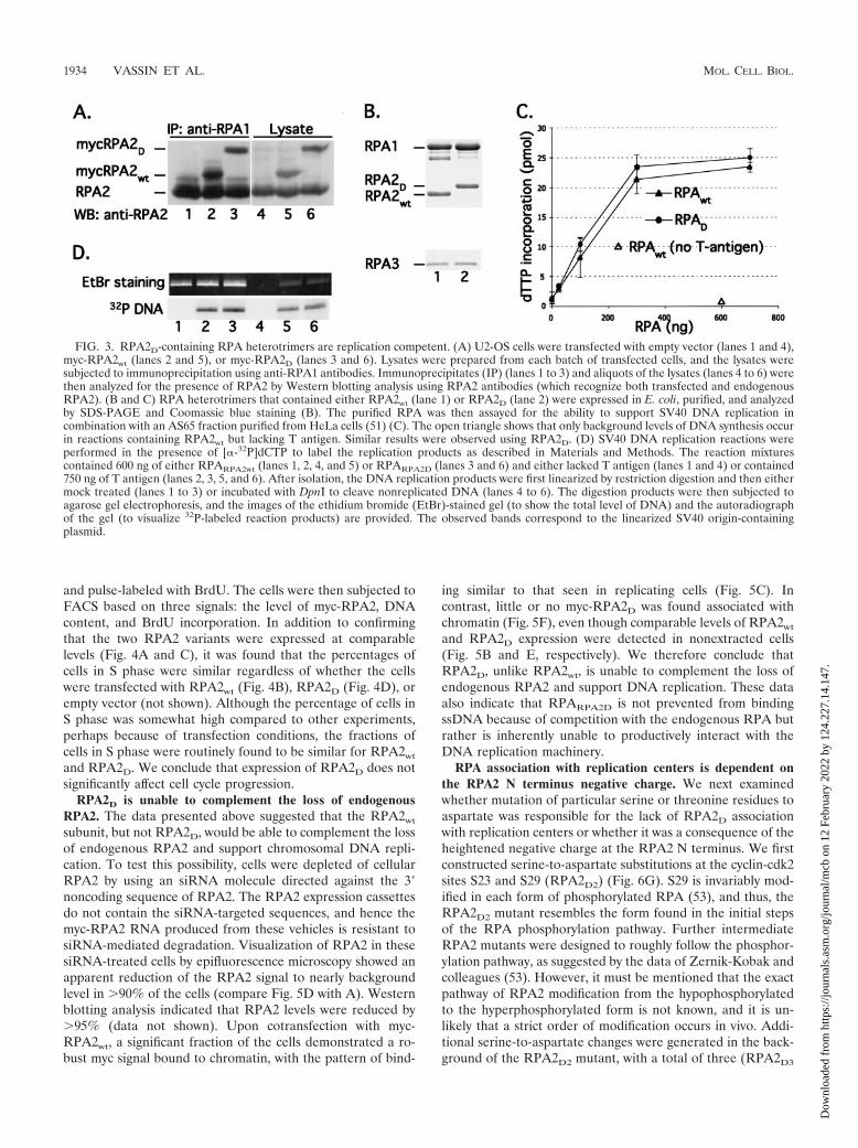
subunit is unable to complex with the other RPA subunits. Toexamine this, lysates prepared from cells transfected withthe RPA2wt and RPA2D expression vectors were subjected toimmunoprecipitation with an anti-RPA1 antibody and im-munoblotted for the presence of RPA2. The two myc-RPA2variants, as well as the endogenous RPA2, efficiently copre-cipitated with the RPA1 subunit (Fig. 3A, lanes 1 to 3). TheRPA2D protein was also found in the lysate at levels com-parable to those of RPA2wt, suggesting that the two proteinshave similar stabilities (lanes 5 and 6). Because RPA1 andRPA2 complex formation requires the RPA3 subunit (24,44), these data indicate that the two mutants form RPAheterotrimers with equivalent efficiencies.
To establish if heterotrimeric RPA containing the RPA2D
subunit (RPARPA2D) was inherently unable to function inDNA replication, we tested the abilities of RPARPA2D andRPARPA2wt to support SV40 DNA replication in vitro. Withthe exception of the viral large T antigen, SV40 replication is
FIG. 1. The myc-RPA2wt subunit colocalizes with endogenous RPA1 and DNA replication centers. U2-OS cells were transfected with a vectorexpressing myc-RPA2wt. To allow visualization of chromatin-bound myc-RPA, the cells were first extracted with 0.5% Triton X-100 for 5 min andthen fixed with formaldehyde. (E to H) To detect sites of DNA replication, BrdU was added to the medium 10 min before the cells were preparedfor epifluorescence microscopy. As indicated, the cells were then stained with anti-myc (A and F), anti-RPA1 (B), or anti-BrdU (E) antibody. Theextent of myc-RPA2wt and endogenous RPA1 colocalization is shown (C), with enlargement of a particular nuclear region (boxed) (D). BrdU andmyc-RPA2wt colocalization are similarly shown (G and H).
1932 VASSIN ET AL. MOL. CELL. BIOL.
Dow
nloa
ded
from
http
s://j
ourn
als.
asm
.org
/jour
nal/m
cb o
n 12
Feb
ruar
y 20
22 b
y 12
4.22
7.14
.147
.
catalyzed by host cell components (8, 22). The SV40 systemcan thus provide a relatively comprehensive test of the abilityof RPA to interact functionally with ssDNA and the DNAreplication machinery. Previous work by J. Hurwitz and col-leagues has shown that separation of human cell extracts byammonium sulfate precipitation yields two required fractions(AS30 and AS65), with RPA found to be the only essentialfactor within the AS30 fraction (51). Because the AS65 frac-tion lacks RPA, the activities of different RPA variants canbe assayed by their abilities to complement the AS65 frac-tion in supporting SV40 DNA replication. The RPARPA2D
and RPARPA2wt variants were produced in E. coli and purifiedto homogeneity (Fig. 3B). Use of the AS65 fraction aloneshowed no significant DNA replication activity (Fig. 3C). Theaddition of either heterotrimeric RPA complex supported T-antigen-dependent viral DNA replication to similar extents,and the activities of the two RPA variants were similar over arange of levels (Fig. 3C). The reaction products synthesized in
the presence of RPARPA2D or RPARPA2wt were equally resis-tant to DpnI, demonstrating that they were bona fide DNAreplication products and not due to repair synthesis (Fig. 3D).RPARPA2D is therefore functionally active in supporting DNAreplication in vitro. RPARPA2D was also found to bind nor-mally to short ssDNA oligonucleotides (4). These results arenot completely unexpected, as it was shown previously that theRPA phosphorylation state does not appreciably affect theability of RPA to function in viral DNA replication or in DNArepair (2, 7, 36). In sum, mutation of the seven serines and onethreonine in the N terminus of RPA2 to negatively chargedaspartate residues does not have any apparent effect on theinherent activity of the heterotrimeric protein.
We next examined the possibility that expression of theRPA2D mutant generates a signal that shuts down cellularDNA synthesis and thus indirectly prevents RPA2D from as-sociating with chromatin. To address this issue, cells weretransfected with the RPA2wt or RPA2D expression construct
FIG. 2. Lack of association of the RPA2D mutant with chromatin in unstressed cells. U2-OS cells were transfected with a vector expressingmyc-RPA2wt (A to D), myc-RPA2D (E to H), or myc-RPA2A (I to L). (C, D, G, H, K, and L) To allow visualization of chromatin-bound myc-RPA,cells were first extracted with 0.5% Triton X-100 for 5 min and then fixed with formaldehyde (� extraction). (A, B, E, F, I, and J) To assay fortransfection efficiency, cells were also fixed without prior extraction ( extraction). The cells were then stained with anti-myc (B, D, F, H, J, andL) or anti-RPA1 (A, C, E, G, I, and K) antibody.
VOL. 24, 2004 REGULATION OF RPA BINDING TO REPLICATION CENTERS 1933
Dow
nloa
ded
from
http
s://j
ourn
als.
asm
.org
/jour
nal/m
cb o
n 12
Feb
ruar
y 20
22 b
y 12
4.22
7.14
.147
.
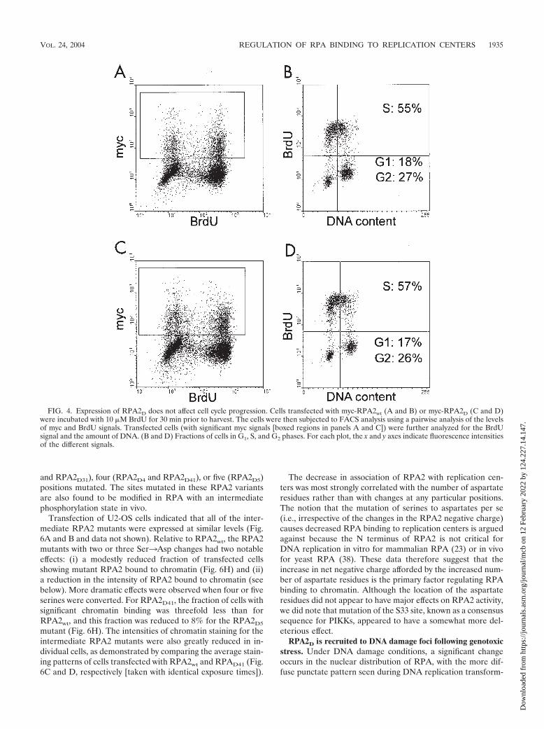
and pulse-labeled with BrdU. The cells were then subjected toFACS based on three signals: the level of myc-RPA2, DNAcontent, and BrdU incorporation. In addition to confirmingthat the two RPA2 variants were expressed at comparablelevels (Fig. 4A and C), it was found that the percentages ofcells in S phase were similar regardless of whether the cellswere transfected with RPA2wt (Fig. 4B), RPA2D (Fig. 4D), orempty vector (not shown). Although the percentage of cells inS phase was somewhat high compared to other experiments,perhaps because of transfection conditions, the fractions ofcells in S phase were routinely found to be similar for RPA2wt
and RPA2D. We conclude that expression of RPA2D does notsignificantly affect cell cycle progression.
RPA2D is unable to complement the loss of endogenousRPA2. The data presented above suggested that the RPA2wt
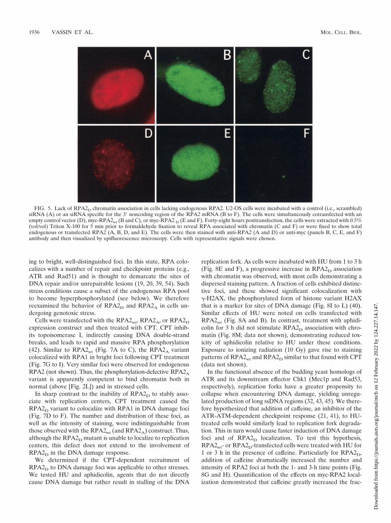
subunit, but not RPA2D, would be able to complement the lossof endogenous RPA2 and support chromosomal DNA repli-cation. To test this possibility, cells were depleted of cellularRPA2 by using an siRNA molecule directed against the 3�noncoding sequence of RPA2. The RPA2 expression cassettesdo not contain the siRNA-targeted sequences, and hence themyc-RPA2 RNA produced from these vehicles is resistant tosiRNA-mediated degradation. Visualization of RPA2 in thesesiRNA-treated cells by epifluorescence microscopy showed anapparent reduction of the RPA2 signal to nearly backgroundlevel in �90% of the cells (compare Fig. 5D with A). Westernblotting analysis indicated that RPA2 levels were reduced by�95% (data not shown). Upon cotransfection with myc-RPA2wt, a significant fraction of the cells demonstrated a ro-bust myc signal bound to chromatin, with the pattern of bind-
ing similar to that seen in replicating cells (Fig. 5C). Incontrast, little or no myc-RPA2D was found associated withchromatin (Fig. 5F), even though comparable levels of RPA2wt
and RPA2D expression were detected in nonextracted cells(Fig. 5B and E, respectively). We therefore conclude thatRPA2D, unlike RPA2wt, is unable to complement the loss ofendogenous RPA2 and support DNA replication. These dataalso indicate that RPARPA2D is not prevented from bindingssDNA because of competition with the endogenous RPA butrather is inherently unable to productively interact with theDNA replication machinery.
RPA association with replication centers is dependent onthe RPA2 N terminus negative charge. We next examinedwhether mutation of particular serine or threonine residues toaspartate was responsible for the lack of RPA2D associationwith replication centers or whether it was a consequence of theheightened negative charge at the RPA2 N terminus. We firstconstructed serine-to-aspartate substitutions at the cyclin-cdk2sites S23 and S29 (RPA2D2) (Fig. 6G). S29 is invariably mod-ified in each form of phosphorylated RPA (53), and thus, theRPA2D2 mutant resembles the form found in the initial stepsof the RPA phosphorylation pathway. Further intermediateRPA2 mutants were designed to roughly follow the phosphor-ylation pathway, as suggested by the data of Zernik-Kobak andcolleagues (53). However, it must be mentioned that the exactpathway of RPA2 modification from the hypophosphorylatedto the hyperphosphorylated form is not known, and it is un-likely that a strict order of modification occurs in vivo. Addi-tional serine-to-aspartate changes were generated in the back-ground of the RPA2D2 mutant, with a total of three (RPA2D3
FIG. 3. RPA2D-containing RPA heterotrimers are replication competent. (A) U2-OS cells were transfected with empty vector (lanes 1 and 4),myc-RPA2wt (lanes 2 and 5), or myc-RPA2D (lanes 3 and 6). Lysates were prepared from each batch of transfected cells, and the lysates weresubjected to immunoprecipitation using anti-RPA1 antibodies. Immunoprecipitates (IP) (lanes 1 to 3) and aliquots of the lysates (lanes 4 to 6) werethen analyzed for the presence of RPA2 by Western blotting analysis using RPA2 antibodies (which recognize both transfected and endogenousRPA2). (B and C) RPA heterotrimers that contained either RPA2wt (lane 1) or RPA2D (lane 2) were expressed in E. coli, purified, and analyzedby SDS-PAGE and Coomassie blue staining (B). The purified RPA was then assayed for the ability to support SV40 DNA replication incombination with an AS65 fraction purified from HeLa cells (51) (C). The open triangle shows that only background levels of DNA synthesis occurin reactions containing RPA2wt but lacking T antigen. Similar results were observed using RPA2D. (D) SV40 DNA replication reactions wereperformed in the presence of [�-32P]dCTP to label the replication products as described in Materials and Methods. The reaction mixturescontained 600 ng of either RPARPA2wt (lanes 1, 2, 4, and 5) or RPARPA2D (lanes 3 and 6) and either lacked T antigen (lanes 1 and 4) or contained750 ng of T antigen (lanes 2, 3, 5, and 6). After isolation, the DNA replication products were first linearized by restriction digestion and then eithermock treated (lanes 1 to 3) or incubated with DpnI to cleave nonreplicated DNA (lanes 4 to 6). The digestion products were then subjected toagarose gel electrophoresis, and the images of the ethidium bromide (EtBr)-stained gel (to show the total level of DNA) and the autoradiographof the gel (to visualize 32P-labeled reaction products) are provided. The observed bands correspond to the linearized SV40 origin-containingplasmid.
1934 VASSIN ET AL. MOL. CELL. BIOL.
Dow
nloa
ded
from
http
s://j
ourn
als.
asm
.org
/jour
nal/m
cb o
n 12
Feb
ruar
y 20
22 b
y 12
4.22
7.14
.147
.
and RPA2D31), four (RPA2D4 and RPA2D41), or five (RPA2D5)positions mutated. The sites mutated in these RPA2 variantsare also found to be modified in RPA with an intermediatephosphorylation state in vivo.
Transfection of U2-OS cells indicated that all of the inter-mediate RPA2 mutants were expressed at similar levels (Fig.6A and B and data not shown). Relative to RPA2wt, the RPA2mutants with two or three Ser3Asp changes had two notableeffects: (i) a modestly reduced fraction of transfected cellsshowing mutant RPA2 bound to chromatin (Fig. 6H) and (ii)a reduction in the intensity of RPA2 bound to chromatin (seebelow). More dramatic effects were observed when four or fiveserines were converted. For RPA2D41, the fraction of cells withsignificant chromatin binding was threefold less than forRPA2wt, and this fraction was reduced to 8% for the RPA2D5
mutant (Fig. 6H). The intensities of chromatin staining for theintermediate RPA2 mutants were also greatly reduced in in-dividual cells, as demonstrated by comparing the average stain-ing patterns of cells transfected with RPA2wt and RPAD41 (Fig.6C and D, respectively [taken with identical exposure times]).
The decrease in association of RPA2 with replication cen-ters was most strongly correlated with the number of aspartateresidues rather than with changes at any particular positions.The notion that the mutation of serines to aspartates per se(i.e., irrespective of the changes in the RPA2 negative charge)causes decreased RPA binding to replication centers is arguedagainst because the N terminus of RPA2 is not critical forDNA replication in vitro for mammalian RPA (23) or in vivofor yeast RPA (38). These data therefore suggest that theincrease in net negative charge afforded by the increased num-ber of aspartate residues is the primary factor regulating RPAbinding to chromatin. Although the location of the aspartateresidues did not appear to have major effects on RPA2 activity,we did note that mutation of the S33 site, known as a consensussequence for PIKKs, appeared to have a somewhat more del-eterious effect.
RPA2D is recruited to DNA damage foci following genotoxicstress. Under DNA damage conditions, a significant changeoccurs in the nuclear distribution of RPA, with the more dif-fuse punctate pattern seen during DNA replication transform-
FIG. 4. Expression of RPA2D does not affect cell cycle progression. Cells transfected with myc-RPA2wt (A and B) or myc-RPA2D (C and D)were incubated with 10 �M BrdU for 30 min prior to harvest. The cells were then subjected to FACS analysis using a pairwise analysis of the levelsof myc and BrdU signals. Transfected cells (with significant myc signals [boxed regions in panels A and C]) were further analyzed for the BrdUsignal and the amount of DNA. (B and D) Fractions of cells in G1, S, and G2 phases. For each plot, the x and y axes indicate fluorescence intensitiesof the different signals.
VOL. 24, 2004 REGULATION OF RPA BINDING TO REPLICATION CENTERS 1935
Dow
nloa
ded
from
http
s://j
ourn
als.
asm
.org
/jour
nal/m
cb o
n 12
Feb
ruar
y 20
22 b
y 12
4.22
7.14
.147
.
ing to bright, well-distinguished foci. In this state, RPA colo-calizes with a number of repair and checkpoint proteins (e.g.,ATR and Rad51) and is thought to demarcate the sites ofDNA repair and/or unrepairable lesions (19, 20, 39, 54). Suchstress conditions cause a subset of the endogenous RPA poolto become hyperphosphorylated (see below). We thereforereexamined the behavior of RPA2D and RPA2A in cells un-dergoing genotoxic stress.
Cells were transfected with the RPA2wt, RPA2A, or RPA2D
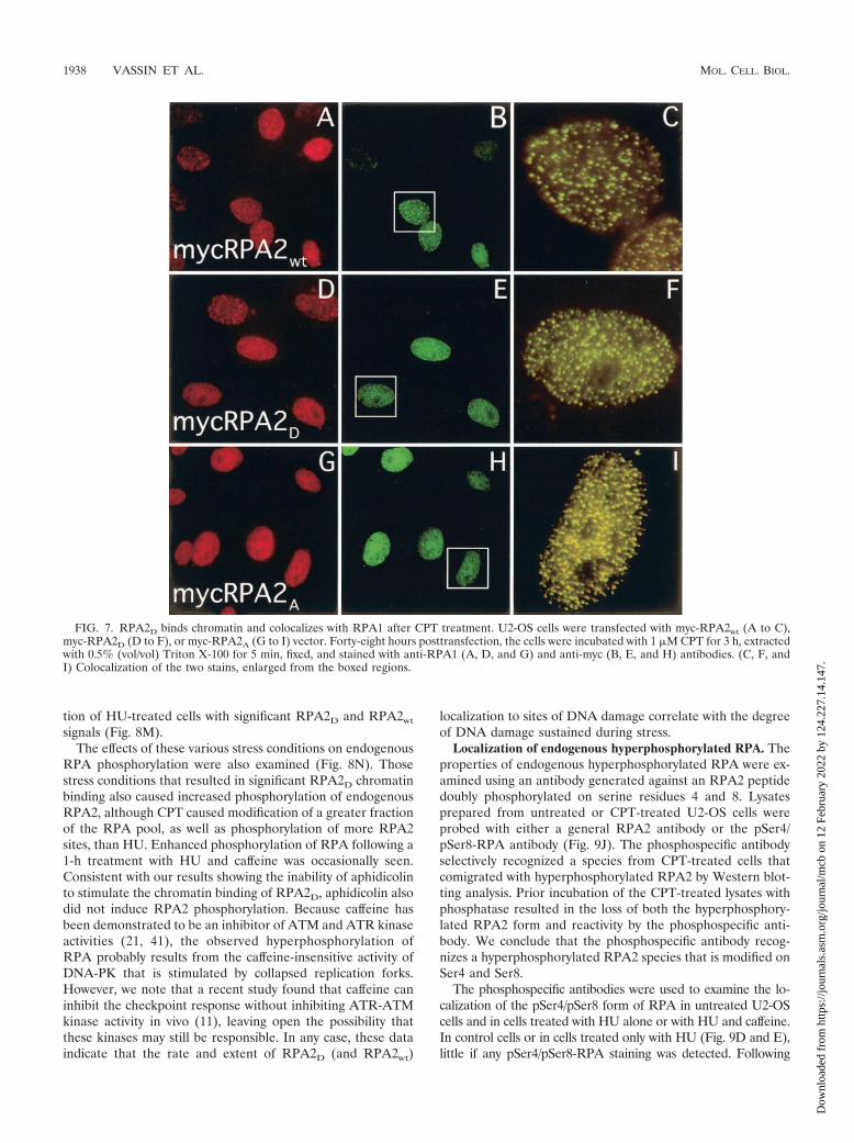
expression construct and then treated with CPT. CPT inhib-its topoisomerase I, indirectly causing DNA double-strandbreaks, and leads to rapid and massive RPA phosphorylation(42). Similar to RPA2wt (Fig. 7A to C), the RPA2A variantcolocalized with RPA1 in bright foci following CPT treatment(Fig. 7G to I). Very similar foci were observed for endogenousRPA2 (not shown). Thus, the phosphorylation-defective RPA2A
variant is apparently competent to bind chromatin both innormal (above [Fig. 2L]) and in stressed cells.
In sharp contrast to the inability of RPA2D to stably asso-ciate with replication centers, CPT treatment caused theRPA2D variant to colocalize with RPA1 in DNA damage foci(Fig. 7D to F). The number and distribution of these foci, aswell as the intensity of staining, were indistinguishable fromthose observed with the RPA2wt (and RPA2A) construct. Thus,although the RPA2D mutant is unable to localize to replicationcenters, this defect does not extend to the involvement ofRPA2D in the DNA damage response.
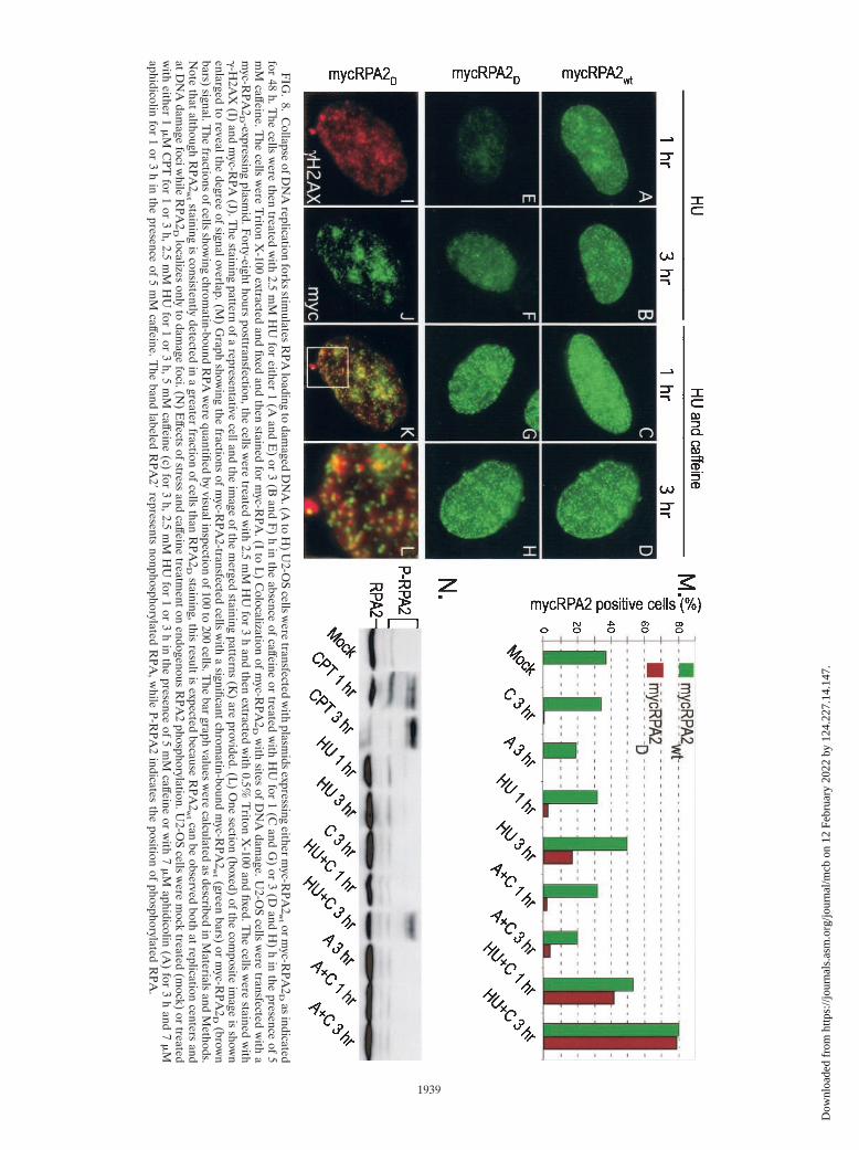
We determined if the CPT-dependent recruitment ofRPA2D to DNA damage foci was applicable to other stresses.We tested HU and aphidicolin, agents that do not directlycause DNA damage but rather result in stalling of the DNA
replication fork. As cells were incubated with HU from 1 to 3 h(Fig. 8E and F), a progressive increase in RPA2D associationwith chromatin was observed, with most cells demonstrating adispersed staining pattern. A fraction of cells exhibited distinc-tive foci, and these showed significant colocalization with�-H2AX, the phosphorylated form of histone variant H2AXthat is a marker for sites of DNA damage (Fig. 8I to L) (40).Similar effects of HU were noted on cells transfected withRPA2wt (Fig. 8A and B). In contrast, treatment with aphidi-colin for 3 h did not stimulate RPA2D association with chro-matin (Fig. 8M; data not shown), demonstrating reduced tox-icity of aphidicolin relative to HU under these conditions.Exposure to ionizing radiation (10 Gy) gave rise to stainingpatterns of RPA2wt and RPA2D similar to that found with CPT(data not shown).
In the functional absence of the budding yeast homologs ofATR and its downstream effector Chk1 (Mec1p and Rad53,respectively), replication forks have a greater propensity tocollapse when encountering DNA damage, yielding unregu-lated production of long ssDNA regions (32, 43, 45). We there-fore hypothesized that addition of caffeine, an inhibitor of theATR-ATM-dependent checkpoint response (21, 41), to HU-treated cells would similarly lead to replication fork degrada-tion. This in turn would cause faster induction of DNA damagefoci and of RPA2D localization. To test this hypothesis,RPA2wt- or RPA2D-transfected cells were treated with HU for1 or 3 h in the presence of caffeine. Particularly for RPA2D,addition of caffeine dramatically increased the number andintensity of RPA2 foci at both the 1- and 3-h time points (Fig.8G and H). Quantification of the effects on myc-RPA2 local-ization demonstrated that caffeine greatly increased the frac-
FIG. 5. Lack of RPA2D chromatin association in cells lacking endogenous RPA2. U2-OS cells were incubated with a control (i.e., scrambled)siRNA (A) or an siRNA specific for the 3� noncoding region of the RPA2 mRNA (B to F). The cells were simultaneously cotransfected with anempty control vector (D), myc-RPA2wt (B and C), or myc-RPA2 D (E and F). Forty-eight hours posttransfection, the cells were extracted with 0.5%(vol/vol) Triton X-100 for 5 min prior to formaldehyde fixation to reveal RPA associated with chromatin (C and F) or were fixed to show totalendogenous or transfected RPA2 (A, B, D, and E). The cells were then stained with anti-RPA2 (A and D) or anti-myc (panels B, C, E, and F)antibody and then visualized by epifluorescence microscopy. Cells with representative signals were chosen.
1936 VASSIN ET AL. MOL. CELL. BIOL.
Dow
nloa
ded
from
http
s://j
ourn
als.
asm
.org
/jour
nal/m
cb o
n 12
Feb
ruar
y 20
22 b
y 12
4.22
7.14
.147
.
FIG
.6.
Increasein
negativecharge
atthe
RPA
2N
terminus
decreasesfrequency
ofm
yc-positivecells.
(Ato
F)
Cells
were
transfectedw
ithm
yc-RPA
2w
tor
myc-R
PA2
D41
asindicated.
Representative
epifluorescenceim
agesshow
ingtotalm
yc-RPA
2staining
(noextraction
[Aand
B]),chrom
atin-boundm
yc-RPA
2(C
andD
),andchrom
atin-boundR
PA1
(Eand
F)
areshow
n.(Gand
H)
Serinesand
threonine(boldface
andunderlined)
atthepotentialphosphorylation
sitesin
theN
terminus
oftheR
PA2
subunitwere
replacedw
ithalanines
(A)
oraspartates
(D)
asindicated.
Cells
expressingthese
myc-tagged
RPA
2m
utantsw
erethen
analyzedby
imm
unofluorescencem
icroscopyfor
thepresence
ofmyc-R
PA2
boundto
chromatin
and,inparallelreactions,for
transfectionefficiency.T
herelative
frequenciesof
myc-R
PA2
positivecells
were
calculatedas
describedin
Materials
andM
ethods.
1937
Dow
nloa
ded
from
http
s://j
ourn
als.
asm
.org
/jour
nal/m
cb o
n 12
Feb
ruar
y 20
22 b
y 12
4.22
7.14
.147
.
tion of HU-treated cells with significant RPA2D and RPA2wt
signals (Fig. 8M).The effects of these various stress conditions on endogenous
RPA phosphorylation were also examined (Fig. 8N). Thosestress conditions that resulted in significant RPA2D chromatinbinding also caused increased phosphorylation of endogenousRPA2, although CPT caused modification of a greater fractionof the RPA pool, as well as phosphorylation of more RPA2sites, than HU. Enhanced phosphorylation of RPA following a1-h treatment with HU and caffeine was occasionally seen.Consistent with our results showing the inability of aphidicolinto stimulate the chromatin binding of RPA2D, aphidicolin alsodid not induce RPA2 phosphorylation. Because caffeine hasbeen demonstrated to be an inhibitor of ATM and ATR kinaseactivities (21, 41), the observed hyperphosphorylation ofRPA probably results from the caffeine-insensitive activity ofDNA-PK that is stimulated by collapsed replication forks.However, we note that a recent study found that caffeine caninhibit the checkpoint response without inhibiting ATR-ATMkinase activity in vivo (11), leaving open the possibility thatthese kinases may still be responsible. In any case, these dataindicate that the rate and extent of RPA2D (and RPA2wt)
localization to sites of DNA damage correlate with the degreeof DNA damage sustained during stress.
Localization of endogenous hyperphosphorylated RPA. Theproperties of endogenous hyperphosphorylated RPA were ex-amined using an antibody generated against an RPA2 peptidedoubly phosphorylated on serine residues 4 and 8. Lysatesprepared from untreated or CPT-treated U2-OS cells wereprobed with either a general RPA2 antibody or the pSer4/pSer8-RPA antibody (Fig. 9J). The phosphospecific antibodyselectively recognized a species from CPT-treated cells thatcomigrated with hyperphosphorylated RPA2 by Western blot-ting analysis. Prior incubation of the CPT-treated lysates withphosphatase resulted in the loss of both the hyperphosphory-lated RPA2 form and reactivity by the phosphospecific anti-body. We conclude that the phosphospecific antibody recog-nizes a hyperphosphorylated RPA2 species that is modified onSer4 and Ser8.
The phosphospecific antibodies were used to examine the lo-calization of the pSer4/pSer8 form of RPA in untreated U2-OScells and in cells treated with HU alone or with HU and caffeine.In control cells or in cells treated only with HU (Fig. 9D and E),little if any pSer4/pSer8-RPA staining was detected. Following
FIG. 7. RPA2D binds chromatin and colocalizes with RPA1 after CPT treatment. U2-OS cells were transfected with myc-RPA2wt (A to C),myc-RPA2D (D to F), or myc-RPA2A (G to I) vector. Forty-eight hours posttransfection, the cells were incubated with 1 �M CPT for 3 h, extractedwith 0.5% (vol/vol) Triton X-100 for 5 min, fixed, and stained with anti-RPA1 (A, D, and G) and anti-myc (B, E, and H) antibodies. (C, F, andI) Colocalization of the two stains, enlarged from the boxed regions.
1938 VASSIN ET AL. MOL. CELL. BIOL.
Dow
nloa
ded
from
http
s://j
ourn
als.
asm
.org
/jour
nal/m
cb o
n 12
Feb
ruar
y 20
22 b
y 12
4.22
7.14
.147
.
FIG
.8.
Collapse
ofDN
Areplication
forksstim
ulatesR
PAloading
todam
agedD
NA
.(Ato
H)
U2-O
Scells
were
transfectedw
ithplasm
idsexpressing
eitherm
yc-RPA
2w
t orm
yc-RPA
2D
asindicated
for48
h.The
cellsw
erethen
treatedw
ith2.5
mM
HU
foreither
1(A
andE
)or
3(B
andF
)h
inthe
absenceof
caffeineor
treatedw
ithH
Ufor
1(C
andG
)or
3(D
andH
)h
inthe
presenceof
5m
Mcaffeine.T
hecells
were
Triton
X-100
extractedand
fixedand
thenstained
form
yc-RPA
.(Ito
L)
Colocalization
ofm
yc-RPA
2D
with
sitesof
DN
Adam
age.U2-O
Scells
were
transfectedw
itha
myc-R
PA2
D -expressingplasm
id.Forty-eight
hoursposttransfection,the
cellsw
eretreated
with
2.5m
MH
Ufor
3h
andthen
extractedw
ith0.5%
Triton
X-100
andfixed.T
hecells
were
stainedw
ith�-H
2AX
(I)and
myc-R
PA(J).T
hestaining
patternof
arepresentative
cellandthe
image
ofthe
merged
stainingpatterns
(K)
areprovided.(L
)O
nesection
(boxed)of
thecom
positeim
ageis
shown
enlargedto
revealthedegree
ofsignaloverlap.(M
)G
raphshow
ingthe
fractionsof
myc-R
PA2-transfected
cellsw
itha
significantchrom
atin-boundm
yc-RPA
2w
t (greenbars)
orm
yc-RPA
2D
(brown
bars)signal.T
hefractions
ofcellsshow
ingchrom
atin-boundR
PAw
erequantified
byvisualinspection
of100to
200cells.T
hebar
graphvalues
were
calculatedas
describedin
Materials
andM
ethods.N
otethat
althoughR
PA2
wt staining
isconsistently
detectedin
agreater
fractionof
cellsthan
RPA
2D
staining,thisresult
isexpected
becauseR
PA2
wt can
beobserved
bothat
replicationcenters
andatD
NA
damage
fociwhile
RPA
2D
localizesonly
todam
agefoci.(N
)E
ffectsofstress
andcaffeine
treatmenton
endogenousR
PA2
phosphorylation.U2-O
Scells
were
mock
treated(m
ock)or
treatedw
itheither
1�
MC
PTfor
1or
3h,2.5
mM
HU
for1
or3
h,5m
Mcaffeine
(c)for
3h,2.5
mM
HU
for1
or3
hin
thepresence
of5
mM
caffeineor
with
7�
Maphidicolin
(A)
for3
hand
7�
Maphidicolin
for1
or3
hin
thepresence
of5
mM
caffeine.The
bandlabeled
RPA
2�represents
nonphosphorylatedR
PA,w
hileP-R
PA2
indicatesthe
positionof
phosphorylatedR
PA.
1939
Dow
nloa
ded
from
http
s://j
ourn
als.
asm
.org
/jour
nal/m
cb o
n 12
Feb
ruar
y 20
22 b
y 12
4.22
7.14
.147
.
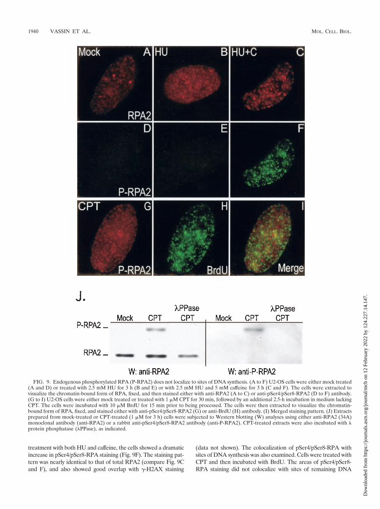
treatment with both HU and caffeine, the cells showed a dramaticincrease in pSer4/pSer8-RPA staining (Fig. 9F). The staining pat-tern was nearly identical to that of total RPA2 (compare Fig. 9Cand F), and also showed good overlap with �-H2AX staining
(data not shown). The colocalization of pSer4/pSer8-RPA withsites of DNA synthesis was also examined. Cells were treated withCPT and then incubated with BrdU. The areas of pSer4/pSer8-RPA staining did not colocalize with sites of remaining DNA
FIG. 9. Endogenous phosphorylated RPA (P-RPA2) does not localize to sites of DNA synthesis. (A to F) U2-OS cells were either mock treated(A and D) or treated with 2.5 mM HU for 3 h (B and E) or with 2.5 mM HU and 5 mM caffeine for 3 h (C and F). The cells were extracted tovisualize the chromatin-bound form of RPA, fixed, and then stained either with anti-RPA2 (A to C) or anti-pSer4/pSer8-RPA2 (D to F) antibody.(G to I) U2-OS cells were either mock treated or treated with 1 �M CPT for 30 min, followed by an additional 2.5-h incubation in medium lackingCPT. The cells were incubated with 10 �M BrdU for 15 min prior to being processed. The cells were then extracted to visualize the chromatin-bound form of RPA, fixed, and stained either with anti-pSer4/pSer8-RPA2 (G) or anti-BrdU (H) antibody. (I) Merged staining pattern. (J) Extractsprepared from mock-treated or CPT-treated (1 �M for 3 h) cells were subjected to Western blotting (W) analyses using either anti-RPA2 (34A)monoclonal antibody (anti-RPA2) or a rabbit anti-pSer4/pSer8-RPA2 antibody (anti-P-RPA2). CPT-treated extracts were also incubated with �protein phosphatase (�PPase), as indicated.
1940 VASSIN ET AL. MOL. CELL. BIOL.
Dow
nloa
ded
from
http
s://j
ourn
als.
asm
.org
/jour
nal/m
cb o
n 12
Feb
ruar
y 20
22 b
y 12
4.22
7.14
.147
.
synthesis to any significant degree (Fig. 9L). A majority of theRPA pool is hyperphosphorylated under these conditions (Fig.8N), rendering similar experiments using general RPA2 antibod-ies uninformative. We conclude that the hyperphosphorylatedform of RPA localizes only to chromatin following DNA damageand is not significantly associated with sites of chromosomal DNAsynthesis.
DISCUSSION
We find that the RPA2D mutant that mimics the hyperphos-phorylated state is prevented from stable association with rep-lication centers in vivo. The lack of association with sites ofDNA synthesis is also observed for endogenous hyperphospho-rylated RPA and is not a result of competition with the non-phosphorylated protein. Importantly, the RPARPA2D proteinhas activity equivalent to the wild-type protein both in ssDNAbinding (4) and in SV40 DNA replication in vitro. The inher-ent activity of hyperphosphorylated RPA or RPARPA2D in vivoalso appears normal because genotoxic stress causes theseRPA species to localize to DNA damage foci similarly to en-dogenous RPA2 and RPA2wt. Our data therefore indicate thatthe chromosomal DNA replication machine has the ability todiscriminate between RPA species with different phosphoryla-tion states. In addition to providing a means to regulate RPAloading and hence DNA replication, RPA phosphorylationalso has the potential to mark sites of DNA damage or repli-cation stress for recruitment of repair factors.
Our data suggest a novel feature of eukaryotic DNA repli-cation, namely, that RPA is actively loaded onto the ssDNA bythe chromosomal replication machinery. This model arisesfrom the fact that RPARPA2D, and by inference hyperphospho-rylated RPA, is inherently active in binding the ssDNA at aDNA replication fork but is unable to do so in vivo. The mostlogical explanation is that, as the duplex DNA is unwound bythe advancing DNA helicase, the hypophosphorylated RPA isloaded onto the ssDNA by protein components of the replica-tion fork machinery. One could easily envision, for example,that the minichromosome maintenance (MCM) complex, sug-gested to be the eukaryotic replicative helicase (29) and knownto interact with RPA (55), would load RPA molecules in astep-by-step fashion as the ssDNA is generated. Selective bind-ing of nonphosphorylated RPA (i.e., endogenous RPA,RPARPA2wt, or RPARPA2A) to MCM would therefore allowthis RPA species to bind only to unwound DNA. (The MCMcomplex is not involved in SV40 DNA replication.) How-ever, RPA interacts with various proteins, including the DNApolymerase �-DNA primase complex (14), and RPA phos-phorylation has been found to inhibit the association withDNA polymerase � (34). Thus, discrimination of the RPAphosphorylation state can be achieved by these or other rep-lication factors. One alternative model that does not requireconcerted RPA loading would involve a discrimination filterthat prevents access of the phosphorylated RPA to the repli-cation fork. The nature of such a filter would be difficult toenvisage.
DNA-damaging stress relieves the inhibition of RPARPA2D
chromatin binding and causes RPARPA2D association withDNA damage foci, as evident by colocalization with �-H2AX.That HU causes RPARPA2D foci to form and increases the
level of RPA2 phosphorylation while aphidicolin does neitherindicates that replication fork blockage is not sufficient forRPARPA2D chromatin binding but that the presence of DNAdamage or aberrant replication fork structures is also required.This conclusion is strengthened by our observation that inhi-bition of ATR- or ATM-mediated checkpoint response bycaffeine stimulates the rate of RPA association with DNAdamage foci. Mutation of MEC1, the Saccharomyces cerevisiaeATR homolog, is known to cause the collapse of DNA repli-cation forks that have been stalled by treatment with HU ormethyl methanesulfonate (32, 45), and such treatment leads tothe production of long ssDNA regions (43). Because of thehigh affinity of RPA for ssDNA (27, 52), we propose that theincreased availability of ssDNA releases the constraints onRPA loading seen during normal S-phase progression. Thus,under damage conditions, the RPA phosphorylation state nolonger regulates the association of RPA with chromatin.
Our data indicate that hyperphosphorylated RPA is prefer-entially associated with sites of DNA damage. The specificassociation of repair factors with this modified form of RPAwould therefore provide a mechanism to recruit repair factorsto sites of DNA damage. Interestingly, the ATRIP-ATR com-plex has been found to sense damaged DNA by recognition ofRPA-ssDNA complexes. Clearly, RPA phosphorylation hasthe potential to regulate the binding of ATRIP-ATR andthereby modify the cellular checkpoint response. Although ourexamination of RPA2D expression did not detect any notableeffects on cell cycle progression, it will be interesting to exam-ine whether RPA2D and RPA2A expression in cells lackingendogenous RPA alters cellular proliferative capacity or re-sponse to DNA damage.
Finally, our data indicate that hyperphosphorylation of RPAcan limit its ability to support chromosomal DNA replication. It isunlikely that this mechanism alone could cause significant reduc-tions in the level of DNA synthesis during genotoxic stress. Undersevere stress conditions, such as 1-h exposure to 1 �M CPT (Fig.8N) or irradiation with 30 J of UV light/m2 (53), the hyperphos-phorylated form of RPA contributes �50% of the total RPA poolprepared from asynchronous cells. Even though the fraction ofhyperphosphorylated RPA may be higher in S-phase cells, thesedata suggest that enough hypophosphorylated RPA would beavailable to sustain DNA replication. That being said, weand others have found that stress conditions also lead to theinhibition of RPA activity by other processes (9, 30, 48),including sequestration of RPA by nucleolin (12, 47). Com-bined, these data suggest that inhibition of RPA activity bymultiple mechanisms can serve to repress chromosomalDNA replication following stress.
ACKNOWLEDGMENTS
We thank Kyung Kim and Diana Dimitrova for helpful discussionsduring the course of these experiments, Kristine Carta for expert technicalassistance, and John Hirsch for assistance with FACS analysis.
J.A.B. was supported by NIH grant AI29963, DOD Breast CancerResearch Program DAMD17-03-1-0299, Philip Morris grant 15-B0001-42171, and the NYU Cancer Institute and the Rita J. andStanley Kaplan Comprehensive Cancer Center (NCI P30CA16087).M.S.W. was supported by NIH grant GM44721.
REFERENCES
1. Abraham, R. T. 2001. Cell cycle checkpoint signaling through the ATM andATR kinases. Genes Dev. 15:2177–2196.
VOL. 24, 2004 REGULATION OF RPA BINDING TO REPLICATION CENTERS 1941
Dow
nloa
ded
from
http
s://j
ourn
als.
asm
.org
/jour
nal/m
cb o
n 12
Feb
ruar
y 20
22 b
y 12
4.22
7.14
.147
.
2. Ariza, R. R., S. M. Keyse, J. G. Moggs, and R. D. Wood. 1996. Reversibleprotein phosphorylation modulates nucleotide excision repair of damagedDNA by human cell extracts. Nucleic Acids Res. 24:433–440.
3. Bartek, J., and J. Lukas. 2001. Mammalian G1- and S-phase checkpoints inresponse to DNA damage. Curr. Opin. Cell Biol. 13:738–747.
4. Binz, S. K., Y. Lao, D. F. Lowry, and M. S. Wold. 2003. The phosphorylationdomain of the 32-kDa subunit of replication protein A modulates RPA-DNA interactions: evidence for an intersubunit interaction. J. Biol. Chem.278:35584–35591.
5. Borowiec, J. 1992. Inhibition of structural changes in the simian virus 40 coreorigin of replication by mutation of essential origin sequences. J. Virol.66:5248–5255.
6. Borowiec, J. A., F. B. Dean, and J. Hurwitz. 1991. Differential induction ofstructural changes in the simian virus 40 origin of replication by T antigen.J. Virol. 65:1228–1235.
7. Brush, G. S., C. W. Anderson, and T. J. Kelly. 1994. The DNA-activatedprotein kinase is required for the phosphorylation of replication protein Aduring simian virus 40 DNA replication. Proc. Natl. Acad. Sci. USA 91:12520–12524.
8. Bullock, P. A. 1997. The initiation of simian virus 40 DNA replication in vitro.Crit. Rev. Biochem. Mol. Biol. 32:503–568.
9. Carty, M. P., M. Zernik-Kobak, S. McGrath, and K. Dixon. 1994. UVlight-induced DNA synthesis arrest in HeLa cells is associated with changesin phosphorylation of human single-stranded DNA-binding protein. EMBOJ. 13:2114–2123.
10. Chan, D. W., S. C. Son, W. Block, R. Ye, K. K. Khanna, M. S. Wold, P.Douglas, A. A. Goodarzi, J. Pelley, Y. Taya, M. F. Lavin, and S. P. Lees-Miller. 2000. Purification and characterization of ATM from human pla-centa. A manganese-dependent, wortmannin-sensitive serine/threonine pro-tein kinase. J. Biol. Chem. 275:7803–7810.
11. Cortez, D. 2003. Caffeine inhibits checkpoint responses without inhibitingthe ataxia-telangiectasia-mutated (ATM) and ATM- and Rad3-related(ATR) protein kinases. J. Biol. Chem. 278:37139–37145.
12. Daniely, Y., and J. A. Borowiec. 2000. Formation of a complex betweennucleolin and replication protein A after cell stress prevents initiation ofDNA replication. J. Cell Biol. 149:799–810.
13. Dimitrova, D. S., and D. M. Gilbert. 2000. Stability and nuclear distributionof mammalian replication protein A heterotrimeric complex. Exp. Cell Res.254:321–327.
14. Dornreiter, I., L. F. Erdile, I. U. Gilbert, D. von Winkler, T. J. Kelly, and E.Fanning. 1992. Interaction of DNA polymerase �-primase with cellular rep-lication protein A and SV40 T antigen. EMBO J. 11:769–776.
15. Durocher, D., and S. P. Jackson. 2001. DNA-PK, ATM and ATR assensors of DNA damage: variations on a theme? Curr. Opin. Cell Biol.13:225–231.
16. Dutta, A., and B. Stillman. 1992. cdc2 family kinases phosphorylate a humancell DNA replication factor, RPA, and activate DNA replication. EMBO J.11:2189–2199.
17. Fotedar, R., and J. M. Roberts. 1992. Cell cycle regulated phosphorylation ofRPA-32 occurs within the replication initiation complex. EMBO J. 11:2177–2187.
18. Gately, D. P., J. C. Hittle, G. K. T. Chan, and T. J. Yen. 1998. Character-ization of ATM expression, localization, and associated DNA-dependentprotein kinase activity. Mol. Biol. Cell 9:2361–2374.
19. Golub, E. I., R. C. Gupta, T. Haaf, M. S. Wold, and C. M. Radding. 1998.Interaction of human rad51 recombination protein with single-strandedDNA binding protein, RPA. Nucleic Acids Res. 26:5388–5393.
20. Haaf, T., E. Raderschall, G. Reddy, D. C. Ward, C. M. Radding, and E. I.Golub. 1999. Sequestration of mammalian Rad51-recombination proteininto micronuclei. J. Cell Biol. 144:11–20.
21. Hall-Jackson, C. A., D. A. Cross, N. Morrice, and C. Smythe. 1999. ATR isa caffeine-sensitive, DNA-activated protein kinase with a substrate specificitydistinct from DNA-PK. Oncogene 18:6707–6713.
22. Hassell, J. A., and B. T. Brinton. 1996. SV40 and polyomavirus DNA rep-lication, p. 639–677. In M. L. DePamphilis (ed.), DNA replication in eukary-otic cells. Cold Spring Harbor Laboratory Press, Cold Spring Harbor, N.Y.
23. Henricksen, L. A., T. Carter, A. Dutta, and M. S. Wold. 1996. Phosphoryla-tion of human replication protein A by the DNA-dependent protein kinaseis involved in the modulation of DNA replication. Nucleic Acids Res. 24:3107–3112.
24. Henricksen, L. A., C. B. Umbricht, and M. S. Wold. 1994. Recombinantreplication protein A: expression, complex formation, and functional char-acterization. J. Biol. Chem. 269:11121–11132.
25. Huang, W., and R. L. Erikson. 1994. Constitutive activation of Mek1 bymutation of serine phosphorylation sites. Proc. Natl. Acad. Sci. USA 91:8960–8963.
26. Iftode, C., and J. A. Borowiec. 1998. Unwinding of origin-specific structuresby human replication protein A occurs in a two-step process. Nucleic AcidsRes. 26:5636–5643.
27. Iftode, C., Y. Daniely, and J. A. Borowiec. 1999. Replication protein A(RPA): the eukaryotic SSB. Crit. Rev. Biochem. Mol. Biol. 34:141–180.
28. Kenny, M. K., U. Schlegel, H. Furneaux, and J. Hurwitz. 1990. The role of
human single-stranded DNA binding protein and its individual subunits insimian virus 40 DNA replication. J. Biol. Chem. 265:7693–7700.
29. Lei, M., and B. K. Tye. 2001. Initiating DNA synthesis: from recruiting toactivating the MCM complex. J. Cell Sci. 114:1447–1454.
30. Liu, J. S., S. R. Kuo, M. M. McHugh, T. A. Beerman, and T. Melendy. 2000.Adozelesin triggers DNA damage response pathways and arrests SV40 DNAreplication through replication protein A inactivation. J. Biol. Chem. 275:1391–1397.
31. Liu, V. F., and D. T. Weaver. 1993. The ionizing radiation-induced replica-tion protein A phosphorylation response differs between ataxia telangiecta-sia and normal human cells. Mol. Cell. Biol. 13:7222–7231.
32. Lopes, M., C. Cotta-Ramusino, A. Pellicioli, G. Liberi, P. Plevani, M. Muzi-Falconi, C. S. Newlon, and M. Foiani. 2001. The DNA replication checkpointresponse stabilizes stalled replication forks. Nature 412:557–561.
33. Niu, H., H. Erdjument-Bromage, Z. Q. Pan, S. H. Lee, P. Tempst, and J.Hurwitz. 1997. Mapping of amino acid residues in the p34 subunit of humansingle-stranded DNA-binding protein phosphorylated by DNA-dependentprotein kinase and Cdc2 kinase in vitro. J. Biol. Chem. 272:12634–12641.
34. Oakley, G. G., S. M. Patrick, J. Yao, M. P. Carty, J. J. Turchi, and K. Dixon.2003. RPA phosphorylation in mitosis alters DNA binding and protein-protein interactions. Biochemistry 42:3255–3264.
35. Pan, Z.-Q., A. A. Amin, E. Gibbs, H. Niu, and J. Hurwitz. 1994. Phosphor-ylation of the p34 subunit of human single-stranded-DNA-binding protein incyclin A-activated G1 extracts is catalyzed by cdk-cyclin A complex andDNA-dependent protein kinase. Proc. Natl. Acad. Sci. USA 91:8343–8347.
36. Pan, Z.-Q., C. H. Park, A. A. Amin, J. Hurwitz, and A. Sancar. 1995.Phosphorylated and unphosphorylated forms of human single-strandedDNA-binding protein are equally active in simian virus 40 DNA replicationand in nucleotide excision repair. Proc. Natl. Acad. Sci. USA 92:4636–4640.
37. Park, J. S., S. J. Park, X. Peng, M. Wang, M. A. Yu, and S. H. Lee. 1999.Involvement of DNA-dependent protein kinase in UV-induced replicationarrest. J. Biol. Chem. 274:32520–32527.
38. Philipova, D., J. R. Mullen, H. S. Maniar, J. Lu, C. Gu, and S. J. Brill. 1996.A hierarchy of SSB protomers in replication protein A. Genes Dev. 10:2222–2233.
39. Raderschall, E., E. I. Golub, and T. Haaf. 1999. Nuclear foci of mammalianrecombination proteins are located at single-stranded DNA regions formedafter DNA damage. Proc. Natl. Acad. Sci. USA 96:1921–1926.
40. Redon, C., D. Pilch, E. Rogakou, O. Sedelnikova, K. Newrock, and W.Bonner. 2002. Histone H2A variants H2AX and H2AZ. Curr. Opin. Genet.Dev. 12:162–169.
41. Sarkaria, J. N., E. C. Busby, R. S. Tibbetts, P. Roos, Y. Taya, L. M. Karnitz,and R. T. Abraham. 1999. Inhibition of ATM and ATR kinase activities bythe radiosensitizing agent, caffeine. Cancer Res. 59:4375–4382.
42. Shao, R. G., C. X. Cao, H. Zhang, K. W. Kohn, M. S. Wold, and Y. Pommier.1999. Replication-mediated DNA damage by camptothecin induces phos-phorylation of RPA by DNA-dependent protein kinase and dissociates RPA:DNA-PK complexes. EMBO J. 18:1397–1406.
43. Sogo, J. M., M. Lopes, and M. Foiani. 2002. Fork reversal and ssDNAaccumulation at stalled replication forks owing to checkpoint defects. Sci-ence 297:599–602.
44. Stigger, E., F. B. Dean, J. Hurwitz, and S.-H. Lee. 1994. Reconstitution offunctional human single-stranded DNA-binding protein from individual sub-units expressed by recombinant baculoviruses. Proc. Natl. Acad. Sci. USA91:579–583.
45. Tercero, J. A., and J. F. Diffley. 2001. Regulation of DNA replication forkprogression through damaged DNA by the Mec1/Rad53 checkpoint. Nature412:553–557.
46. Wang, H., J. Guan, A. R. Perrault, Y. Wang, and G. Iliakis. 2001. Replicationprotein A2 phosphorylation after DNA damage by the coordinated action ofataxia telangiectasia-mutated and DNA-dependent protein kinase. CancerRes. 61:8554–8563.
47. Wang, Y., J. Guan, H. Wang, D. Leeper, and G. Iliakis. 2001. Regulation ofDNA replication after heat shock by replication protein A-nucleolin inter-actions. J. Biol. Chem. 276:20579–20588.
48. Wang, Y., A. R. Perrault, and G. Iliakis. 1998. Replication protein A as apotential regulator of DNA replication in cells exposed to hyperthermia.Radiat. Res. 149:284–293.
49. Wittekind, M., J. Reizer, J. Deutscher, M. H. Saier, and R. E. Klevit. 1989.Common structural changes accompany the functional inactivation of HPrby seryl phosphorylation or by serine to aspartate substitution. Biochemistry28:9908–9912.
50. Wobbe, C. R., F. Dean, L. Weissbach, and J. Hurwitz. 1985. In vitro repli-cation of duplex circular DNA containing the simian virus 40 DNA originsite. Proc. Natl. Acad. Sci. USA 82:5710–5714.
51. Wobbe, C. R., L. Weissbach, J. A. Borowiec, F. B. Dean, Y. Murakami, P.Bullock, and J. Hurwitz. 1987. Replication of simian virus 40 origin-containingDNA in vitro with purified proteins. Proc. Natl. Acad. Sci. USA 84:1834–1838.
1942 VASSIN ET AL. MOL. CELL. BIOL.
Dow
nloa
ded
from
http
s://j
ourn
als.
asm
.org
/jour
nal/m
cb o
n 12
Feb
ruar
y 20
22 b
y 12
4.22
7.14
.147
.

52. Wold, M. S. 1997. Replication protein A: a heterotrimeric, single-strandedDNA-binding protein required for eukaryotic DNA metabolism. Annu. Rev.Biochem. 66:61–92.
53. Zernik-Kobak, M., K. Vasunia, M. Connelly, C. W. Anderson, and K. Dixon.1997. Sites of UV-induced phosphorylation of the p34 subunit of replicationprotein A from HeLa cells. J. Biol. Chem. 272:23896–23904.
54. Zou, L., and S. J. Elledge. 2003. Sensing DNA damage through ATRIPrecognition of RPA-ssDNA complexes. Science 300:1542–1548.
55. Zou, L., and B. Stillman. 2000. Assembly of a complex containing Cdc45p,replication protein A, and Mcm2p at replication origins controlled by S-phase cyclin-dependent kinases and Cdc7p-Dbf4p kinase. Mol. Cell. Biol.20:3086–3096.
VOL. 24, 2004 REGULATION OF RPA BINDING TO REPLICATION CENTERS 1943
Dow
nloa
ded
from
http
s://j
ourn
als.
asm
.org
/jour
nal/m
cb o
n 12
Feb
ruar
y 20
22 b
y 12
4.22
7.14
.147
.