Embed Size (px)
Citation preview
Articles
Replication Protein A Interactions with DNA. III. Molecular Basis of Recognitionof Damaged DNA†
Ye Lao,‡ Xavier V. Gomes,‡,§ Yingjie Ren,| John-Stephen Taylor,| and Marc S. Wold*,‡
Department of Biochemistry, UniVersity of Iowa College of Medicine, 51 Newton Road, Iowa City, Iowa 52242-1109, andDepartment of Chemistry, Washington UniVersity, St. Louis, Missouri 63130-4899
ReceiVed July 23, 1999; ReVised Manuscript ReceiVed NoVember 16, 1999
ABSTRACT: Human replication protein A (RPA) is a heterotrimeric single-stranded DNA-binding protein(subunits of 70, 32, and 14 kDa) that is required for cellular DNA metabolism. RPA has been reportedto interact specifically with damaged double-stranded DNA and to participate in multiple steps of nucleotideexcision repair (NER) including the damage recognition step. We have examined the mechanism of RPAbinding to both single-stranded and double-stranded DNA (ssDNA and dsDNA, respectively) containingdamage. We show that the affinity of RPA for damaged dsDNA correlated with disruption of the doublehelix by the damaged bases and required RPAs ssDNA-binding activity. We conclude that RPA isrecognizing single-stranded character caused by the damaged nucleotides. We also show that RPA bindsspecifically to damaged ssDNA. The specificity of binding varies with the type of damage with RPAhaving up to a 60-fold preference for a pyrimidine(6-4)pyrimidone photoproduct. We show that thisspecific binding was absolutely dependent on the zinc-finger domain in the C-terminus of the 70-kDasubunit. The affinity of RPA for damaged ssDNA was 5 orders of magnitude higher than that of thedamage recognition protein XPA (xeroderma pigmentosum group A protein). These findings suggest thatRPA probably binds to both damaged and undamaged strands in the NER excision complex. RPA bindingmay be important for efficient excision of damaged DNA in NER.
The eukaryotic single-stranded DNA-binding protein,replication protein A is essential for multiple processes incellular DNA metabolism including DNA replication, DNArepair, and recombination (reviewed in refs1 and2). Humanreplication protein A (RPA)1 is composed of subunits of 70,32, and 14 kDa (RPA70, RPA32, and RPA14, respectively).RPA binds to single-stranded DNA (ssDNA) with lowspecificity and high affinity (Ka ) ∼1010 M-1) (3). Muta-tional and structural analysis have identified three domainsof RPA that directly interact with single-stranded DNA: aweak ssDNA-binding domain in RPA32, a high-affinitycentral ssDNA-binding domain of RPA70 (which is com-posed of two copies of an oligonucleotide/oligosaccharidebinding motif; OB fold), and the C-terminal domain ofRPA70 (which contains a highly conserved zinc-finger motif)(Figure 1) (ref4 and references therein). The central DNA-
binding domain and the zinc-finger domain are both neces-sary and sufficient for high-affinity single-stranded bindingactivity (ref 4 and references therein). RPA has been shownto bind dsDNA and to promote helix destabilization (un-winding). The first two papers of this series (4, 5) demon-strated that these DNA binding activities also depend on thecentral DNA-binding domain and zinc-finger domain ofRPA70, and that double-stranded DNA binding is causedby RPA destabilizing the double helix and then binding tothe resulting single-stranded regions in duplex DNA.
RPA has been shown to bind directly to damaged dsDNA.Initial studies demonstrated that RPA was able to bind tocisplatin-modified dsDNA (6). Subsequently it has been
† These studies were supported by Grant GM44721 (M.S.W.) fromthe National Institutes of Health General Medicine Institute andCA40463 (J.S.T.) from the National Institutes of Health National CancerInstitute. Initial stages of this project were supported by the Roy J.Carver Charitable Trust.
* To whom correspondence should be addressed: phone (319) 335-6784; Fax (319) 335-9570; Email [email protected].
‡ University of Iowa College of Medicine.§ Present address: Department of Biochemistry and Molecular
Biophysics, Washington University, School of Medicine, St. Louis, MO63110-1093.
| Department of Chemistry, Washington, University.
1 Abbreviations: RPA, human replication protein A; RPA70, RPA32,RPA14, 70-, 32-, and 14-kDa subunits of human replication proteinA; RPA‚70(Zn*), human replication protein A complex containing(C500S‚C503S) 70-kDa subunit; ssDNA, single-stranded DNA; ds-DNA, double-stranded DNA; NER, nucleotide excision repair; XPA,XPG, XPC, xeroderma pigmentosum complementation groups A, G,and C; TFIIH, transcription factor IIH; ERCC1/XPF, ERCC1/xerodermapigmentosum complementation group F complex; DTT, dithiothreitol;GMSA, gel mobility shift assay; SDS-PAGE, sodium dodecyl sulfate-polyacrylamide gel electrophoresis; nt, nucleotide; bp, base pair; SSB,single stranded DNA-binding protein; (6-4) product, pyrimidine(6-4)pyrimidone product; CPD, cyclobutane pyrimidine dimer; c,s,cis,synstereochemistry; t,s-I,trans,syn-I stereochemistry; t,s-II,trans,syn-IIstereochemistry; Dewar product, Dewar valence isomer of the (6-4)product.
850 Biochemistry2000,39, 850-859
10.1021/bi991704s CCC: $19.00 © 2000 American Chemical SocietyPublished on Web 01/12/2000
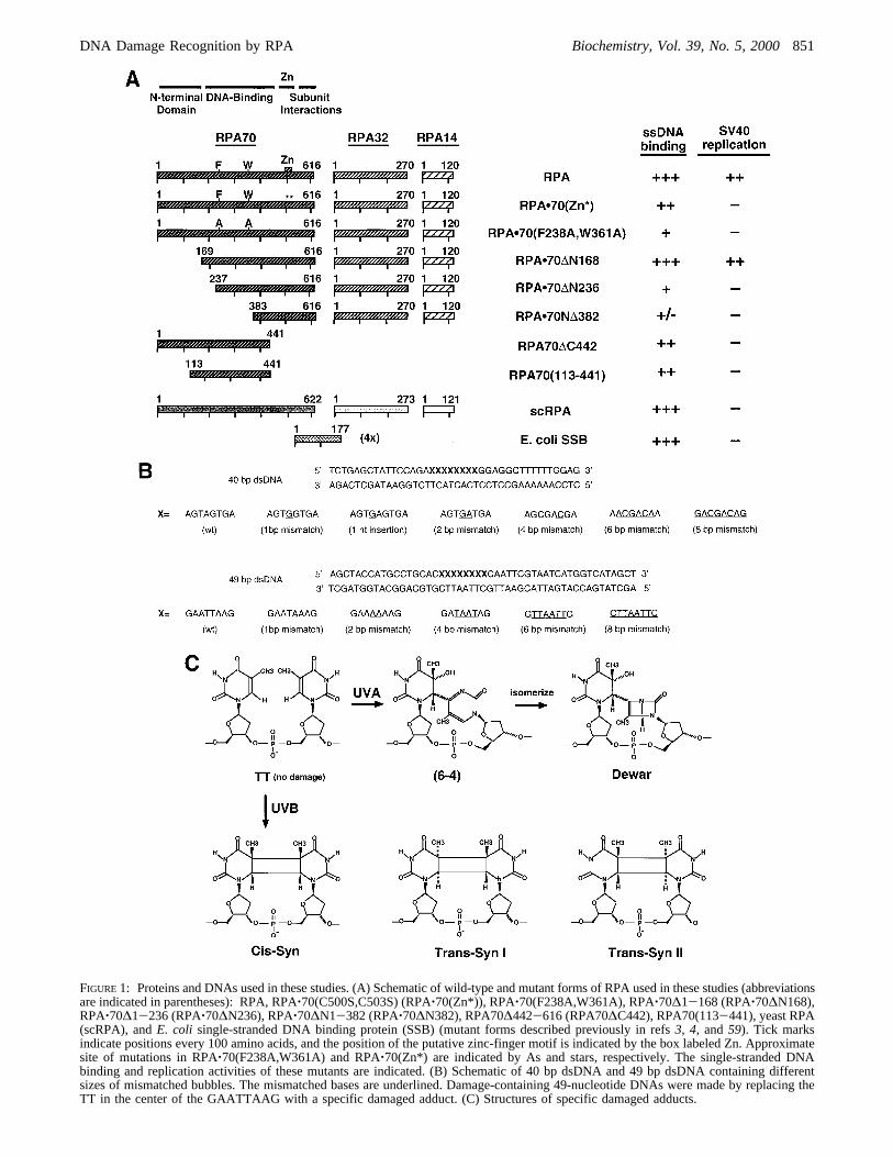
FIGURE 1: Proteins and DNAs used in these studies. (A) Schematic of wild-type and mutant forms of RPA used in these studies (abbreviationsare indicated in parentheses): RPA, RPA‚70(C500S,C503S) (RPA‚70(Zn*)), RPA‚70(F238A,W361A), RPA‚70∆1-168 (RPA‚70∆N168),RPA‚70∆1-236 (RPA‚70∆N236), RPA‚70∆N1-382 (RPA‚70∆N382), RPA70∆442-616 (RPA70∆C442), RPA70(113-441), yeast RPA(scRPA), andE. coli single-stranded DNA binding protein (SSB) (mutant forms described previously in refs3, 4, and59). Tick marksindicate positions every 100 amino acids, and the position of the putative zinc-finger motif is indicated by the box labeled Zn. Approximatesite of mutations in RPA‚70(F238A,W361A) and RPA‚70(Zn*) are indicated by As and stars, respectively. The single-stranded DNAbinding and replication activities of these mutants are indicated. (B) Schematic of 40 bp dsDNA and 49 bp dsDNA containing differentsizes of mismatched bubbles. The mismatched bases are underlined. Damage-containing 49-nucleotide DNAs were made by replacing theTT in the center of the GAATTAAG with a specific damaged adduct. (C) Structures of specific damaged adducts.
DNA Damage Recognition by RPA Biochemistry, Vol. 39, No. 5, 2000851

shown that RPA also binds preferentially to UV-damageddsDNA and dsDNA modified byN-acetoxy-2-acetylami-nofluorene (7, 8). RPA has also been purified as a cisplatin-damaged dsDNA binding protein (9). Binding to double-stranded damaged DNA is less sensitive to salt and magnesiumconcentration than RPA binding to undamaged dsDNA (9).
Nucleotide excision repair (NER) is a primary pathwayfor repairing DNA damage caused by ultraviolet (UV) lightand environmental mutagens. NER is a multistep process inwhich damage is first recognized and excised and theresulting gap filled in through a replicative process (reviewedin refs10-13). RPA is required for NER and is thought tobe involved in all steps of this process. Recognition of sitesof damage is critical for efficient NER. Initial studiessuggested that damage was recognized by xeroderma pig-mentosa group A protein (XPA) (14). This protein has noknown enzymatic activity but binds to double-stranded DNAcontaining modified/damaged bases (14-16). Subsequentlyit was shown that XPA binding to damage was enhanced byRPA (7, 17) and that nonhybridizing base pairs are involvedin recognition and binding (18, 19). Recent studies have alsoimplicated XPC in damage recognition and initiation of NER(20, 21); although in in vitro model reactions, excisionoccurred faster when XPA and RPA were the first damagerecognition factors present (21). The proteins involved indamage recognition and repair interact with each other,complicating the elucidation of the mechanism of damagerecognition. For example, RPA interacts with XPA, XPG,and ERCC1/XPF (22-25). RPA can also stimulate theendonuclease activities of both XPG and ERCC1/XPF (25,26). These interactions are thought to be essential for damagerecognition, formation of the excision complex, and damageexcision during NER(21, 27; also reviewed in ref56).
To try to gain additional insights into the role of RPA innucleotide excision repair and to understand the basis of RPAbinding to damage-containing DNA, we have examined RPAbinding to both double-stranded and single-stranded formsof DNA containing specific types of damage and comparedthose interactions with those of XPA. We show that RPAhas an increased affinity for dsDNA containing DNA damagebut that its affinity is lower than that of XPA. The affinityof RPA correlated with the change in melting temperaturecaused by a particular form of damage. We conclude thatenhanced RPA binding primarily depends on the disruptionof the duplex DNA caused by the damage. Surprisingly, wealso found that RPA bound specifically to ssDNA containingdamage. This specificity was dependent on the type ofdamage, with RPA having the highest affinity for pyrimidine-(6-4)pyrimidone photoproducts. Specificity for binding todamaged ssDNA was absolutely dependent on the zinc-fingerdomain of RPA70. RPA had a higher affinity than XPA forsingle-stranded damage containing DNA. These resultssuggest that RPA directly interacts with damaged ssDNAstrand in the NER excision complex.
EXPERIMENTAL PROCEDURES
Materials. Restriction endonucleases, polynucleotide ki-nase, and Klenow fragment were purchased from NewEngland Biolabs and Life Technologies, Inc. [γ-32P]ATP(4,500 Ci/mmol) and [R-32P]dATP (3,000 Ci/mmol) wereobtained from Amersham.O-Phenanthroline was obtained
from Sigma. Oligonucleotides were purchased from Bio-Synthesis, Inc.Escherichia coliDH5R cells were from LifeTechnologies, Inc.E. coli expression strain BL21(DE3) wasfrom W. Studier (28). E. coli single-stranded DNA-bindingprotein (SSB) was the generous gift of T. Lohman, Wash-ington University, St. Louis.
HI buffer contains 30 mM HEPES (diluted from 1 M stockat pH 7.8), 1 mM dithiothreitol, 0.25 mM EDTA, 0.5% (w/v) inositol, and 0.01% (v/v) Nonidet-P40. HI was supple-mented with different concentrations of salt as indicated. FBBbuffer (1×) contains 30 mM HEPES (pH 7.8), 100 mMNaCl, 5 mM MgCl2, 0.5% inositol, and 1 mM DTT. Tris-acetate/EDTA (TAE) gel buffer (1×) contained 40 mMTris-acetate and 2 mM EDTA, pH 8.5, and Tris-borate/EDTA (TBE) gel buffer (1×) contained 89 mM Tris base,89 mM boric acid, and 2 mM EDTA (29).
Mutant Forms of RPA.Recombinant human RPA and theyeast homologue of RPA were expressed in BL21 (DE3)cells and purified as previously described (30, 31). Deletionand mutational forms of RPA used in these studies are shownschematically in Figure 1. All mutant forms of RPA wereexpressed and purified as described previously (4).
DNA Substrates Containing Site-Specific Damage.Site-specifically damaged DNA was prepared as describedpreviously (32). Briefly, d(AATTAA) was irradiated with254 nm light, and the photoproducts were separated by C-18HPLC. The major peak with an absorption maximum near325 nm was collected and confirmed by NMR to be d(AA-[6-4]AA). The Dewar valence isomer of the (6-4) product,d(AA[Dewar]AA), was prepared in essentially quantitativeyield by irradiation of d(AA[6-4]AA) with Pyrex andMylar-filtered medium-pressure mercury lamp light. Oligo-nucleotides containing stereoisomers of the cyclobutanethymine dimer, d(CGAAT[c,s]TAAGC), d(AAT[t,s-I]TAA),and d(AAT[t,s-II]TAA), were prepared by solid-phasesynthesis using TT photoproduct building blocks (33-35)and purified by anion-exchange and C-18 HPLC. Thephotoproduct-containing hexamers were individually enzy-matically ligated to 18 nt and 25 nt oligonucleotides in thepresence of a complementary 34 nt scaffold. Thecis,syndimer-containing decamer was similarly ligated to 16 nt and23 nt oligonucleotides. Finally, the photoproduct-containing49 nt oligonucleotides were isolated by preparative acryla-mide gel electrophoresis. The sequence of the 49 residueoligonucleotide is shown in Figure 1B.
DNA Templates.The dsDNA fragment used as a probefor the UV-damage assay was generated by digesting pUC19with HindIII and radiolabeled with [R-32P]ATP. The DNAwas subsequently digested withAflIII and the ∼400 bpfragment was isolated.
Single-stranded specific damaged 49 nt or parental 49 ntoligonucleotides used in ssDNA binding reactions werelabeled with [γ-32P]ATP by T4 polynucleotide kinase (29).Labeled DNA was separated from free ATP with a Nensorbcolumn following the manufacturer’s protocol.
Double-stranded DNA containing either undamaged orspecific damaged bases were made by individually annealingthe labeled single-stranded 49 nt oligonucleotides (top strand,Figure 1B) with equal amounts of the complementary strand(lower strand in Figure 1B). Annealing reactions [50 mMTris-HCl (pH 7.5), 100 mM NaCl, and 10 mM MgCl2] wereincubated for 3 min at 95°C and slowly cooled to room
852 Biochemistry, Vol. 39, No. 5, 2000 Lao et al.
temperature. The efficiency of annealing was examined bya 1× TBE 15% polyacrylamide gel followed by autorad-iography. For all experiments, more than 95% of labeledDNA was in double-stranded form. Double-stranded frag-ments of 40 bp were made by the same protocol as for the49 bp fragments. Sequences of the 40 bp fragments usedare shown in Figure 1B, and the structures of the photo-products used are shown in Figure 1C.
Gel Mobility Shift Assays.Gel mobility shift assays wereperformed as described previously with slight modifications(36). The indicated amounts of protein were incubated with2 fmol of labeled DNA and 50 ng/µL BSA in 15µL of either1× FBB or the indicated buffer for 20 min at 25°C. Bindingreactions were brought to a final concentration of 4% glyceroland 0.01% bromophenol blue and electrophoresed on 1%agarose gel in 0.1× TAE at 10 V/cm for 1.5 h or as indicated.The gels were then dried on DE81 paper and radioactivebands were visualized by autoradiography. The radioactivityin each band was quantitated using a Packard instant imager.The resulting binding isotherms were analyzed by nonlinearleast-squares fitting to the Langmuir binding equation withKaleidaGraph (Abelbeck Software) as described previously(37). In all cases the apparent binding constants shown isthe average of multiple independent titrations.
RESULTS
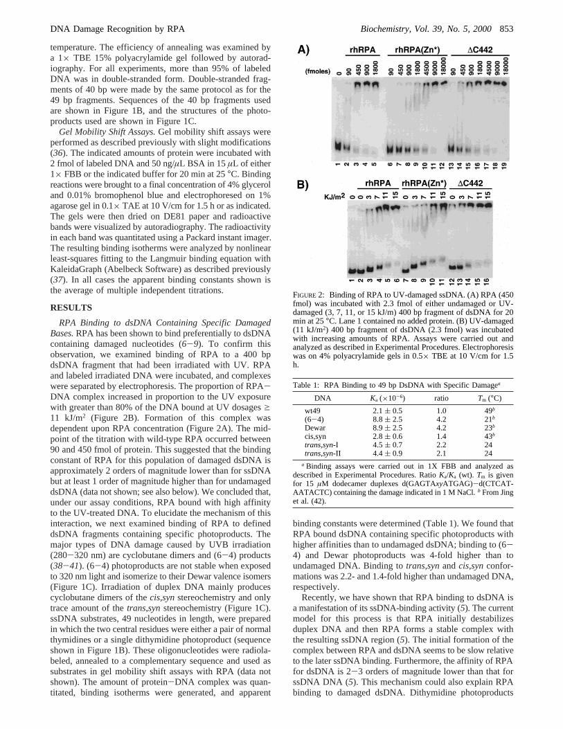
RPA Binding to dsDNA Containing Specific DamagedBases.RPA has been shown to bind preferentially to dsDNAcontaining damaged nucleotides (6-9). To confirm thisobservation, we examined binding of RPA to a 400 bpdsDNA fragment that had been irradiated with UV. RPAand labeled irradiated DNA were incubated, and complexeswere separated by electrophoresis. The proportion of RPA-DNA complex increased in proportion to the UV exposurewith greater than 80% of the DNA bound at UV dosagesg11 kJ/m2 (Figure 2B). Formation of this complex wasdependent upon RPA concentration (Figure 2A). The mid-point of the titration with wild-type RPA occurred between90 and 450 fmol of protein. This suggested that the bindingconstant of RPA for this population of damaged dsDNA isapproximately 2 orders of magnitude lower than for ssDNAbut at least 1 order of magnitude higher than for undamageddsDNA (data not shown; see also below). We concluded that,under our assay conditions, RPA bound with high affinityto the UV-treated DNA. To elucidate the mechanism of thisinteraction, we next examined binding of RPA to defineddsDNA fragments containing specific photoproducts. Themajor types of DNA damage caused by UVB irradiation(280-320 nm) are cyclobutane dimers and (6-4) products(38-41). (6-4) photoproducts are not stable when exposedto 320 nm light and isomerize to their Dewar valence isomers(Figure 1C). Irradiation of duplex DNA mainly producescyclobutane dimers of thecis,synstereochemistry and onlytrace amount of thetrans,synstereochemistry (Figure 1C).ssDNA substrates, 49 nucleotides in length, were preparedin which the two central residues were either a pair of normalthymidines or a single dithymidine photoproduct (sequenceshown in Figure 1B). These oligonucleotides were radiola-beled, annealed to a complementary sequence and used assubstrates in gel mobility shift assays with RPA (data notshown). The amount of protein-DNA complex was quan-titated, binding isotherms were generated, and apparent
binding constants were determined (Table 1). We found thatRPA bound dsDNA containing specific photoproducts withhigher affinities than to undamaged dsDNA; binding to (6-4) and Dewar photoproducts was 4-fold higher than toundamaged DNA. Binding totrans,synandcis,synconfor-mations was 2.2- and 1.4-fold higher than undamaged DNA,respectively.
Recently, we have shown that RPA binding to dsDNA isa manifestation of its ssDNA-binding activity (5). The currentmodel for this process is that RPA initially destabilizesduplex DNA and then RPA forms a stable complex withthe resulting ssDNA region (5). The initial formation of thecomplex between RPA and dsDNA seems to be slow relativeto the later ssDNA binding. Furthermore, the affinity of RPAfor dsDNA is 2-3 orders of magnitude lower than that forssDNA DNA (5). This mechanism could also explain RPAbinding to damaged dsDNA. Dithymidine photoproducts
FIGURE 2: Binding of RPA to UV-damaged ssDNA. (A) RPA (450fmol) was incubated with 2.3 fmol of either undamaged or UV-damaged (3, 7, 11, or 15 kJ/m) 400 bp fragment of dsDNA for 20min at 25°C. Lane 1 contained no added protein. (B) UV-damaged(11 kJ/m2) 400 bp fragment of dsDNA (2.3 fmol) was incubatedwith increasing amounts of RPA. Assays were carried out andanalyzed as described in Experimental Procedures. Electrophoresiswas on 4% polyacrylamide gels in 0.5× TBE at 10 V/cm for 1.5h.
Table 1: RPA Binding to 49 bp DsDNA with Specific Damagea
DNA Ka (×10-6) ratio Tm (°C)
wt49 2.1( 0.5 1.0 49b
(6-4) 8.8( 2.5 4.2 21b
Dewar 8.9( 2.5 4.2 23b
cis,syn 2.8( 0.6 1.4 43b
trans,syn-I 4.5 ( 0.7 2.2 24trans,syn-II 4.4 ( 0.9 2.1 24
a Binding assays were carried out in 1X FBB and analyzed asdescribed in Experimental Procedures. RatioKa/Ka (wt). Tm is givenfor 15 µM dodecamer duplexes d(GAGTAxyATGAG)-d(CTCAT-AATACTC) containing the damage indicated in 1 M NaCl. b From Jinget al. (42).
DNA Damage Recognition by RPA Biochemistry, Vol. 39, No. 5, 2000853
disrupt the duplex structure of DNA and reduce the meltingtemperature of the DNA (42). The resulting changes inmelting temperature correlated with the affinity of RPA, withthe hierarchy of binding (in order of decreasing affinity)being (6-4) and Dewar> ts-I and ts-II> cs> undamagedDNA. (6-4) and Dewar photoproducts caused large de-creases in melting temperature and had the highest affinityfor RPA (Table 1).cis,synandtrans,synconformations haveintermediate melting temperatures and intermediate affinitiesfor RPA. The correlation between the logKa and the meltingtemperature was not linear (data not shown), suggesting thatfactors other than duplex stability contribute to RPA recogni-tion of damaged DNA.
To elucidate the role of unpaired bases in the binding ofRPA to ssDNA, we examined RPA binding to double-stranded oligonucleotides containing various numbers ofmismatched bases. We found that small mismatched bubbles(1-4 nt) caused small changes in the Tm (e 3 degrees) andhad no effect on RPA binding (Table 2).2 In contrast,mismatched bubbles of 6 or 8 nt caused decreases inTm of6-13 °C and caused the affinity for RPA to increase by asmuch as 10-fold (Table 2). Similar results were obtained fortwo independent series of oligonucleotides indicating thatthese observations are probably general effects caused bythe mismatched nucleotides. In addition, these results areconsistent with previous reports that RPA binds with highaffinity to DNA duplexes containing 6 more unpairednucleotides (43, 44). We conclude that the single-strandedcharacter introduced by damaged or mismatched basescontributed to the enhanced binding of RPA. To try tounderstand these interactions in more detail, we examinedthe binding of RPA to ssDNA containing damaged bases.
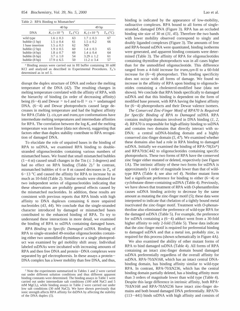
RPA Binding to Specific Damaged ssDNA.Binding ofRPA to single-stranded 49-residue oligonucleotides contain-ing either two unmodified thymidines or a single photoprod-uct was examined by gel mobility shift assay. Individuallabeled ssDNAs were incubated with increasing amounts ofRPA and then free DNA and protein-DNA complexes wereseparated by gel electrophoresis. In these assays a protein-DNA complex has a lower mobility than free DNA, and thus,
binding is indicated by the appearance of low-mobility,radioactive complexes. RPA bound to all forms of single-stranded, damaged DNA (Figure 3). RPA has an occludedbinding site size of 30 nt (31, 45). Therefore the two bandswith lower mobility observed correspond to singly anddoubly liganded complexes (Figure 3). The amounts of freeand RPA-bound ssDNA were quantitated, binding isothermswere generated, and apparent binding constants were deter-mined (Table 3). The affinity of RPA for oligonucleotidescontaining thymidine photoproducts was in all cases higherthan for the unmodified oligonucleotide. This differenceranged from a 4-fold increase fortrans,syn-II to a 60-foldincrease for (6-4) photoproduct. This binding specificitydoes not occur with all forms of damage. We found noincrease in the affinity of RPA upon binding to oligonucle-otides containing a cholesterol-modified base (data notshown). We conclude that RPA binds specifically to damagedssDNA and that this binding is dependent on the form ofmodified base present, with RPA having the highest affinityfor (6-4) photoproducts and their Dewar valence isomers.
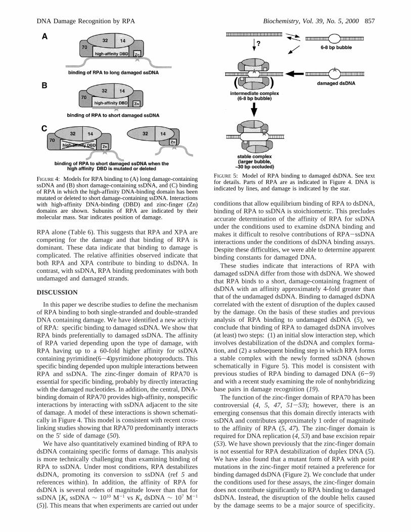
The ConserVed Zinc-Finger Motif of RPA70 Is Requiredfor Specific Binding of RPA to Damaged ssDNA.RPAcontains multiple domains involved in DNA binding (1, 2,4). RPA70 is responsible for high-affinity binding to ssDNAand contains two domains that directly interact with ss-DNA: a central ssDNA-binding domain and a highlyconserved zinc-finger domain (5, 47). We examined whetherthese domains also had a role in RPA binding to damagedssDNA. Initially we examined the binding of RPA‚70(Zn*)and RPA70∆C442 to oligonucleotides containing specificphotoproducts. These two forms of RPA have the conservedzinc finger either mutated or deleted, respectively (see Figure1A). The intrinsic affinity of these two forms for ssDNA isapproximately 1 order of magnitude lower than that of wild-type RPA (Table 4; see also ref4). Neither mutant formhad a significant preference for binding to either (6-4) orcyclobutane dimer-containing ssDNA (Table 4). Previously,we have shown that treatment of RPA withO-phenanthrolinecauses ssDNA binding activity to decrease by the sameamount as mutating the zinc-finger motif (5); these data wereinterpreted to indicate that chelation of a tightly bound metalinactivated the zinc-finger motif. Treatment withO-phenan-throline also eliminated the preference of wild-type RPA forthe damaged ssDNA (Table 5). For example, the preferencefor ssDNA containing a (6-4) adduct went from a 30-foldhigher affinity to only 2-fold (Table 5). These data indicatethat the zinc-finger motif is required for preferential bindingto damaged ssDNA and that a metal ion, probably zinc, isrequired for this process (shown schematically in Figure 4A).
We also examined the ability of other mutant forms ofRPA to bind damaged ssDNA (Table 4). All forms of RPAcontaining an intact zinc-finger domain bound damagedssDNA preferentially regardless of the overall affinity forssDNA. RPA‚70∆N168, which has an intact central DNA-binding domain, has binding affinity similar to wild-typeRPA. In contrast, RPA‚70∆N236, which has the centralbinding domain partially deleted, has a binding affinity morethan 3 orders of magnitude lower than wild type (Table 4).Despite this large difference in intrinsic affinity, both RPA‚70∆N168 and RPA‚70∆N236 have intact zinc-finger do-mains and both bound damaged DNA preferentially. RPA70-(113-441) binds ssDNA with high affinity and consists of
2 Note the experiments summarized in Tables 1 and 2 were carriedout under different solution conditions and thus different apparentbinding constants were determined. The binding assays in Table 1 werecarried out under intermediate salt conditions (100 mM NaCl and 5mM MgCl2), while binding assays in Table 2 were carried out underlow salt conditions (30 mM NaCl). We have shown previously thationic strength affects RPA affinity for dsDNA by affecting the stabilityof the DNA duplex (5).
Table 2: RPA Binding to Mismatchesa
40 bp 49 bp
DNA Ka (×10-9) Tm (°C) Ka (×10-9) Tm (°C)
wild type 1.6( 0.3 63 1.7( 0.3 67bubble (1 bp) 1.5( 0.4 61 1.5( 0.2 661 base insertion 1.5( 0.3 62 NDbubble (2 bp) 1.9( 0.5 60 1.4( 0.3 65bubble (4 bp) 1.8( 0.7 60 1.4( 0.4 64bubble (6 bp) 3.7( 1.0 54 3.29( 1.2 61bubble (8 bp) 17.9( 6.5 50 11.2( 3.4 57
a Binding assays were carried out in HI buffer containing 30 mMKCl and analyzed as described in Experimental Procedures.Tm’sdetermined as in ref5.
854 Biochemistry, Vol. 39, No. 5, 2000 Lao et al.
only the central DNA-binding domain of RPA70. Thismutant form does not contain the zinc-finger domain andhad no specificity for damaged DNA (Table 4). These dataconfirm that the zinc-finger domain is necessary for specificbinding to damaged DNA and suggest that it directly interactswith damaged bases. These studies do not distinguish whetherthe zinc-finger motif is directly interacting with the damagedbases or is required for maintaining the structure of thisdomain of RPA70. We also found that when the zinc fingeris present without the central DNA-binding domain, as inRPA‚70∆N382, stable protein-DNA complexes were notobserved with either parental ssDNA or specific damagedssDNA (data not shown). We conclude that the central DNA-binding domain of RPA70 plays a predominant role indetermining the overall affinity for DNA but does notcontribute to specificity of binding to damage.
To determine whether other single-stranded DNA-bindingproteins also preferentially bound to damaged oligonucle-otides, we examined the binding ofE. coli single-strandedDNA binding protein (SSB) and the yeast RPA homologue,scRPA.E. coli SSB does not contain a zinc-finger motif (48)and only showed a 3-fold higher affinity for the Dewaradduct containing ssDNA (Table 4). In contrast, scRPA,which contains a conserved zinc-finger motif, had ag22-fold higher affinity for Dewar photoproduct-containing
ssDNA. This indicated that preferential binding for damageddsDNA is a general property of RPA homologues but notof all single-stranded DNA binding proteins.
Taken together, our studies suggest that the specificity ofRPA binding to damaged ssDNA arises because of thecombined action of the nonspecific, high-affinity centralDNA-binding domain and the low-affinity, damage-specificzinc-finger domain. The central binding domain directlyinteracts with 8 nt (49). Thus, we predicted that shortoligonucleotides should show a weaker preference forbinding to damage because they will be too short to interactfully with both domains (shown schematically in Figure 4B).Therefore, we tested our hypothesis by examining the bindingof wild-type and mutant forms of RPA to a 12-residueoligonucleotide. RPA bound to the 12-residue oligonucleotidewith lower affinity than observed with longer oligonucle-otides (Table 5; see also ref37). No significant preferencefor binding a damage-containing 12-residue oligonucleotidewas observed (Table 5). [The 2-fold preference observed withthe 12-nt damaged oligonucleotide was equivalent to thatseen with longer oligonucleotides and RPA‚70(Zn*) orO-phenanthroline treated RPA.] Treatment withO-phenan-throline still caused a 10-fold decrease in affinity of RPA(Table 5). This suggests that a 12-nt oligonucleotide is longenough to partially interact with the zinc-finger domain.Alternatively, disruption of the zinc-finger motif by removalof associated metal could cause a change in the structure ofthe central binding domain. As was seen with longeroligonucleotides, RPA70∆C442 showed no preference forbinding damaged ssDNA and binding was insensitive toO-phenanthroline (Table 5). We also examined binding byRPA‚70(F238A,W361A). This mutant form of RPA has twopoint mutations in the central binding domain of RPA70 thatpartially disrupt its structure and cause a reduction ofapproximately 3 orders of magnitude in the affinity forssDNA (4). RPA‚70(F238A,W361A) had a 10-fold higher
FIGURE 3: Binding of wild-type RPA to specific photodamaged ssDNA. Assays were carried out and analyzed as described in ExperimentalProcedures. The positions of the shifted complexes, free oligonucleotides, and free label are indicated. The type of specific damage containedin each oligonucleotide DNA is also indicated.
Table 3: Wild-Type RPA Binding to ssDNA with SpecificDamagea
Ka (×10-8) ratio
wt49 57( 17 1(6-4) 3500( 1920 62Dewar 1550( 270 27cis,syn 320( 100 6trans,syn-I 420 ( 230 8trans,syn-II 230 ( 60 4
a Binding assays were carried out in 1× FBB and analyzed asdescribed in Experimental Procedures. Ratio) Ka/Ka (wt).
DNA Damage Recognition by RPA Biochemistry, Vol. 39, No. 5, 2000855
affinity for the (6-4) adduct-containing 12-nt oligonucleotidethan for the undamaged 12-nt oligonucleotide (Table 5). Thispreference was sensitive toO-phenanthroline. The simplestexplanation for these results is that when the central bindingdomain is partially disrupted, the short oligonucleotide is ableto associate with the zinc-finger domain and preferentialbinding to damaged ssDNA is observed (shown schemati-cally in Figure 4C). These data indicate that there is acompetition between the central binding domain and the zinc-finger domain for ssDNA and that it is the interplay betweenthese two sites that leads to the observed binding propertiesof RPA.
Binding of XPA and RPA to Damaged DNA.RPA andXPA are both involved in the damage recognition step of
NER (7). Therefore, we also examined the binding of XPAprotein to both single- and double-stranded forms of thedamaged oligonucleotides. We found that XPA had anapparent binding constant for double-stranded, undamagedDNA of 1.2 × 10⁷ (Table 6). This is approximately an orderof magnitude higher than that of RPA under the sameconditions. We observed additive binding to double-strandedDNA when RPA and XPA were mixed (Table 6). In contrast,XPA binds to ssDNA with an affinity 3 orders of magnitudelower than RPA and did not show any specificity for bindingsingle-stranded, damaged oligonucleotides (Table 6 and datanot shown). When binding of a mixture of RPA and XPAto single-stranded oligonucleotides was examined, the bind-ing constants observed were slightly reduced from those of
Table 4: Binding of Mutant Forms of RPA to ssDNA with Specific Damagea
RPAundamagedKa (× 10-8)
DewarKa (× 10-8) ratio
cis,synKa (×10-8) ratio
RPA 57( 17 1550( 270 27 320( 100 6RPA70∆C442 6.9( 1.1 6.9( 0.9 1.0 6.3( 0.6 0.9RPA‚70(Zn*) 3.12( 0.7 4.9( 1.1 1.5 2.8( 0.8 0.9RPA‚70∆N168 49( 5 g3500 72b 440( 180 8.9RPA‚70∆N236 0.0086( 0.0004 0.23( 0.04 27 0.11( 0.02 13RPA70(113-441) 2.8( 0.3 5.4( 0.6 1.9 2.7( 0.3 1.0scRPA 34( 5 g770 22b 220( 50 6.2E. coli SSB 13( 2 41( 9 3.0 15( 3 1.1
a Binding assays were carried out in 1× FBB and analyzed as described in Experimental Procedures.b Binding is under stoichiometric conditions,so binding constants were estimated from the concentration of RPA at half-saturation. Ratio) ratio of binding constants for damaged ssDNA/undamaged ssDNA.
Table 5: Binding of Wild-Type and Mutant Forms of RPA to Specific Damaged ssDNA( Phenanthrolinea
49 nt oligonucleotide
protein phenanthrolineundamaged DNA
Ka (×10-8)damaged DNA
Ka (×10-8) ratio
wtRPA - 52 ( 28 725( 250 (t,s-II) 14wtRPA + 1.3( 0.8 3.0( 1.6 (t,s-II) 2wtRPA - 39 ( 8 1200( 350 (6-4) 30wtRPA + 1.8( 0.7 3.5( 2.4 (6-4) 2
12 nt oligonucleotide
protein phenanthrolineundamagedKa (×10-7)
(6-4) damagedKa (×10-7) ratio
wtRPA - 11 ( 1.5 24( 5 2.3wtRPA + 1.1( 0.2 1.3( 0.3 1.1RPA70∆C442 - 1.5( 0.2 2.4( 0.7 1.5RPA70∆C442 + 1.0( 0.1 1.6( 0.2 1.5RPA‚70(F238A,W361A) - 0.12( 0.03 1.2( 0.1 9.7RPA‚70(F238A,W361A) + 0.022( 0.015 0.027( 0.014 1.2
a Binding assays with 49 nt oligonucleotide were carried out in 1× FBB. Binding assays with 12 nt oligonucleotide [5′ GCGTATTATGCG 3′,with underlined TT being a (6-4) photoproduct] were carried out in 200 mM NaCl, 1 mM MgCl2, 25 mM Hepes, 4% glycerol, 0.5 mM DTT,0.01% NP-40 (58). The sequence of the analysis was carried out as described in Experimental Procedures. Ratio) ratio of binding constants fordamaged ssDNA/undamaged ssDNA.
Table 6: Comparison of Binding Constants for XPA and RPAa
Ka (×10-7)
DNA RPA XPA XPA + RPA
ds49 bp 0.21( 0.05 1.2( 0.3 1.4( 0.4ds49 bp,cis,syn 0.28( 0.06 3.0( 0.5 1.4( 0.3ds49 bp, (6-4) 0.88( 0.25 3.3( 0.5 4.2( 0.4
ss49 nt 570( 170 0.46( 0.13 99( 26ss49 nt,cis,syn 3200( 1000 0.55( 0.11 420( 110ss49 nt, (6-4) 35,000( 19,000 0.18( 0.05 3000( 1100
a Binding assays were carried out in 1× FBB and analyzed as described in Experimental Procedures.
856 Biochemistry, Vol. 39, No. 5, 2000 Lao et al.
RPA alone (Table 6). This suggests that RPA and XPA arecompeting for the damage and that binding of RPA isdominant. These data indicate that binding to damage iscomplicated. The relative affinities observed indicate thatboth RPA and XPA contribute to binding to dsDNA. Incontrast, with ssDNA, RPA binding predominates with bothundamaged and damaged strands.
DISCUSSION
In this paper we describe studies to define the mechanismof RPA binding to both single-stranded and double-strandedDNA containing damage. We have identified a new activityof RPA: specific binding to damaged ssDNA. We show thatRPA binds preferentially to damaged ssDNA. The affinityof RPA varied depending upon the type of damage, withRPA having up to a 60-fold higher affinity for ssDNAcontaining pyrimidine(6-4)pyrimidone photoproducts. Thisspecific binding depended upon multiple interactions betweenRPA and ssDNA. The zinc-finger domain of RPA70 isessential for specific binding, probably by directly interactingwith the damaged nucleotides. In addition, the central, DNA-binding domain of RPA70 provides high-affinity, nonspecificinteractions by interacting with ssDNA adjacent to the siteof damage. A model of these interactions is shown schemati-cally in Figure 4. This model is consistent with recent cross-linking studies showing that RPA70 predominantly interactson the 5′ side of damage (50).
We have also quantitatively examined binding of RPA todsDNA containing specific forms of damage. This analysisis more technically challenging than examining binding ofRPA to ssDNA. Under most conditions, RPA destabilizesdsDNA, promoting its conversion to ssDNA (ref5 andreferences within). In addition, the affinity of RPA fordsDNA is several orders of magnitude lower than that forssDNA [Ka ssDNA ∼ 1010 M-1 vs Ka dsDNA ∼ 107 M-1
(5)]. This means that when experiments are carried out under
conditions that allow equilibrium binding of RPA to dsDNA,binding of RPA to ssDNA is stoichiometric. This precludesaccurate determination of the affinity of RPA for ssDNAunder the conditions used to examine dsDNA binding andmakes it difficult to resolve contributions of RPA-ssDNAinteractions under the conditions of dsDNA binding assays.Despite these difficulties, we were able to determine apparentbinding constants for damaged DNA.
These studies indicate that interactions of RPA withdamaged ssDNA differ from those with dsDNA. We showedthat RPA binds to a short, damage-containing fragment ofdsDNA with an affinity approximately 4-fold greater thanthat of the undamaged dsDNA. Binding to damaged dsDNAcorrelated with the extent of disruption of the duplex causedby the damage. On the basis of these studies and previousanalysis of RPA binding to undamaged dsDNA (5), weconclude that binding of RPA to damaged dsDNA involves(at least) two steps: (1) an initial slow interaction step, whichinvolves destabilization of the dsDNA and complex forma-tion, and (2) a subsequent binding step in which RPA formsa stable complex with the newly formed ssDNA (shownschematically in Figure 5). This model is consistent withprevious studies of RPA binding to damaged DNA (6-9)and with a recent study examining the role of nonhybridizingbase pairs in damage recognition (19).
The function of the zinc-finger domain of RPA70 has beencontroversial (4, 5, 47, 51-53); however, there is anemerging consensus that this domain directly interacts withssDNA and contributes approximately 1 order of magnitudeto the affinity of RPA (5, 47). The zinc-finger domain isrequired for DNA replication (4, 53) and base excision repair(53). We have shown previously that the zinc-finger domainis not essential for RPA destabilization of duplex DNA (5).We have also found that a mutant form of RPA with pointmutations in the zinc-finger motif retained a preference forbinding damaged dsDNA (Figure 2). We conclude that underthe conditions used for these assays, the zinc-finger domaindoes not contribute significantly to RPA binding to damageddsDNA. Instead, the disruption of the double helix causedby the damage seems to be a major source of specificity.
FIGURE 4: Models for RPA binding to (A) long damage-containingssDNA and (B) short damage-containing ssDNA, and (C) bindingof RPA in which the high-affinity DNA-binding domain has beenmutated or deleted to short damage-containing ssDNA. Interactionswith high-affinity DNA-binding (DBD) and zinc-finger (Zn)domains are shown. Subunits of RPA are indicated by theirmolecular mass. Star indicates position of damage.
FIGURE 5: Model of RPA binding to damaged dsDNA. See textfor details. Parts of RPA are as indicated in Figure 4. DNA isindicated by lines, and damage is indicated by the star.
DNA Damage Recognition by RPA Biochemistry, Vol. 39, No. 5, 2000857
Presumably this is because the disrupted helix acts as a“nucleation site” for RPA during the slow initial bindingevent (Figure 5). Direct interactions with the zinc-fingerdomain appear to occur only with single-stranded DNA andthus occur after initial binding events that lead to unwindingthe dsDNA. This model is consistent with the finding that amutant form of RPA with a disrupted zinc-finger domainwas functional in NER but was able to support only about50% the level of excision as compared to wild-type RPA(53). These data indicate that, while not essential for NER,the zinc-finger domain is necessary for optimal excision ofdamage. We hypothesize that interactions between the zinc-finger domain and specific damage help align the excisionnucleases and increase the efficiency of excision.
These studies provide new insights into the function ofRPA during NER. RPA is required for the damage recogni-tion step in NER (7, 54, 55). We present evidence that therole of RPA during damage recognition is to bind to thedisruptions in the DNA duplex caused by damaged nucle-otides. This activity requires RPA’s ssDNA-binding activityand is closely related to RPA’s dsDNA-binding and helix-destabilizing activities. The specificity of RPA for damageddsDNA is modest; nevertheless, this interaction coupled withthat of other damage binding proteins leads to the formationof a specific repair complex at the site of damage. Damagerecognition also requires XPA (14-16) and XPC (12, 20)and components of TFIIH (12). Individually, the specificitiesof these proteins for binding sites of damage are also modest(12, 14, 16). Therefore, it seems likely that high levels ofspecificity are achieved through the combined binding ofmultiple proteins with modest specificity (12, 19) or throughthe binding of a “repair complex” containing several (or all)of these proteins (56).
After the initial damage recognition step, a proteincomplex forms at the site of damage and causes localizedunwinding of the duplex and excision of the damage. RPAis also required for these processes. Most models proposethat when the DNA is unwound, RPA binds to the undam-aged strand while XPA binds to the damaged strand (27,54). Our studies show that this model is likely to be incorrect.The affinities of RPA for undamaged and damaged ssDNAare 3-5 orders of magnitude higher than those of XPA(Table 6; see also ref19). Therefore RPA should easilycompete with XPA and bind to both unwound strands in theexcision complex. Furthermore, RPA interacts with both thenucleases involved with excision, XPG and ERCC1-XPF (7,25). RPA can stimulate excision by XPG and ERCC1-XPF(27). This stimulation is enhanced in an orientation consistentwith RPA binding to the undamaged strand; however, thesestudies did not rule out binding to both strands (27). Wepropose that RPA is bound to both strands in the excisioncomplex and that specific protein-protein interactions in thiscomplex are essential for efficient excision. The affinity ofRPA is high enough that RPA should bind to both strandseven in the absence of specific interactions with the damagednucleotides. This could explain why the zinc-finger domainis not essential for NER (53). Mutant forms of RPA thatbind ssDNA with high affinity but do not bind damagespecifically [e.g., RPA‚70(Zn*)] can still bind to bothunwound strands in the excision complex; however, suchmutant forms will not be positioned as precisely, leading toa reduced efficiency of excision. It is also possible that steric
constraints or specific protein interactions may limit RPAbinding in the damage excision complex. Additional studieswill be needed to examine this possibility.
Both human and yeast RPA have conserved zinc-fingerdomains and bind specifically to damaged ssDNA. Incontrast,E. coli SSB does not contain a zinc-finger motifand does not bind damaged ssDNA specifically. Functionally,both RPA homologues are essential for NER and involvedin damage recognition, whileE. coli SSB is not required forthe equivalent pathway (uvrABC) inE. coli. These homolo-gies suggest that there may be a general role for damagerecognition in DNA repair processes. The single-strandedDNA-binding protein from phage T4, gene 32 protein, alsocontains a zinc finger and has been shown to bind damageddsDNA (57). It will be interesting to determine whether gene32 protein also binds damaged ssDNA specifically.
ACKNOWLEDGMENT
We thank the members of the Wold laboratory forscientific discussions and critical reading of the manuscriptand Mu Wang for preparing the 49 nt substrates. We thankRichard Wood for the XPA expression plasmid. We thankAziz Sancar for communication of data prior to publication.We also thank the University of Iowa DNA Core Facilityfor oligonucleotide synthesis and DNA sequencing.
REFERENCES
1. Wold, M. S. (1997)Annu. ReV. Biochem. 66, 61-92.2. Iftode, C., Daniely, Y., and Borowiec, J. A. (1999)CRC Crit.
ReV. Biochem. 34, 141-180.3. Gomes, X. V., and Wold, M. S. (1996)Biochemistry 35,
10558-10568.4. Walther, A. P., Gomes, X. V., Lee, C. G., and Wold, M. S.
(1999)Biochemistry 38, 3963-3973.5. Lao, Y., Lee, C. G., and Wold, M. S. (1999)Biochemistry
38, 3963-3973.6. Clugston, C. K., McLaughlin, K., Kenny, M. K., and Brown,
R. (1992)Cancer Res. 52, 6375-6379.7. He, Z., Henricksen, L. A., Wold, M. S., and Ingles, C. J. (1995)
Nature 374, 566-569.8. Burns, J. L., Guzder, S. N., Sung, P., Prakash, S., and Prakash,
L. (1996)J. Biol. Chem. 271, 11607-11610.9. Patrick, S. M., and Turchi, J. J. (1998)Biochemistry 37, 8808-
8815.10. Sancar, A. (1996)Annu. ReV. Biochem. 65, 43-81.11. Wood, R. D. (1996)Annu. ReV. Biochem. 65, 135-167.12. Wood, R. D. (1999)Biochimie 81, 39-44.13. Petit, C., and Sancar, A. (1999)Biochimie 81, 15-25.14. Jones, C. J., and Wood, R. D. (1993)Biochemistry 32, 12096-
12104.15. Robins, P., Jones, C. J., Biggerstaff, M., Lindahl, T., and
Wood, R. D. (1991)EMBO J. 10, 3913-3921.16. Asahina, H., Kuraoka, I., Shirakawa, M., Morita, E. H., Miura,
N., Ohtsuka, E., Okada, Y., and Tanaka, K. (1994)Mutat. Res.315, 229-237.
17. Li, L., Lu, X. Y., Peterson, C. A., and Legerski, R. J. (1995)Mol. Cell. Biol. 15, 5396-5402.
18. Buschta-Hedayat, N., Buterin, T., Hess, M. T., Nissura, M.,and Naegeli, H. (1999)Proc. Natl. Acad. Sci. U.S.A. 96, 6090-6095.
19. Buschta-Hedayat, N., Buterin, T., Hess, M. T., Missura, M.,and Naegeli, H. (1999)Proc. Natl. Acad. Sci. U.S.A. 96, 6090-6095.
20. Sugasawa, K., Ng, J. M., Masutani, C., Iwai, S., Van der Spek,P. J., Eker, A. P., Bootsma, D., and Hoeijmakers, J. H. (1998)Mol. Cell 2, 223-232.
21. Wakasugi, M., and Sancar, A. (1999)J. Biol. Chem. 274,18759-18768.
858 Biochemistry, Vol. 39, No. 5, 2000 Lao et al.
22. Coverley, D., Kenny, M. K., Lane, D. P., and Wood, R. D.(1992)Nucleic Acids Res. 20, 3873-3880.
23. Heyer, W.-D., Rao, M. R. S., Erdile, L. F., Kelly, T. J., andKolodner, R. D. (1990)EMBO J. 9, 2321-2329.
24. Moore, S. P., Erdile, L., Kelly, T., and Fishel, R. (1991)Proc.Natl. Acad. Sci. U.S.A. 88, 9067-9071.
25. Matsunaga, T., Park, C. H., Bessho, T., Mu, D., and Sancar,A. (1996)J. Biol. Chem. 271, 11047-11050.
26. Bessho, T., Sancar, A., Thompson, L. H., and Thelen, M. P.(1997)J. Biol. Chem. 272, 3833-3837.
27. De Laat, W. L., Appeldoorn, E., Sugasawa, K., Weterings,E., Jaspers, N. G. J., and Hoeijmakers, J. H. J. (1998)GenesDeV. 12, 2598-2609.
28. Studier, F. W., Rosenberg, A. H., Dunn, J. J., and Dubendorff,J. W. (1990)Methods Enzymol. 185, 60-89.
29. Ausubel, F. M., Brent, R., Kingston, R. E., Moore, D. D.,Seidman, J. G., Smith, J. A., and Struhl, K. (1989)Currentprotocols in molecular biology, John Wiley and Sons, NewYork.
30. Henricksen, L. A., Umbricht, C. B., and Wold, M. S. (1994)J. Biol. Chem. 269, 11121-11132.
31. Sibenaller, Z. A., Sorensen, B. R., and Wold, M. S. (1998)Biochemistry 37, 12496-12506.
32. Smith, C. A., Wang, M., Jiang, N., Che, L., Zhao, X., andTaylor, J.-S. (1996)Biochemistry 35, 4146-4154.
33. Taylor, J.-S., Brockie, I. R., and O’Day, C. L. (1987)J. Am.Chem. Soc. 109, 6735-6742.
34. Taylor, J.-S., and Brockie, I. R. (1988)Nucleic Acids Res.16, 5123-5136.
35. Kao, J. L.-F., Nadji, S., and Taylor, J.-S. (1993)Chem. Res.Toxicol. 6, 561-567.
36. Kim, C., Snyder, R. O., and Wold, M. S. (1992)Mol. Cell.Biol. 12, 3050-3059.
37. Kim, C., Paulus, B. F., and Wold, M. S. (1994)Biochemistry33, 14197-14206.
38. Wang, S. Y. (1976) inPhotochemistry and Photobiology ofNucleic Acids Volume I, Chemistry, Academic Press, Inc., NewYork.
39. Cadet, J., and Vigny, P. (1990) inBiooorganic Photochemistry(Morrison, H., Ed.) pp 1-272, John Wiley and Sons, NewYork.
40. Smith, C. A., and Taylor, J.-S. (1993)J. Biol. Chem. 268,11143-11151.
41. Taylor, J.-S. (1994)Acc. Chem. Res. 27, 76-82.42. Jing, Y., Kao, J. F., and Taylor, J. S. (1998)Nucleic Acids
Res. 26, 3845-3853.43. Iftode, C., and Borowiec, J. A. (1997)Mol. Cell. Biol. 17,
3876-3883.44. Iftode, C., and Borowiec, J. A. (1998)Nucleic Acids Res. 26,
5636-5643.45. Kim, C., and Wold, M. S. (1995)Biochemistry 34, 2058-
2064.46. Walther, A. P., Bjerke, M. P., and Wold, M. S. (1999)Nucleic
Acids Res. 27, 656-664.47. Brill, S. J., and Bastin-Shanower, S. (1998)Mol. Cell. Biol.
18, 7225-7234.48. Lohman, T. M., and Ferrari, M. E. (1994)Annu. ReV. Biochem.
63, 527-570.49. Bochkarev, A., Pfuetzner, R. A., Edwards, A. M., and Frappier,
L. (1997)Nature 385, 176-181.50. Schweizer, U., Hey, T., Lipps, G., and Krauss, G. (1999)
Nucleic Acids Res. 27, 3183-3189.51. Lin, Y. L., Chen, C., Keshav, K. F., Winchester, E., and Dutta,
A. (1996)J. Biol. Chem. 271, 17190-17198.52. Dong, J. W., Park, J. S., and Lee, S. H. (1999)Biochem. J.
337, 311-317.53. Lin, Y. L., Shivji, M. K. K., Chen, C., Kolodner, R., Wood,
R. D., and Dutta, A. (1998)J. Biol. Chem. 273, 1453-1461.54. Mu, D., Hsu, D. S., and Sancar, A. (1996)J. Biol. Chem. 271,
8285-8294.55. Aboussekhra, A., Biggerstaff, M., Shivji, M. K. K., Vilpo, J.
A., Moncollin, V., Podust, V. N., Protic, M., Hu¨bscher, U.,Egly, J.-M., and Wood, R. D. (1995)Cell 80, 859-868.
56. Araujo, S. J., and Wood, R. D. (1999)Mutat. Res. DNA Repair435, 23-33.
57. Toulme, J. J., Behmoaras, T., Guigues, M., and Helene, C.(1983)EMBO J. 2, 505-510.
58. Pfuetzner, R. A., Bochkarev, A., Frappier, L., and Edwards,A. M. (1997) J. Biol. Chem. 272, 430-434.
59. Gomes, X. V., and Wold, M. S. (1995)J. Biol. Chem. 270,4534-4543.
60. Herrmann, G., Lindahl, T., and Scha¨r, P. (1998)EMBO J. 17,4188-4198.
BI991704S
DNA Damage Recognition by RPA Biochemistry, Vol. 39, No. 5, 2000859