Embed Size (px)
Citation preview
Renal histopathology and crystal deposits in patientswith small bowel resection and calcium oxalatestone diseaseAndrew P. Evan1, James E. Lingeman2, Elaine M. Worcester3, Sharon B. Bledsoe1, Andre J. Sommer4,James C. Williams Jr1, Amy E. Krambeck5, Carrie L. Philips6 and Fredric L. Coe3
1Department of Anatomy and Cell Biology, Indiana University School of Medicine, Indianapolis, Indiana, USA; 2International KidneyStone Institute, Methodist Clarian Hospital, Indianapolis, Indiana, USA; 3Department of Medicine, University of Chicago, Chicago, Illinois,USA; 4Department of Chemistry and Biochemistry, Miami University, Oxford, Ohio, USA; 5Division of Urology, Mayo Clinic, Rochester,Minnesota, USA and 6Department of Pathology, Clarian Health Partners, Indianapolis, Indiana, USA
We present here the anatomy and histopathology of kidneys
from 11 patients with renal stones following small bowel
resection, including 10 with Crohn’s disease and 1 resection
in infancy for unknown cause. They presented predominantly
with calcium oxalate stones. Risks of formation included
hyperoxaluria (urine oxalate excretion greater than 45 mg
per day) in half of the cases, and acidic urine of reduced
volume. As was found with ileostomy and obesity bypass,
inner medullary collecting ducts (IMCDs) contained crystal
deposits associated with cell injury, interstitial inflammation,
and papillary deformity. Cortical changes included modest
glomerular sclerosis, tubular atrophy, and interstitial fibrosis.
Randall’s plaque (interstitial papillary apatite) was abundant,
with calcium oxalate stone overgrowth similar to that seen in
ileostomy, idiopathic calcium oxalate stone formers, and
primary hyperparathyroidism. Abundant plaque was
compatible with the low urine volume and pH. The IMCD
deposits all contained apatite, with calcium oxalate present
in three cases, similar to findings in patients with obesity
bypass but not an ileostomy. The mechanisms for calcium
oxalate stone formation in IMCDs include elevated urine and
presumably tubule fluid calcium oxalate supersaturation, but
a low calcium to oxalate ratio. However, the mechanisms for
the presence of IMCD apatite remain unknown.
Kidney International (2010) 78, 310–317; doi:10.1038/ki.2010.131;
published online 28 April 2010
KEYWORDS: clinical nephrology; kidney stones; renal biopsy; renal pathology;
renal physiology
We have shown elsewhere1,2 that bowel disease can produce awide range of renal histopathology and entrain multiplemechanisms for renal stone production and formation ofintrarenal crystal deposits. In patients with obesity bypassprocedures, calcium oxalate (CaOx) stones are found free inthe urinary collecting system. Within the kidney, the onlydeposits are occasional apatite plugs within inner medullarycollecting ducts (IMCDs), rare microscopic linear deposits ofCaOx over apical surfaces of otherwise normal-appearingIMCD cells, and a single small IMCD CaOx plug in each oftwo of the five cases that we have studied.1,2 Patients withileostomy produce stones containing both uric acid andCaOx.3 In their kidneys, we have found interstitial apatite(Randall’s) plaque, and IMCD plugs composed of apatite andthe sodium and ammonium acid salts of uric acid.3 SomeCaOx stones were found growing over regions of plaque, as isseen in idiopathic CaOx stone formers (ICSFs) and stoneformers (SFs) with primary hyperparathyroidism.4–6 Otherstones were found free in the collecting system.
We have little difficulty in explaining the plaque, stones,and urate IMCD deposits that we have thus far encounteredin bowel patients. Bypass patients produce no excess ofplaque above control subjects and their stones never growattached to renal papillae. The lack of plaque is consistentwith our earlier findings that plaque is promoted by scantyacidic urine and hypercalciuria; bypass patients have highurine volumes and are not hypercalciuric.1,7 Their urineoxalate excretion is high, and we can presume with reason-able certainty that hyperoxaluria (urine oxalate excretion445 mg per day) increased urine supersaturation (SS) withrespect to CaOx and led to crystal formation, perhaps in freesolution. By contrast, ileostomy patients produce scanty andacidic urine, form abundant plaque, and produce attachedCaOx stones over areas of papillary plaque. Their urine has ahigh uric acid SS that accounts for their uric acid stones.Urine sodium and ammonium urate SS was present in threeof the five ileostomy patients with these salts in their IMCDs,indicating that, for urate species, IMCDs and urine were not
o r i g i n a l a r t i c l e http://www.kidney-international.org
& 2010 International Society of Nephrology
Received 1 December 2009; revised 3 February 2010; accepted 3 March
2010; published online 28 April 2010
Correspondence: Andrew P. Evan, Department of Anatomy and Cell
Biology, Indiana University School of Medicine, 635 Barnhill Drive, MS 5055S,
Indianapolis, Indiana 46223, USA. E-mail: [email protected]
310 Kidney International (2010) 78, 310–317
discordant. Altogether, the types of stones formed by bypassand ileostomy patients that we have presented to date, as wellas urate IMCD deposits, can be reasonably explained by theirurine SS, and plaque abundance has seemed to follow urinevolume, calcium, and pH as in ICSFs and normal subjects.
What we cannot easily explain is the discordance betweenapatite IMCD deposits and both urine SS and compositionsof stones formed. Though CaOx makes up the bulk of stonesin bypass patients, and they are hyperoxaluric, apatite is thepredominant IMCD crystal,2 even though their average urinepH was below 5.5, and, correspondingly, their SS with respectto calcium phosphate (CaP) was below 1. Discordance is evengreater with ileostomy. Although stones were composed ofuric acid and CaOx, urine pH was low, and CaP SS was wellbelow 1, apatite was the predominant IMCD crystal. In otherwords, in both diseases, the IMCD environment seems toproduce and support apatite that would not be stable orpredicted in their urine. Stones reflect the urine conditions,whereas IMCD deposits do not.
Discordance is by no means the rule. ICSFs have verymodest CaP SS in urine and never produce any IMCD crystaldeposits.2 Brushite SFs have higher urine CaP SS and produceIMCD apatite deposits;8 the same is true for SFs with primaryhyperparathyroidism5,9 or distal renal tubular acidosis.10
Hence, apatite deposits can occur, or not, in accord withurine SS; it is in our two bowel diseases that we haveencountered a unique disconnect between urine and IMCD,the tubule fluid of which must closely approximate the finalurine under normal circumstances.
One way to advance the problem is to study additionalgroups of patients with bowel disease, in the hope that withinthe groups, we will eventually be able to discern mechanismsfor the dissociation of IMCD apatite from final urineconditions and from stones. In this study, we present ourwork on 11 patients who form CaOx stones and have smallbowel resection due to mainly Crohn’s disease. For the firsttime, we can, in some patients, identify sizable IMCD CaOxdeposits that are consistent with both urine SS and the stonesbeing formed. However, in these deposits, we also always findapatite, as unexpected as in ileostomy and bypass patients.
The details of these patients, taken along with those frombypass and ileostomy patients, lead to an initial hypothesisthat might be of value for future research into themechanisms for IMCD deposit formation, and completepresent knowledge of crystal formation in tissues of SFs withbowel disease. In addition, these patients form interstitialplaque, and their urine findings support our past workindicating that scanty and acidic urine can promote plaque inthe absence of overt hypercalciuria.
RESULTSClinical findings
In total, 11 patients were studied (Table 1); most patientswere men and all but one (case 3) had Crohn’s disease. Bowelsurgeries numbered from one to seven procedures; theamount of bowel resected is not known to us. None hadileostomy or colostomy. Stone formation began before orafter the surgery with a wide variation in timing (Table 1).Numbers of stones and procedures were often quite high.Serum creatinine values were not elevated; metabolic acidosisand hypokalemia were not observed. Stones were over-whelmingly CaOx monohydrate.
Surgical pathology
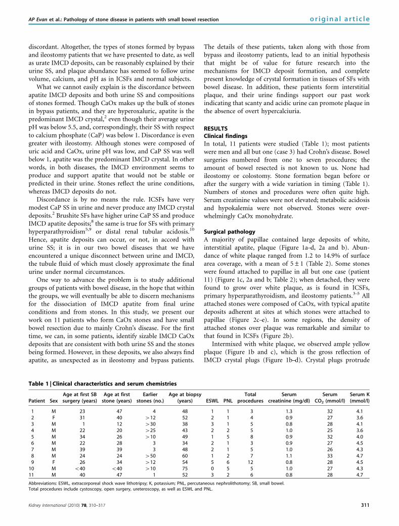
A majority of papillae contained large deposits of white,interstitial apatite, plaque (Figure 1a–d, 2a and b). Abun-dance of white plaque ranged from 1.2 to 14.9% of surfacearea coverage, with a mean of 5±1 (Table 2). Some stoneswere found attached to papillae in all but one case (patient11) (Figure 1c, 2a and b; Table 2); when detached, they werefound to grow over white plaque, as is found in ICSFs,primary hyperparathyroidism, and ileostomy patients.3–5 Allattached stones were composed of CaOx, with typical apatitedeposits adherent at sites at which stones were attached topapillae (Figure 2c–e). In some regions, the density ofattached stones over plaque was remarkable and similar tothat found in ICSFs (Figure 2b).
Intermixed with white plaque, we observed ample yellowplaque (Figure 1b and c), which is the gross reflection ofIMCD crystal plugs (Figure 1b–d). Crystal plugs protrude
Table 1 | Clinical characteristics and serum chemistries
Patient SexAge at first SBsurgery (years)
Age at firststone (years)
Earlierstones (no.)
Age at biopsy(years) ESWL PNL
Totalprocedures
Serumcreatinine (mg/dl)
SerumCO2 (mmol/l)
Serum K(mmol/l)
1 M 23 47 4 48 1 1 3 1.3 32 4.12 F 31 40 412 52 2 1 4 0.9 27 3.63 M 1 12 430 38 3 1 5 0.8 28 4.14 M 22 20 425 43 2 2 5 1.0 25 3.65 M 34 26 410 49 1 5 8 0.9 32 4.06 M 22 28 3 34 2 1 3 0.9 27 4.57 M 39 39 3 48 2 1 5 1.0 26 4.38 M 24 24 450 60 1 2 7 1.1 33 4.79 F 26 34 412 54 5 6 12 0.8 28 4.5
10 M o40 o40 410 75 0 5 5 1.0 27 4.311 M 40 47 1 52 3 2 6 0.8 28 4.7
Abbreviations: ESWL, extracorporeal shock wave lithotripsy; K, potassium; PNL, percutaneous nephrolithotomy; SB, small bowel.Total procedures include cystoscopy, open surgery, ureteroscopy, as well as ESWL and PNL.
Kidney International (2010) 78, 310–317 311
AP Evan et al.: Pathology of stone disease in patients with small bowel resection o r i g i n a l a r t i c l e
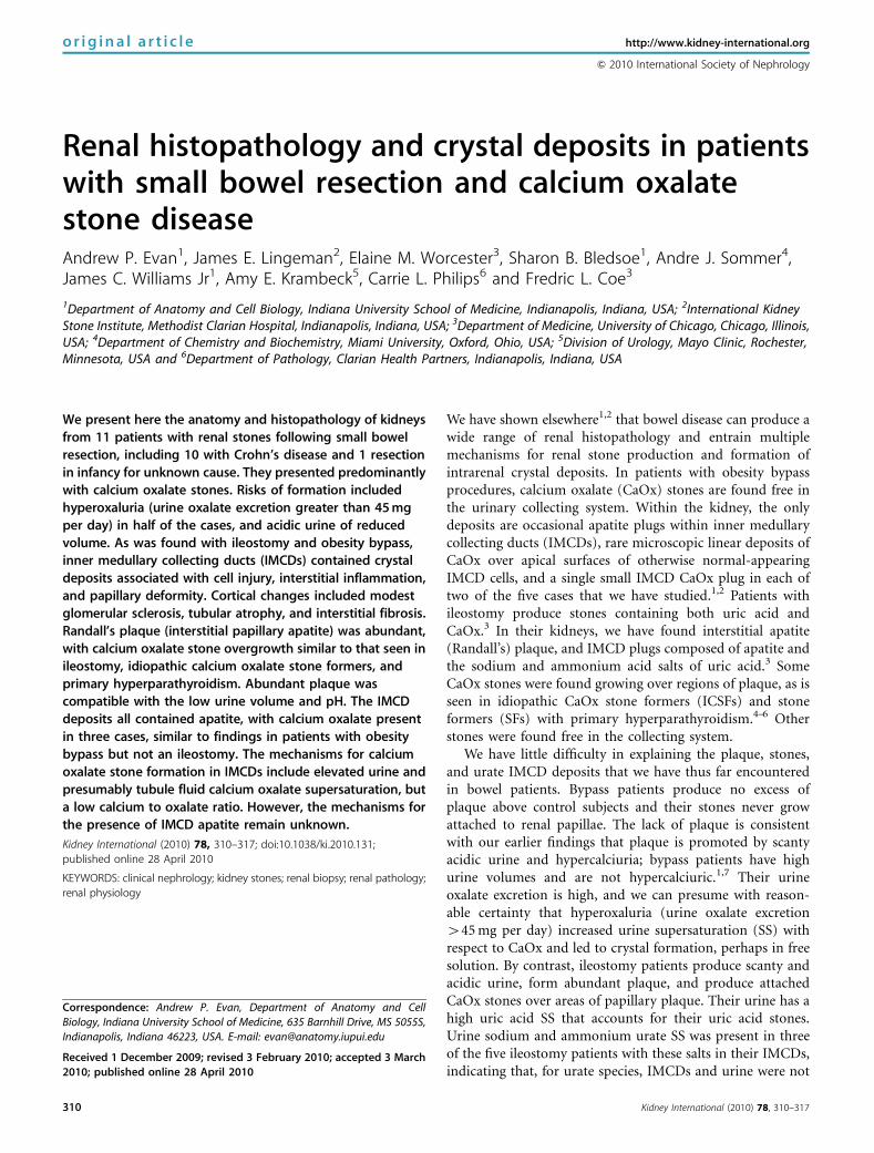
(Figure 1b–d) from mouths of dilated Bellini ducts (BDs).Dilated BDs were most abundant in flattened and deformedpapillae (Table 2). The amount of deformity was less thanthat found in brushite SFs,8 but greater than that found inileostomy patients.
Histopathology
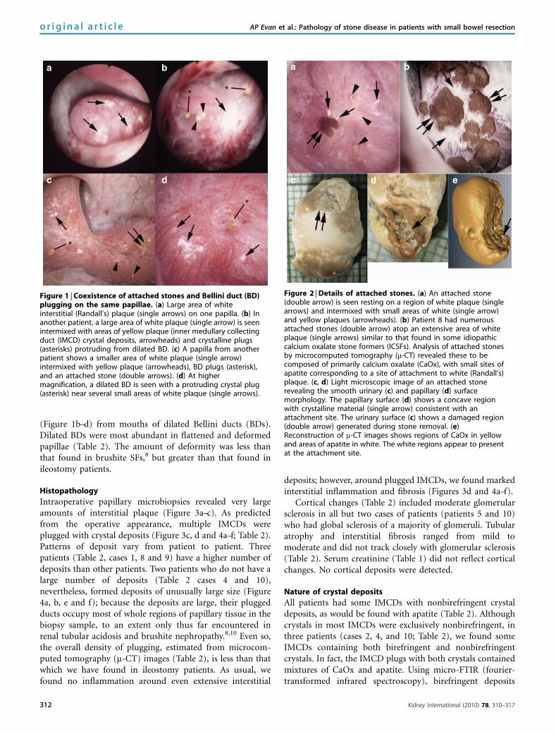
Intraoperative papillary microbiopsies revealed very largeamounts of interstitial plaque (Figure 3a–c). As predictedfrom the operative appearance, multiple IMCDs wereplugged with crystal deposits (Figure 3c, d and 4a–f; Table 2).Patterns of deposit vary from patient to patient. Threepatients (Table 2, cases 1, 8 and 9) have a higher number ofdeposits than other patients. Two patients who do not have alarge number of deposits (Table 2 cases 4 and 10),nevertheless, formed deposits of unusually large size (Figure4a, b, e and f); because the deposits are large, their pluggedducts occupy most of whole regions of papillary tissue in thebiopsy sample, to an extent only thus far encountered inrenal tubular acidosis and brushite nephropathy.8,10 Even so,the overall density of plugging, estimated from microcom-puted tomography (m-CT) images (Table 2), is less than thatwhich we have found in ileostomy patients. As usual, wefound no inflammation around even extensive interstitial
deposits; however, around plugged IMCDs, we found markedinterstitial inflammation and fibrosis (Figures 3d and 4a–f).
Cortical changes (Table 2) included moderate glomerularsclerosis in all but two cases of patients (patients 5 and 10)who had global sclerosis of a majority of glomeruli. Tubularatrophy and interstitial fibrosis ranged from mild tomoderate and did not track closely with glomerular sclerosis(Table 2). Serum creatinine (Table 1) did not reflect corticalchanges. No cortical deposits were detected.
Nature of crystal deposits
All patients had some IMCDs with nonbirefringent crystaldeposits, as would be found with apatite (Table 2). Althoughcrystals in most IMCDs were exclusively nonbirefringent, inthree patients (cases 2, 4, and 10; Table 2), we found someIMCDs containing both birefringent and nonbirefringentcrystals. In fact, the IMCD plugs with both crystals containedmixtures of CaOx and apatite. Using micro-FTIR (fourier-transformed infrared spectroscopy), birefringent deposits
*
*
**
a b
c d
Figure 1 | Coexistence of attached stones and Bellini duct (BD)plugging on the same papillae. (a) Large area of whiteinterstitial (Randall’s) plaque (single arrows) on one papilla. (b) Inanother patient, a large area of white plaque (single arrow) is seenintermixed with areas of yellow plaque (inner medullary collectingduct (IMCD) crystal deposits, arrowheads) and crystalline plugs(asterisks) protruding from dilated BD. (c) A papilla from anotherpatient shows a smaller area of white plaque (single arrow)intermixed with yellow plaque (arrowheads), BD plugs (asterisk),and an attached stone (double arrows). (d) At highermagnification, a dilated BD is seen with a protruding crystal plug(asterisk) near several small areas of white plaque (single arrows).
a
c d e
b
Figure 2 | Details of attached stones. (a) An attached stone(double arrow) is seen resting on a region of white plaque (singlearrows) and intermixed with small areas of white (single arrow)and yellow plaques (arrowheads). (b) Patient 8 had numerousattached stones (double arrow) atop an extensive area of whiteplaque (single arrows) similar to that found in some idiopathiccalcium oxalate stone formers (ICSFs). Analysis of attached stonesby microcomputed tomography (m-CT) revealed these to becomposed of primarily calcium oxalate (CaOx), with small sites ofapatite corresponding to a site of attachment to white (Randall’s)plaque. (c, d) Light microscopic image of an attached stonerevealing the smooth urinary (c) and papillary (d) surfacemorphology. The papillary surface (d) shows a concave regionwith crystalline material (single arrow) consistent with anattachment site. The urinary surface (c) shows a damaged region(double arrow) generated during stone removal. (e)Reconstruction of m-CT images shows regions of CaOx in yellowand areas of apatite in white. The white regions appear to presentat the attachment site.
312 Kidney International (2010) 78, 310–317
o r i g i n a l a r t i c l e AP Evan et al.: Pathology of stone disease in patients with small bowel resection
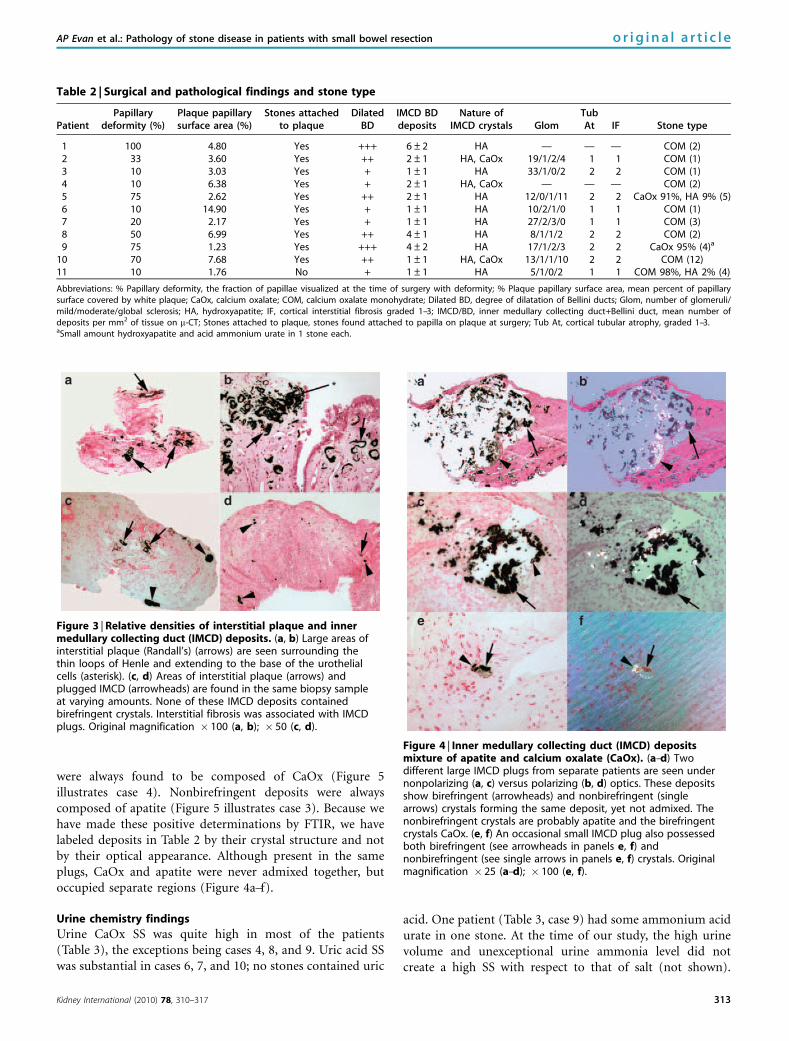
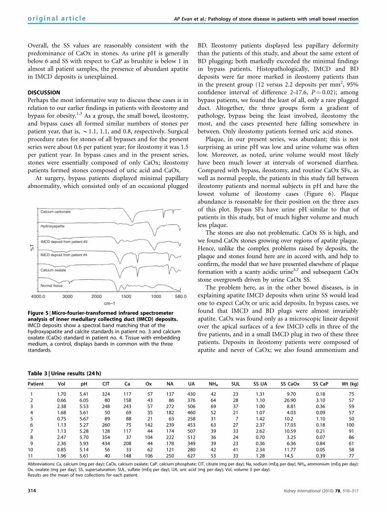
were always found to be composed of CaOx (Figure 5illustrates case 4). Nonbirefringent deposits were alwayscomposed of apatite (Figure 5 illustrates case 3). Because wehave made these positive determinations by FTIR, we havelabeled deposits in Table 2 by their crystal structure and notby their optical appearance. Although present in the sameplugs, CaOx and apatite were never admixed together, butoccupied separate regions (Figure 4a–f).
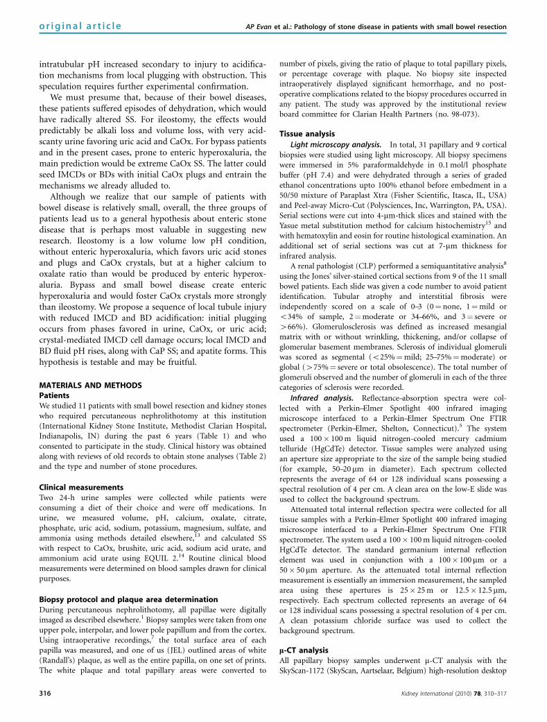
Urine chemistry findings
Urine CaOx SS was quite high in most of the patients(Table 3), the exceptions being cases 4, 8, and 9. Uric acid SSwas substantial in cases 6, 7, and 10; no stones contained uric
acid. One patient (Table 3, case 9) had some ammonium acidurate in one stone. At the time of our study, the high urinevolume and unexceptional urine ammonia level did notcreate a high SS with respect to that of salt (not shown).
Table 2 | Surgical and pathological findings and stone type
PatientPapillary
deformity (%)Plaque papillarysurface area (%)
Stones attachedto plaque
DilatedBD
IMCD BDdeposits
Nature ofIMCD crystals Glom
TubAt IF Stone type
1 100 4.80 Yes +++ 6±2 HA — — — COM (2)2 33 3.60 Yes ++ 2±1 HA, CaOx 19/1/2/4 1 1 COM (1)3 10 3.03 Yes + 1±1 HA 33/1/0/2 2 2 COM (1)4 10 6.38 Yes + 2±1 HA, CaOx — — — COM (2)5 75 2.62 Yes ++ 2±1 HA 12/0/1/11 2 2 CaOx 91%, HA 9% (5)6 10 14.90 Yes + 1±1 HA 10/2/1/0 1 1 COM (1)7 20 2.17 Yes + 1±1 HA 27/2/3/0 1 1 COM (3)8 50 6.99 Yes ++ 4±1 HA 8/1/1/2 2 2 COM (2)9 75 1.23 Yes +++ 4±2 HA 17/1/2/3 2 2 CaOx 95% (4)a
10 70 7.68 Yes ++ 1±1 HA, CaOx 13/1/1/10 2 2 COM (12)11 10 1.76 No + 1±1 HA 5/1/0/2 1 1 COM 98%, HA 2% (4)
Abbreviations: % Papillary deformity, the fraction of papillae visualized at the time of surgery with deformity; % Plaque papillary surface area, mean percent of papillarysurface covered by white plaque; CaOx, calcium oxalate; COM, calcium oxalate monohydrate; Dilated BD, degree of dilatation of Bellini ducts; Glom, number of glomeruli/mild/moderate/global sclerosis; HA, hydroxyapatite; IF, cortical interstitial fibrosis graded 1–3; IMCD/BD, inner medullary collecting duct+Bellini duct, mean number ofdeposits per mm2 of tissue on m-CT; Stones attached to plaque, stones found attached to papilla on plaque at surgery; Tub At, cortical tubular atrophy, graded 1–3.aSmall amount hydroxyapatite and acid ammonium urate in 1 stone each.
*a b
c d
Figure 3 | Relative densities of interstitial plaque and innermedullary collecting duct (IMCD) deposits. (a, b) Large areas ofinterstitial plaque (Randall’s) (arrows) are seen surrounding thethin loops of Henle and extending to the base of the urothelialcells (asterisk). (c, d) Areas of interstitial plaque (arrows) andplugged IMCD (arrowheads) are found in the same biopsy sampleat varying amounts. None of these IMCD deposits containedbirefringent crystals. Interstitial fibrosis was associated with IMCDplugs. Original magnification � 100 (a, b); � 50 (c, d).
a b
c d
e f
Figure 4 | Inner medullary collecting duct (IMCD) depositsmixture of apatite and calcium oxalate (CaOx). (a–d) Twodifferent large IMCD plugs from separate patients are seen undernonpolarizing (a, c) versus polarizing (b, d) optics. These depositsshow birefringent (arrowheads) and nonbirefringent (singlearrows) crystals forming the same deposit, yet not admixed. Thenonbirefringent crystals are probably apatite and the birefringentcrystals CaOx. (e, f) An occasional small IMCD plug also possessedboth birefringent (see arrowheads in panels e, f) andnonbirefringent (see single arrows in panels e, f) crystals. Originalmagnification � 25 (a–d); � 100 (e, f).
Kidney International (2010) 78, 310–317 313
AP Evan et al.: Pathology of stone disease in patients with small bowel resection o r i g i n a l a r t i c l e
Overall, the SS values are reasonably consistent with thepredominance of CaOx in stones. As urine pH is generallybelow 6 and SS with respect to CaP as brushite is below 1 inalmost all patient samples, the presence of abundant apatitein IMCD deposits is unexplained.
DISCUSSION
Perhaps the most informative way to discuss these cases is inrelation to our earlier findings in patients with ileostomy andbypass for obesity.1,3 As a group, the small bowel, ileostomy,and bypass cases all formed similar numbers of stones perpatient year, that is, B1.1, 1.1, and 0.8, respectively. Surgicalprocedure rates for stones of all bypasses and for the presentseries were about 0.6 per patient year; for ileostomy it was 1.5per patient year. In bypass cases and in the present series,stones were essentially composed of only CaOx; ileostomypatients formed stones composed of uric acid and CaOx.
At surgery, bypass patients displayed minimal papillaryabnormality, which consisted only of an occasional plugged
BD. Ileostomy patients displayed less papillary deformitythan the patients of this study, and about the same extent ofBD plugging; both markedly exceeded the minimal findingsin bypass patients. Histopathologically, IMCD and BDdeposits were far more marked in ileostomy patients thanin the present group (12 versus 2.2 deposits per mm2, 95%confidence interval of difference 2–17.6, P¼ 0.02); amongbypass patients, we found the least of all, only a rare pluggedduct. Altogether, the three groups form a gradient ofpathology, bypass being the least involved, ileostomy themost, and the cases presented here falling somewhere inbetween. Only ileostomy patients formed uric acid stones.
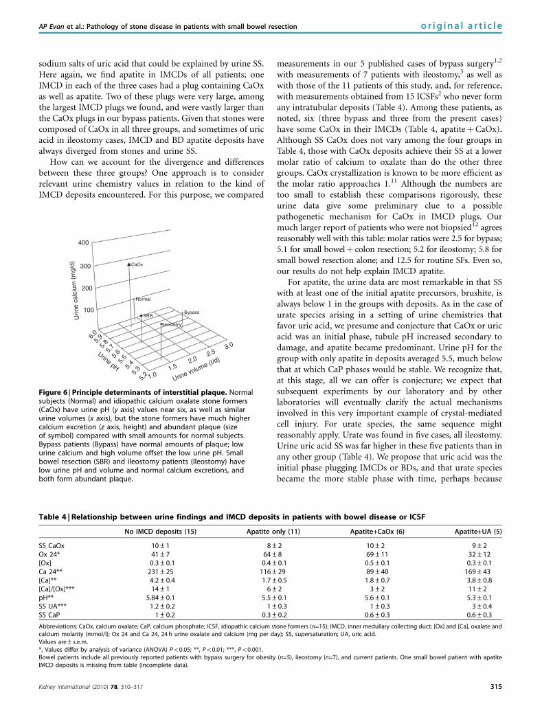
Plaque, in our present series, was abundant; this is notsurprising as urine pH was low and urine volume was oftenlow. Moreover, as noted, urine volume would most likelyhave been much lower at intervals of worsened diarrhea.Compared with bypass, ileostomy, and routine CaOx SFs, aswell as normal people, the patients in this study fall betweenileostomy patients and normal subjects in pH and have thelowest volume of ileostomy cases (Figure 6). Plaqueabundance is reasonable for their position on the three axesof this plot. Bypass SFs have urine pH similar to that ofpatients in this study, but of much higher volume and muchless plaque.
The stones are also not problematic. CaOx SS is high, andwe found CaOx stones growing over regions of apatite plaque.Hence, unlike the complex problems raised by deposits, theplaque and stones found here are in accord with, and help toconfirm, the model that we have presented elsewhere of plaqueformation with a scanty acidic urine3,7 and subsequent CaOxstone overgrowth driven by urine CaOx SS.
The problem here, as in the other bowel diseases, is inexplaining apatite IMCD deposits when urine SS would leadone to expect CaOx or uric acid deposits. In bypass cases, wefound that IMCD and BD plugs were almost invariablyapatite. CaOx was found only as a microscopic linear depositover the apical surfaces of a few IMCD cells in three of thefive patients, and in a small IMCD plug in two of these threepatients. Deposits in ileostomy patients were composed ofapatite and never of CaOx; we also found ammonium and
580.010001500
cm–1
200030004000.0
Normal tissue
Calcium oxalate
IMCD deposit from patient #4
IMCD deposit from patient #3
Hydroxyapatite
Calcium carbonate
%T
Figure 5 | Micro-fourier-transformed infrared spectrometeranalysis of inner medullary collecting duct (IMCD) deposits.IMCD deposits show a spectral band matching that of thehydroxyapatite and calcite standards in patient no. 3 and calciumoxalate (CaOx) standard in patient no. 4. Tissue with embeddingmedium, a control, displays bands in common with the threestandards.
Table 3 | Urine results (24 h)
Patient Vol pH CIT Ca Ox NA UA NH4 SUL SS UA SS CaOx SS CaP Wt (kg)
1 1.70 5.41 324 117 57 137 430 42 23 1.31 9.70 0.18 752 0.66 6.05 80 158 43 86 376 64 28 1.10 26.90 3.10 573 2.38 5.53 248 243 57 272 506 69 37 1.00 8.81 0.36 594 1.68 5.61 50 69 35 182 460 52 21 1.07 4.03 0.09 575 0.75 5.67 89 88 21 63 258 31 7 1.42 10.2 1.10 506 1.13 5.27 260 75 142 239 453 63 27 2.37 17.03 0.18 1007 1.13 5.28 128 117 44 174 507 39 33 2.62 10.59 0.21 918 2.47 5.70 354 37 104 222 512 36 24 0.70 3.25 0.07 869 2.36 5.93 434 208 44 178 349 39 23 0.36 6.36 0.84 61
10 0.85 5.14 56 33 62 121 280 42 41 2.34 11.77 0.05 5811 1.96 5.61 40 148 106 250 627 53 33 1.28 14.5 0.39 77
Abbreviations: Ca, calcium (mg per day); CaOx, calcium oxalate; CaP, calcium phosphate; CIT, citrate (mg per day); Na, sodium (mEq per day); NH4, ammonium (mEq per day);Ox, oxalate (mg per day); SS, supersaturation; SUL, sulfate (mEq per day); UA, uric acid (mg per day); Vol, volume (l per day).Results are the mean of two collections for each patient.
314 Kidney International (2010) 78, 310–317
o r i g i n a l a r t i c l e AP Evan et al.: Pathology of stone disease in patients with small bowel resection
sodium salts of uric acid that could be explained by urine SS.Here again, we find apatite in IMCDs of all patients; oneIMCD in each of the three cases had a plug containing CaOxas well as apatite. Two of these plugs were very large, amongthe largest IMCD plugs we found, and were vastly larger thanthe CaOx plugs in our bypass patients. Given that stones werecomposed of CaOx in all three groups, and sometimes of uricacid in ileostomy cases, IMCD and BD apatite deposits havealways diverged from stones and urine SS.
How can we account for the divergence and differencesbetween these three groups? One approach is to considerrelevant urine chemistry values in relation to the kind ofIMCD deposits encountered. For this purpose, we compared
measurements in our 5 published cases of bypass surgery1,2
with measurements of 7 patients with ileostomy,3 as well aswith those of the 11 patients of this study, and, for reference,with measurements obtained from 15 ICSFs2 who never formany intratubular deposits (Table 4). Among these patients, asnoted, six (three bypass and three from the present cases)have some CaOx in their IMCDs (Table 4, apatiteþCaOx).Although SS CaOx does not vary among the four groups inTable 4, those with CaOx deposits achieve their SS at a lowermolar ratio of calcium to oxalate than do the other threegroups. CaOx crystallization is known to be more efficient asthe molar ratio approaches 1.11 Although the numbers aretoo small to establish these comparisons rigorously, theseurine data give some preliminary clue to a possiblepathogenetic mechanism for CaOx in IMCD plugs. Ourmuch larger report of patients who were not biopsied12 agreesreasonably well with this table: molar ratios were 2.5 for bypass;5.1 for small bowelþ colon resection; 5.2 for ileostomy; 5.8 forsmall bowel resection alone; and 12.5 for routine SFs. Even so,our results do not help explain IMCD apatite.
For apatite, the urine data are most remarkable in that SSwith at least one of the initial apatite precursors, brushite, isalways below 1 in the groups with deposits. As in the case ofurate species arising in a setting of urine chemistries thatfavor uric acid, we presume and conjecture that CaOx or uricacid was an initial phase, tubule pH increased secondary todamage, and apatite became predominant. Urine pH for thegroup with only apatite in deposits averaged 5.5, much belowthat at which CaP phases would be stable. We recognize that,at this stage, all we can offer is conjecture; we expect thatsubsequent experiments by our laboratory and by otherlaboratories will eventually clarify the actual mechanismsinvolved in this very important example of crystal-mediatedcell injury. For urate species, the same sequence mightreasonably apply. Urate was found in five cases, all ileostomy.Urine uric acid SS was far higher in these five patients than inany other group (Table 4). We propose that uric acid was theinitial phase plugging IMCDs or BDs, and that urate speciesbecame the more stable phase with time, perhaps because
Table 4 | Relationship between urine findings and IMCD deposits in patients with bowel disease or ICSF
No IMCD deposits (15) Apatite only (11) Apatite+CaOx (6) Apatite+UA (5)
SS CaOx 10±1 8±2 10±2 9±2Ox 24* 41±7 64±8 69±11 32±12[Ox] 0.3±0.1 0.4±0.1 0.5±0.1 0.3±0.1Ca 24** 231±25 116±29 89±40 169±43[Ca]** 4.2±0.4 1.7±0.5 1.8±0.7 3.8±0.8[Ca]/[Ox]*** 14±1 6±2 3±2 11±2pH** 5.84±0.1 5.5±0.1 5.6±0.1 5.3±0.1SS UA*** 1.2±0.2 1±0.3 1±0.3 3±0.4SS CaP 1±0.2 0.3±0.2 0.6±0.3 0.6±0.3
Abbreviations: CaOx, calcium oxalate; CaP, calcium phosphate; ICSF, idiopathic calcium stone formers (n=15); IMCD, inner medullary collecting duct; [Ox] and [Ca], oxalate andcalcium molarity (mmol/l); Ox 24 and Ca 24, 24 h urine oxalate and calcium (mg per day); SS, supersaturation; UA, uric acid.Values are±s.e.m.*, Values differ by analysis of variance (ANOVA) Po0.05; **, Po0.01; ***, Po0.001.Bowel patients include all previously reported patients with bypass surgery for obesity (n=5), ileostomy (n=7), and current patients. One small bowel patient with apatiteIMCD deposits is missing from table (incomplete data).
3.02.5
2.0
1.5
1.0 Urine volume (I/d)
5.25.
35.45.
55.65.
75.85.
96.0
Urine pH
100
200
300
400
Urin
e ca
lciu
m (
mg/
d)
Bypass
Ileostomy
SBR
Normal
CaOx
Figure 6 | Principle determinants of interstitial plaque. Normalsubjects (Normal) and idiopathic calcium oxalate stone formers(CaOx) have urine pH (y axis) values near six, as well as similarurine volumes (x axis), but the stone formers have much highercalcium excretion (z axis, height) and abundant plaque (sizeof symbol) compared with small amounts for normal subjects.Bypass patients (Bypass) have normal amounts of plaque; lowurine calcium and high volume offset the low urine pH. Smallbowel resection (SBR) and ileostomy patients (Ileostomy) havelow urine pH and volume and normal calcium excretions, andboth form abundant plaque.
Kidney International (2010) 78, 310–317 315
AP Evan et al.: Pathology of stone disease in patients with small bowel resection o r i g i n a l a r t i c l e
intratubular pH increased secondary to injury to acidifica-tion mechanisms from local plugging with obstruction. Thisspeculation requires further experimental confirmation.
We must presume that, because of their bowel diseases,these patients suffered episodes of dehydration, which wouldhave radically altered SS. For ileostomy, the effects wouldpredictably be alkali loss and volume loss, with very acid-scanty urine favoring uric acid and CaOx. For bypass patientsand in the present cases, prone to enteric hyperoxaluria, themain prediction would be extreme CaOx SS. The latter couldseed IMCDs or BDs with initial CaOx plugs and entrain themechanisms we already alluded to.
Although we realize that our sample of patients withbowel disease is relatively small, overall, the three groups ofpatients lead us to a general hypothesis about enteric stonedisease that is perhaps most valuable in suggesting newresearch. Ileostomy is a low volume low pH condition,without enteric hyperoxaluria, which favors uric acid stonesand plugs and CaOx crystals, but at a higher calcium tooxalate ratio than would be produced by enteric hyperox-aluria. Bypass and small bowel disease create enterichyperoxaluria and would foster CaOx crystals more stronglythan ileostomy. We propose a sequence of local tubule injurywith reduced IMCD and BD acidification: initial pluggingoccurs from phases favored in urine, CaOx, or uric acid;crystal-mediated IMCD cell damage occurs; local IMCD andBD fluid pH rises, along with CaP SS; and apatite forms. Thishypothesis is testable and may be fruitful.
MATERIALS AND METHODSPatientsWe studied 11 patients with small bowel resection and kidney stoneswho required percutaneous nephrolithotomy at this institution(International Kidney Stone Institute, Methodist Clarian Hospital,Indianapolis, IN) during the past 6 years (Table 1) and whoconsented to participate in the study. Clinical history was obtainedalong with reviews of old records to obtain stone analyses (Table 2)and the type and number of stone procedures.
Clinical measurementsTwo 24-h urine samples were collected while patients wereconsuming a diet of their choice and were off medications. Inurine, we measured volume, pH, calcium, oxalate, citrate,phosphate, uric acid, sodium, potassium, magnesium, sulfate, andammonia using methods detailed elsewhere,13 and calculated SSwith respect to CaOx, brushite, uric acid, sodium acid urate, andammonium acid urate using EQUIL 2.14 Routine clinical bloodmeasurements were determined on blood samples drawn for clinicalpurposes.
Biopsy protocol and plaque area determinationDuring percutaneous nephrolithotomy, all papillae were digitallyimaged as described elsewhere.1 Biopsy samples were taken from oneupper pole, interpolar, and lower pole papillum and from the cortex.Using intraoperative recordings,7 the total surface area of eachpapilla was measured, and one of us (JEL) outlined areas of white(Randall’s) plaque, as well as the entire papilla, on one set of prints.The white plaque and total papillary areas were converted to
number of pixels, giving the ratio of plaque to total papillary pixels,or percentage coverage with plaque. No biopsy site inspectedintraoperatively displayed significant hemorrhage, and no post-operative complications related to the biopsy procedures occurred inany patient. The study was approved by the institutional reviewboard committee for Clarian Health Partners (no. 98-073).
Tissue analysis
Light microscopy analysis. In total, 31 papillary and 9 corticalbiopsies were studied using light microscopy. All biopsy specimenswere immersed in 5% paraformaldehyde in 0.1 mol/l phosphatebuffer (pH 7.4) and were dehydrated through a series of gradedethanol concentrations upto 100% ethanol before embedment in a50/50 mixture of Paraplast Xtra (Fisher Scientific, Itasca, IL, USA)and Peel-away Micro-Cut (Polysciences, Inc, Warrington, PA, USA).Serial sections were cut into 4-mm-thick slices and stained with theYasue metal substitution method for calcium histochemistry15 andwith hematoxylin and eosin for routine histological examination. Anadditional set of serial sections was cut at 7-mm thickness forinfrared analysis.
A renal pathologist (CLP) performed a semiquantitative analysis8
using the Jones’ silver-stained cortical sections from 9 of the 11 smallbowel patients. Each slide was given a code number to avoid patientidentification. Tubular atrophy and interstitial fibrosis wereindependently scored on a scale of 0–3 (0¼ none, 1¼mild oro34% of sample, 2¼moderate or 34–66%, and 3¼ severe or466%). Glomerulosclerosis was defined as increased mesangialmatrix with or without wrinkling, thickening, and/or collapse ofglomerular basement membranes. Sclerosis of individual glomeruliwas scored as segmental (o25%¼mild; 25–75%¼moderate) orglobal (475%¼ severe or total obsolescence). The total number ofglomeruli observed and the number of glomeruli in each of the threecategories of sclerosis were recorded.
Infrared analysis. Reflectance–absorption spectra were col-lected with a Perkin–Elmer Spotlight 400 infrared imagingmicroscope interfaced to a Perkin–Elmer Spectrum One FTIRspectrometer (Perkin–Elmer, Shelton, Connecticut).5 The systemused a 100� 100 m liquid nitrogen-cooled mercury cadmiumtelluride (HgCdTe) detector. Tissue samples were analyzed usingan aperture size appropriate to the size of the sample being studied(for example, 50–20 mm in diameter). Each spectrum collectedrepresents the average of 64 or 128 individual scans possessing aspectral resolution of 4 per cm. A clean area on the low-E slide wasused to collect the background spectrum.
Attenuated total internal reflection spectra were collected for alltissue samples with a Perkin–Elmer Spotlight 400 infrared imagingmicroscope interfaced to a Perkin–Elmer Spectrum One FTIRspectrometer. The system used a 100� 100 m liquid nitrogen-cooledHgCdTe detector. The standard germanium internal reflectionelement was used in conjunction with a 100� 100mm or a50� 50 mm aperture. As the attenuated total internal reflectionmeasurement is essentially an immersion measurement, the sampledarea using these apertures is 25� 25 m or 12.5� 12.5 mm,respectively. Each spectrum collected represents an average of 64or 128 individual scans possessing a spectral resolution of 4 per cm.A clean potassium chloride surface was used to collect thebackground spectrum.
l-CT analysisAll papillary biopsy samples underwent m-CT analysis with theSkyScan-1172 (SkyScan, Aartselaar, Belgium) high-resolution desktop
316 Kidney International (2010) 78, 310–317
o r i g i n a l a r t i c l e AP Evan et al.: Pathology of stone disease in patients with small bowel resection
m-CT system, allowing nondestructive mapping of the locationand size of the crystalline deposits within a biopsy specimen.5 Thism-CT system can generate a tissue window so that both the mineraldeposit and tissue organization are seen at the same time. For thisprotocol, biopsy samples are quickly dipped in a 1:10 dilution ofHypaque (50%, Nycomed, Princeton, NJ)/phosphate-bufferedsaline, coated with a thin layer of paraffin, and mounted in thecenter of a small chuck, which is then locked into place in themachine. The sample was positioned in the center of the beam, andthe system configuration was set at 35 kV, 209 mA, 1801 rotation,with flatfield correction. Images were saved in compact disks andreconstructed with cone-reconstruction software by SkyScan. Theseimages were then reconstructed into three-dimensional images withSkyScan’s CTAnþCTVol software. These images allowed us toproperly orient each biopsy sample for future light microscopicanalysis. Three separate scans from each patient were used todetermine the number of sites of intraluminal deposits per squaremillimeter.
Stones obtained from these patients were analyzed using theSkyScan 1172 with reconstruction voxel sizes of 10–20 mm. Three-dimensional reconstructions of stones were examined using ImageJ(http://rsb.info.nih.gov/ij/) and Voxx2 (http://www.nephrology.iupui.edu/imaging/voxx/) softwares to verify the composition.Surface renderings were carried out using Amira program (VisageImaging, Carlsbad, CA).
DISCLOSUREAll the authors declared no competing interests.
ACKNOWLEDGMENTSThis work was funded by NIH PO1 DK56788.
REFERENCES1. Evan AP, Lingeman JE, Coe FL et al. Randall’s plaque of patients with
nephrolithiasis begins in basement membranes of thin loops of Henle.J Clin Invest 2003; 111: 607–616.
2. Evan AP, Coe FL, Gillen D et al. Renal intratubular crystals and hyaluronanstaining occur in stone formers with bypass surgery but not withidiopathic calcium oxalate stones. Anat Rec 2008; 291: 325–334.
3. Evan AP, Lingeman JE, Coe FL et al. Intra-tubular deposits, urine andstone composition are divergent in patients with ileostomy. Kidney Int2009; 76: 1081–1088.
4. Miller NL, Gillen DL, Williams Jr JC et al. A formal test of the hypothesisthat idiopathic calcium oxalate stones grow on Randall’s plaque. BJU Int2009; 103: 966–971.
5. Evan AE, Lingeman JE, Coe FL et al. Histopathology and surgical anatomyof patients with primary hyperparathyroidism and calcium phosphatestones. Kidney Int 2008; 74: 223–229.
6. Evan AP, Coe FL, Lingeman JE et al. Mechanism of formation of humancalcium oxalate renal stones on Randall’s plaque. Anat Rec 2007; 290:1315–1323.
7. Kuo RL, Lingeman JE, Evan AP et al. Urine calcium and volume predictcoverage of renal papilla by Randall’s plaque. Kidney Int 2003; 64: 2150–2154.
8. Evan AP, Lingeman JE, Coe FL et al. Crystal-associated nephropathy inpatients with brushite nephrolithiasis. Kidney Int 2005; 67: 576–591.
9. Parks JH, Coe FL, Evan AP et al. Clinical and laboratory characteristics ofcalcium stone-formers with and without primary hyperparathyroidism.BJU Int 2009; 103: 670–678.
10. Evan AP, Lingeman J, Coe F et al. Renal histopathology of stone-formingpatients with distal renal tubular acidosis. Kidney Int 2007; 71: 795–801.
11. Nancollas GH, Singh RP. In vitro system for calcium stone formation: theconstant composition model. Contrib Nephrol 1987; 58: 49–58.
12. Parks JH, Worcester EM, O’Connor RC et al. Urine stone risk factors innephrolithiasis patients with and without bowel disease. Kidney Int 2003;63: 255–265.
13. Parks JH, Goldfisher E, Asplin JR et al. A single 24-h urine collection isinadequate for the medical evaluation of nephrolithiasis. J Urol 2002; 167:1607–1612.
14. Werness PG, Brown CM, Smith LH et al. Equil 2: a basic computer programfor the calculation of urinary saturation. J Urol 1985; 134: 1242–1244.
15. Yasue T. Histochemical identification of calcium oxalate. Acta HistochemCytochem 1969; 2: 83–95.
Kidney International (2010) 78, 310–317 317
AP Evan et al.: Pathology of stone disease in patients with small bowel resection o r i g i n a l a r t i c l e


![Reduction of Oxalate Levels in Tomato Fruit and … of Oxalate Levels in Tomato Fruit and Consequent Metabolic Remodeling Following Overexpression of a Fungal Oxalate Decarboxylase1[W]](https://img.dokumen.tips/doc/110x75/5af8e5787f8b9aff288c704b/reduction-of-oxalate-levels-in-tomato-fruit-and-of-oxalate-levels-in-tomato.jpg)