Embed Size (px)
Citation preview
- -
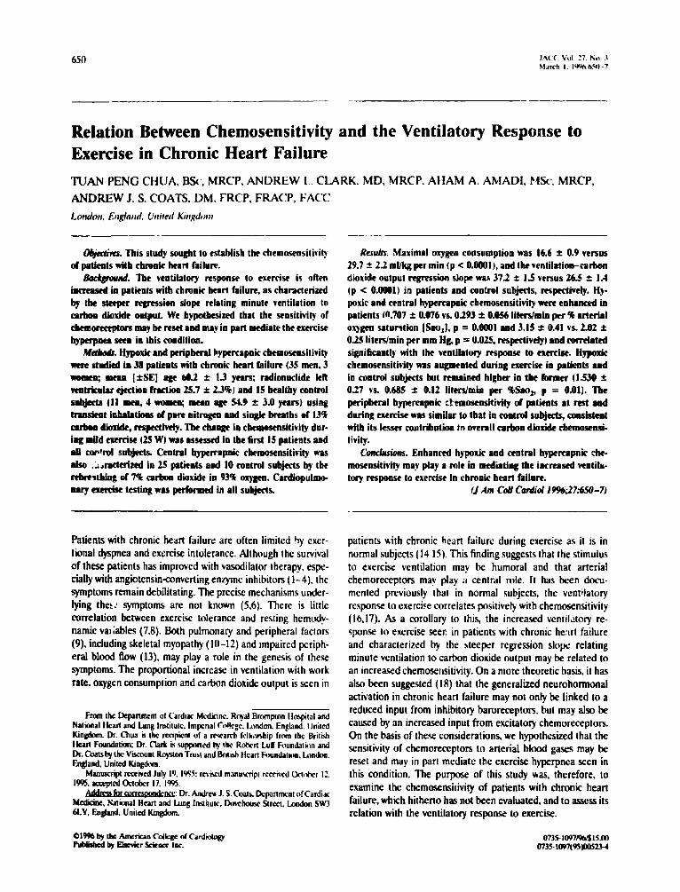
Relation Between Chemosensitivity and the Ventilatory Response to Exercise in Chronic Heart Failure TUAN PENG CHUA, BSc. MRCP. ANDREW I.. CLARK, MD, MRCP, AHAM A. AMADI. t4Sc. MRCP, ANDREW J. S. COATS. DM, FRCP. FRACP, FACT
ObiddilQ. ‘Ihis study sought to establish the chemosensitivity cd patkats witb chroak hea failure.
i%ukgmd. The veatibtory response to exetise is often bmased in patknts with chmnk beart failure, as chamcterized by the steeper regression slope relating minute ventilation to cahu dioxide o&put. We hypothrsized that the sensitivity of cbem~ors may bc reset and nay in part mediate the exercise hypepna sew in this coadition.
Me&& Hypoxk and peripheral bypercspllk cbemosensitivity WYe studied in 38 patients with chroak heart failure (35 me& 3 rromcn; mesa [*SE] age Y3 * 1.3 years: radionuclide left vwtrkulnr ejection fmctioa 25.7 f 23%) and 15 healthy control wbjects (II pep, 4 s mean age 54.9 f 3.0 yean) using bansiat iohalatbos of pare nitrogen and sin& breaths of 13% carboa dkuide, respe&ely. Tbe change io mitivity dur- iag alld exercise (2!5 W) was assessed in the first 15 patients and all coNml snbjects. Central bypempak cbemosensitivity was also .dactwlzed in 25 patients and IO coontml subjects by the rehsmablug of 7% carbon dioxide in 93% oxygen. Cardiof~lmo- nary exe&se ttsting was performed in all subjects.
Patients with chronic heart failure are often limited % exer- tional dyspnea and exercise intolerance. Although the survival of these patients has improved with vasodilator therapy, espe- cially with angiotensinconvct-ting enzyme inhibitors (l-4). the symptoms remain debilitating. The precise mechanisms under- lying the: symptoms are not known (5.6). There is little correlation between exerciw tolerance and resting hem@- namic val ;ables (7,8). Both pulmonary and peripheral facton (9), including skeletal myopathy (10-12) and Impaired periph- eral blood Bow (13), may play a role in the genesis of these symptoms. The proportional increase in ventilation with work rate, oxygen consumption and carbon dioxide output is seen in
From the Department of Cardw Mcdkinc. Royal Bromprcm Hmpiral and Nafioaal Heari and Lung Irr~ituic. lmpcrial C4c:r. London. England. I lnitcd Kingdmr. Dr. Chua is the wpicrn of a wxawh klhxsh~p frmn the Britnh Heart FnundaGax Dr. Clark b wppwlcd ty !k R&VI Lull Foundailcm and Dr. Coalsby !hc Virwn~ Royslon Trusi and Bntuh Hcan Founda~rm. Lmdon. Engtand, United Kingdwn.
Manuuript mxwed Juty 19. I’M: r&cd manuwip~ rco5wd Oct14w I?. 195. aaqlwd ckldlcl17.1995.
-for Dr. Awtrcw J. S. Coats. Dcpartmcm ofCardix Matick. Nahnd Heart and Luq trmiww. Dwchousc Prwt. bndon SW3 6t.Y. Enghd. United Kingdum.
019% hy I& Amerkan Cd- d Cardidqy PubadKdby~rSEicDarInc.
---
&M&S. Maximal oxygen consumption was 16.6 2 0.9 versus 29.7 * 2.2 ml/kg per min tp < O.OMM), and the ventilation-carbon dioxide output rrgressioo sbpe WM 37.2 i I.5 vtrsus 26.5 + I.4 (p < O.WOl) in patients and control subjects, respec+ively. Hy- poxk and central hypercapak ebemosensitivity were enhanced in patients m.707 f 0.076 VL 0.293 f 0.056 litersMa prr % arterial oxygen satumtion [Sao,], p = O.OOOl and 3.15 f 0.41 vs 2.02 f 0.25 litcrs/min per mm Hg. p = U.025, nqectively) and carrelated significantly dtb the vralilatwy respoase to exercise. Hypoxk chemosensitivity was r-ted during exercise in patients aad io control subjects but remained higher in the lorPer (I.!% 2 0.27 vs. 0.685 f 0.12 litcrslmin per %SaO, p = 0.01). llw periplseral hyp-capnk r%mc~~sitivity of patients at rest aad during exercise was similar to that in cootrol snbjects, coaststeat with its lesser co~tributioa tn overall earboo dk&k cbemoseasi- tivity.
Conrlusions. Enhanced hypoxk osd central hypercapnk de- mosensitivity may play a mle in mediating the iacreased ventila- tory response to exercise in cbronk beart failore.
(I Am Cdl Caniid 19!b;27.W-7)
patients with chronic heart failure during exercise as it IS in normal subjects (14 15). This finding suggests that the stimulus to exercise ventilation may be humoral and that arterial chemoreceptors may play ;I central role. It has been docu- mented previously that in normal subjects, the ventilatory response to cxcrcice correlates positively with chemosensitivity (16.17). As a corollary to this, the increased vcntilatoty re- sponse to exercise seer. in patients with chronic he:irt failure and characterized by the steeper regression slog relating minute ventilation to carbon dioxide output may be related to an increased chemosensitivity. On a more theoretic basis. it has also been suggested (18) that the generalized neurohormonal activation in chronic heart failure may not only be linked to a reduced input from inhibitory baroreceptors, but may also be caused by an increased input from excitatory chemoreceptors. On the basis of these considerations, we hypothesized that the sensitivity of chemoreceptors to afterial blood gases may he reset and may in part mediate the exercise hyperpnea seen in this condition. The purpose of this study was, therefore, to examine the chemosensitivity of patients with chronic heart failure, which hitherto has MM been evaluated, and to assess its relation with the vcntilatory responx to exercise.
Methods Subjects. Thirty-eight patients *Gth chronic heart failure
between 40 and 75 years of age (mean age [‘SE] ho.2 ? 1.3 years: 35 men. 3 women) participated in the study. Patients with a known history of pulmo?dT and ncurologic disease were excluded from the study. AL patients had been in helit failure for >3 months. They were all treated with diuretic drugs, and most received angiotensinconvertinp enzyme inhib- itors. Nineteen patients were in New York Heart Association functional class II. and 16 in class III. No patient was limited by angina. Patient characteristics are summarized in Table I. A control group of 15 healthy subjects (31 10 73 years old, mean age 54.9 2 3.0 years; 11 men. 4 women) was also studied.
Hypoxic and peripheral hypercapnic chernosensitivit)l. bolh mediated by the carotid chemoreceptors. was assessed at rest in all subjects. The first I5 consecutive patients with chronic heart failure recruited into the study also had these asses+ merits made during mild exercise on a cycle ergometer (Tun-
turi) at 15 W for 10 min on two separate occasions lo evaluate the change in chemosensitivity on exercise. A bed exlerna’ work rate ~‘2% used based on the obscrvatilln that for a parlicular work Kite. patients with chronic heart failure have a higher ventillitory rc?lponsc: than normal subjects (14). In addilion, ccnrral hypercapnic chemosensitivity was assessed in IO control subjects and 3 patients to assess the role of central (medullarv) chcmorcccplnrs. AH rubjccls performed rdrdio- pulmonan: cxerciwz te$ling it) as~“i% the vcnlilaton response to exerciw. Subjects were told 14) avoid caffeinated products on the morning of the tesls. The study had been approved by Ihe local clllin commil~ee. and all sibjccta gave written informed consent.
Transient hypoxic wntilatwy mponse ttst. Thcrc are three principal methods of asxcsing hypoxic chemcMnsidvi9 using steady slate (IY). progressive (X.21) and transient (22) hypoxia. respectively. The absolute values obtained from each method arc different. hut relatively. fhey reflect the same indexes of chcmwn$itiviry (23.24). The transient hypoxic method was chosen in this study for practical ;1, 1 safety reasons because patienls were not subjected to 1 olonged episodes of hypoxia. cspeciallv during exercise. and Ine depres- sant effects of prolonged hypoxia on central respiratory drive are also avoided.
The transient hypoxic chemosensitivity tesr was performed while tuhjects were qieaierl and after a period of quiel hreath- ing. Each subject wore a nose clip and.breathcd through a pneumatic respiratory valve (Innovision). which separated the expir-te from the inspirate. The inspilate porr wz further connected 10 a T-valve placed behind the subject. and, depend- ing on the position of the T-valve. the subject breathed either room air or pure nitrogen from a 4-liter reservoir bag that was quietly refilled from a gas cyliqder containing pure nitrogen. Minute ventilation was measured breath by breath using a heated pneumotachograph. and continuous monitoring of oxygen and carbon dioxide was done at the mouth by w spectrometry (Amis 2000, Innovision). The pneumolachom- eler and mass spectromeicr were calihrzted &fore each test. Arterial oxygen saturation was measured using a pulse oxime- tcr (model N-IOOE. Nelkor), set at fast mode uith a response time of 2 to 3 s and a lightweight ear probe clipped gently on the subject‘s right earlobe. After the subject breathed room air for several minutss. the T-valve was turned surreptitiously during the expiralory phase of the previous brealb so that pure nitrogen was inhaled for two to eight breaths. This was repeated IO lo I5 times to provide a wide range of arterial oxygen saturations from 70% to IO%. with -3min intervals of air breathing between eizres to dlow anerial oqp saturation and end-tidal carbon dioxide to return to the subject’s baseline. The maximal minute ventilation after each peti of nitrogen inhalation was obtained by aleraging the two largest consecutive breaths. We found t!w two-breath period optimal in maintaining sensitivity and r:praiwbility of the transient hypcrxic test as did Edelman et al. (3). The timal ventilation was then plotted against th: lowest arterial oxygen saturation reached for eveT period of nitrogen inha-
lation. The transient hypoxic chemoscnsitivity was ohtaincd as the slope of the best-fit line that related ventilation IO
arterial oxygen saturation. calculated by loa+squarch linear regression analysis and expressed in terms of liters per minute per percent oxygen saturation (litcrsimin per BSao,). For the transient hypoxic test during cxercisc. on average, only six episodes of transient hypoxia were @en in view of the duration of the exercise. A delay of 3 rnlhl after the onset of exercise was given before the hypoic chcmo- sensitivity testing to allow steady state (X,27). For safety reasons, the degree of arterial oxygen desaturation was deliberately kept X30%. All patients were monitored clcc- trocardiographically during exercise.
Siwth carbon dioxide ventilatoy response test. Peripnera! hypercapnic chemosensitivity was axwssed using the single-breath carbon dioxide test as described by McClean et al, (28). The apparatus includingthe T-valve was the same as that described before. A smaller 2-liter reservoir hag was used. which was quietly refilled after each inhalation with a gas mixture containing 139 carbon dioxide in air. After a period of quiet breathing, the T-valve was turned during the expiratoq ph;w of the previous breath so that the subject inhaled a single breath of 13% carbon dioxide in air. On average, IO breaths were administered at -2-min intervals. As before. minute ventilation was measured. with continuous monitoring of car- bon dioxide done at the mouth. The mean minute ventilation of the preceding five breaths before the stimulus carbon dioxide breath was calculated and taken as the control venti- lation [v(C)]. The mean end-tidal fraction of carbon dioxide of these breaths was also cakulatcd and taken as the control end-tidal fraction of carbon dioxide [Ft~co,(C)]. The rcspmx ventilation after the stimulus catin dioxide hreath [v(S)1 was calculated by averaging the two largest conxcutive breaths but, unlike before, within 20 z after the stimulus breath. Breaths beyond this time limit were assumed to be affected by central chemoreceptors and therefore excluded from analysis (28). The end-tidal carbon dioxide concentration of the stim- ulus breath was considered the stimulus end-tidal fraction of carbon diixide [FtTcx)JS)l, The single-breath ca&m dioxide response was calculated using the previous variables as follows:
where P, is the atmospheric pressure (mm Hg), and 47 is the saturated water vapor pressure (mm Hg). The mean of 10 responses was considered the subject’s single-breath carbon dii response at rest and expressed in liters per minute per millimeter Hg (liters/min per mmHg). For the IO-min exercise period at 25 W on the cycle ergometer, an average of six single breaths of carbon dioxide were administered at shorter time intervals in view of increased minute ventilation and circula- tion during exercise. These were given 3 min after the onset of exerck to allow steady state.
m dbGdc rdn~!&@ ttebn&. Central hypercap- nit chemasensitivity was assersed using rebreathing of carbon
dioxide (ZY). During the tot. expired carbon dioxide is con- stantly returned to Ihc lungs, and as carbon dioxide accumu- lirtes. thih provides ti progrcs\ive carbon dioxide stimulus lo vrntilation. After ti period of quiet breathing, subjects re- breathed through a &liter hag containing a gas mixture of 7ci carbon diosidc in oxvgcn for 1 min; the test was stopped sooner tf they wcrc too hrcathlrL3 IO continue or if P~-T(I)~ exceeded Ill’r. II has hecn shoun (?Y) that by rehreathingof 7% carbon dioxide (which approximates IO venous PC o?) in oxygen. a Pco, equilibrium is developed rapidly in the mixed venous blood. arterial blood. gas in the lung and gas in the breathing bag. Thus. Ihe PC o2 in any one of these compartments is rcpresen- lativc of the other compartments. including the brain tissue. With thr high oxygen concentration. the peripheral hypercap- nit respnsc is known IO be very small or negligihle (3U.31). Minute ventilation was measured breath by breath. and con- tinuous monitoring of oxygen and carbon dioxide was done at the mouth by mass spectrometry. The central hypercapnic chemuscnsitivity was obtained irs the slope that related minute ventilation to PETCO~ calculated by linear regression analysis ,md expressed in terms of liters per minute per millimeter Hg (literslmin prr mm Hg). We did not perform this test during exercise because of the uncertainties of Pco, equilibrium during exercise in all the compartments (nixed venous blood. arterial bkwd, gas in the lung and gas ill the breathing bag) using the same mixture of 7?i carbon dioxide in oxygen.
Studies of reproducibility. The transient hypoxic. singlr- breath carbon dioxide and rehreathing carbon dioxide tests were repeated in seven healthy control subjects and tive patients with chronic heart failure lo assess reproducibility and the coefticient of variation. There was g& agreement he- !ween repeated mcasuremcnts (r = 11.93, O.hY and 0.97, respectively. p c: 0.0s for all three tests), with the mean coe@cient nf variation of the respective tests being 21.4 2 3.w (. 21.0 2 4.Y? and 14.6 2 4.4%. accordingly, comparable to other studies (24,28,2Y). Similarly. the mean value of the respective tests in our control subjects compared favorably with previous data (24.25,28.2Y).
CanGopulmma~ exercise testing, Cardiopulmonary exer- cise testing was performed in all subjects on a separate day IO
determine the exercise ventilation. All exercised to exhaustion (respiratory exchange ratio > 1.1) using the Bruce protocol (32). with the addition of a “stage 0” at 1.0 mph and 5% gradient tor the patients with chronic heart failure. Respiratory gas exchange analysis was carried out by means of a respiratory mass spectrometer (Amis 2OtKL Innovision) using the inert gas dilution technique (33). The slope of the regression line relating minute ventilation to carbon dioxide output was used as an index of the vcntilatory response to exercise (IS).
Statistieal analysis. Results are presented as mean value 2 SE. Two-tailed Student I test was used where appropriate to asess the significance of results. The relation between vari- ables was assessed using linear regression analysis; p < 0.05 was considered significant.
2.5 ,
I
cmlrob CHF
Figure I. Transient hypoxic chcmmensil’vi~ in pa~icols with chronic heart failure (CHF) versus normal suhjccls. !%a02 = aflerial onpcn saturation.
Results Cardiopulmoaory exe&se ttsting Mean age. height and
weight of control subjects and patients with chronic heart failure did not differ significantly (Table I). Also shown in Table I, patients with chronic heart failure had a lower maximal oxygen consumption (p < O.oOnI ) and higher venti- latory response to exercise as judged by the higher ventilation- carbon dioxide output regression slope compared with that fol control subjects (p < 0.0001). There was no significant differ- ence in maximal oxygen consumption (16.4 2 0.X vs. 17.3 2 2.3 ml/kg per min). ventilation-carbon dioxide output rcgres- sion slope (38.13 z 1.70 vs. 34.M 5 3.40) or left ventricular ejection fraction (27. I 2 2.8 vs. 22.7 2 4.5%) between patients with chronic heart failure from ischemic heart disease and idiopathic dilated cardiomyopathy who participated in the study.
Hypoxic cbemoscasitivity. Figure 1 shows a significanrly higher transient hypoxic venrilatory response in patients with chronic heart failure (0.707 ? 0.076 litenimin per %aol) than in control subjects (0.293 % 0.056 lilrrs/min per ‘iSac),). When the results of the transient hypoxic ventilarory test were analyzed separately for patierlts with chronic hearl failure from ischemic heart disease and idiopathic dilated cardiomyopathy. there was no significant difference between the two groups (0.642 ? 0,088 vs. 0.819 ? 0.14 literslmin per Mao,, p = 03).
H-pie rkao6cpsitivity. Tbe peripheral hypercapnic chemosensitivity was higher in patients with chronic heart failure (0.388 2 0.04 vs. 0.310 2r 0.051 litersimin per mm Hg). but this did not reach statistical significance (Fig. 2). On the amtrary. when central hypercapnic chemosensilivity was as- sessed in 25 of the patients with chronic heart failure. there was a significant difference between patients and conboA subw (3.15 2 0.41 vs. 2.02 2 025 lite&nin per mm Hg. p = 0X5)
1.2
(Fig. 3). These 25 paiients had similar age (57.‘:! z 1.7 wan). maximal oxygen consumption (17.8 lr 1.1 ,ni,kg per mm). bcntilation-carbon dioxide output regression siop (37.8 z 2.0) and left ventricular ejection fraction (23.4 z WTi ) whrn compared with the total patient group studied. Fourteen patients had ischemic heart disease. and nine had idiopathic cardiomyopathy. There W;IF no difference in central hypercap nit chemosensitivity between these two groups of patients (3.20 ? 0.51 vs. 3.27 Z 0.83 liter5’min per mm Hg. p = 0.95). Of thcr;e 25 patients. I I were in New York HerrI Association functional class II and I3 in clau III.
Ckmosensittity, hncti4aaI impairacat and exercise veo- tilatwy mpoase. To see whether there wti any relation between hyxic and central hypercapnic chemosensitiviry and functional Impairment. we compared the rmpective chemosen- sitivity in patients in functional classes II and III. as shown in Figure 4. Although the hypotic chemosensitivity was higher in
F$rrr 3. Central hypcrcapnic chrmo!&Glivity az, mravurcd by cat-bon dioxide (CO!) rehrrathing in paticntswilh chronic hian failure (CHF) WMUI normal auhjccts
-K) ’ c - 0.015
P = 0.41 ,---- -.
II 3
0
B ’ a
cwn 7$1rc A Tc,I. Relation hctwccn hypoxic chemtvjrns~~ivity and fu.w lional capacity showing a higher chcmwnsitiviry in NW Vorl; Iirarr Atiation functional class III. although not statistically significant because of much ovcrtapMwccn paticnis in clases II and III. llotlom Relation Mwccn central hypcrcapnic chrmowwitiviry and functional cqacity. Ahhreviatirbm a, in Figurt3 I and 1.
functional class III than class II, this was not statistically significant (0.724 + 0.10~s. 0.W z 0.11 litcdmin per 5; Sao:. p = 0.41). In contrast, central hypercapnic chemmnsitivifv tended lo k more discriminatory. with patients in functional class I11 having a significantly higher chcmosensitivity than
?&we 5. Relation hctwrcn ventWon-carbon dioxide OUI~UI (VE- VCOL?! rcgrcssion slope and tranaicnt hypoxic chcrntwnsiti~ it! do wt. Other ahhrcriatiom as in Figure I
# .
50 ” r.o.00 ?~O.OOl
thosr in cl;~ss II (4.17 : tI.M, vs. 2.M ? 0.14 litets/min per mm ffg. p = 0.007) (also shown in Fig, 4).
A significam corrclaGun was seen between the vent&tory response to exercise and hypoxic chcmosensitivity. as shown in Figurl. 5 (r = (1.N p = tl.001). There was 61~ a significant correlation between the ventilatoty response to exercise and central hypercapnic chemosensitivity. as shown in Figure h (r = O&i. p < 0.001). There was no correlation ktween the vcntitatory response to exercise and the peripheral hyprcap- nit chemusensitivity (r = O.OK. p = 0.94).
Chemusensitivify during mild pxehse. The I5 patients Nith chronic heart failure who had their chemoscnsitivity assessed during mild exercise showed similar has&z charac- teristics (age 60.7 * 1.3 years: maximal oxygen consumption 17.9 2 I.4 mlkg p’r min: ventilation-carbon dioxide output regression slope 34. I.1 2 2..W left ventricular ejection fraction 27.5 t 3.6%) compared with all patients with chronic heart faibc as a group. As shown in Table 7. the tr,ln.Gent hypoxic
Table 2. Summary of Chcmcncnsitivitv RLXJIIS LI Rtit and During Mild Excrcir in Norms1 Control Suh&\ ulld Patirnts With Chronic Hcan Failurr
P~wnl\ Wtth (‘HF P (n - ltil \‘dUL
response during exercise in these 15 patients was I.531 + 030 litersimin per GSao: compared with O.tM 2 ft.)20 litedmin per SSSao, in normal subjects. Despite augmentation of the hypoxic chemosendtivity during exercise in both control sub- jects and patients with chronic heart farhue. the transrent hypoxic response remained significantly higher m patients. Also shown in Table 2. the peripheral hypcrcapnic chemoscn- sitivity was also augmented during cxcrcise in control subjects and patients. but there was no significant ?ifferenic between the two groups.
When the hypoxic chemosensitivit! I’nCdwrLX! during mild exercise was cotrrlated with ventilation-carhjn dioxide output regression slope. the relation ktween these two variab!rs remained intact. with a correlation coelTicient of 0.41 (p = 0.024). A weak association hctween the: Gngle-breath czrhon dioxide response during exercise and venttlation-carhon dioa- idc output regression slope was also seen (r = 0.U. p = O.DY).
Discussion General findings. Little is known about chcmosensitivity in
chronic heart failure. It has been documented th3t centrar hypercapnic chemoser,:,itivity is enhanced in patients with chronic heart failure with central sleep apnea. but hypoxic chcmosensitivity was not studied (34). We demonstrated in the present study that there is increased hvpoxs and central hypercapnic chemosensitivitv in patients with chronic heart failure. We showed that patients in functional class 111 had a higher chemosensitivrty than those in class II. although thiswas not statistically significant with respect to hypoxic chcmoscn- sitivity. The lack of diflerentiation may be because there is much overlap in hypoxic ch;mosonsitivity Ltween patrents in functional classes II and II: or perhaps h.Ypoxic chemosensi- tivity is less associated with the functional capacity of patients. It has to be acknowledged that even in normal subjects. both hypoxlc and hypercapnic chemosensitivity may va:y consider- ably between subjects (35). However, the central hypercapnir chemosensitivity was significanth higher in ,p&ients in a I ighcr functional class and may therefore relate more to the severi!! of chronic heart failure.
The absence of a significant increase in single-breath car- bon dioxide response in patients with chronic heart failure ic surprismg although no; totally unexpected. The single-breath carbon dioxide responx test measures the pcr;pheral hypcr- capnic chemosensitivity. Functional hypercapnic chemosens; tivity is predominantly mediated by the central citemurczep ton. Some studies (.W) have shown that the peripheral hypercapnic chemt~nsitivity recounts for ortry chwt one tenth of overall carbon dioxide chen~naitivity.
hlechmisrns causing bcrtwud elnmomsitii. There arc several possible mechanisms causing increased hypoxic chemosensitivity in patients with chronic heart failure. It is recognized that there are neurohormonaf changes (.%.37) in patients with chronic heart failure. and these include increased catecholamine Icvek. Catecholamines are known lo potentiale che~nsitivity (38.39). Peripheral bland flow is also known
lo hc rcduccd in chronic hrart failure (13). and this may be related to redaced cardiac:~utput or tncreawd vasomotor tone. it has been prcvious!y +uggcsted that a decrcasc ill chemorc- ceptor Mood How. causing an ;schemic hvxoxia. may alw mcdiat.: an increay: in chemotinGvity (40).
Mechanisms causing altered central hyxercapnic chcmtnen- sitivitv arc ;F3rc obscure. Alteration in central nrurogenic input or si;nab from muscl: erpcrccepton (41.42) may induce ch;tlgC\ in central hypercapnic cnenloscnsitivrty. Mtcmativcly. signals from muscle ergoreceptors may feed directly into the re\piratoF control centers. causmg ceMral augmsntJtion of both medu’tary and carotid chemoreceptor input. We have recently shoun that !he contribution of ergorcceptors to the vcntilato? rcsponsc to cxercix IS higher in patients with chronic heart failure than in control subjects (43). Early lactic acidosis during exerciw may reduce the buffering ability ir. patients with chrouic heart failure aith consequent inr:e;ud cScmorcceptor acti+ but this cannot explain the increased chemocensitivity at rest. Perhaps the interaction of carbon dioxide with other ventitatory control mechanisms may offer clue.. It may b: that the respiratory control centers are similarly and nluthpcifically more responsive to vdrioiis stimuli includinu carbon dioxide and exercise (4).
ReMioll llellmn cbcmoscnsittiIy ad excmise wltilhoa. The role of chemcrsen~itivity in mediating the ventilalory rcsponw: to cxrrcice is not known in chronic heart failure. !n normJ sutjrcts. it has been shown in one study that carbon dioxide &mtKcnsitivity. a? measllrcd by the rebrcathing method, is related to the ventilato5 response to exercise (In). In another study. exercise ventilation has been shown to be pos*‘ivrly corr&a~ed with both hyprxic and hyvrcapnic che- mosensitivity in healthy subjects (17). The mechanisms @ which chsmorzceptors may mediate exercise hyperpnra re- main unckar. becz~use there is litttc Ytituation in arterial Mood gscs. the puurtive stimuli. dui,ng exercise. Suggested rtechanisms include an increased gain in chermrsensilivity during cxrrcisc (4S.a). Augment,.ion in chcmosemitivity during exercise has been well documented in normal subjects (38.47). In other words. for a given partial pressure: of arterial blood gases. it represenls a higher stimdus to ventitalion than otherwise, had the chemosensrtivity not ken enhanced. Thus. the absence of significant hypoxemia or hypercapnia does n01 exclude the existence of increard chemoxnrilivity. In uur study, we showed an increase in rest hypoxic and cenlral hyprcapnic chcmosensitivity in patients with chronic heart failure crbmpared with that in nrrmal subjects. There WAS ab, a significant degree of correlation between resting chemosefl- ritivity and rhe rentilatory responu IO exercise. Hypxxic and hvprcapnic (at least the peripheral component) chemosensi- ti& were shown IO be augmented in p&ems with chronic heart failure during excrcisc. * augmentation in chemosen- sitivity during exe&c may be explained by a further increase in catccholamiocs during cxercisc i-38). ekzatron of arterial potGum level (48) or perhaps early lactic acidosis duriog exgd. Increased odations in dtierial carbon dtidc. own and pH caused b exerck and exaggerated in hcuC
6.56 (‘HUA Fr AL JAW Vdd 27. NW. 3 INCREASED (‘HtMOStHSITIVl~tY IN HEART FAILUHI: March I. IvK65iL7
failure may also be a possible mechanism of augmentation of meosuremrnt of atierial blood samples (56.57). Furthermore, chernosensitivity during exercise. The potenti importance of as shown by Shaw et al. (24). the transient method can give as such oscillations as a mechanism by which chemoreceptors good an index of hypoxic chemosensitivity as the progressive
method. may a&t exercise hyperpnea has been documented previously (4950). In chronic heart failure, the increased ventilatory response to exercise is often attributed to the increase in dead space ventilation (51). However, it remains 10 k answered how this dead space is sensed by the body, particularly if arterial blood gases remam little changed in chronic heart failure (52). Increased physiologic dead space and ventilation- perfusion mismatch may well increase the amplitude of oscil- lations in blood gases that is sensed by chemoreceptors. They may therefore also by important in mediating the effects of increased dead space and ventilation-perfuaior. mismatch in chronic heart failure.
Coeelns~ The increase in chemosensitivity may serve as a compensatory mechanicm producing an increase in ventila- tory response during exercise and thereby preserving blood g* homecntasis. including maintaining arterial oxygen concentra- tion. In fact. it has been shown (53) that increasing inspired oxygen concentration during exercise improves breathlessness and reduces minute ventilation in these patients. Other inves- tigators (54) have reported mechanisms such as increased erylhropoiesis that directly improve arterial oxygen content In chronic heart failure patients to compensate for reducrd systemic arterial oxygen deliver).
In conclusion. there is increased hypoxic and central hyper- capnic chemosensitivity in patients with chronic heart failure. Both hypoxic and hypercapnic chemosensitivity appear to correlate well with the ventilatory response to exercise. Al- though this association does not necessarily mean causation, arterial chemoreceptors may mediate the exercise hyperppea seen in patients with this condition. The control of ventilation during exercise is complex, probably more so in chronic heart failure. Therefore, although increased chemosensitivily may play a role in the exercise hyperpnea in chronic heart failure, there are likely IO be other factors involved. Furthermore, although this study shows the association between enhanced chemosensitivity and the increased ventilatory response to exercise, it should be emphaswd that the latter is not synon- ymous with the sensation of dyspnea. It thus remains to be seen whether drugs that suppress chemosensitivity, such as mild opiates (55). would be of therapeutic benefit in retieving the debilitating symptoms of breathlessness in chronic heart fail- ure.
Umit&b~ oftbe at&y. With the transient hypoxic chc- mc6ensitivity test, both the arterial oxygen desaturation and the resultant ventilatory response are fleeting in nature. The measurement of arterial oxygen saturation and the use of a Wreath period to define maximal minute ventilation in such eatditions. although IWEWIYY under the constraints of the method. may give rise to errors. However. a pulse oximeter (Mkor NGOOE) with a fast response time of 2 to 3 s was used in W Study. Previous Studies have demonstraIed the accuracy (23%) and reliability Of arterial oxygen saturation obtained by puk oximeter of this partidar mu&l in comparison to direct
From the results of the first 15 consecutive patients with chronic heart failu e. we found that the hypoxic chemosensi- tivity was significantly higher in chronic heart failure, kth at rest and during exercise. We therefore discontinued assessing the exercise chemosensitivity of the remaining patients. pri- marily kcause hypoxia during exercise was unpleasant to patients and had a certain associated risk. These 15 patients had similar age. exercise capacity, tadionuclide ejection frac- tion and resting chcmosensitivity compared with all the pa- tients with chronic heart failure we studied as a whole. Despite only a randomI}- chten subgroup of subjects having had the carbon dioxide rebreathing test. we were able to demonstrate that the central hypercapnic chemoscnnsitivity was increased in patients with chronic heart failure. be did not attempt to measure central hypercapnic chemosnsitivity during exercise., for the reawns stated, which may have otherwise added more information lo our St+.
Finally. because of the obvious lin+ation of using ventila- tory changes to quantitatc chemosensir;vity. it is not possible to distinguish whether the increased ventilatory responses to hypoxia or hypercapma are solety retated to an increased gain in intrinsic chemoreccptor tunction or whether they reflect the sitered kinetics caused by an increased dead space in heart failure. Howcber. 3% discussed earlier. there are several abnor- malities in chronic heart failure that may cause an increased chemosensitivity. thus making it unlikely that the results in this study arc largely caused by an increased dead spuce. Further- more, there is a fundamental difference between the ventila- tory response to metabolic changes during exercise and the vc,ltilatory response to hypo!.ia or hypercapnia. as in the tests of chemosensitivity, in relation to dead space. The ventilatory rqonse lo exercise represents a response to an endogenous load, whereas rhe latter represents that to an external stimulus. An increased dead space may actually diminish the level of the external stimulation reaching the arterial chemoreceptors. That there was an increase in chemosensitivity thcrcfore suggests that there is an inherent change in chemoreceptor function.
References Cohn JN. Archibald ffi, ZIC&C S. PI al. EPa 111 vandilatw ~hrrafy on mortality III chmnlc cunpiivc heart failure. Rcwlts of a Vctrram A&n- itiration mqcntivc audy. N bgl J Mcd lu1(b314:1547-52. CONSENSUS Trial Sk+ Gmup. Elca+ c$cnalapnl on mt~~A~ty in WVPJL- congcuivc hcan faihrc. Rmula of the cmpcrawc north Eandinabian cnalapril swvival study (CONSENSUS). N Eql J Mcd 19X:31b:l42%35 SOLVD lnvcstiyton Ekct of cnaIapnl WI suwiw in pwnl\wlth rcduccd kft vcnlrirular cjcc~in lr~linm and cr>s~irc karr kilurc N En@ J Mcd 1991:3ZX%-LC.
Cohn IN. Jtthnwn G. Zicshc S. ct rl. A mmpuislm of cnalqril with hydraluinc-iwc&& &nitrate in the treatment ofchronic nmgc&vc heart faiturr. N Eqgl J MLd lWl:3~:3O.~IO. Clark AL Cmk AK. The mcchanii unhtyiq tk incrc;rrrd walilnnfy W+U~C 10 CXIXC~ inchronic hc~l ftilurc. Eur lkar~ J IW~l3:I~-7Wl.
h. Wilwn JR, Marbzini DM. Factcm nmtrihuting to the cxcrcir hmitatinn 14 hcan failure. J Am Cull Cardiol 1993:22(Suppl A)z93A-RA.
7. Fnnciusa JA. Park M. Levine TB. Lack of corrclati!:n kwccn cxcrciY‘ cdpaciry and indexn of resting kft untri~~ar perfnrmam~ in hc&l h!luie Am J Cardin IvRI:4733-9.
8. Higgmhotham MB. Morris KG. COM EH. Cakman RE. Cot+ FR. D&r. minantr uf variabk eaereiw performance among patlrntr with wcrc kh vcnrricular ~functtun. Am J Cardid lhU5l:S2-NI.
9. Krxmcr MD, Kuh. SH. Rector TS. BrunswId N. B;lnl AJ. Pulmwwy and pcriphcral vascular factors are itrpwtant dclcrrninanl~ of ped). cxcrcw oxygn uptake in patients with hcarr failure J Am Cull C.~r&d lW.?2l: 641-w.
10. Ma& BM. Conway M. Yottge R. F~.titick S. Lcdmgharn J. Shcklrl muuk mclah&t during cxcrwe under ‘&twmtc nnditmn, in ctmge&.c caidldE failure Circulatin I9lUl;7ll:Xt-6.
I I. Miwttl JR.Chtistuph 1. Oka R. Wcincr MW. Wells L Marslc BM. Impa:rcd slrcktal mu& fun&m in patients with congestive heart failure. Relation- shp to s+mtk cxmise pctforrnancc. J Clin Inv&t 199I;KR:J’77-82.
I?. Wibn JR Mamni DM. Duntmln WR. txcr~itwnrl fatigue due IO Qektal musck dnfuncticm in patients with heart failure. C~rcuhlion 1993$7:Jnl-5.
13. Levlnc TB. LcTinc M. Regimwl Mud fluw supply wtd demand in hc.vt failure. Am Heart J lY9&l~l547-5I.
14. Wchcr KT. Kmauzwitz bT. Jankki IS. Fnhman AP. Oxyn ~~illufnm and ventilation during ccxcrc& in patients with chnmk car&c failur?. Clrcul+ lion lQ2:tiIZlfD
IS. &Ilkr NP. Hwde-Wilwn PA Mcrhanixm$of incrzavd vcntilatory r~yutrw tn excnw in pstrnts with chmnic heart failure. Br Heart J IWltt13.31-3.
16. Rclwck AS. Junn NL Campbell EJM. Vcathlur) wpun~! to cwcnc and IO (Cl, rehttathmg tn normal sub~ecls. Clin Sci 1972;43:LI-7.
17. Martin BJ. Wcil IV. Spa& IE. McCullough RE. Gnwcr RF. Ewrti ventilation correlatr~ positivc~ with bentilatory chemwe\pnmivem. J Appl ptlytiul 197W:45:557-M.
18. Floras JS. Clinical ayccts of ympailwic activation and pJrasynpath&c withdrawal in heart hdute. J Am Coil Cardio! 1993:Z~S~~ppl A).7LA-WA.
19 Ccumalr RS. Cunningham DJC. Gee JBL The egecli6 cadwt dioxide on rk reytratn~ roplmc to want nf oxygen. Q J Exp Phy.d I957-41:3I.~W.
111. Wcil JV. +-Quinn E.Sudal IEet al. HypuxicrrntiLtwyd~+~ in ttunnal man. J Clin Ittvcst lJ7u;49: IO61 -72
21. R&k AS. Campbell EJM. A dinical rncthal for av\\ing the vcntilato~ rusponsc tn hpplna. rim Rev Repir Di* 1979:lt)9..U.~50.
12. Edehnan NH. bhni S. Bra& L Chemwk NS. Ftiman .+P. The blunted ventilatnty responw to hyfnlria in cyarwtic clmgcnital heart drjes. N Engl J Med I9il0l?R?:Ut5-I 1.
23. tbmcnkrg R. Hamiltw FN. Gahel R. Hicky R. Read DJC. Scvcnnghau~ 1. Compariwn c$ fhrec mrthldr fw quantilaling reyirator). mpm-e to wa in man. Reap Ph+d 1972;16:llW.
24. Shaw RA Scknkld SA. Whifwnth M Pr0grw.n~ and iranwnt hyox;C vcntihtnry drive tcsb in h&thy d+t*. Am Rev Rcyir Dis IPCIZ:IZh:37- 40.
25, Edclman NH Epstein PE bhiri S. Chcmiack NS Vcntilatory responvelb~ tranwcnt bpnla and hyrcrcapnla in man Rw Ph+l 197117 n2-14.
3. Wawrman IL Hanrn JE. Sue DY. Whlpp BJ. Ca..buri R. Pritwpk of Exrrciw TkQting and lntrrpretatinr 2nd id. Phdacklphu: Lea & FcCrpcr. I9+4:51-79.
27. Sirtvmr KE. Ben-hw I. Zhang YY. Sullivan C. Wawmtan K. elh cf oxygen upakc hr whnaximal cxereixe and n~~ne~ m pltrnb nth clunnic heart faihrc. (bzst Ipo(:llF:lhu~711l.
X. McCkan PA flnllifwn EA Mti~rtcz D. Zamel Y. sir& hrea*h of CO: a\ J chnical lest of the pcriphcnl chenmlefler J A@ Physrd I98n:bl:iu-9.
I9 Reti DJC. A clinical tncthod br zw&tg lhe wntilatt? ~w I&I m-tmn dimidc. Australin tin Sled l%6:16:20- 12.
.30. Gel6nd R Lunbetin CJ. Dynamic roprat~ respuw in abrup r* of mqired catkm dimik af nmnal and hi RI> J Appl Pt+ IP’US: 903-il.
31 Sekn P. Barthckm) L M&m P. CO: chermrrfkx driw d wnttlattun m man: cfrrtr rJ ~ruxla and YX dikmncv. Rmpiralwt wuk57:?64-7.
32. Bnrs‘RA.BldinurJRk~lW.Frrrciu~rc;l~inrJultnrmrl~m and Fudir @mL l%tdwcs l%UE742- 55.
33.m~N.OcoimDM.Tbe~rrmnaulrrr~ga~~aad minute vcnli&ina hy m85a IpNumrcr) rkrr L5p ph+ 197PM:Zhl-7