Embed Size (px)
Citation preview

7/29/2019 Regulation of Endodermal Differentiation of Human
http://slidepdf.com/reader/full/regulation-of-endodermal-differentiation-of-human 1/13
Regulation of endodermal differentiation of humanembryonic stem cells through integrin-ECMinteractions
DA Brafman*,1, C Phung1, N Kumar2 and K Willert*,1
Many cellular responses during development are regulated by interactions between integrin receptors and extracellular matrixproteins (ECMPs). Although the majority of recent studies in human embryonic stem cell (hESC) differentiation have focused onthe role of growth factors, such as FGF, TGFb, and WNT, relatively little is known about the role of ECMP-integrin signaling in thisprocess. Moreover, current strategies to direct hESC differentiation into various lineages are inefficient and have yet to producefunctionally mature cells in vitro . This suggests that additional factors, such as ECMPs, are required for the efficientdifferentiation of hESCs. Using a high-throughput multifactorial cellular array technology, we investigated the effect of hundredsof ECMP combinations and concentrations on differentiation of several hPSC lines to definitive endoderm (DE), an earlyembryonic cell population fated to give rise to internal organs such as the lung, liver, pancreas, stomach, and intestine. From thisscreen we identified fibronectin (FN) and vitronectin (VTN) as ECMP components that promoted DE differentiation. Analysis of
integrin expression revealed that differentiation toward DE led to an increase in FN-binding integrin a5 (ITGA5) and VTN-bindingintegrin aV (ITGAV). Conditional short hairpin RNA-mediated knockdown of ITGA5 and ITGAV disrupted hESC differentiationtoward DE. Finally, fluorescence-based cell sorting for ITGA5 and ITGAV significantly enriched cells with gene expressionsignatures associated with DE, demonstrating that these cell surface proteins permit isolation and enrichment of DE fromhESCs. These data provide evidence that FN and VTN promote endoderm differentiation of hESCs through interaction with ITGA5and ITGAV, and that ECMP-integrin interactions are required for hESC differentiation into functionally mature cells.Cell Death and Differentiation (2013) 20, 369–381; doi:10.1038/cdd.2012.138; published online 16 November 2012
Human embryonic stem cells (hESCs), with their ability to
differentiate into mature cell types, represent a novel system
to study human development and disease, and assess safety
and efficacy of drugs before clinical trials. In addition, thesecells provide an unlimited source of ‘raw material’ for
regenerative medicine therapies of many incurable diseases,
including diabetes and heart disease. However, applications
of hESCs in basic research, pharmaceutical, and regenerative
medicine are hampered by the lack of well-defined conditions
for their directed differentiation and insufficient methods for
the purification of lineage-specific cell types from hetero-
geneous cell populations. Cells derived from definitive
endoderm (DE), including those comprising the gut, lung,
andpancreas, areof significant interest for many regenerative
medicine purposes. Previous studies have identified condi-
tions to generate DE from hESCs through growth factor or
small molecule modulation of various soluble signaling
pathways including TGFb, Wnt and AKT/PI3K.1–4 However,
most DE differentiation protocols yield heterogeneous cell
populations,4 suggesting that additional factors, such as
extracellular matrix proteins (ECMPs), are required for the
specification of hESCs to specific fates.
While many studies have focused on the roles of signaling
molecules in the differentiation of hESCs, relatively little isknown about the role of ECMPs and their interactions with
integrins in controlling hESC fate. In mammals, 24 hetero-
dimeric integrin receptors consisting of one of 18 a-subunits
and one of 8 b-subunits have been identified. In addition to
mediating binding to specific ECMPs, which provide a scaffold
for cell growth,5,6 activation of integrins through interactions
with local ECMPs influence cellular processes during
embryonic development, including cell survival, proliferation,
motility and differentiation. Furthermore, the bidirectional
(i.e. inside-out and outside-in) nature of integrin signaling
serves as a link between the extracellular and intracellular
environments and in turn modulates various downstream
signaling pathways and components, such as MEK–ERK,
PI3-kinase, and SRC.7 Moreover, many of these downstream
pathways have previously been implicated in regulating
hESC self-renewal, proliferation, and differentiation.7
1Department of Cellular and Molecular Medicine, Stem Cell Program, University of California, La Jolla, CA, USA and 2Department of Bioengineering, University ofCalifornia, La Jolla, CA, USA*Corresponding authors: D Brafman or K Willert, Department of Cellular and Molecular Medicine, Stem Cell Program, University of California, UCSD, 9500 Gilman Drive,La Jolla, CA 92093-0695, USA. Tel: þ 858 822 3235; Fax: þ 858 246 1579; E-mail: [email protected] or [email protected]
Received 29.3.12; revised 21.8.12; accepted 2.10.12; Edited by R De Maria; published online 16.11.12
Keywords: human embryonic stem cells; arrayed cellular microenvironments; extracellular matrix proteins; integrin signaling; endoderm developmentAbbreviations: Acta, Activin A; COL I, collagen I; COL III, collagen III; COL IV, collagen IV; COL V, collagen V; DE, definitive endoderm; DOX, doxycycline; ECM,extracellular matrix; ECMP, extracellular matrix protein; FN, fibronectin; hESC, human embryonic stem cell; IF, immunofluorescence; ITGA5, integrin a5; ITGAV, integrinaV; LN, laminin; MGEL, Matrigel; MMP, metalloproteinase; PE, pancreatic endoderm; PF, posterior foregut; PGT, primitive gut tube; qPCR, quantitative reversetranscription PCR; shRNA, short hairpin RNA; VTN, vitronectin; W3A, Wnt3a
Cell Death and Differentiation (2013) 20, 369–381
& 2013 Macmillan Publishers Limited All rights reserved 1350-9047/13
www.nature.com/cdd

7/29/2019 Regulation of Endodermal Differentiation of Human
http://slidepdf.com/reader/full/regulation-of-endodermal-differentiation-of-human 2/13
Therefore, the study of ECMP-integrin signaling is important
in understanding the mechanisms that control hESC
differentiation.
Current endodermal differentiation strategies involve guid-
ing hESCs through sequential, staged protocols that mimic
early embryonic signaling events known to control primitive
streak formation and gastrulation. However, these protocolsare often variable and inefficient, yielding only 30–40% cells
expressing endodermal markers, such as SOX17. A potential
strategy for improving hESC differentiation efficiency involves
a two-pronged approach in which hESCs are differentiated to
DE and then isolated andenriched using cell surface markers.
This strategy not only relies on developing methods for
improving the efficiency of DE differentiation from hESCs but
also on methods for isolating endodermal cells from hetero-
geneous differentiating hESC cultures.
The majority of hESC differentiation protocols utilize poorly
defined matrices, such as Matrigel (MGEL, BD Biosciences,
San Jose, CA, USA), which is a protein mixture produced by
EHS mouse sarcoma cells. While such protein extracts
provide extracellular components necessary to support cell
adhesion, they fail to mimic the specialized microenviron-
ments to which cells are exposed in vivo . In this study, we
employed a high-throughput combinatorial ECMP array plat-
form to identify fibronectin (FN) and vitronectin (VTN) as
components that improve differentiation of hESCs to DE.
Furthermore, we show that the integrin receptors that engage
FN and VTN are required for hESC differentiation to DE.
Finally, we identified a novel DE integrin ‘signature’ that allows
for fluorescence-based cell sorting methods to purify endo-
dermal progeny from differentiating hESC cultures. Thus, our
studies demonstrate the utility of investigating ECMP-integrin
interactions to improve of hESC differentiation.
Results
High-throughput cellular microarray screen to identify
matrix components that promote endodermal differen-
tiation. In an effort to further define and optimize current
differentiation protocols of hESCs, we chose to examine the
staged differentiation protocol toward pancreatic endoderm
(PE, Figure 1a), the first stage of which involves Wnt3a and
Activin A treatment to differentiate hESCs to DE,2,8,9 as
assessed by expression of SOX17, FOXA2, and CXCR4.
Although certain hESC lines, such as CyT49, efficiently
differentiate into DE,2,8,9 other cell lines yield variable
amounts of cells expressing DE marker genes, ranging from
32–65% (Supplementary Figure 1), suggesting that addi-
tional factors are required for DE differentiation. We sought to
investigate to what extent the extracellular milieu, specifically
ECMPs, affects endodermal differentiation of three hESC
lines, H9, HUES1, and HUES9. We employed a cellular
microarray screening platform previously developed in our
laboratory.10–13 All possible combinations of seven ECMPs,
collagen I (COL I), collagen III (COL III), collagen IV (COL IV),
collagen V (COL V), FN, laminin (LN) and VTN were printed on
arrays as described (see Materials and Methods). For compar-
ison we included MGEL (BD Biosciences), which is commonlyemployed in differentiation protocols of adherent hESC cultures.
These arrays were seeded with hESCs, the medium was
supplemented with Wnt3a and Activin A to promote endodermal
differentiation, and cells were fixed, stained, and imaged for the
DE marker SOX17 and DNA (Hoechst Stain 33342) (Figure 1b).
Hierarchical clustering of data sets revealed eight well-
defined clusters (Figure 1c; for raw data see Supplementary
Table 1), representing ECMP combinations that either
promoted high normalized SOX17 expression in all three
hESC lines tested (Cluster I), two out of three hESC lines
tested (clusters II, III, and IV), one out of the three hESC lines
tested (cluster V, VI, VII), or in none of the hESC lines tested
(cluster VIII). Analysis of the Pearson correlation coefficient
between independent array experiments demonstrated the
consistent effects of ECMP combinations within each hESC
line but also revealed that the efficiency of some ECMP
conditions in promoting DE differentiation was cell line-
specific (Supplementary Figures 2a and b). To identify the
ECMPs that most effectively promoted DE formation, we
performed a full factorial analysis,14 which revealed FN and
VTN as the most common DE promoting ECMPs (Figure 1d).
Other ECMPs hadeither no effect (e.g. LN) or negative effects
(e.g. COL V) on DE differentiation (Figure 1d).
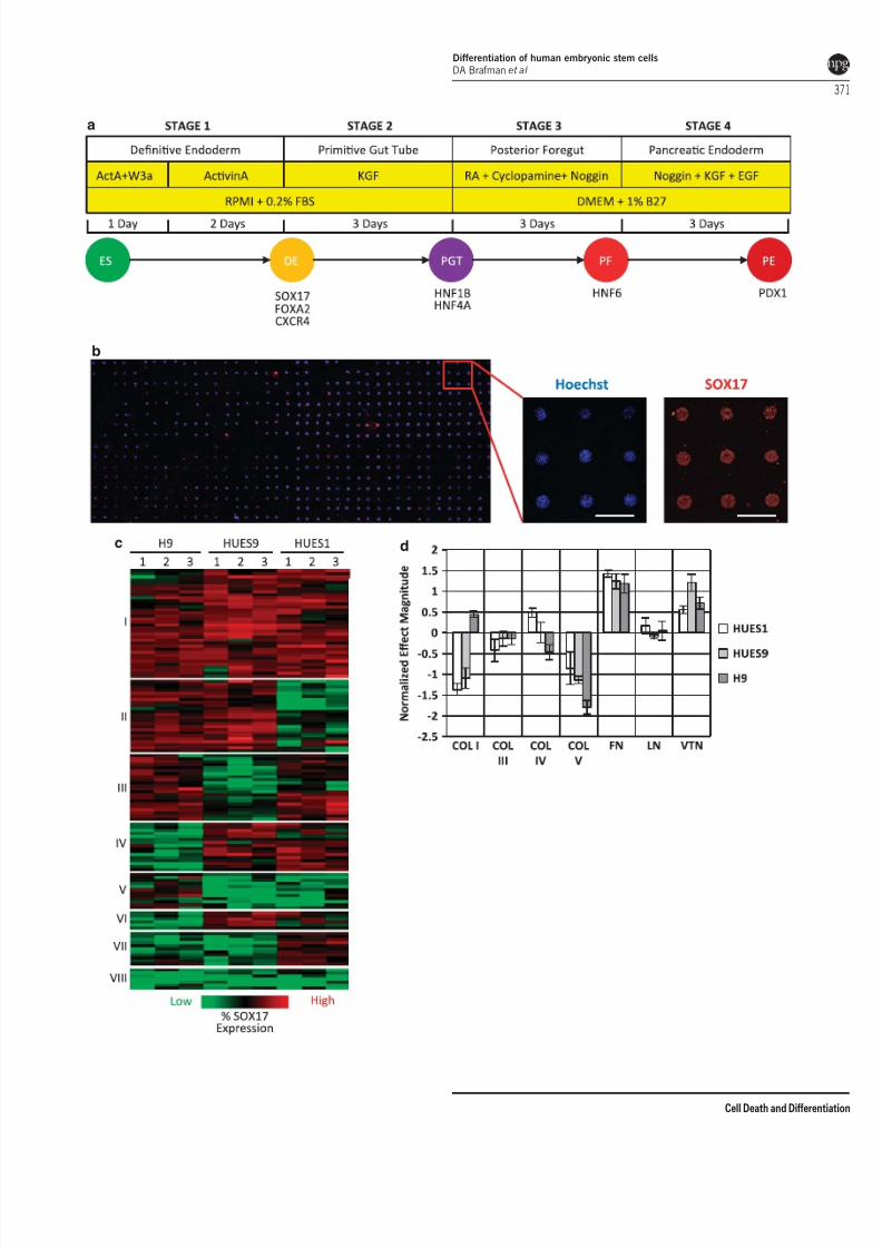
FN and VTN promote endodermal differentiation. To
confirm that FN and VTN promoted DE differentiation of
hESCs we compared their effects to that of MGEL inconventional cell culture formats. HUES9 hESCs were plated
on MGEL and the combination of FN and VTN (FN þVTN)
and differentiated to DE. Immunofluorescent (IF) staining
demonstrated that FNþVTN caused a statistically significant
increase in the percentage of cells expressing the DE marker
SOX17 (Figure 2a). Moreover, culture on FNþVTN
increased the total number of SOX17þ cells, as well as
the total cell number (Figure 2b). Flow cytometry revealed
that culture on FNþVTN produced an increase in the
percentage of cells expressing the DE marker CXCR4
(Figure 2c). Finally, quantitative PCR (qPCR; Figure 2d) of
DE markers SOX17 , FOXA2 , and CXCR4 showed that
FNþVTN increased the efficiency of DE differentiation
relative to MGEL. We also observed that FN þVTN resulted
in increased DE differentiation in two additional hESC lines,
HUES1 and H9 (Supplementary Figures 3a and b). Finally,
our analysis revealed that FNþVTN increased the efficiency
Figure 1 High-throughput ECMP screen reveals the influence of ECMPs in DE differentiation. (a) Schematic of the four stage differentiation protocol from hESC (ES), toDE, PGT, posterior PF, and finally PE. The soluble factors and culture media used at each stage are shown. (b) hESCs (H9, HUES1, HUES9) were cultured on ECMP arraysusing previously published DE differentiation conditions.8 On day 3, arrays were fixed and stained with Hoechst and an antibody to SOX17, a marker for DE (scalebar¼ 450mm). (c) Heat map representing the cell number normalized SOX17 expression of each ECMP combination (rows) for each independent array experiment. Threeindependent array experiments were performed with each hESC line. Columns were mean normalized and scaled to one unit S.D. Hierarchical clustering of ECMP conditionswas performed using Pearson correlation coefficient as a similarity metric. Clustering segregated ECMP combinations into eight groups based on the normalized SOX17expression induced in each hESC line. (d) Magnitude of the main effects from a full factorial analysis of the ECMP array data reveals that specific ECMP components, FN andVTN, have largest positive effects on DE differentiation efficiency ( n ¼ 3 independent array experiments; error bars, S.E.M.)
Differentiation of human embryonic stem cells
DA Brafman et al
370
Cell Death and Differentiation
7/29/2019 Regulation of Endodermal Differentiation of Human
http://slidepdf.com/reader/full/regulation-of-endodermal-differentiation-of-human 3/13
a
b
c d
Differentiation of human embryonic stem cells
DA Brafman et al
371
Cell Death and Differentiation
7/29/2019 Regulation of Endodermal Differentiation of Human
http://slidepdf.com/reader/full/regulation-of-endodermal-differentiation-of-human 4/13
of DE differentiation over that observed when hESCs were
differentiated on either ECMP alone (FN, VTN) or on MGEL
(Supplementary Figures 3c and d).
To address whether culture on FNþVTN improved
differentiation of hESC to more mature endodermal lineages,
HUES9 were cultured on FNþVTN and MGEL and
a b
cd
e f g
Figure 2 ECMPs improve efficiency of hESC differentiation to DE, PGT, PF endoderm, and PE. HESCs were cultured on MGEL and FN and VTN (FN þVTN) usingpreviously published protocols.2,8,9 (a) Representative images of aSOX17 immunofluorescence of HUES9 hESCs differentiated to DE on MGEL and FNþVTN(mean±S.E.M.). (b) Quantification of HUES9 hESCs stained by SOX17 cells out of total cell number (n ¼ 3; mean±S.E.M.). (c) Flow cytometric analysis of CXCR4expression of HUES9 hESCsdifferentiatedto DE on MGELand FN andVTN (FNþVTN). Gene expression analysis formarkers of (d) DE (SOX17, FOXA2, CXCR4), (e) PGT(HNF1b, HNF4a), and (f and g) PF and PE (HNF6, PDX1) of HUES9 hESCs differentiated to DE, PGT, PF, and PE on MGEL and FN þVTN. (n ¼ 3; error bars, S.E.M.).Asterisks indicate statistical significance relative to MGEL as determined by a two tail t -test
Differentiation of human embryonic stem cells
DA Brafman et al
372
Cell Death and Differentiation
7/29/2019 Regulation of Endodermal Differentiation of Human
http://slidepdf.com/reader/full/regulation-of-endodermal-differentiation-of-human 5/13
differentiated to primitive gut tube (PGT), posterior foregut
(PF), and pancreatic endoderm (PE) using previously
published protocols (Figure 1a).9 Expression of HNF1b and
HNF4 a, markers of both pancreas and liver development, was
higher in cells differentiated on FNþVTN compared with
those differentiated on MGEL (Figure 2e). Expression of the
PF marker HNF6 (Figure 2f) and the PE marker PDX1(Figure 2g) was higher in FNþVTN versus MGEL cultures.
These results demonstrate that culture on FNþVTN increases
differentiation efficiency toward endodermal lineages.
Integrin expression in hESC differentiation. Having
established that FN and VTN were critical ECMP compo-
nents to promote DE differentiation, we studied the role of
integrin receptors in hESC differentiation. HESCs were
differentiated to the three germ layers—endoderm, meso-
derm, and ectoderm—using previously established proto-
cols2,15,16 and analyzed for integrin gene expression.
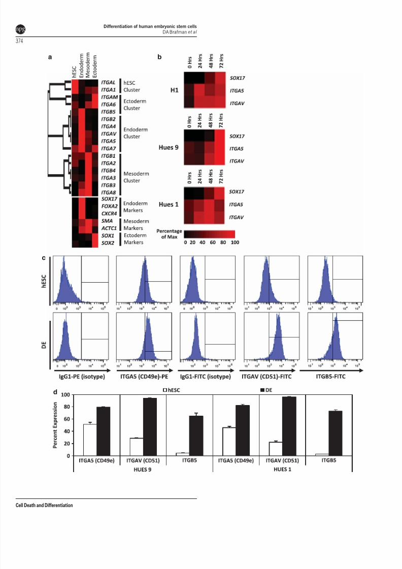
Hierarchical clustering of integrin gene expression revealed
specific integrin ‘signatures’ that defined each differentiated
cell population, with a set of integrin genes ( ITGA4 , ITGA5
(integrin a5), ITGA7 , ITGAV (integrin aV), ITGB2 , ITGB5 )
being upregulated in cells expressing endodermal markers
(SOX17 , FOXA2 , CXCR4 ) relative to cells expressing either
mesodermal (SMA, ACTC1) or ectodermal (SOX1, SOX2 )
marker genes (Figure 3a). Importantly, among these
integrins, ITGA4, ITGA5, ITGAV, and ITGB5 encode sub-
units of heterodimeric integrin receptors that bind FN and
VTN.17–21
As ITGA5 is required for hESC binding to FN and ITGAV,
and ITGB5 are required for binding to VTN,22 we investigated
the expression levels of these integrin subunits as hESCs
differentiate to DE. A time course of hESCs differentiating to
DE revealed that ITGA5 and ITGAV expression is upregulatedin a dynamically similar manner to that of SOX17 (Figure 3b).
Furthermore, expression of ITGB5 is also upregulated as cells
differentiate to DE (Figure 3c), suggesting that hESCs display
functional FN and VTN receptors as they differentiate to DE.
By contrast, expression of the gene encoding subunits of the
LN receptor, ITGA6 , is significantly downregulated during
endoderm differentiation of (Supplementary Figure 4a).
Using flow cytometry, we confirmed that expression of
these three integrin receptors, detected by antibodies that
bind ITGA5 (CD49e), ITGAV (CD51), andITGB5, is increased
in DE versus undifferentiated hESCs (Figures 3c and d). In
comparison, flow cytometry of ITGA6 and ITGB1, integrin
subunits that comprise the LN receptor, were either down-
regulated or unchanged as hESCs differentiated to DE
(Supplementary Figures 4b and c). Taken together, these
results suggest that hESCs differentiating to DE significantly
upregulate cell surface expression of the subunits that
comprise the integrin receptors that bind FN and VTN, the
two ECMP components that we identified in our cellular
microarray screen to promote DE differentiation.
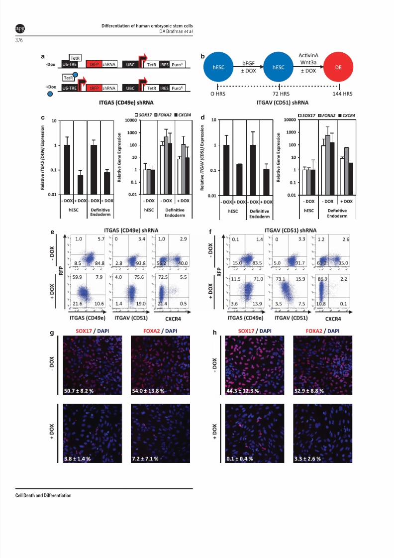
Knockdown of ITGA5 and ITGAV impairs endoderm
formation. To determine to what extent expression of the
FN and VTN integrin receptors is functionally important
during endodermal differentiation, we used a short hairpin
RNA (shRNA) approach to knockdown expression of either
ITGA5 or ITGAV. HESCs stably harboring doxycycline
(DOX) inducible shRNAs (Figure 4a) to either gene—
referred to as ITGA5shRNA or ITGAVshRNA hESCs—were
treated for 3 days with DOX (1 mg/ml) before induction of
endodermal differentiation (see flowchart in Figure 4b). DOX
treatment of either ITGA5shRNA or ITGAVshRNA hESCs and
DE led to a significant increase in expression of redfluorescent protein (Supplementary Figure 5a), the expres-
sion of which was driven from the same DOX-inducible
promoter as the shRNAs. QPCR analysis confirmed that
expression of ITGA5 and ITGAV was significantly decreased
in DOX-treated undifferentiated hESC and DE cell popula-
tions (Figures 4c and d). Flow cytometry revealed that cell
surface protein expression of ITGA5 and ITGAV was
decreased in DOX-treated DE cell populations (Figures 4e
and f). We confirmed that DOX treatment of ITGA5shRNA
hESCs had no effect on ITGAV gene (Supplementary
Figure 5b) or cell surface protein expression (Figure 4e).
Similarly, DOX treatment of ITGAVshRNA hESCs had no
effect on ITGA5 gene (Supplementary Figure 5c) or cell
surface protein expression (Figure 4f). Additionally, DOX
treatment alone was not responsible for decreases in integrin
expression as DOX treatment of wild-type hESCs had no
effect on ITGA5 or ITGAV expression (Supplementary
Figure 5d). Importantly, expression of the endodermal
marker genes, SOX17 , FOXA2, and CXCR4 , was signifi-
cantly decreased in hESC-derived DE in which ITGA5 or
ITGAV expression was knocked-down by the shRNAs
(Figures 4c and d). Furthermore, flow cytometry revealed
that CXCR4 cell surface expression was almost absent in
DOX-treated DE cells (Figures 4e and f). IF analysis
demonstrated that knockdown of either ITGA5 or ITGAV
resulted in a significant reduction in SOX17 or FOXA2
staining at DE (Figures 4g and h). These results suggest thatexpression of FN and VN integrin receptors ITGA5 and
ITGAV is necessary for differentiation of hESCs to DE.
ITGA5 and ITGAV as cell surface markers for the
isolation of endodermal progeny from differentiating
hESCs. As increases in ITGA5 (CD49e) and ITGAV (CD51)
expression correlate with endodermal differentiation, we
tested whether cell separation by flow cytometry for these
integrin receptors could be employed to isolate cells with
endodermal gene expression signatures (Figure 5a). HESC-
derived DE was sorted for ITGA5 (CD49e) and ITGAV
(CD51) (Figure 5b) and analyzed for expression of SOX17
(Figure 5c and Supplementary Figure 6). This analysis
revealed that double-positive ITGA5(CD49e)þ /ITGAV
(CD51)þ cells expressed higher amounts of the DE maker
gene SOX17 than single-positive ITGA5(CD49e)þ /ITGAV
(CD51)À or ITGA5(CD49e)À /ITGAV(CD51)þ cells or dou-
ble-negative ITGA5(CD49e)À /ITGAV(CD51)À cells (Supple-
mentary Figure 6). Furthermore, expression of additional DE
marker genes FOXA2 and CXCR4 was increased in double-
positive cells compared with double-negative cells
(Figure 5c). We next investigated if double-positive
ITGA5(CD49e)þ /ITGAV(CD51)þ were capable of differen-
tiating into more mature endodermal progeny. Double-
positive ITGA5(CD49e)þ /ITGAV(CD51)þ and double-nega-
tive ITGA5(CD49e)À /ITGAV(CD51)À cells were replated
Differentiation of human embryonic stem cells
DA Brafman et al
373
Cell Death and Differentiation
7/29/2019 Regulation of Endodermal Differentiation of Human
http://slidepdf.com/reader/full/regulation-of-endodermal-differentiation-of-human 6/13
a
c
d
b
Differentiation of human embryonic stem cells
DA Brafman et al
374
Cell Death and Differentiation
7/29/2019 Regulation of Endodermal Differentiation of Human
http://slidepdf.com/reader/full/regulation-of-endodermal-differentiation-of-human 7/13
after cell sorting and differentiated to PGT. Subsequent gene
expression analysis revealed that expression of HNF1b and
HNF4 a, markers of pancreas and liver development, were
enriched in the ITGA5(CD49e)þ /ITGAV(CD51)þ population
relative to the ITGA5(CD49e)À /ITGAV(CD51)- population
(Figure 5d). Therefore, cell enrichment strategies for ITGA5
(CD49e) and ITGAV (CD51) significantly increase the yield ofcells with DE gene expression patterns from differentiating
hESC cultures.
Remodeling of the extracellular matrix during endoder-
mal differentiation. In addition to exploring the effects of
exogenous ECMP on DE formation, we wanted to investigate
the role of endogenous ECMP production and remodeling
during endodermal differentiation. To that end, we measured
endogenous ECMP gene expression of hESCs differentiated
to DE (Figure 6a). In general, expression of endogenous
ECMPs increased as cells differentiated to DE on MGEL and
FNþVTN substrates. Specifically, we observed a statisti-
cally significant increased expression of several COLs(COL4A2 , COL5A1, COL6A1, COL7A1, COL8A1, COL11A1,
COL12A1), FN1, VTN , and LN subunits (LAMA3 , LAMAB1,
LAMAB3 ) as hESCs differentiated to DE.
Previous studies have shown ECMP degradation and
proteolysis have a critical role in endoderm development
and cell differentiation.23,24 Therefore, we wanted to deter-
mine if ECM remodeling anddegradation through the action of
matrix metalloproteinases (MMPs) was required for differ-
entiation of hESCs to DE. While expression of MMP 1, 3, 7,
10, 12, or 13 was not detected in hESCs or DE (data not
shown), we observed a statistically significant increased
expression of MMP 2, 8, 9, 14, and 15 during DE differentia-
tion (Figure 6b). To test whether MMP activity was required for
DE differentiation, we treated cells with broad-spectrum smallmolecule inhibitors of MMP (Baritasmat, Marimastat,
CP471474) and the glycoprotein tissue inhibitor of metallo-
proteinases (TIMP1) during DE differentiation. Gene expres-
sion analysis of DE markers SOX17 , FOXA2 , and CXCR4
revealed that MMP inhibition does not inhibit formation of DE
(Figure 6c). Therefore, even though we observe changes in
the composition of the extracellular environment during DE
differentiation, including endogenous deposition of ECMPs
and secretion of MMPs, pharmacological inhibition of MMPs
does not appear to disrupt endodermal differentiation of
hESCs.
Discussion
The selection of the appropriate extracellular matrix is critical
for hESC self-renewal and proliferation,13,22 and here we
show that the ECMP composition also potently influences
hESC differentiation to DE. By systematically screening
hundreds of ECMP combinations, we identified two ECMPs,
FN, and VTN, which significantly improve the efficiency of
hESC differentiation to DE, thereby overcoming the need for
poorly defined and non-human biological components, such
as MGEL, in the manipulation of hESCs.While certain studies have explored the role of physical
properties of the microenvironment, including three-dimen-
sional culture25 and substrate rigidity,26 we focused our
analysis on the role of the ECM in differentiation and found
that changes in the composition of the ECM profoundly
affected the differentiation of hESCs to endoderm. It is
particularly important to note that the effects of the growth
factors inducing DE (Wnt3a and Activin A) are significantly
influenced by ECMP composition. Our results suggest that
appropriately defining the ECMP substrate in addition to the
soluble signaling molecule environment is critical for improv-
ing the differentiation of hESCs to specific lineages.
The differentiation of hESCs to DE resembles that of
primitive streak formation and gastrulation where cells
invaginate and generate mesendodermal cell populations.
These movements of epiblast cells require several growth
factor signaling pathways, as well as an ECM. In this process
the ECM does not merely function as a scaffold through which
cells migrate. Rather, as determined by computational and
optical methods, migrating cells move in concert with the ECM
with little cellular movement relative to the ECM.27 This study
supports the notion that the ECM has a more active role in
development than previously appreciated and further under-
scores the importance of performing screens to identify
optimal ECM compositions that promote specific develop-
mental processes.
Consistent with our identification of FN and VTN ascritical components that promote endodermal differentiation
in cell culture, several studies in model organisms have
provided compelling evidence that these ECM components
are critical constituents of the microenvironment guiding the
processes of primitive streak formation and gastrulation. For
example, injection of agents that disrupt integrin-FN interac-
tions, such as RGDS peptides or antibodies and Fab’
fragments directed against FN, into chick embryos perturb
gastrulation movements.28 Such microinjection experiments
in frog embryos have led to similar observations.29 Mice
lacking FN-binding integrins die early in development and fail
to extend the anterior–posterior axis.30–32 Earlier defects,
such as during gastrulation, are likely not uncovered due to
rescue by maternally contributed FN message and protein.
Together, countless studies in a variety of model organisms
support the concept that the ECM, and specifically FN, has an
Figure 3 Expression of integrin genes in hESC differentiation. (a) HUES9 hESCs were differentiated in vitro to the three germ layers (ectoderm, endoderm, andmesoderm) as previously described.2,15,16 QPCR analysis of integrin gene expression was performed. The data is displayed as a heat map where black corresponds tominimum expression levels and red corresponds to maximum levels. Hierarchical clustering of integrin gene expression resulted in segregation of integrins into four groupsbased on their expression levels in hESCs or germ layer-specific cell types. (b) Time course of DE marker SOX17 and FN/VTN specific subunits ITGA5 (CD49e) and ITGAV (CD51) gene expressionduring hESC (H1, HUES1,and HUES9)differentiation to DE.This analysis reveals that SOX17 , ITGA5 (CD49e) , and ITGAV (CD51) geneexpressionincrease in a dynamically similar manner. (c) Representative flow cytometry histograms of cell surface protein expression of FN/VTN specific subunits ITGA5 (CD49e), ITGAV(CD51), and ITGB5in hESCs and DE. (d) Quantification of percentage of cellsurface protein expression of ITGA5þ (CD49eþ ), ITGAVþ (CD51þ ), and ITGB5þ hESCsandDE (HUES9 and HUES1; n ¼ 3; error bars, S.E.M.)
Differentiation of human embryonic stem cells
DA Brafman et al
375
Cell Death and Differentiation
7/29/2019 Regulation of Endodermal Differentiation of Human
http://slidepdf.com/reader/full/regulation-of-endodermal-differentiation-of-human 8/13
a
c
e f
g h
d
b
Differentiation of human embryonic stem cells
DA Brafman et al
376
Cell Death and Differentiation
7/29/2019 Regulation of Endodermal Differentiation of Human
http://slidepdf.com/reader/full/regulation-of-endodermal-differentiation-of-human 9/13
important and instructive function in early embryogenesis.
However, it should be stressed that in our studies only the initial
matrix compositions are specified. Cells exposed to these
ECMPs remodel the underlying matrix and begin secreting their
own ECMPs. Even so, the observed cellular responses are a
result of their exposure to the initial composition of the ECM.
Previous studies demonstrated that undifferentiated hESCsexpress a variety of integrins, including integrins a1, 2, 3, 5, 6,
7, 11, E, and V, and b1, 2, 3, and 5.33–35 We extended these
studies by examining integrin gene expression in undiffer-
entiated hESCs and hESC differentiated to each of the three
primitive germ layers—endoderm, mesoderm, and ectoderm.
This analysis revealed a specific integrin ‘signature’ that was
unique to each of these cell populations. Specifically, we
found that ITGA5 and ITGAV gene expression was highly
upregulated and ITGA6 expression was significantly down-
regulated in the endodermal lineage. Treatment with specific
integrin blocking antibodies revealed that blocking ITGA5
impaired adhesion to FN, blocking ITGAV and ITGB5 reduced
the binding to VTN, and blocking ITGA6 inhibited binding
to LN.22 Furthermore, ITGA6 binding to LN has been
implicated as having a critical role in the self-renewal and
maintenance of pluripotent hESCs.36 In this study, we
implemented an inducible shRNA system to demonstrate
that knockdown of ITGA5 and ITGAV impaired endoderm
formation. During development, integrin switching, rapid
changes in the proportions of specific integrin subunits
expressed at the cell surface, has been implicated as a
mechanism that regulates cell differentiation.37,38 Together
our results suggest a possible mechanism in which hESCs
differentiating to DE undergo an integrin switch from an ITGA6
signature which favors binding LN, and thereby maintenance
of pluripotency, to an ITGA5 and ITGAV signature, which
allows for interaction with FN and VTN and subsequentdifferentiation to DE.
Mouse models have been used extensively to interrogate
integrin expression and functionality during embryonic devel-
opment.37,39,40 Interestingly, mouse embryos stained for
different integrin subunits at E6.5 revealed that Itga5 expres-
sion was mainly restricted to endoderm,41 which is consistent
with the ITGA5 expression patterns that we identified in
hESC-derived DE. Furthermore, Itga5 and ItgaV are widely
expressed during development of many organs of endoder-
mal origin, such as the pancreas, liver, and lungs.42–44
Knockout of Itga5 or ItgaV resulted in embryonic lethality,32,45
while tissue specific deletion of Itga5 or ItgaV resulted in
vasculature and neuronal defects.18,32,45,46 Therefore, the
novel findings presented here, which demonstrate knockdown
of ITGA5 and ITGAV in hESCs impaired endoderm formation
in hESCs, suggests that similar integrin knockdown strategies
in hESCs can be used to interrogate the function of various
integrin-ECMP interactions during the earliest stages of
human development.
Current hESC differentiation protocols are insufficient in
creating pure cell populations, which are required for under-
standing human development and creating disease relevant
models. Therefore, developing sorting strategies for flowcytometry-based isolation of highly pure populations of cells
from differentiating hESC cultures is of particular interest.47–50
By investigating the role of integrin-ECMP interactions in
hESC differentiation to DE, we identified a panel of novel
surface integrins, ITGA5 (CD49e) and ITGAV (CD51), that
allow for the FACS-based isolation of endodermal cells. In the
future, similar integrin ‘signatures’ could be developed that
would permit the isolation of lineage committed cells from
mixed differentiated hESC cultures.
Materials and MethodsArrayed cellular microenvironment fabrication. Arrayed cellularmicroenvironment (ACME) slides were fabricated as previously described. Briefly,glass slides were cleaned, silanized, and then functionalized with a polyacrylamidegel layer. Stock solutions of human COL I, COl III, COl IV, COl V, FN, LN (Sigma-Aldrich, St Louis, MO, USA) and VTN (EMD-Millipore, Billerica, MA, USA) wereprepared in an ECMP-printing buffer (200 mM acetate, 10 mM EDTA, 40% (v/v)glycerol, and 0.5% (v/v) triton-X-100 in MQH2O, with pH adjusted to 4.9 usingglacial acetic acid). All ECMP combinations were premixed at a constant proteinconcentration of 250 mg/ml in polypropylene 384-well plates. SMP 3.0 spottingpins (Telechem Corp., Atlanta, GA, USA) were washed with 90% ethanol. Allprintings were performed with a SpotArray 24 (Perkin-Elmer, Waltham, MA, USA)at room temperature (RT) with 65% relative humidity. The printing conditions werea 1000-ms inking time and a 250-ms stamping time. To control for variability, eachECMP combination was printed in replicates of five spots. Each spot had adiameter of 150–200mm, and neighboring microenvironments were separated bya center-to-center distance of 450 mm. A single slide carried 6400 spots arrangedin sixteen 20Â 20 matrices so that one slide carried 1280 unique ECMP
conditions. Slides were inspected manually under a light microscope for consistentand uniform ECMP deposition. ECMP spotting was characterized using generalprotein stain (SYPRO Ruby gel stain, Life Technologies, Carlsbad, CA, USA) orprotein specific antibodies (Sigma-Aldrich) as previously described. A single slidefrom each batch of printed arrays was seeded with HEK-293 (2.5 Â 105 cells perslide) to ensure that each ECMP spot supported cell adhesion.
Cells and culture conditions. The following media were used: mouseembryonic fibroblast (MEF) (1Â high glucose DMEM, 10% fetal bovine serum(FBS), 1% (v/v) L-glutamine penicillin/streptomycin); H9/WA09 hESCs (1ÂDMEM-F12, 20% (v/v) Knockout Serum Replacement, 1% (v/v) non-essentialamino acids, 0.5% (v/v) glutamine, 120 mM 2-mercaptoethanol (Sigma-Aldrich));HUES1 and nine hESCs (1ÂKnockout DMEM, 10% (v/v) Knockout SerumReplacement, 10% (v/v) human plasmanate (Chapin Healthcare, Anaheim, CA,USA), 1% (v/v) non-essential amino acids, 1% (v/v) penicillin/streptomycin, 1%(v/v) Gluta-MAX, 55 mM 2-mercaptoethanol (Sigma-Aldrich)). All media compo-
nents are from Life Technologies unless indicated otherwise. H9, HUES9, andHUES1 hESC lines were maintained on feeder layers of mitotically inactivatedMEFs (2Â 104 /cm2; Millipore). All hESC cultures were supplemented with 30 ng/ ml bFGF (Life Technologies). MEF-CM was produced by culturing the appropriate
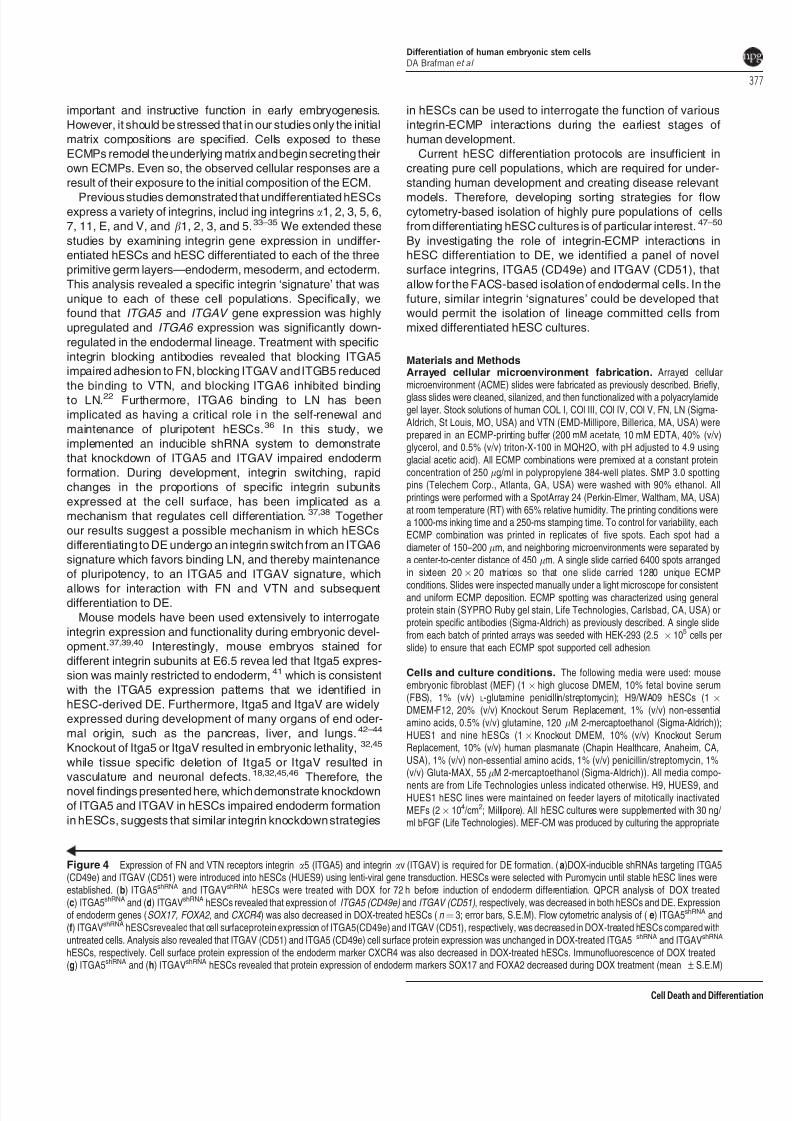
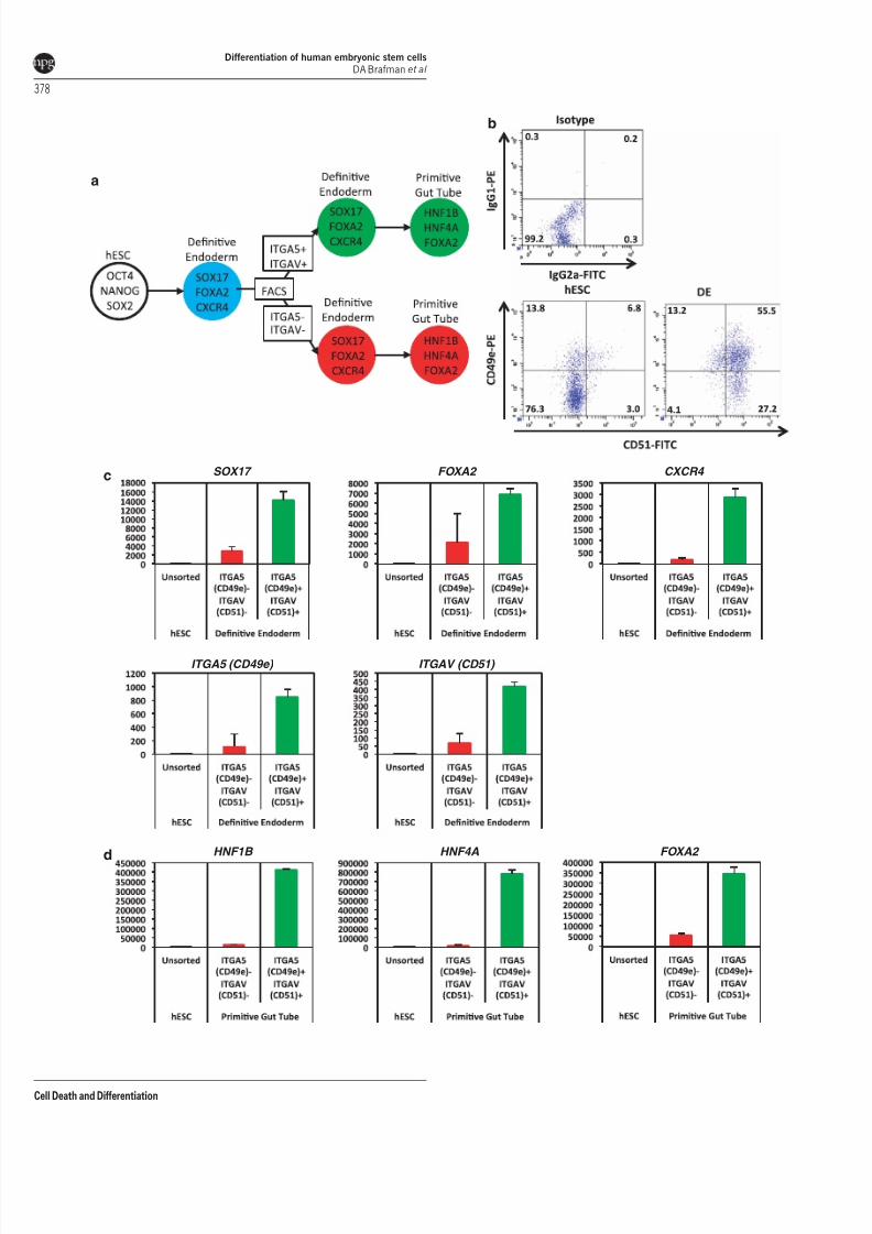
Figure 4 Expression of FN and VTN receptors integrin a5 (ITGA5) and integrin av (ITGAV) is required for DE formation. (a)DOX-inducible shRNAs targeting ITGA5(CD49e) and ITGAV (CD51) were introduced into hESCs (HUES9) using lenti-viral gene transduction. HESCs were selected with Puromycin until stable hESC lines wereestablished. (b) ITGA5shRNA and ITGAVshRNA hESCs were treated with DOX for 72 h before induction of endoderm differentiation. QPCR analysis of DOX treated(c) ITGA5shRNA and (d) ITGAVshRNA hESCs revealed that expression of ITGA5 (CD49e) and ITGAV (CD51) , respectively, was decreased in both hESCs and DE. Expressionof endoderm genes (SOX17, FOXA2, and CXCR4 ) was also decreased in DOX-treated hESCs (n ¼ 3; error bars, S.E.M). Flow cytometric analysis of (e) ITGA5shRNA and(f) ITGAVshRNA hESCsrevealed that cell surfaceprotein expression of ITGA5(CD49e) and ITGAV (CD51), respectively, was decreased in DOX-treated hESCs compared withuntreated cells. Analysis also revealed that ITGAV (CD51) and ITGA5 (CD49e) cell surface protein expression was unchanged in DOX-treated ITGA5shRNA and ITGAVshRNA
hESCs, respectively. Cell surface protein expression of the endoderm marker CXCR4 was also decreased in DOX-treated hESCs. Immunofluorescence of DOX treated(g) ITGA5shRNA and (h) ITGAVshRNA hESCs revealed that protein expression of endoderm markers SOX17 and FOXA2 decreased during DOX treatment (mean±S.E.M)
Differentiation of human embryonic stem cells
DA Brafman et al
377
Cell Death and Differentiation
7/29/2019 Regulation of Endodermal Differentiation of Human
http://slidepdf.com/reader/full/regulation-of-endodermal-differentiation-of-human 10/13
a
b
c
d
SOX17
ITGA5 (CD49e)
HNF1B HNF4A FOXA2
ITGAV (CD51)
FOXA2 CXCR4
Differentiation of human embryonic stem cells
DA Brafman et al
378
Cell Death and Differentiation
7/29/2019 Regulation of Endodermal Differentiation of Human
http://slidepdf.com/reader/full/regulation-of-endodermal-differentiation-of-human 11/13
hESC medium on MEFs for 24 h. Cells were routinely passaged with Acutase(Millipore), washed, and replated at a density 4.25Â 104 /cm2).
Endoderm induction on ACME slides. Before their use, slides weresoaked in PBS while being exposed to UVC germicidal radiation in a sterile flowhood for 10 min. Before seeding onto the ACME slides, hESCs were cultured fortwo passages on MGEL (BD) with MEF-CM supplemented with 30ng/ml bFGF to
remove residual feeder cells. HESCs were then acutase-passaged onto the ACMEslides (5.0Â 105 cells per slide) and allowed to settle on the spots for 18 h. Arrayslides were then gently washed twice with RPMI (Life Technologies) to remove celldebris and residual hESC media. The medium was then changed to RPMIsupplemented with 1% (v/v) Gluta-MAX and 100 ng/ml recombinant human ActivinA (R&D Systems, Minneapolis, MN, USA). Cells were cultured for 3 days, withFBS concentrations at 0% for the first day and 0.2% for the second and third days.Cultures were supplemented with 30 ng/ml purified mouse Wnt3a for the first day.
Endoderm induction on defined ECMPs. H9, HUES9 and HUES1were cultured on MGEL (BD) with MEF-CM supplemented with 30 ng/ml bFGF for2 passages to remove residual MEFs. The human ECMP-coated plates wereprepared by coating tissue culture plates in the ECMP (diluted in 10 mM aceticacid) overnight, followed by air drying. 10 mg of total protein was plated per cm2 ofculture dish surface. Human ECMP-coated plates were used immediately after airdrying. HESCs were passaged at a density of 2.5Â 105 cells/ml onto human
ECMP or MGEL-coated plates in order to achieve confluency the following day.HESCs were then gently washed twice with RPMI (Life Technologies) to removecell debris and residual hESC media. The medium was then changed to RPMIsupplemented with 1% (v/v) Gluta-MAX and 100 ng/ml recombinant human ActivinA (R&D Systems). Cells were cultured for 3 days, with FBS concentrations at 0%for the first day and 0.2% for the second and third days. Cultures weresupplemented with 30 ng/ml purified mouse Wnt3a for the first day. For furtherdifferentiation to PGT, the medium was changed to RPMI with 0.2% FBS
a b
c
C O L 1
A 1
C O L 4
A 1
C O L 5
A 1
C O L 6
A 1
C O L 6
A 2
C O L 7
A 1
C O L 8
A 1
C O L 1 1
A 1
C O L 1 2
A 1
C O L 1 4
A 1
C O L 1 6
A 1
F N 1
L A M
A 1
L A M
A 2
L A M
A 3
L A M
B 1
L A M
B 3
L A M C 1
V T N
M M
P 2
M M
P 8
M M
P 9
M M P
1 1
M M P
1 4
M M P
1 5
M M P
1 6
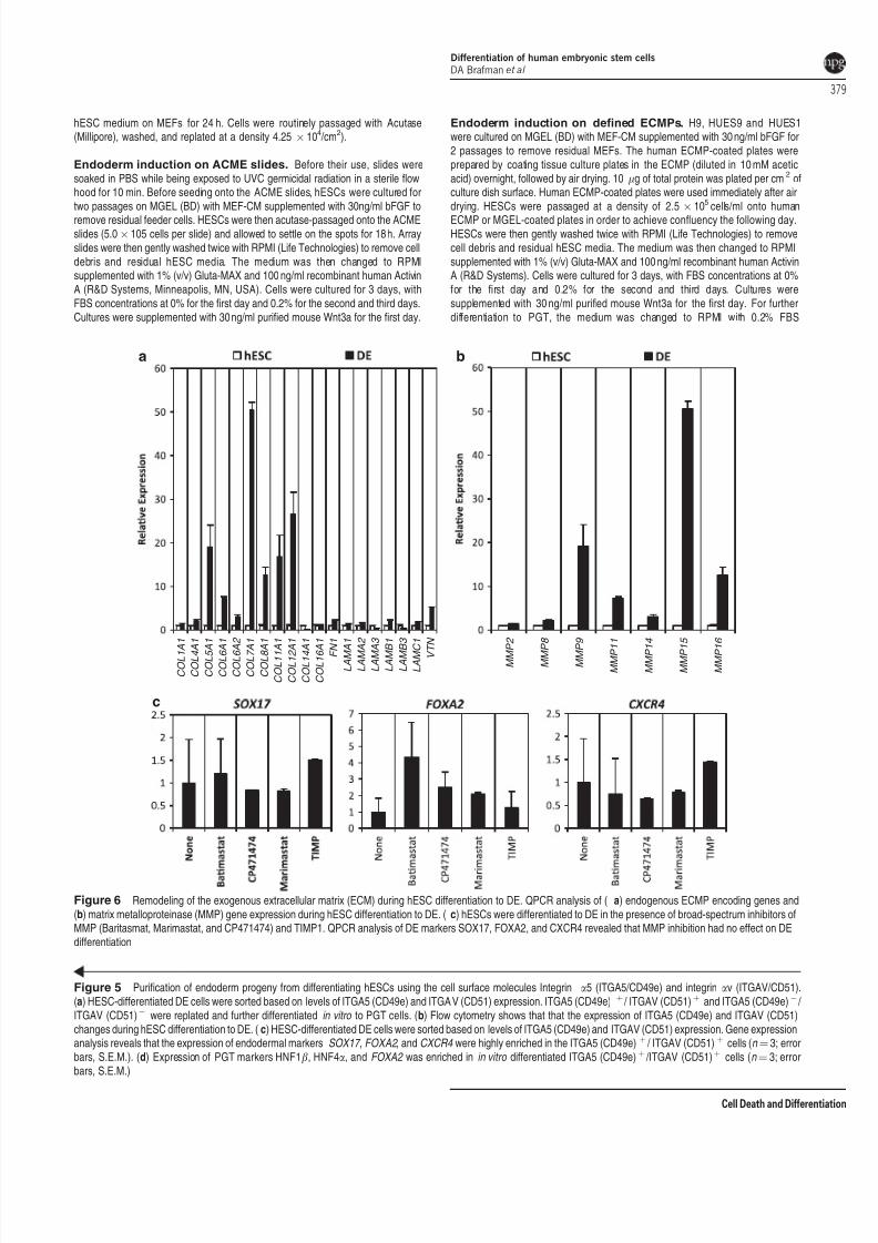
Figure 6 Remodeling of the exogenous extracellular matrix (ECM) during hESC differentiation to DE. QPCR analysis of ( a) endogenous ECMP encoding genes and(b) matrix metalloproteinase (MMP) gene expression during hESC differentiation to DE. ( c) hESCs were differentiated to DE in the presence of broad-spectrum inhibitors ofMMP (Baritasmat, Marimastat, and CP471474) and TIMP1. QPCR analysis of DE markers SOX17, FOXA2, and CXCR4 revealed that MMP inhibition had no effect on DEdifferentiation
Figure 5 Purification of endoderm progeny from differentiating hESCs using the cell surface molecules Integrin a5 (ITGA5/CD49e) and integrin av (ITGAV/CD51).(a) HESC-differentiated DE cells were sorted based on levels of ITGA5 (CD49e) and ITGAV (CD51) expression. ITGA5 (CD49e) þ / ITGAV (CD51)þ and ITGA5 (CD49e)À / ITGAV (CD51)À were replated and further differentiated in vitro to PGT cells. (b) Flow cytometry shows that that the expression of ITGA5 (CD49e) and ITGAV (CD51)changes during hESC differentiation to DE. (c) HESC-differentiated DE cells were sorted based on levels of ITGA5 (CD49e) and ITGAV (CD51) expression. Gene expressionanalysis reveals that the expression of endodermal markers SOX17 , FOXA2 , and CXCR4 were highly enriched in the ITGA5 (CD49e)þ / ITGAV (CD51) þ cells (n ¼ 3; errorbars, S.E.M.). (d) Expression of PGT markers HNF1b, HNF4a, and FOXA2 was enriched in in vitro differentiated ITGA5 (CD49e)þ /ITGAV (CD51)þ cells (n ¼ 3; errorbars, S.E.M.)
Differentiation of human embryonic stem cells
DA Brafman et al
379
Cell Death and Differentiation
7/29/2019 Regulation of Endodermal Differentiation of Human
http://slidepdf.com/reader/full/regulation-of-endodermal-differentiation-of-human 12/13
supplemented with 50ng/ml recombinant human KGF (R&D Systems) for 3 days.For differentiation to PF endoderm, the medium was changed to DMEM with1ÂB27 (Life Technologies), 50ng/ml recombinant human Noggin (R&DSystems), 0.25mM KAAD-cyclopamine (Tocris Biosciences, Bristol, UK), and2mM retinoic acid (Sigma-Aldrich) for 3 days. Finally, for differentiation to PE themedium was changed to DMEM with 1Â B27 (Invitrogen) supplemented with50 ng/ml recombinant human Noggin, 50 ng/ml recombinant human KGF, and
50 ng/ml recombinant human EGF (R&D Systems) and for 3 days.
Ectoderm and mesoderm differentiation. To differentiate hESCs toectoderm we modified several previously published protocols.9,15,51 To initiateectoderm differentiation, hPSCs were cultured on MGEL in MEF-CM supple-mented with 30 ng/ml FGF2. Cells were then detached with treatment with acutase(Millipore) for 5 min and resuspended in ectoderm embryoid body (EB) media(10% FBS/1% N2/1% B27/DMEM:F12) supplemented with 5 mM Y-267632(Stemgent, Cambridge, MA, USA), 50 ng/ml recombinant mouse Noggin (R&DSystems), 0.5mM Dorsomorphin (Tocris Biosciences). Next, 7.5 Â 105 cells werepipetted to each well of several 6-well ultra low attachment plates (Corning, Lowell,MA, USA). The plates were then placed on an orbital shaker set at 95 r.p.m. in a37 1C/5% CO2 tissue culture incubator. The next day, the cells formed sphericalclusters and the media was changed to ectoderm EB media without FBSsupplemented with 50 ng/ml recombinant mouse Noggin and 0.5 mM Dorsomor-
phin. The media was subsequently changed every other day. After 5 days insuspension culture, the EBs were then transferred to a 10-cm dish coated (3 Â 6wells per 10cm dish) with growth factor reduced MGEL (1 : 25 in KnockOutDMEM; BD Biosciences) for attachment. The plated EBs were cultured inectoderm EB media without FBS supplemented with 50 ng/ml recombinant mouseNoggin and 0.5 mM Dorsomorphin for an additional 3 days. To diff erentiate hESCto mesoderm, we modified several previously published protocols.16,52 To initiatemesoderm differentiation, hPSCs were cultured on MGEL in MEF-CMsupplemented with 30 ng/ml FGF2 until they reached 60% confluency. Cellswere then gently washed with RMPI media to remove residual hESC media. Themedium was then changed to RPMI supplemented with 0.5% B27 supplement.Cells were cultured for 5 days supplemented with 100 ng/ml recombinant humanActivin A (R&D systems) for the first day, and 10 ng/ml BMP4 for days 2–5.
Immunofluorescence. ACME slides and cultures were gently washed twicewith staining buffer (PBS w/ 1% (w/v) BSA) before fixation. Cultures were then
fixed for 15 min at RT with fresh paraformaldeyde (4% (w/v)). The cultures werewashed twice with staining buffer and permeabilized with 0.2% (v/v) triton-X-100 instain buffer for 20 min at 4 1C. Cultures were then washed twice with stainingbuffer. Primary antibodies were incubated overnight at 4 1C and then washedtwice with stain buffer at RT. Secondary antibodies were incubated at RT for 1 h.Antibodies used are listed in Supplementary Table 2. Nucleic acids were stainedfor DNA with Hoechst 33342 (2 mg/ml; Life Technologies) for 5 min at RT. Imagingof was performed using an automated confocal microscope (Olympus Fluoview1000 (Olympus America, Center Valley, PA, USA) with motorized stage andincubation chamber). Images of ACME slides were quantified using GenePixsoftware (MDS Analytical Technologies, Molecular Devices, Sunnyvale, CA, USA).Quantification of additional images was performed by counting a minimum of ninefields at  20 magnification.
Flow cytometry and cell replating. Cells were dissociated Acutase(Millipore) for 5 min at 37 1C, triturated, and passed through a 40 mm cell strainer.Cells were then washed twice with FACS buffer (PBS, 10 mM EDTA, and 2%FBS) and resuspended at a maximum concentration of 5Â 106 cells per 100 ml.One test volume of antibody was added for each 100ml cell suspension(Supplementary Table 2). Cells were stained for 30 min on ice, washed, andresuspended in stain buffer. Cells were analyzed and sorted with a FACSCanto orFACSAria (BD Biosciences). FACS data was analyzed with FACSDiva software(BD Biosciences). For replating experiments, DE cells were stained with CD49eand CD51 and sorted into FACS buffer with 10 nm Y27632 (Stemgent). Sortedcells were replated at a density of 1Â 105 cells/cm2 in stage 2 PGT media with10 nM Y27632 and differentiated for 4 days.
MMP inhibition during endoderm differentiation. Before endodermdifferentiation, hESC were treated with 0.5 mM Batimastat (Tocris Biosciences),0.5mM CP471474 (Tocris Biosciences), 0.5 mM Marimastat (Tocris Biosciences),or recombinant 130nM human TIMP1 for 48 h. The medium was then changed to
RPMI supplemented with 1% (v/v) Gluta-MAX and 100 ng/ml recombinant humanActivin A (R&D Systems). Cells were cultured for 3 days, with FBS concentrationsat 0% for the first day and 0.2% for the second and third days. Cultures weresupplemented with 30 ng/ml purified mouse Wnt3a for the first day. MMP inhibitioncontinued throughout the duration of endoderm differentiation.
Quantitative PCR. RNA was isolated from cells using TRIzol (Life
Technologies), and treated with DNase I (Life Technologies) to remove tracesof DNA. Reverse transcription was performed by means of qScript cDNASupermix (Quanta Biosciences, Gaithersburg, MA, USA). QPCR was carried outusing TaqMan probes (Life Technologies) and TaqMan Fast Universal PCRMaster Mix (Life Technologies) on a 7900HT Real Time PCR machine (LifeTechnologies), with a 10-min gradient to 95 1C followed by 40 cycles at 95 1C for15s and 60 1C for 1 min. Taqman gene expression assay primers (LifeTechnologies; Supplementary Table 3) were used. Gene expression wasnormalized to 18S rRNA levels. Delta C t values were calculated as C t
targetÀ
C t18s. All experiments were performed with three technical replicates. Rela tive fold
changes in gene expression were calculated using the 2ÀDDC t method.53 Dataare presented as the average of the biological replicates±S.E.M.
Generation of inducible shRNA hESCs. The lenti constructs that wereused to generate the inducible shRNA lines were obtained from Open Biosystems
(ITGA5:1334333, ITGAV:133468). High titer lenti virus was produced as previouslydescribed.54,55 HUES9 hESCs were infected overnight with lenti virus and treatedwith puromycin (0.5 mg/ml) for 2 weeks.
Statistical analysis. For statistical analysis, unpaired t -test were used and aP -valueo0.05 was considered statistically significant. All values were presentedas mean±S.E.M. unless otherwise noted. For each ACME experiment, the ratio(R i) of the log2 of the SOX17 signal and the DNA signal was calculated for eachspot. From this a differentiation z -score was calculated for each spot Z DIF¼(R iÀmDIF)/ sDIF, where R i was the ratio for the spot, mDIF was the average of therations for all spots on each array, and sDIF was the S.D. of the ratios for all spotson each array. Differentiation z -scores from replication spots (n ¼ 5 per ECMPcondition) were averaged for each ECMP condition on the array. The replicateaverage z -scores were displayed in a heat map with rows corresponding toindividual ECMP conditions and columns representing independent arrayexperiments. For each array experiment, all columns were mean-centered and
normalized to one unit S.D. The rows were clustered using Pearson correlationsas a metric of similarity.56 All clustering was performed using Gene Cluster.56 Theresults were displayed using a color code with red and green representing anincrease and decrease, respectively, relative to the global mean. All heat mapswere created using Tree View.56 Normalized effect magnitudes were calculated aspreviously described.14
Conflict of Interest
The authors declare no conflict of interest.
Acknowledgements. DAB was supported by funding from the UCSD StemCell Program and a gift from Michael and Nancy Kaehr. This research wassupported in part by the NIDDK Beta Cell Biology Consortium (5U01DK089567-02)and the California Institute for Regenerative Medicine (RB1-01406).
Author contributionsDAB and KW designed the experiments. DAB, CP, and NK performed theexperiments. DAB, CP, and KW analyzed the results. DAB and KW wrote themanuscript.
1. Borowiak M, Maehr R, Chen S, Chen AE, Tang W, Fox JL et al. Small molecules efficiently
direct endodermal differentiation of mouse and human embryonic stem cells. Cell Stem
Cell 2009; 4: 348–358.
2. D’Amour KA, Agulnick AD, Eliazer S, Kelly OG, Kroon E, Baetge EE. Efficient
differentiation of human embryonic stem cells to definitive endoderm. Nat Biotechnol 2005;
23: 1534–1541.
3. McLean AB, D’Amour KA, Jones KL, Krishnamoorthy M, Kulik MJ, Reynolds DM et al.
Activin a efficiently specifies definitive endoderm from human embryonic stem cells only
when phosphatidylinositol 3-kinase signaling is suppressed. Stem Cells 2007; 25: 29–38.
Differentiation of human embryonic stem cells
DA Brafman et al
380
Cell Death and Differentiation
7/29/2019 Regulation of Endodermal Differentiation of Human
http://slidepdf.com/reader/full/regulation-of-endodermal-differentiation-of-human 13/13
4. Osafune K, Caron L, Borowiak M, Martinez RJ, Fitz-Gerald CS, Sato Y et al. Marked
differences in differentiation propensity among human embryonic stem cell lines. Nat
Biotechnol 2008; 26: 313–315.
5. Berrier AL, Yamada KM. Cell-matrix adhesion. J Cell Physiol 2007; 213: 565–573.
6. Humphries JD, Byron A, Humphries MJ. Integrin ligands at a glance. J Cell Sci 2006; 119:
3901–3903.
7. Prowse AB, Chong F, Gray PP, Munro TP. Stem cell integrins: implications for ex-vivo
culture and cellular therapies. Stem Cell Res 2011; 6: 1–12.
8. Kroon E, Martinson LA, Kadoya K, Bang AG, Kelly OG, Eliazer S et al. Pancreatic
endoderm derived from human embryonic stem cells generates glucose-responsive
insulin-secreting cells in vivo . Nat Biotechnol 2008; 26: 443–452.
9. Schulz TC, Young HY, Agulnick AD, Babin MJ, Baetge EE, Bang AG et al. A scalable
system for production of functional pancreatic progenitors from human embryonic stem
cells. PLoS One 2012; 7: e37004.
10. Brafman DA, Chang CW, Fernandez A, Willert K, Varghese S, Chien S. Long-term human
pluripotent stem cell self-renewal on synthetic polymer surfaces. Biomaterials 2010; 31:
9135–9144.
11. Brafman DA, Chien S, Willert K. Arrayed cellular microenvironments for identifying culture
and differentiation conditions for stem, primary and rare cell populations. Nat Protoc 2012;
7: 703–717.
12. Brafman DA, de Minicis S, Seki E, Shah KD, Teng D, Brenner D et al. Investigating the role
of the extracellular environment in modulating hepatic stellate cell biology with arrayed
combinatorial microenvironments. Integr Biol 2009; 1: 513–524.
13. Brafman DA, Shah KD, Fellner T, Chien S, Willert K. Defining long-term maintenance
conditions of human embryonic stem cells with arrayed cellular microenvironment
technology. Stem Cells Dev 2009; 18: 1141–1154.14. Box G et al. Statistics for Experimenters . 1st edn. Wiley: New York, 1978.
15. Chambers SM, Fasano CA, Papapetrou EP, Tomishima M, Sadelain M, Studer L. Highly
efficient neural conversion of human ES and iPS cells by dual inhibition of SMAD signaling.
Nat Biotechnol 2009; 27: 275–280.
16. Yang L, Soonpaa MH, Adler ED, Roepke TK, Kattman SJ, Kennedy M et al. Human
cardiovascular progenitor cells develop from a KDRþ embryonic-stem-cell-derived
population. Nature 2008; 453: 524–528.
17. Huang X,GriffithsM, WuJ, FareseRV Jr,Sheppard D.Normaldevelopment, woundhealing,
and adenovirus susceptibility in beta5-deficient mice. Mol Cell Biol 2000; 20: 755–759.
18. McCarty JH, Lacy-Hulbert A, Charest A, Bronson RT, Crowley D, Housman D et al.
Selective ablation of alphav integrins in the central nervous system leads to cerebral
hemorrhage, seizures, axonal degeneration and premature death. Development 2005;
132: 165–176.
19. Munger JS, Huang X, Kawakatsu H, Griffiths MJ, Dalton SL, Wu J etal. The integrin alpha v
beta 6 binds and activates latent TGF beta 1: a mechanism for regulating pulmonary
inflammation and fibrosis. Cell 1999; 96: 319–328.
20. Yang JT, Hynes RO. Fibronectin receptor functions in embryonic cells deficient in
alpha 5 beta 1 integrin can be replaced by alpha V integrins. Mol Biol Cell 1996; 71737–1748.
21. Yang JT, Rayburn H, Hynes RO. Cell adhesion events mediated by alpha 4 integrins are
essential in placental and cardiac development. Development 1995; 121: 549–560.
22. BraamSR, Zeinstra L, Litjens S, Ward-van Oostwaard D, van denBrink S, van Laake L et al.
Recombinant vitronectin is a functionally defined substrate that supports human embryonic
stem cell self-renewal via alphavbeta5 integrin. Stem Cells 2008; 26: 2257–2265.
23. Behrendtsen O, Alexander CM, Werb Z. Cooperative interactions between extracellular
matrix, integrins and parathyroid hormone-related peptide regulate parietal endoderm
differentiation in mouse embryos. Development 1995; 121: 4137–4148.
24. Behrendtsen O, Werb Z. Metalloproteinases regulate parietal endoderm differentiating and
migrating in cultured mouse embryos. Dev Dyn 1997; 208: 255–265.
25. Chen SS, Fitzgerald W, Zimmerberg J, Kleinman HK, Margolis L. Cell-cell and cell-
extracellular matrix interactions regulate embryonic stem cell differentiation. Stem Cells
2007; 25: 553–561.
26. Engler AJ, Sen S, Sweeney HL, Discher DE. Matrix elasticity directs stem cell lineage
specification. Cell 2006; 126: 677–689.
27. Zamir EA, Rongish BJ, Little CD. The ECM moves during primitive streak formation –computation of ECM versus cellular motion. PLoS Biol 2008; 6: e247.
28. Brown AJ, Sanders EJ. Interactions between mesoderm cells and the extracellular matrix
following gastrulation in the chick embryo. J Cell Sci 1991; 99(Part 2): 431–441.
29. Boucaut JC, Darribere T, Li SD, Boulekbache H, Yamada KM, Thiery JP et al. Evidence for
the role of fibronectin in amphibian gastrulation. J Embryol Exp Morphol 1985; 89(Suppl):
211–227.
30. George EL, Georges-Labouesse EN, Patel-King RS, Rayburn H, Hynes RO. Defects in
mesoderm, neural tube and vascular development in mouse embryos lacking fibronectin.
Development 1993; 119: 1079–1091.
31. Takahashi S, Leiss M, Moser M, Ohashi T, Kitao T, Heckmann D et al. The RGD motif in
fibronectin is essential for development but dispensable for fibril assembly. J Cell Biol 2007;
178: 167–178.
32. Yang JT, Rayburn H, Hynes RO. Embryonic mesodermal defects in alpha 5 integrin-
deficient mice. Development 1993; 119: 1093–1105.
33. Alavi SM, Rodina M, Viveiros AT, Cosson J, Gela D, Boryshpolets S et al. Effects of
osmolality on sperm morphology, motility and flagellar wave parameters in Northern pike
(Esox lucius L.). Theriogenology 2009; 72: 32–43.
34. Prowse AB, Doran MR, Cooper-White JJ, Chong F, Munro TP, Fitzpatrick J etal. Long term
culture of human embryonic stem cells on recombinant vitronectin in ascorbate free media.
Biomaterials 2010; 31: 8281–8288.
35. Rowland TJ, Miller LM, Blaschke AJ, Doss EL, Bonham AJ, Hikita ST et al. Roles of
integrins in human induced pluripotent stem cell growth on Matrigel and vitronectin. Stem
Cells Dev 2010; 19: 1231–1240.
36. Xu C et al. Feeder-free growth of undifferentiated human embryonic stem cells. Nat
Biotechnol 2001; 19: 971–974.
37. Meighan CM, Schwarzbauer JE. Temporal and spatial regulation of integrins during
development. Curr Opin Cell Biol 2008; 20: 520–524.
38. Zaidel-Bar R, Geiger B. The switchable integrin adhesome. J Cell Sci 2010; 123:
1385–1388.
39. Bokel C, Brown NH. Integrins in development: moving on,respondingto, and sticking to the
extracellular matrix. Dev Cell 2002; 3: 311–321.
40. Hynes RO. Integrins: bidirectional, allosteric signaling machines. Cell 2002; 110: 673–687.
41. Liu J, He X, Corbett SA, Lowry SF, Graham AM, Fassler R et al. Integrins are required for
the differentiation of visceral endoderm. J Cell Sci 2009; 122: 233–242.
42. Coraux C, Delplanque A, Hinnrasky J, Peault B, Puchelle E, Gaillard D. Distribution ofintegrins during human fetal lung development. J Histochem Cytochem 1998; 46: 803–810.
43. Shiojiri N, Sugiyama Y. Immunolocalization of extracellular matrix components and
integrins during mouse liver development. Hepatology 2004; 40: 346–355.
44. Wang R, Li J, Lyte K, Yashpal NK, Fellows F, Goodyer CG. Role for beta1 integrin and its
associated alpha3, alpha5, and alpha6 subunits in development of the human fetal
pancreas. Diabetes 2005; 54: 2080–2089.
45. van der Flier A, Badu-Nkansah K, Whittaker CA, Crowley D, Bronson RT, Lacy-Hulbert A
et al. Endothelial alpha5 and alphav integrins cooperate in remodeling of the vasculature
during development. Development 2010; 137: 2439–2449.
46. Marchetti G, Escuin S, van der Flier A, De Arcangelis A, Hynes RO, Georges-Labouesse E
et al. Integrin alpha5beta1 is necessary for regulation of radial migration of cortical neurons
during mouse brain development. Eur J Neurosci 2010; 31: 399–409.
47. Dubois NC, Craft AM, Sharma P, Elliott DA, Stanley EG, Elefanty AG et al. SIRPA is a
specific cell-surface marker for isolating cardiomyocytes derived from human pluripotent
stem cells. Nat Biotechnol 2011; 29: 1011–1018.
48. Elliott DA, Braam SR, Koutsis K, Ng ES, Jenny R, Lagerqvist EL et al. NKX2-5(eGFP/w)
hESCs for isolation of human cardiac progenitors and cardiomyocytes. Nat Methods 2011;
8: 1037–1040.49. Wang P, Rodriguez RT, Wang J, Ghodasara A, Kim SK. Targeting SOX17 in human
embryonic stem cells creates unique strategies for isolating and analyzing developing
endoderm. Cell Stem Cell 2011; 8: 335–346.
50. Yuan SH, Martin J, Elia J, Flippin J, Paramban RI, Hefferan MP et al. Cell-surface marker
signatures for the isolation of neural stem cells, glia and neurons derived from human
pluripotent stem cells. PLoS One 2011; 6: e17540.
51. Watanabe K, Kamiya D, Nishiyama A, Katayama T, Nozaki S, Kawasaki H et al. Directed
differentiation of telencephalic precursors from embryonic stem cells. Nat Neurosci 2005; 8:
288–296.
52. Laflamme MA, Chen KY, Naumova AV, Muskheli V, Fugate JA, Dupras SK et al.
Cardiomyocytes derived from human embryonic stem cells in pro-survival factors enhance
function of infarcted rat hearts. Nat Biotechnol 2007; 25: 1015–1024.
53. VanGuilder HD, Vrana KE, Freeman WM. Twenty-five years of quantitative PCR for gene
expression analysis. Biotechniques 2008; 44: 619–626.
54. Miyoshi H etal. Development of a self-inactivating lentivirus vector. J Virol 1998; 72: 8150–8157.
55. Zufferey R, Dull T, Mandel RJ, Bukovsky A, Quiroz D, Naldini L et al. Self-inactivating
lentivirus vector for safe and efficient in vivo gene delivery. J Virol 1998; 72: 9873–9880.56. Eisen MB, Spellman PT, Brown PO, Botstein D. Cluster analysis and display of genome-
wide expression patterns. Proc Natl Acad Sci USA 1998; 95: 14863–14868.
This work is licensed under the Creative Commons
Attribution-NonCommercial-No Derivative Works 3.0
Unported License. To view a copy of this license, visit http://
creativecommons.org/licenses/by-nc-nd/3.0/
Supplementary Information accompanies the paper on Cell Death and Differentiation website ( http://www.nature.com/cdd)
Differentiation of human embryonic stem cells
DA Brafman et al
381
Cell Death and Differentiation