Embed Size (px)
Citation preview
2
Regulation of DNA Replication Origin Licensing
Srikripa Chandrasekaran, Karen T. Reidy and Jeanette Gowen Cook University of North Carolina at Chapel Hill
United States of America
1. Introduction
DNA replication is a fundamental biological process that serves to create two copies of the genetic material during each cell division. Complete and precise replication enables identical sets of genes to be faithfully delivered to daughter cells during each cell division. To achieve rapid duplication of the entire genome, eukaryotic cells initiate DNA replication at multiple locations on each chromosome termed origins of DNA replication. Origin DNA is unwound and complementary DNA is then synthesized from bi-directionally moving replication forks. The replication forks eventually merge to form two identical chromosomes. The cell expends tremendous energy ensuring that a single origin of replication does not initiate replication twice within the same cell cycle. One of the most highly regulated steps in DNA replication is assembly of pre-replication complexes (pre-RCs). Pre-RC assembly begins as cells exit mitosis and continues through G1 phase, culminating in chromosomes poised for replication by the end of G1. At the onset of S phase, origins fire and replication begins. During this time, several overlapping mechanisms prevent pre-RC assembly on origins that have already fired to avoid utilizing any origins twice. An abnormal situation in which replication is triggered multiple times from the same origin during a single cell cycle is termed re-replication (Figure 1). Re-replication is detrimental to genome stability, because it generates multiple replication forks on the same DNA strand. Ultimately such structures result in double strand breaks, genome instability, and in some cases, tumorigenesis (Arentson et al., 2002; Karakaidos et al., 2004; Xouri et al., 2004; Liontos et al., 2007). This chapter focuses on mechanisms to prevent re-replication during normal and perturbed cell cycles.
2. Pre-replication complex (pre-RC) assembly
To faithfully replicate its genomic information in a timely manner, a cell must initiate replication at thousands of sites across the genome. These origins of replication are prepared for replication through assembly of pre-RC complexes, beginning in late mitosis and continuing through G1 phase of the cell cycle. Origins with a fully assembled pre-RC are said to be “licensed” for replication. It is essential that origins assemble pre-RCs only in G1 because assembly of pre-RCs in S or G2 can lead to re-replication. Pre-RC assembly begins when the six-subunit origin recognition complex (ORC) binds to an origin of replication (Figure 2). ORC is composed of the constitutively-expressed subunits Orc2-6, as well as the cell cycle-regulated Orc1 protein, and acts as an ATPase (Dhar et al.,
www.intechopen.com
Fundamental Aspects of DNA Replication
14
2001; Vashee et al., 2001; Bowers et al., 2004; Mendez et al., 2002). Once bound to origins, ORC recruits the remaining licensing factors Cdc6 and Cdt1 to origins (Cocker et al., 1996; Nishitani et al., 2000).
G1
S/G2
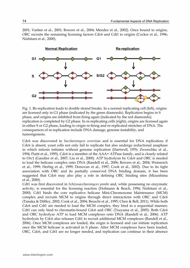
Re-replication
G1
S
G2
Normal Replication
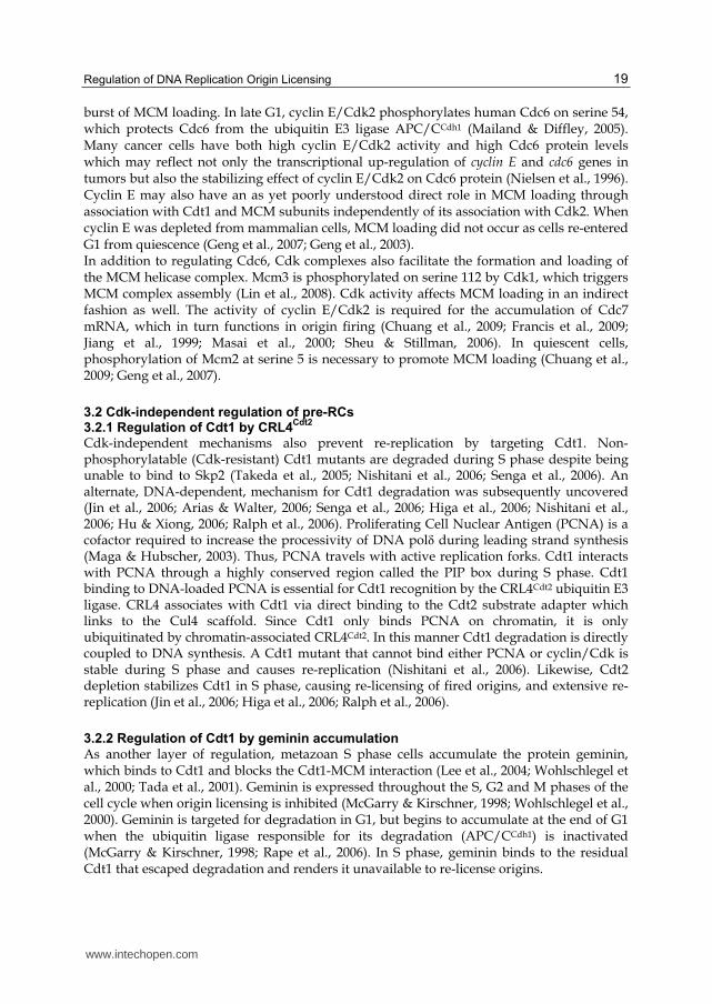
Fig. 1. Re-replication leads to double strand breaks. In a normal replicating cell (left), origins are licensed only in G1 phase (indicated by the green diamonds). Replication begins in S phase, and origins are inhibited from firing again (indicated by the red diamonds); replication is completed by G2 phase. In re-replicating cells (right), origins are licensed again in either S or G2 phase, leading to origin re-firing and re-replicated stretches of DNA. The consequences of re-replication include DNA damage, genome instability, and tumorigenesis.
Cdc6 was discovered in Saccharomyces cerevisiae and is essential for DNA replication; if Cdc6 is absent, yeast cells not only fail to replicate but also undergo reductional anaphase in which mitosis initiates without genome replication (Hartwell, 1976; Zwerschke et al., 1994; Piatti et al., 1995). Cdc6 is a member of the AAA+ ATPase family, and is closely related to Orc1 (Gaudier et al., 2007; Liu et al., 2000). ATP hydrolysis by Cdc6 and ORC is needed to load the helicase complex onto DNA (Randell et al., 2006; Bowers et al., 2004; Weinreich et al., 1999; Herbig et al., 1999; Donovan et al., 1997; Cook et al., 2002). Due to its tight association with ORC and its partially conserved DNA binding domain, it has been suggested that Cdc6 may also play a role in defining ORC binding sites (Mizushima et al., 2000). Cdt1 was first discovered in Schizosaccharomyces pombe and, while possessing no enzymatic activity, is essential for the licensing reaction (Hofmann & Beach, 1994; Nishitani et al., 2000). Cdt1 binds the core replicative helicase Mini-Chromosome Maintenance (MCM) complex and recruits MCM to origins through direct interactions with ORC and Cdc6 (Tanaka & Diffley, 2002; Cook et al., 2004; Bruschi et al., 1995; Chen & Bell, 2011). While both Cdc6 and Cdt1 are needed to load the MCM complex, they bind in a sequential manner; Cdt1 can only bind to chromatin-bound Cdc6 and ORC (Tsuyama et al., 2005). Both Cdc6 and ORC hydrolyze ATP to load MCM complexes onto DNA (Randell et al., 2006). ATP hydrolysis by Cdc6 also releases Cdt1 to recruit additional MCM complexes (Randell et al., 2006). Once MCM complexes are loaded, the origin is licensed and can initiate replication once the MCM helicase is activated in S phase. After MCM complexes have been loaded, ORC, Cdc6, and Cdt1 are no longer needed, and replication can continue in their absence
www.intechopen.com

Regulation of DNA Replication Origin Licensing
15
(Donovan et al., 1997; Rowles et al., 1999; Maiorano et al., 2000). This property of the loaded MCM complex is key to preventing re-replication because, as discussed below, ORC, Cdc6, and Cdt1 are inactivated beginning in S phase. At each origin, at least two MCM hexamer complexes are loaded at a time, with multiple rounds of loading at each origin (Evrin et al., 2009; Remus et al., 2009; Edwards et al., 2002; Lei et al., 1996). The exact mechanism of MCM loading is not currently understood, but electron microscopy images suggest ORC and Cdc6 form a structure similar to known clamp loaders such as RFC (Chen et al., 2008; Speck et al., 2005). While multiple MCM complexes can be loaded at each origin, perhaps as many ten copies per origin, the majority of the MCM complexes that associate with chromatin do not travel with the replication fork suggesting that they are not normally activated (Edwards et al., 2002; Krude et al., 1996; Dimitrova et al., 1999). These additional MCM complexes may be loaded as a backup mechanism to ensure that a sufficient number of origins fire in S phase (Ge & Blow, 2010).
ORC
Cdc6 MCMMCM
Cdt1
MCM
ORC
Cdc6
Cdt1
MCMMCM MCM
ORC
origin
Fig. 2. Pre-RC assembly in G1 phase. Pre-RC assembly begins when the Origin Recognition Complex (ORC) binds to origin DNA. ORC recruits Cdc6, which in turn recruits Cdt1 bound to the Mini-Chromosome Maintenance (MCM) core helicase complex. Through the ATPase activity of ORC and Cdc6, the MCM complex is loaded onto DNA and the origin is licensed for replication.
MCM loading is highly regulated by multiple overlapping mechanisms (summarized in Table 1). Cdc6 and Cdt1 protein levels peak at different stages of the cell cycle; Cdt1 levels peak in G1 phase whereas Cdc6 peaks in S/G2 phase in mammalian cells (Nishitani et al., 2000; Petersen et al., 2000). Additionally, a member of the ORC complex, Orc1, is degraded or inactivated at the onset of S phase (Mendez et al., 2002; Li & DePamphilis, 2002; Li et al., 2004).
www.intechopen.com
Fundamental Aspects of DNA Replication
16
Due in part to these alternating protein levels, there are only two short windows in the cell cycle when pre-RC formation can occur. Pre-RC assembly begins at the end of mitosis, before the Anaphase Promoting Complex/Cyclosome (APC/C) becomes active in G1, and targets Cdc6 for degradation. The second round of pre-RC assembly occurs in late G1 phase when activated Cdk2 stabilizes Cdc6 but before Cdt1 is degraded at the onset of S phase (Diffley, 2004). Furthermore, the MCM subunits undergo post-translational modifications that facilitate MCM complex formation as well as their ability to be loaded onto DNA (Lin et al., 2008; Chuang et al., 2009). These mechanisms will be discussed in depth in the subsequent sections with specific emphasis on the regulation of metazoan pre-RC assembly.
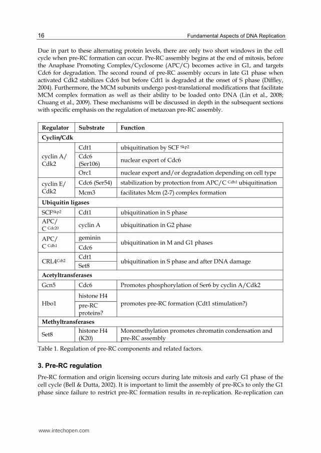
Regulator Substrate Function
Cyclin/Cdk
cyclin A/ Cdk2
Cdt1 ubiquitination by SCF Skp2
Cdc6 (Ser106)
nuclear export of Cdc6
Orc1 nuclear export and/or degradation depending on cell type
cyclin E/ Cdk2
Cdc6 (Ser54) stabilization by protection from APC/C Cdh1 ubiquitination
Mcm3 facilitates Mcm (2-7) complex formation
Ubiquitin ligases
SCFSkp2 Cdt1 ubiquitination in S phase
APC/ C Cdc20
cyclin A ubiquitination in G2 phase
APC/ C Cdh1
geminin ubiquitination in M and G1 phases
Cdc6
CRL4Cdt2 Cdt1
ubiquitination in S phase and after DNA damage Set8
Acetyltransferases
Gcn5 Cdc6 Promotes phosphorylation of Ser6 by cyclin A/Cdk2
Hbo1
histone H4
promotes pre-RC formation (Cdt1 stimulation?) pre-RC proteins?
Methyltransferases
Set8 histone H4 (K20)
Monomethylation promotes chromatin condensation and pre-RC assembly
Table 1. Regulation of pre-RC components and related factors.
3. Pre-RC regulation
Pre-RC formation and origin licensing occurs during late mitosis and early G1 phase of the
cell cycle (Bell & Dutta, 2002). It is important to limit the assembly of pre-RCs to only the G1
phase since failure to restrict pre-RC formation results in re-replication. Re-replication can
www.intechopen.com
Regulation of DNA Replication Origin Licensing
17
cause increases in double strand breaks and ploidy. Over time, damaged DNA could lead to
genome instability, ultimately increasing the propensity for tumorigenesis. In this section
we will discuss several mechanisms by which metazoan cells avoid re-replication.
3.1 Regulation of pre-RCs by cyclin-dependent kinases
Cyclin-dependent kinases (Cdks) are a family of serine-threonine protein kinases essential
for timely and appropriate progression through different stages of the cell cycle. Cdks are
activated by association with cyclins, whose expression and stability are cell cycle-regulated.
In budding and fission yeast, a single Cdk controls the G1/S and G2/M transitions, while in
metazoans different Cdks are active in different phases of the cell cycle (Figure 3, reviewed
in Malumbres & Barbacid, 2009). In metazoans, passage through G1 phase is governed by
cyclin D/Cdk4 (or cyclin D/Cdk6) and cyclin E/Cdk2 (Braden et al., 2008; van den Heuvel
& Harlow, 1993). S phase, and therefore DNA replication, is regulated by cyclin A/Cdk2
(Wheeler et al., 2008). Finally, mitotic entry is triggered by Cdk1 first binding to cyclin A
and then cyclin B.
DNA replication is both positively and negatively governed by Cdk activity. High Cdk2 and
Cdk1 activities, which are found from early S phase through mid-mitosis, block pre-RC
assembly; thus pre-RC assembly begins as cells exit from mitosis when these kinases are not
active. Cyclin E protein peaks in early S phase, triggering Cdk2 activity and replication
initiation from licensed origins; replication begins also with the help of a dedicated
replication kinase, Cdc7/Dbf4 (Bochman & Schwacha, 2009; Remus et al., 2009). (Additional
chapters related to origin firing and S phase progression appear elsewhere in this book.)
Simultaneously, S phase Cdk activity inhibits pre-RC formation at origins that have already
fired. Premature expression of cyclins E or A during G1 phase blocks normal pre-RC
assembly (Wheeler et al., 2008; Ekholm-Reed et al., 2004). Thus S phase Cdks promote
replication initiation but block pre-RC assembly after G1 (described below), resulting in one
genome duplication per cell cycle.
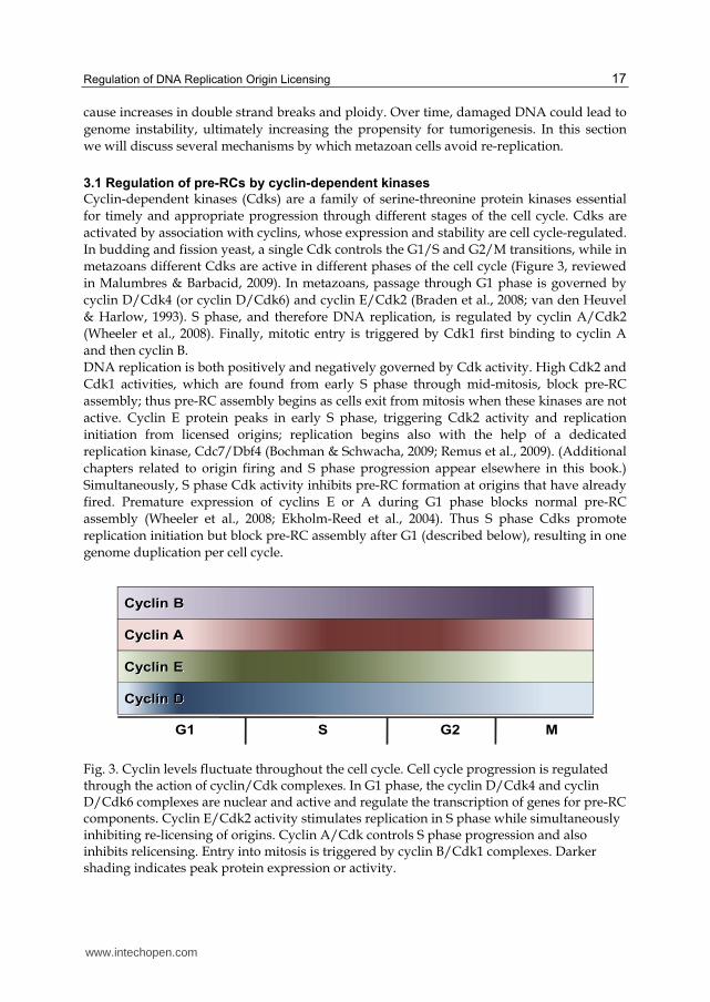
Cyclin D
Cyclin E
Cyclin A
G1 S G2 M
Cyclin B
Fig. 3. Cyclin levels fluctuate throughout the cell cycle. Cell cycle progression is regulated through the action of cyclin/Cdk complexes. In G1 phase, the cyclin D/Cdk4 and cyclin D/Cdk6 complexes are nuclear and active and regulate the transcription of genes for pre-RC components. Cyclin E/Cdk2 activity stimulates replication in S phase while simultaneously inhibiting re-licensing of origins. Cyclin A/Cdk controls S phase progression and also inhibits relicensing. Entry into mitosis is triggered by cyclin B/Cdk1 complexes. Darker shading indicates peak protein expression or activity.
www.intechopen.com
Fundamental Aspects of DNA Replication
18
3.1.1 Cyclin-Cdks as negative regulators of pre-RC assembly
Several experimental findings support a role for Cdks in preventing re-replication. For example, transient over-expression of the Cdk inhibitor, p21, in G2 caused re-replication (Bates et al., 1998). In addition, ORC and MCM complexes were recruited to human chromatin in G2 when Cdk activity was inhibited genetically or pharmacologically (Coverley et al., 1996; Fujita et al., 1998; Ballabeni et al., 2004; Li et al., 2004; Sugimoto et al., 2004). Finally, chemical inactivation of Cdk1, combined with genetic ablation of Cdk2, allowed pre-RCs to assemble inappropriately during mitosis (Ballabeni et al., 2004). These observations highlight a role for Cdks in preventing inappropriate origin licensing. Re-licensing of origins during S phase is prevented, in part, by Cdk2, in association with cyclin A (Wheeler et al., 2008). Cdt1 interacts with the S phase cyclin A/Cdk2 complex, which results in Cdt1 phosphorylation at threonine 29 (Li et al., 2004; Liu et al., 2004). Phosphorylated Cdt1 binds to the F-box protein, Skp2, the substrate receptor for the ubiquitin ligase SCFSkp2. Cdt1 is polyubiquitinated by SCFSkp2 and targeted for degradation by the 26S proteasome, thus reducing the pool of Cdt1 protein available to participate in origin licensing (Takeda et al., 2005; Sugimoto et al., 2004; Kim & Kipreos, 2007). Coincident with Cdt1 destruction in S phase, Cdc6 is acetylated by Gcn5 on lysines 92, 105, and 109 which promotes cyclin A/Cdk2 phosphorylation on Cdc6 at serine 106 (Paolinelli et al., 2009; Mailand & Diffley, 2005). Serine 106 phosphorylation results in exclusion of Cdc6 protein from the nucleus, preventing re-replication (Paolinelli et al., 2009; Saha et al., 1998; Coverley et al., 2000; Kim et al., 2007; Fujita et al., 1999; Jiang et al., 1999; Petersen et al., 1999). The small amount of Cdc6 that remains nuclear throughout S phase is chromatin-bound and likely participates in the ATR-dependent intra-S phase checkpoint by mechanisms that are not yet understood (Mendez & Stillman, 2000; Lau et al., 2006). Additionally, the Orc1 subunit of ORC is phosphorylated by cyclin A/Cdk1 during S phase, and this phosphorylation promotes Orc1 degradation in HeLa cells (Mendez et al., 2002). The same phosphorylation on Orc1 in Chinese Hamster Ovary cells (CHO) does not affect Orc1 stability, but lowers the affinity of Orc1 for chromatin (Li et al., 2004). In both HeLa and CHO cells, Orc1 phosphorylation allows the export of Orc1 to the cytoplasm (Saha et al., 2006). Over-expression of cyclin A from Kaposi’s Sarcoma-associated herpes virus also facilitates re-localization of Orc1 to the cytoplasm. These results show that Orc1 is subject to phosphorylation by cyclin A/Cdk1, and this event modulates the stability and/or localization of Orc1, thereby contributing to the prevention of re-replication. Recent evidence from S. cerevisiae suggests that Orc2 and Orc6 may also be targets of cyclin/Cdk inhibition. Phosphorylation of these subunits leads to a marked decrease in MCM loading (Green et al., 2006; Tanny et al., 2006; Nguyen et al., 2001). Interaction between Orc6 and the S phase Cdk, Clb5, is needed to prevent MCM loading outside of G1 phase; this interaction occludes the Cdt1 binding site on the ORC complex (Wilmes et al., 2004; Tanny et al., 2006; Chen & Bell, 2011). In addition to steric hindrance, Clb5 phosphorylates Orc6; this modification also partially blocks the Cdt1 binding site and prevents MCM loading (Chen & Bell, 2011). It remains to be determined if similar mechanisms also apply to Cdk regulation of mammalian ORC (DePamphilis, 2005).
3.1.2 Cyclin-Cdks as positive regulators of pre-RC assembly
Cdc6 protein levels are very low in both quiescent cells and in early/mid G1 phase cells due to ubiquitin-mediated proteolysis, but Cdc6 protein accumulates in late G1 just prior to a
www.intechopen.com
Regulation of DNA Replication Origin Licensing
19
burst of MCM loading. In late G1, cyclin E/Cdk2 phosphorylates human Cdc6 on serine 54, which protects Cdc6 from the ubiquitin E3 ligase APC/CCdh1 (Mailand & Diffley, 2005). Many cancer cells have both high cyclin E/Cdk2 activity and high Cdc6 protein levels which may reflect not only the transcriptional up-regulation of cyclin E and cdc6 genes in tumors but also the stabilizing effect of cyclin E/Cdk2 on Cdc6 protein (Nielsen et al., 1996). Cyclin E may also have an as yet poorly understood direct role in MCM loading through association with Cdt1 and MCM subunits independently of its association with Cdk2. When cyclin E was depleted from mammalian cells, MCM loading did not occur as cells re-entered G1 from quiescence (Geng et al., 2007; Geng et al., 2003). In addition to regulating Cdc6, Cdk complexes also facilitate the formation and loading of the MCM helicase complex. Mcm3 is phosphorylated on serine 112 by Cdk1, which triggers MCM complex assembly (Lin et al., 2008). Cdk activity affects MCM loading in an indirect fashion as well. The activity of cyclin E/Cdk2 is required for the accumulation of Cdc7 mRNA, which in turn functions in origin firing (Chuang et al., 2009; Francis et al., 2009; Jiang et al., 1999; Masai et al., 2000; Sheu & Stillman, 2006). In quiescent cells, phosphorylation of Mcm2 at serine 5 is necessary to promote MCM loading (Chuang et al., 2009; Geng et al., 2007).
3.2 Cdk-independent regulation of pre-RCs 3.2.1 Regulation of Cdt1 by CRL4
Cdt2
Cdk-independent mechanisms also prevent re-replication by targeting Cdt1. Non-phosphorylatable (Cdk-resistant) Cdt1 mutants are degraded during S phase despite being unable to bind to Skp2 (Takeda et al., 2005; Nishitani et al., 2006; Senga et al., 2006). An alternate, DNA-dependent, mechanism for Cdt1 degradation was subsequently uncovered (Jin et al., 2006; Arias & Walter, 2006; Senga et al., 2006; Higa et al., 2006; Nishitani et al., 2006; Hu & Xiong, 2006; Ralph et al., 2006). Proliferating Cell Nuclear Antigen (PCNA) is a cofactor required to increase the processivity of DNA polδ during leading strand synthesis (Maga & Hubscher, 2003). Thus, PCNA travels with active replication forks. Cdt1 interacts with PCNA through a highly conserved region called the PIP box during S phase. Cdt1 binding to DNA-loaded PCNA is essential for Cdt1 recognition by the CRL4Cdt2 ubiquitin E3 ligase. CRL4 associates with Cdt1 via direct binding to the Cdt2 substrate adapter which links to the Cul4 scaffold. Since Cdt1 only binds PCNA on chromatin, it is only ubiquitinated by chromatin-associated CRL4Cdt2. In this manner Cdt1 degradation is directly coupled to DNA synthesis. A Cdt1 mutant that cannot bind either PCNA or cyclin/Cdk is stable during S phase and causes re-replication (Nishitani et al., 2006). Likewise, Cdt2 depletion stabilizes Cdt1 in S phase, causing re-licensing of fired origins, and extensive re-replication (Jin et al., 2006; Higa et al., 2006; Ralph et al., 2006).
3.2.2 Regulation of Cdt1 by geminin accumulation
As another layer of regulation, metazoan S phase cells accumulate the protein geminin, which binds to Cdt1 and blocks the Cdt1-MCM interaction (Lee et al., 2004; Wohlschlegel et al., 2000; Tada et al., 2001). Geminin is expressed throughout the S, G2 and M phases of the cell cycle when origin licensing is inhibited (McGarry & Kirschner, 1998; Wohlschlegel et al., 2000). Geminin is targeted for degradation in G1, but begins to accumulate at the end of G1 when the ubiquitin ligase responsible for its degradation (APC/CCdh1) is inactivated (McGarry & Kirschner, 1998; Rape et al., 2006). In S phase, geminin binds to the residual Cdt1 that escaped degradation and renders it unavailable to re-license origins.
www.intechopen.com

Fundamental Aspects of DNA Replication
20
Recent biochemical evidence has suggested that geminin-Cdt1 complexes exist in several forms (De Marco et al., 2009). These forms include a licensing-inhibitory heterohexamer that consists of two Cdt1 molecules and four geminin molecules, and a licensing-permissive heterotrimer, comprised of one Cdt1 molecule and two geminin molecules (Lutzmann et al., 2006). Binding of geminin to Cdt1 in a heterohexamer can tether several Cdt1 molecules together, creating chromatin-bound foci that may cooperatively inhibit licensing (Ode et al., 2011). Depending on the amount of geminin in the cell, geminin may switch from being an inhibitor of origin licensing when geminin levels are high to an activator when levels of geminin are low.
3.2.3 APC/C as a regulator of pre-RC assembly
The cell spends a significant amount of energy to ensure that the correct proteins are expressed at the appropriate time. Before one cell cycle phase begins, cells ensure that the previous step has been properly completed and, in many cases, inactivated by controlling protein activity abundance. One mechanism for enforcing the proper order of events is through regulated protein degradation. The Anaphase Promoting Complex/Cyclosome (APC/C) is uniquely tied to cell cycle progression and control of DNA replication as evidenced by the fact its regulation and activity are modulated in every phase of the cell cycle. APC/C is a RING-type E3 ubiquitin ligase originally discovered though its association with its substrates, the mitotic cyclins (Sudakin et al., 1995; King et al., 1995). Two activator subunits, Cdc20 and Cdh1, interact dynamically with the APC/C holoenzyme to influence substrate recognition (Figure 4). APC/CCdh1 targets in G1 include Skp2, a member of the SCF ubiquitin ligase complex, the licensing factor Cdc6, and the inhibitor protein geminin (Wei et al., 2004; Petersen et al., 2000; McGarry & Kirschner, 1998). Degradation of Skp2 results in accumulation of the Cdk2 inhibitors p21 and p27, and prevents premature S phase entry due to low Cdk2 activity (Wei et al., 2004). APC/CCdh1 also acts to limit the amount of Cdc6 that is available in the cell (discussed in 3.1.2). As cells progress through S phase, Cdh1 is phosphorylated by cyclin A/Cdk2 complexes; since hyper-phosphorylated Cdh1 cannot interact with APC/C, the ubiquitin ligase complex is inactive (Zachariae et al., 1998). This inactivation allows geminin to accumulate and bind any remaining Cdt1 (McGarry & Kirschner, 1998). During S phase, APC/CCdh1 is also bound by its inhibitor protein, Emi1 (Hsu et al., 2002). Interestingly, Emi1 accumulation is not needed to begin S phase but is needed to signal the stop of replication and mitotic entry, even though APC/CCdc20 can still ubiquitinate its targets if Emi1 is present in mitosis (Di Fiore & Pines, 2007). Emi1 remains bound until prophase, when it is phosphorylated by Plk1 (Moshe et al., 2004). While Emi1 accumulation is not needed for S phase entry, it is essential to inhibit re-replication. Depletion of Emi1 leads to re-replication in human cells, due to the untimely activation of APC/CCdh1 (Sivaprasad et al., 2007; Machida & Dutta, 2007). This stabilization allows geminin levels to drop when Cdt1 levels are high; at the same time, increased activity of cyclin A/Cdk2 allows Cdc6 to become stabilized. With both licensing factors present, origins are licensed outside of G1 and re-replication occurs.
4. Re-replication causes DNA damage and cell cycle arrest
Manipulation of key components of the replication machinery can induce re-replication. For instance, over-expression of replication proteins Cdt1 and Cdc6 causes re-replication in human cancer cells (Nishitani & Nurse, 1995; Vaziri et al., 2003; Ekholm-Reed et al., 2004;
www.intechopen.com
Regulation of DNA Replication Origin Licensing
21
Ub
Ub
Ub
G2 S
G1M
APC/C
Cdh1Cdc6
Cdc20APC/C
APC/C
Emi1
Cyclin B UbSecurinUb
Geminin
Cdh1P
P
Cdh1
P
P
P
P P P
P
Cdc20
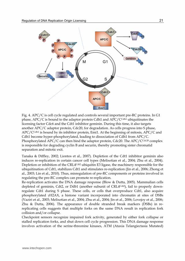
Fig. 4. APC/C is cell cycle regulated and controls several important pre-RC proteins. In G1 phase, APC/C is bound to the adaptor protein Cdh1 and APC/CCdh1 ubiquitinates the licensing factor Cdc6 and the Cdt1 inhibitor geminin. During this time, it also targets another APC/C adaptor protein, Cdc20, for degradation. As cells progress into S phase, APC/CCdh1 is bound by its inhibitor protein, Emi1. At the beginning of mitosis, APC/C and Cdh1 become hyper-phosphorylated, leading to dissociation of Cdh1 from APC/C. Phosphorylated APC/C can then bind the adaptor protein, Cdc20. The APC/CCdc20 complex is responsible for degrading cyclin B and securin, thereby promoting sister chromatid separation and mitotic exit.
Tanaka & Diffley, 2002; Liontos et al., 2007). Depletion of the Cdt1 inhibitor geminin also induces re-replication in certain cancer cell types (Melixetian et al., 2004; Zhu et al., 2004). Depletion or inhibition of the CRL4Cdt2 ubiquitin E3 ligase, the machinery responsible for the ubiquitination of Cdt1, stabilizes Cdt1 and stimulates re-replication (Jin et al., 2006; Zhong et al., 2003; Lin et al., 2010). Thus, misregulation of pre-RC components or proteins involved in regulating the pre-RC complex can promote re-replication. Re-replication activates the DNA damage response (Blow & Dutta, 2005). Mammalian cells
depleted of geminin, Cdt2, or Ddb1 (another subunit of CRL4Cdt2), fail to properly down-
regulate Cdt1 during S phase. These cells, or cells that overproduce Cdt1, also acquire
phosphorylated γH2AX, a histone variant incorporated into chromatin at sites of DSBs
(Vaziri et al., 2003; Melixetian et al., 2004; Zhu et al., 2004; Jin et al., 2006; Lovejoy et al., 2006;
Zhu & Dutta, 2006). The appearance of double stranded break markers (DSBs) in re-
replicating cells suggests that multiple forks on the same DNA result in replication fork
collision and/or collapse.
Checkpoint sensors recognize impaired fork activity, generated by either fork collapse or
stalled replication forks, and shut down cell cycle progression. This DNA damage response
involves activation of the serine-threonine kinases, ATM (Ataxia Telangiectasia Mutated)
www.intechopen.com
Fundamental Aspects of DNA Replication
22
and ATR (ATM-related) (Shiloh, 2003). Single-stranded regions are recognized by ATR,
which preferentially activates and phosphorylates the Chk1 protein kinase (Guo et al., 2000;
Zhao & Piwnica-Worms, 2001; Kramer et al., 2004; Niida et al., 2007; Zou & Elledge, 2003).
Chk1 in turn inactivates the Cdc25 phosphatase causing an intra-S or G2/S phase arrest
(Donzelli & Draetta, 2003; Jin et al., 2003; Ferguson et al., 2005). Inactivation of Cdc25 is also
accomplished by induction of MAPKAP-K2 (MK2), a downstream effector kinase of the p38
MAP kinase pathway, which is stimulated by genotoxic stress (Manke et al., 2005; Reinhardt
et al., 2007; Lemaire et al., 2006; Huard et al., 2008). Without active Cdc25, the mitotic cyclin
B/Cdk1 complex remains phosphorylated and inactive. Thus, the DNA damage response
pathway leads to a G2/M cell cycle arrest (Tang et al., 2006).
Damage caused by DSBs is recognized by ATM (Shechter et al., 2004; Costanzo et al., 2001).
ATM activates and phosphorylates the checkpoint protein kinase Chk2 (Melchionna et al.,
2000; Zhou & Elledge, 2000). Activated Chk2 then phosphorylates and activates the
transcriptional regulator, p53 (Meek, 1994; Milczarek et al., 1997). Active p53 induces
expression of many genes needed for DNA repair and cell cycle arrest, including the Cdk
inhibitor, p21 (Smith et al., 1995). The accumulation of p21 inhibits Cdk2 function and blocks
cells from entering S phase (Taylor & Stark, 2001; Levine, 1997; Vogelstein et al., 2000). DNA
damage in G2 phase also leads to p53-dependent p21 accumulation, inhibition of Cdk1, and
arrest in G2 (Agarwal et al., 1995; Deng et al., 1995; Brugarolas et al., 1995).
Cells can recover from arrest by activating repair pathways and continuing cell division, but
if the damage is too extensive, they die by apoptosis (Hartwell & Weinert, 1989; Weinert &
Hartwell, 1990; Nasmyth, 1996; Abraham, 2001). Re-replication induced by Cdt1
stabilization or geminin depletion results in many of the checkpoint events outlined above,
including phosphorylation of Chk2, Chk1, and p53, failure to enter mitosis or initiation of
apoptosis depending on the degree of re-replication-associated damage (Zhu & Dutta, 2006;
Zhu et al., 2004; McGarry, 2002). G2/M arrest caused by loss of geminin is primarily
mediated by ATR recognition of single-stranded regions generated early in re-replication.
As cells eventually accumulate more damage from more extensive re-replication and
generate DSBs, the ATM pathway is induced (Lin & Dutta, 2007). Such sequential activation
may permit low-level damage to be repaired during an ATR-mediated G2/M arrest prior to
ATM-mediated p53 activation and permanent arrest or cell death.
Interestingly, ATR-deficient cells show a higher propensity to re-replicate than their ATR-
proficient counterparts do (Lin & Dutta, 2007). These observations suggest that the ATR
pathway can restrict re-replication while re-replication simultaneously activates ATR.
Another potential interplay between replication licensing and the ATR pathway is
suggested by the binding of Cdc6 to ATR, which promotes ATR chromatin binding
(Yoshida et al., 2010). Cdc6 can also activate p21-bound Cdk2 and override the DNA
damage checkpoint (Kan et al., 2008).
5. Pre-RC regulation during a DNA damage response
As described above, cyclin/Cdk activity prevents inappropriate formation of pre-RC
complexes in S and G2 phases of the cell cycle. Exposure to DNA-damaging agents or
generation of DSBs during replication elicits the DNA-damage response, which can reduce
Cdk activity, particularly in G2 phase. Therefore, DNA damage can perturb the normal
Cdk-dependent regulation of pre-RC components such that there is potential danger of
www.intechopen.com
Regulation of DNA Replication Origin Licensing
23
initiating re-replication events. To counteract this risk, the DNA damage induces Cdk-
independent mechanisms to inhibit pre-RC assembly.
5.1 Degradation of Cdc6
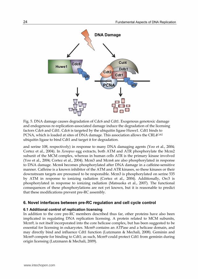
When cells are exposed to various genotoxins, such as ultraviolet (UV) irradiation, ionizing radiation (IR), base alkylating agents, etc., Cdc6 is actively degraded by two mechanisms. As outlined above, the DNA damage response results in p53-dependent induction of the Cdk inhibitor p21Cip1. Reduced Cdk activity prevents phosphorylation of Cdc6 at serine 54 (Duursma & Agami, 2005). Without this protective phosphorylation, Cdc6 can be ubiquitinated by APC/CCdh1 and degraded, particularly in G1 phase due to the high activity of APC/CCdh1 (Mailand & Diffley, 2005). Cdc6 ubiquitination by APC/CCdh1 is further facilitated by the activation of APC/CCdh1 by the DNA damage checkpoint and cyclin destruction (which also reduces Cdc6 serine 54 phosphorylation) (Sudo et al., 2001). Cdc6 is also targeted for degradation by a different E3 ubiquitin ligase, Huwe1 (Figure 5). Recognition of Cdc6 by Huwe1 is independent of both p53 induction of p21 and changes in Cdk-mediated phosphorylation (Hall et al., 2007). Huwe1 is a monomeric HECT family E3 ligase that has been linked to the regulation of many proteins central to cell proliferation control, including maintaining low p53 levels in the absence of DNA damage (Chen et al., 2005). Huwe1-mediated Cdc6 degradation is also activated in response to endogenous re-replication-associated DNA damage ultimately leading to loss of Cdc6 from re-replicating cells (Hall et al., 2008).
5.2 Degradation of Cdt1
When cells are exposed to exogenous DNA damaging agents, Cdt1 is rapidly degraded (Higa et al., 2003; Hu et al., 2004). DNA damage-induced Cdt1 degradation is unaffected by Cdk activity, and is therefore unrelated to ubiquitination by the SCFSkp2 ubiquitin ligase. Instead, Cdt1 is degraded through association with the CRL4Cdt2 ubiquitin ligase (Higa et al., 2003; Hu & Xiong, 2006; Jin et al., 2006; Nishitani et al., 2006; Ralph et al., 2006; Sansam et al., 2006; Senga et al., 2006). PCNA is loaded onto damaged DNA not only during normal replication, but also in response to DNA damage both for DNA repair synthesis and in a checkpoint role (Lee & Myung, 2008). As a result, Cdt1 interacts with loaded PCNA after DNA damage in the same way that it interacts with PCNA during S phase. The interaction of Cdt1 with PCNA at sites of DNA damage recruits CRL4Cdt2 via direct binding of Cdt2 to the Cdt1-PCNA complex, leading to Cdt1 ubiquitination and proteasomal degradation. Like Cdc6, Cdt1 is targeted for destruction in response to both exogenous and endogenous DNA damage. Human or Drosophila cells depleted of geminin also rapidly lose Cdt1 and Cdc6 (Mihaylov et al., 2002; Ballabeni et al., 2004). Since the lack of geminin induces re-replication-related DNA damage, Cdt1 and Cdc6 are simultaneously targeted for degradation by CRL4Cdt2 and Huwe1, respectively (Hall et al., 2008). In cells depleted of either CRL4Cdt2 components or Huwe1, geminin depletion allowed more extensive re-replication, suggesting that the destruction of pre-RC proteins in re-replicating cells protects them from even further re-replication.
5.3 Phosphorylation of pre-RC components by DNA damage-stimulated kinases
Cdt1 and Cdc6 destruction after DNA damage is largely independent of the DNA damage checkpoint, but other pre-RC members are specifically targeted by the ATM and ATR pathways. Mcm2 is phosphorylated in both Xenopus and human cancer cells (on serine 92
www.intechopen.com
Fundamental Aspects of DNA Replication
24
Cdc6Huwe1
Ddb1
Cul4
DNA Damage
Cdt2
PCNA
Ub Cdt1
Ub
Fig. 5. DNA damage causes degradation of Cdc6 and Cdt1. Exogenous genotoxic damage and endogenous re-replication-associated damage induce the degradation of the licensing factors Cdc6 and Cdt1. Cdc6 is targeted by the ubiquitin ligase Huwe1. Cdt1 binds to PCNA, which is loaded at sites of DNA damage. This association allows the CRL4Cdt2 ubiquitin ligase to bind Cdt1 and target it for degradation.
and serine 108, respectively) in response to many DNA damaging agents (Yoo et al., 2004; Cortez et al., 2004). In Xenopus egg extracts, both ATM and ATR phosphorylate the Mcm2 subunit of the MCM complex, whereas in human cells ATR is the primary kinase involved (Yoo et al., 2004; Cortez et al., 2004). Mcm3 and Mcm4 are also phosphorylated in response to DNA damage. Mcm4 becomes phosphorylated after DNA damage in a caffeine-sensitive manner. Caffeine is a known inhibitor of the ATM and ATR kinases, so these kinases or their downstream targets are presumed to be responsible. Mcm3 is phosphorylated on serine 535 by ATM in response to ionizing radiation (Cortez et al., 2004). Additionally, Orc3 is phosphorylated in response to ionizing radiation (Matsuoka et al., 2007). The functional consequences of these phosphorylations are not yet known, but it is reasonable to predict that these modifications prevent pre-RC assembly.
6. Novel interfaces between pre-RC regulation and cell cycle control
6.1 Additional control of replication licensing
In addition to the core pre-RC members described thus far, other proteins have also been implicated in regulating DNA replication licensing. A protein related to MCM subunits, Mcm9, is not itself incorporated into the core helicase complex, but has been suggested to be essential for licensing in eukaryotes. Mcm9 contains an ATPase and a helicase domain, and may directly bind and influence Cdt1 function (Lutzmann & Mechali, 2008). Geminin and Mcm9 compete for binding to Cdt1; as such, Mcm9 could protect Cdt1 from geminin during origin licensing (Lutzmann & Mechali, 2009).
www.intechopen.com
Regulation of DNA Replication Origin Licensing
25
Another protein that plays a role in influencing DNA replication is the acetyltransferase, Hbo1. Hbo1 binds both Orc1 and Mcm2, and is thereby recruited to origins (Miotto & Struhl, 2008; Iizuka et al., 2006; Burke et al., 2001). Hbo1 depletion causes licensing defects in human cells and in Xenopus egg extracts by preventing the loading of MCM complexes but not the chromatin association of ORC, Cdc6, or Cdt1 (Iizuka et al., 2006; Miotto & Struhl, 2008). These defects can be reversed by over-expressing Cdt1, suggesting that Hbo1 is an activator of MCM loading at the Cdt1 step in pre-RC assembly. Hbo1 acetylates histone H4 which may reflect a role for Hbo1 in modulating chromatin at origins (Miotto & Struhl, 2010). Hbo1 also associates with the tumor suppressor, p53, and this association negatively regulates Hbo1 enzymatic activity. Cellular stress agents other than DNA damage, such as hyperosmotic stress, activate p53 (Vogelstein et al., 2000), and this activation was suggested to inhibit the HAT activity of Hbo1 and consequently, origin licensing (Iizuka et al., 2008). Hbo1 can also acetylate multiple pre-RC components in vitro however, so its role in pre-RC assembly may also be through direct modification of licensing proteins (Iizuka et al., 2006). In addition to histone acetylation, monomethylation of histone H4 facilitates pre-RC formation. Tethering the H4 Lys20 methyltransferase, Set8 (PR-Set7), to an ectopic sequence induced pre-RC formation at that site, suggesting that H4K20 methylation may also promote pre-RC assembly at normal chromosomal origins (Tardat et al., 2010). Set8 is ubiquitinated by the CRL4Cdt2 ubiquitin ligase in PCNA-dependent manner during S phase and in response to DNA damage (Jorgensen et al., 2011; Wu & Rice, 2010; Oda et al., 2010). Failure to degrade Set8 in S phase leads to re-replication and lack of chromatin condensation during mitosis (Wu & Rice, 2010; Jorgensen et al., 2011). These data further suggest an involvement of chromatin structure in the regulation of origin licensing. Each of the genes encoding pre-RC components is transcriptionally regulated by the Rb-E2F pathway. Given that tumor cells frequently exhibit high-level expression of E2F target genes, (Rb or p16 loss, cyclin overproduction, etc.) it is not surprising that Cdt1 and Cdc6 are overproduced in many cancers (Ohta et al., 2001; Karakaidos et al., 2004; Pinyol et al., 2006; Di Micco et al., 2006). Overproduction of Cdt1 or Cdc6 in cultured human cells induces re-replication, raising the possibility that tumor cells also re-replicate in vivo. Recently it has been suggested that cancer cells “hyper-replicate” and that this form of replication stress is a driving force in oncogenesis. It has also been suggested that excessive pre-RC assembly may even downregulate expression of the INK4/ARF tumor suppressor locus due to interference between a nearby origin and the INK4 promoter (Gonzalez et al., 2006). Recently, mutations in genes for several components of the pre-RC, including Orc1, Orc4, Orc6, Cdt1, and Cdc6 have been linked to the autosomal recessive primordial dwarfism syndrome, Meier Gorlin syndrome (Guernsey et al., 2011; Bicknell et al., 2011). This is the first report implicating impaired licensing in a developmental disorder. Taken together there are now clear links between pre-RC formation, normal human development, and tumorigenesis.
7. References
Abraham, R.T. (2001). Cell cycle checkpoint signaling through the ATM and ATR kinases. Genes Dev, Vol. 15, No. 17, 2177-2196.
Agarwal, M.L., Agarwal, A., Taylor, W.R. and Stark, G.R. (1995). p53 controls both the G2/M and the G1 cell cycle checkpoints and mediates reversible growth arrest in human fibroblasts. Proc Natl Acad Sci U S A, Vol. 92, No. 18, 8493-8497.
www.intechopen.com
Fundamental Aspects of DNA Replication
26
Arentson, E., Faloon, P., Seo, J., et al. (2002). Oncogenic potential of the DNA replication licensing protein CDT1. Oncogene, Vol. 21, No. 8, 1150-1158.
Arias, E.E. and Walter, J.C. (2006). PCNA functions as a molecular platform to trigger Cdt1 destruction and prevent re-replication. Nat Cell Biol, Vol. 8, No. 1, 84-90.
Ballabeni, A., Melixetian, M., Zamponi, R., Masiero, L., Marinoni, F. and Helin, K. (2004). Human geminin promotes pre-RC formation and DNA replication by stabilizing CDT1 in mitosis. Embo J, Vol. 23, No. 15, 3122-3132.
Bates, S., Ryan, K.M., Phillips, A.C. and Vousden, K.H. (1998). Cell cycle arrest and DNA endoreduplication following p21Waf1/Cip1 expression. Oncogene, Vol. 17, No. 13, 1691-1703.
Bell, S.P. and Dutta, A. (2002). DNA replication in eukaryotic cells. Annu Rev Biochem, Vol. 71, No. 333-374.
Bicknell, L.S., Bongers, E.M., Leitch, A., et al. (2011). Mutations in the pre-replication complex cause Meier-Gorlin syndrome. Nat Genet, Vol. 43, No. 4, 356-359.
Blow, J.J. and Dutta, A. (2005). Preventing re-replication of chromosomal DNA. Nat Rev Mol Cell Biol, Vol. 6, No. 6, 476-486.
Bochman, M.L. and Schwacha, A. (2009). The Mcm complex: unwinding the mechanism of a replicative helicase. Microbiol Mol Biol Rev, Vol. 73, No. 4, 652-683.
Bowers, J.L., Randell, J.C., Chen, S. and Bell, S.P. (2004). ATP hydrolysis by ORC catalyzes reiterative Mcm2-7 assembly at a defined origin of replication. Mol Cell, Vol. 16, No. 6, 967-978.
Braden, W.A., Mcclendon, A.K. and Knudsen, E.S. (2008). Cyclin-dependent kinase 4/6 activity is a critical determinant of pre-replication complex assembly. Oncogene, Vol. 27, No. 56, 7083-7093.
Brugarolas, J., Chandrasekaran, C., Gordon, J.I., Beach, D., Jacks, T. and Hannon, G.J. (1995). Radiation-induced cell cycle arrest compromised by p21 deficiency. Nature, Vol. 377, No. 6549, 552-557.
Bruschi, C.V., Mcmillan, J.N., Coglievina, M. and Esposito, M.S. (1995). The genomic instability of yeast cdc6-1/cdc6-1 mutants involves chromosome structure and recombination. Mol Gen Genet, Vol. 249, No. 1, 8-18.
Burke, T.W., Cook, J.G., Asano, M. and Nevins, J.R. (2001). Replication factors MCM2 and ORC1 interact with the histone acetyltransferase HBO1. J Biol Chem, Vol. 276, No. 18, 15397-15408.
Chen, D., Kon, N., Li, M., Zhang, W., Qin, J. and Gu, W. (2005). ARF-BP1/Mule is a critical mediator of the ARF tumor suppressor. Cell, Vol. 121, No. 7, 1071-1083.
Chen, S. and Bell, S.P. (2011). CDK prevents Mcm2-7 helicase loading by inhibiting Cdt1 interaction with Orc6. Genes Dev, Vol. 25, No. 4, 363-372.
Chen, Z., Speck, C., Wendel, P., Tang, C., Stillman, B. and Li, H. (2008). The architecture of the DNA replication origin recognition complex in Saccharomyces cerevisiae. Proc Natl Acad Sci USA, Vol. 105, No. 30, 10326-10331.
Chuang, L.C., Teixeira, L.K., Wohlschlegel, J.A., et al. (2009). Phosphorylation of Mcm2 by Cdc7 promotes pre-replication complex assembly during cell-cycle re-entry. Mol Cell, Vol. 35, No. 2, 206-216.
Cocker, J.H., Piatti, S., Santocanale, C., Nasmyth, K. and Diffley, J.F. (1996). An essential role for the Cdc6 protein in forming the pre-replicative complexes of budding yeast. Nature, Vol. 379, No. 6561, 180-182.
www.intechopen.com
Regulation of DNA Replication Origin Licensing
27
Cook, J.G., Chasse, D.A. and Nevins, J.R. (2004). The regulated association of Cdt1 with minichromosome maintenance proteins and Cdc6 in mammalian cells. J Biol Chem, Vol. 279, No. 10, 9625-9633.
Cook, J.G., Park, C.H., Burke, T.W., et al. (2002). Analysis of Cdc6 function in the assembly of mammalian prereplication complexes. Proc Natl Acad Sci USA, Vol. 99, No. 3, 1347-1352.
Cortez, D., Glick, G. and Elledge, S.J. (2004). Minichromosome maintenance proteins are direct targets of the ATM and ATR checkpoint kinases. Proc Natl Acad Sci U S A, Vol. 101, No. 27, 10078-10083.
Costanzo, V., Robertson, K., Bibikova, M., et al. (2001). Mre11 protein complex prevents double-strand break accumulation during chromosomal DNA replication. Mol Cell, Vol. 8, No. 1, 137-147.
Coverley, D., Pelizon, C., Trewick, S. and Laskey, R.A. (2000). Chromatin-bound Cdc6 persists in S and G2 phases in human cells, while soluble Cdc6 is destroyed in a cyclin A-cdk2 dependent process. J Cell Sci, Vol. 113 ( Pt 11), No. 1929-1938.
Coverley, D., Wilkinson, H.R. and Downes, C.S. (1996). A protein kinase-dependent block to reinitiation of DNA replication in G2 phase in mammalian cells. Exp Cell Res, Vol. 225, No. 2, 294-300.
De Marco, V., Gillespie, P.J., Li, A., et al. (2009). Quaternary structure of the human Cdt1-Geminin complex regulates DNA replication licensing. Proc Natl Acad Sci U S A, Vol. 106, No. 47, 19807-19812.
Deng, C., Zhang, P., Harper, J.W., Elledge, S.J. and Leder, P. (1995). Mice lacking p21CIP1/WAF1 undergo normal development, but are defective in G1 checkpoint control. Cell, Vol. 82, No. 4, 675-684.
Depamphilis, M.L. (2005). Cell cycle dependent regulation of the origin recognition complex. Cell Cycle, Vol. 4, No. 1, 70-79.
Dhar, S.K., Yoshida, K., Machida, Y., et al. (2001). Replication from oriP of Epstein-Barr virus requires human ORC and is inhibited by geminin. Cell, Vol. 106, No. 3, 287-296.
Di Fiore, B. and Pines, J. (2007). Emi1 is needed to couple DNA replication with mitosis but does not regulate activation of the mitotic APC/C. J Cell Biol, Vol. 177, No. 3, 425-437.
Di Micco, R., Fumagalli, M., Cicalese, A., et al. (2006). Oncogene-induced senescence is a DNA damage response triggered by DNA hyper-replication. Nature, Vol. 444, No. 7119, 638-642.
Diffley, J.F. (2004). Regulation of early events in chromosome replication. Curr Biol, Vol. 14, No. 18, 778-786.
Dimitrova, D.S., Todorov, I.T., Melendy, T. and Gilbert, D.M. (1999). Mcm2, but not RPA, is a component of the mammalian early G1-phase prereplication complex. J Cell Biol, Vol. 146, No. 4, 709-722.
Donovan, S., Harwood, J., Drury, L.S. and Diffley, J.F. (1997). Cdc6p-dependent loading of Mcm proteins onto pre-replicative chromatin in budding yeast. Proc Natl Acad Sci U S A, Vol. 94, No. 11, 5611-5616.
Donzelli, M. and Draetta, G.F. (2003). Regulating mammalian checkpoints through Cdc25 inactivation. EMBO Rep, Vol. 4, No. 7, 671-677.
Duursma, A. and Agami, R. (2005). p53-Dependent regulation of Cdc6 protein stability controls cellular proliferation. Mol Cell Biol, Vol. 25, No. 16, 6937-6947.
www.intechopen.com
Fundamental Aspects of DNA Replication
28
Edwards, M.C., Tutter, A.V., Cvetic, C., Gilbert, C.H., Prokhorova, T.A. and Walter, J.C. (2002). MCM2-7 complexes bind chromatin in a distributed pattern surrounding the origin recognition complex in Xenopus egg extracts. J Biol Chem, Vol. 277, No. 36, 33049-33057.
Ekholm-Reed, S., Mendez, J., Tedesco, D., Zetterberg, A., Stillman, B. and Reed, S.I. (2004). Deregulation of cyclin E in human cells interferes with prereplication complex assembly. J Cell Biol, Vol. 165, No. 6, 789-800.
Evrin, C., Clarke, P., Zech, J., et al. (2009). A double-hexameric MCM2-7 complex is loaded onto origin DNA during licensing of eukaryotic DNA replication. Proc Natl Acad Sci U S A, Vol. 106, No. 48, 20240-20245.
Ferguson, A.M., White, L.S., Donovan, P.J. and Piwnica-Worms, H. (2005). Normal cell cycle and checkpoint responses in mice and cells lacking Cdc25B and Cdc25C protein phosphatases. Mol Cell Biol, Vol. 25, No. 7, 2853-2860.
Francis, L.I., Randell, J.C., Takara, T.J., Uchima, L. and Bell, S.P. (2009). Incorporation into the prereplicative complex activates the Mcm2-7 helicase for Cdc7-Dbf4 phosphorylation. Genes Dev, Vol. 23, No. 5, 643-654.
Fujita, M., Yamada, C., Goto, H., et al. (1999). Cell cycle regulation of human CDC6 protein. Intracellular localization, interaction with the human mcm complex, and CDC2 kinase-mediated hyperphosphorylation. J Biol Chem, Vol. 274, No. 36, 25927-25932.
Fujita, M., Yamada, C., Tsurumi, T., Hanaoka, F., Matsuzawa, K. and Inagaki, M. (1998). Cell cycle- and chromatin binding state-dependent phosphorylation of human MCM heterohexameric complexes. A role for cdc2 kinase. J Biol Chem, Vol. 273, No. 27, 17095-17101.
Gaudier, M., Schuwirth, B.S., Westcott, S.L. and Wigley, D.B. (2007). Structural basis of DNA replication origin recognition by an ORC protein. Science, Vol. 317, No. 5842, 1213-1216.
Ge, X.Q. and Blow, J.J. (2010). Chk1 inhibits replication factory activation but allows dormant origin firing in existing factories. J Cell Biol, Vol. 191, No. 7, 1285-1297.
Geng, Y., Lee, Y.M., Welcker, M., et al. (2007). Kinase-independent function of cyclin E. Mol Cell, Vol. 25, No. 1, 127-139.
Geng, Y., Yu, Q., Sicinska, E., et al. (2003). Cyclin E ablation in the mouse. Cell, Vol. 114, No. 4, 431-443.
Gonzalez, S., Klatt, P., Delgado, S., et al. (2006). Oncogenic activity of Cdc6 through repression of the INK4/ARF locus. Nature, Vol. 440, No. 7084, 702-706.
Green, B.M., Morreale, R.J., Ozaydin, B., Derisi, J.L. and Li, J.J. (2006). Genome-wide mapping of DNA synthesis in Saccharomyces cerevisiae reveals that mechanisms preventing reinitiation of DNA replication are not redundant. Mol Biol Cell, Vol. 17, No. 5, 2401-2414.
Guernsey, D.L., Matsuoka, M., Jiang, H., et al. (2011). Mutations in origin recognition complex gene ORC4 cause Meier-Gorlin syndrome. Nat Genet, Vol. 43, No. 4, 360-364.
Guo, Z., Kumagai, A., Wang, S.X. and Dunphy, W.G. (2000). Requirement for Atr in phosphorylation of Chk1 and cell cycle regulation in response to DNA replication blocks and UV-damaged DNA in Xenopus egg extracts. Genes Dev, Vol. 14, No. 21, 2745-2756.
www.intechopen.com
Regulation of DNA Replication Origin Licensing
29
Hall, J.R., Kow, E., Nevis, K.R., et al. (2007). Cdc6 stability is regulated by the Huwe1 ubiquitin ligase after DNA damage. Mol Biol Cell, Vol. 18, No. 9, 3340-3350.
Hall, J.R., Lee, H.O., Bunker, B.D., et al. (2008). CDT1 and CDC6 are destablized by rereplication-induced DNA damage. J Biol Chem, Vol. No. 25356-25363.
Hartwell, L.H. (1976). Sequential function of gene products relative to DNA synthesis in the yeast cell cycle. J Mol Biol, Vol. 104, No. 4, 803-817.
Hartwell, L.H. and Weinert, T.A. (1989). Checkpoints: controls that ensure the order of cell cycle events. Science, Vol. 246, No. 4930, 629-634.
Herbig, U., Marlar, C.A. and Fanning, E. (1999). The Cdc6 nucleotide-binding site regulates its activity in DNA replication in human cells. Mol Biol Cell, Vol. 10, No. 8, 2631-2645.
Higa, L.A., Banks, D., Wu, M., Kobayashi, R., Sun, H. and Zhang, H. (2006). L2DTL/CDT2 interacts with the CUL4/DDB1 complex and PCNA and regulates CDT1 proteolysis in response to DNA damage. Cell Cycle, Vol. 5, No. 15, 1675-1680.
Higa, L.A., Mihaylov, I.S., Banks, D.P., Zheng, J. and Zhang, H. (2003). Radiation-mediated proteolysis of CDT1 by CUL4-ROC1 and CSN complexes constitutes a new checkpoint. Nat Cell Biol, Vol. 5, No. 11, 1008-1015.
Hofmann, J.F. and Beach, D. (1994). cdt1 is an essential target of the Cdc10/Sct1 transcription factor: requirement for DNA replication and inhibition of mitosis. Embo J, Vol. 13, No. 2, 425-434.
Hsu, J.Y., Reimann, J.D., Sorensen, C.S., Lukas, J. and Jackson, P.K. (2002). E2F-dependent accumulation of hEmi1 regulates S phase entry by inhibiting APC(Cdh1). Nat Cell Biol, Vol. 4, No. 5, 358-366.
Hu, J., Mccall, C.M., Ohta, T. and Xiong, Y. (2004). Targeted ubiquitination of CDT1 by the DDB1-CUL4A-ROC1 ligase in response to DNA damage. Nat Cell Biol, Vol. 6, No. 10, 1003-1009.
Hu, J. and Xiong, Y. (2006). An evolutionarily conserved function of proliferating cell nuclear antigen for Cdt1 degradation by the Cul4-Ddb1 ubiquitin ligase in response to DNA damage. J Biol Chem, Vol. 281, No. 7, 3753-3756.
Huard, S., Elder, R.T., Liang, D., Li, G. and Zhao, R.Y. (2008). Human immunodeficiency virus type 1 Vpr induces cell cycle G2 arrest through Srk1/MK2-mediated phosphorylation of Cdc25. J Virol, Vol. 82, No. 6, 2904-2917.
Iizuka, M., Matsui, T., Takisawa, H. and Smith, M.M. (2006). Regulation of replication licensing by acetyltransferase Hbo1. Mol Cell Biol, Vol. 26, No. 3, 1098-1108.
Iizuka, M., Sarmento, O.F., Sekiya, T., Scrable, H., Allis, C.D. and Smith, M.M. (2008). Hbo1 Links p53-dependent stress signaling to DNA replication licensing. Mol Cell Biol, Vol. 28, No. 1, 140-153.
Jiang, W., Wells, N.J. and Hunter, T. (1999). Multistep regulation of DNA replication by Cdk phosphorylation of HsCdc6. Proc Natl Acad Sci U S A, Vol. 96, No. 11, 6193-6198.
Jin, J., Arias, E.E., Chen, J., Harper, J.W. and Walter, J.C. (2006). A family of diverse Cul4-Ddb1-interacting proteins includes Cdt2, which is required for S phase destruction of the replication factor Cdt1. Mol Cell, Vol. 23, No. 5, 709-721.
Jin, J., Shirogane, T., Xu, L., et al. (2003). SCFbeta-TRCP links Chk1 signaling to degradation of the Cdc25A protein phosphatase. Genes Dev, Vol. 17, No. 24, 3062-3074.
www.intechopen.com
Fundamental Aspects of DNA Replication
30
Jorgensen, S., Eskildsen, M., Fugger, K., et al. (2011). SET8 is degraded via PCNA-coupled CRL4(CDT2) ubiquitylation in S phase and after UV irradiation. J Cell Biol, Vol. 192, No. 1, 43-54.
Kan, Q., Jinno, S., Yamamoto, H., Kobayashi, K. and Okayama, H. (2008). ATP-dependent activation of p21WAF1/CIP1-associated Cdk2 by Cdc6. Proc Natl Acad Sci USA, Vol. 105, No. 12, 4757-4762.
Karakaidos, P., Taraviras, S., Vassiliou, L.V., et al. (2004). Overexpression of the replication licensing regulators hCdt1 and hCdc6 characterizes a subset of non-small-cell lung carcinomas: synergistic effect with mutant p53 on tumor growth and chromosomal instability--evidence of E2F-1 transcriptional control over hCdt1. Am J Pathol, Vol. 165, No. 4, 1351-1365.
Kim, J., Feng, H. and Kipreos, E.T. (2007). C. elegans CUL-4 prevents rereplication by promoting the nuclear export of CDC-6 via a CKI-1-dependent pathway. Curr Biol, Vol. 17, No. 11, 966-972.
Kim, Y. and Kipreos, E.T. (2007). Cdt1 degradation to prevent DNA re-replication: conserved and non-conserved pathways. Cell Div, Vol. 2, No. 18.
King, R.W., Peters, J.M., Tugendreich, S., Rolfe, M., Hieter, P. and Kirschner, M.W. (1995). A 20S complex containing CDC27 and CDC16 catalyzes the mitosis-specific conjugation of ubiquitin to cyclin B. Cell, Vol. 81, No. 2, 279-288.
Kramer, A., Mailand, N., Lukas, C., et al. (2004). Centrosome-associated Chk1 prevents premature activation of cyclin-B-Cdk1 kinase. Nat Cell Biol, Vol. 6, No. 9, 884-891.
Krude, T., Musahl, C., Laskey, R.A. and Knippers, R. (1996). Human replication proteins hCdc21, hCdc46 and P1Mcm3 bind chromatin uniformly before S-phase and are displaced locally during DNA replication. J Cell Sci, Vol. 109 ( Pt 2), No. 309-318.
Lau, E., Zhu, C., Abraham, R.T. and Jiang, W. (2006). The functional role of Cdc6 in S-G2/M in mammalian cells. EMBO Rep, Vol. 7, No. 4, 425-430.
Lee, C., Hong, B., Choi, J.M., et al. (2004). Structural basis for inhibition of the replication licensing factor Cdt1 by geminin. Nature, Vol. 430, No. 7002, 913-917.
Lee, K.Y. and Myung, K. (2008). PCNA modifications for regulation of post-replication repair pathways. Mol Cells, Vol. 26, No. 1, 5-11.
Lei, M., Kawasaki, Y. and Tye, B.K. (1996). Physical interactions among Mcm proteins and effects of Mcm dosage on DNA replication in Saccharomyces cerevisiae. Mol Cell Biol, Vol. 16, No. 9, 5081-5090.
Lemaire, M., Froment, C., Boutros, R., et al. (2006). CDC25B phosphorylation by p38 and MK-2. Cell Cycle, Vol. 5, No. 15, 1649-1653.
Levine, A.J. (1997). p53, the cellular gatekeeper for growth and division. Cell, Vol. 88, No. 3, 323-331.
Li, C.J. and Depamphilis, M.L. (2002). Mammalian Orc1 protein is selectively released from chromatin and ubiquitinated during the S-to-M transition in the cell division cycle. Mol Cell Biol, Vol. 22, No. 1, 105-116.
Li, C.J., Vassilev, A. and Depamphilis, M.L. (2004). Role for Cdk1 (Cdc2)/cyclin A in preventing the mammalian origin recognition complex's largest subunit (Orc1) from binding to chromatin during mitosis. Mol Cell Biol, Vol. 24, No. 13, 5875-5886.
Lin, D.I., Aggarwal, P. and Diehl, J.A. (2008). Phosphorylation of MCM3 on Ser-112 regulates its incorporation into the MCM2-7 complex. Proc Natl Acad Sci USA, Vol. 105, No. 23, 8079-8084.
www.intechopen.com
Regulation of DNA Replication Origin Licensing
31
Lin, J.J. and Dutta, A. (2007). ATR pathway is the primary pathway for activating G2/M checkpoint induction after re-replication. J Biol Chem, Vol. 282, No. 42, 30357-30362.
Lin, J.J., Milhollen, M.A., Smith, P.G., Narayanan, U. and Dutta, A. (2010). NEDD8-targeting drug MLN4924 elicits DNA rereplication by stabilizing Cdt1 in S phase, triggering checkpoint activation, apoptosis, and senescence in cancer cells. Cancer Res, Vol. 70, No. 24, 10310-10320.
Liontos, M., Koutsami, M., Sideridou, M., et al. (2007). Deregulated overexpression of hCdt1 and hCdc6 promotes malignant behavior. Cancer Res, Vol. 67, No. 22, 10899-10909.
Liu, E., Li, X., Yan, F., Zhao, Q. and Wu, X. (2004). Cyclin-dependent kinases phosphorylate human Cdt1 and induce its degradation. J Biol Chem, Vol. 279, No. 17, 17283-17288.
Liu, J., Smith, C.L., Deryckere, D., Deangelis, K., Martin, G.S. and Berger, J.M. (2000). Structure and function of Cdc6/Cdc18: implications for origin recognition and checkpoint control. Mol Cell, Vol. 6, No. 3, 637-648.
Lovejoy, C.A., Lock, K., Yenamandra, A. and Cortez, D. (2006). DDB1 maintains genome integrity through regulation of Cdt1. Mol Cell Biol, Vol. 26, No. 21, 7977-7990.
Lutzmann, M., Maiorano, D. and Mechali, M. (2006). A Cdt1-geminin complex licenses chromatin for DNA replication and prevents rereplication during S phase in Xenopus. Embo J, Vol. 25, No. 24, 5764-5774.
Lutzmann, M. and Mechali, M. (2008). MCM9 binds Cdt1 and is required for the assembly of prereplication complexes. Mol Cell, Vol. 31, No. 2, 190-200.
Lutzmann, M. and Mechali, M. (2009). How to load a replicative helicase onto chromatin: a more and more complex matter during evolution. Cell Cycle, Vol. 8, No. 9, 1309-1313.
Machida, Y.J. and Dutta, A. (2007). The APC/C inhibitor, Emi1, is essential for prevention of rereplication. Genes Dev, Vol. 21, No. 2, 184-194.
Maga, G. and Hubscher, U. (2003). Proliferating cell nuclear antigen (PCNA): a dancer with many partners. J Cell Sci, Vol. 116, No. Pt 15, 3051-3060.
Mailand, N. and Diffley, J.F. (2005). CDKs promote DNA replication origin licensing in human cells by protecting Cdc6 from APC/C-dependent proteolysis. Cell, Vol. 122, No. 6, 915-926.
Maiorano, D., Moreau, J. and Mechali, M. (2000). XCDT1 is required for the assembly of pre-replicative complexes in Xenopus laevis. Nature, Vol. 404, No. 6778, 622-625.
Malumbres, M. and Barbacid, M. (2009). Cell cycle, CDKs and cancer: a changing paradigm. Nat Rev Cancer, Vol. 9, No. 3, 153-166.
Manke, I.A., Nguyen, A., Lim, D., Stewart, M.Q., Elia, A.E. and Yaffe, M.B. (2005). MAPKAP kinase-2 is a cell cycle checkpoint kinase that regulates the G2/M transition and S phase progression in response to UV irradiation. Mol Cell, Vol. 17, No. 1, 37-48.
Masai, H., Matsui, E., You, Z., Ishimi, Y., Tamai, K. and Arai, K. (2000). Human Cdc7-related kinase complex. In vitro phosphorylation of MCM by concerted actions of Cdks and Cdc7 and that of a criticial threonine residue of Cdc7 bY Cdks. J Biol Chem, Vol. 275, No. 37, 29042-29052.
Matsuoka, S., Ballif, B.A., Smogorzewska, A., et al. (2007). ATM and ATR substrate analysis reveals extensive protein networks responsive to DNA damage. Science, Vol. 316, No. 5828, 1160-1166.
Mcgarry, T.J. (2002). Geminin deficiency causes a Chk1-dependent G2 arrest in Xenopus. Mol Biol Cell, Vol. 13, No. 10, 3662-3671.
www.intechopen.com

Fundamental Aspects of DNA Replication
32
Mcgarry, T.J. and Kirschner, M.W. (1998). Geminin, an inhibitor of DNA replication, is degraded during mitosis. Cell, Vol. 93, No. 6, 1043-1053.
Meek, D.W. (1994). Post-translational modification of p53. Semin Cancer Biol, Vol. 5, No. 3, 203-210.
Melchionna, R., Chen, X.B., Blasina, A. and Mcgowan, C.H. (2000). Threonine 68 is required for radiation-induced phosphorylation and activation of Cds1. Nat Cell Biol, Vol. 2, No. 10, 762-765.
Melixetian, M., Ballabeni, A., Masiero, L., et al. (2004). Loss of Geminin induces rereplication in the presence of functional p53. J Cell Biol, Vol. 165, No. 4, 473-482.
Mendez, J. and Stillman, B. (2000). Chromatin association of human origin recognition complex, cdc6, and minichromosome maintenance proteins during the cell cycle: assembly of prereplication complexes in late mitosis. Mol Cell Biol, Vol. 20, No. 22, 8602-8612.
Mendez, J., Zou-Yang, X.H., Kim, S.Y., Hidaka, M., Tansey, W.P. and Stillman, B. (2002). Human origin recognition complex large subunit is degraded by ubiquitin-mediated proteolysis after initiation of DNA replication. Mol Cell, Vol. 9, No. 3, 481-491.
Mihaylov, I.S., Kondo, T., Jones, L., et al. (2002). Control of DNA replication and chromosome ploidy by geminin and cyclin A. Mol Cell Biol, Vol. 22, No. 6, 1868-1880.
Milczarek, G.J., Martinez, J. and Bowden, G.T. (1997). p53 Phosphorylation: biochemical and functional consequences. Life Sci, Vol. 60, No. 1, 1-11.
Miotto, B. and Struhl, K. (2008). HBO1 histone acetylase is a coactivator of the replication licensing factor Cdt1. Genes Dev, Vol. 22, No. 19, 2633-2638.
Miotto, B. and Struhl, K. (2010). HBO1 histone acetylase activity is essential for DNA replication licensing and inhibited by Geminin. Mol Cell, Vol. 37, No. 1, 57-66.
Mizushima, T., Takahashi, N. and Stillman, B. (2000). Cdc6p modulates the structure and DNA binding activity of the origin recognition complex in vitro. Genes Dev, Vol. 14, No. 13, 1631-1641.
Moshe, Y., Boulaire, J., Pagano, M. and Hershko, A. (2004). Role of Polo-like kinase in the degradation of early mitotic inhibitor 1, a regulator of the anaphase promoting complex/cyclosome. Proc Natl Acad Sci U S A, Vol. 101, No. 21, 7937-7942.
Nasmyth, K. (1996). Viewpoint: putting the cell cycle in order. Science, Vol. 274, No. 5293, 1643-1645.
Nguyen, V.Q., Co, C. and Li, J.J. (2001). Cyclin-dependent kinases prevent DNA re-replication through multiple mechanisms. Nature, Vol. 411, No. 6841, 1068-1073.
Nielsen, N.H., Arnerlov, C., Emdin, S.O. and Landberg, G. (1996). Cyclin E overexpression, a negative prognostic factor in breast cancer with strong correlation to oestrogen receptor status. Br J Cancer, Vol. 74, No. 6, 874-880.
Niida, H., Katsuno, Y., Banerjee, B., Hande, M.P. and Nakanishi, M. (2007). Specific role of Chk1 phosphorylations in cell survival and checkpoint activation. Mol Cell Biol, Vol. 27, No. 7, 2572-2581.
Nishitani, H., Lygerou, Z., Nishimoto, T. and Nurse, P. (2000). The Cdt1 protein is required to license DNA for replication in fission yeast. Nature, Vol. 404, No. 6778, 625-628.
Nishitani, H. and Nurse, P. (1995). p65cdc18 plays a major role controlling the initiation of DNA replication in fission yeast. Cell, Vol. 83, No. 3, 397-405.
www.intechopen.com
Regulation of DNA Replication Origin Licensing
33
Nishitani, H., Sugimoto, N., Roukos, V., et al. (2006). Two E3 ubiquitin ligases, SCF-Skp2 and DDB1-Cul4, target human Cdt1 for proteolysis. Embo J, Vol. 25, No. 5, 1126-1136.
Oda, H., Hubner, M.R., Beck, D.B., et al. (2010). Regulation of the histone H4 monomethylase PR-Set7 by CRL4(Cdt2)-mediated PCNA-dependent degradation during DNA damage. Mol Cell, Vol. 40, No. 3, 364-376.
Ode, K.L., Fujimoto, K., Kubota, Y. and Takisawa, H. (2011). Inter-origin cooperativity of geminin action establishes an all-or-none switch for replication origin licensing. Genes Cells, Vol. 16, No. 4, 380-396.
Ohta, S., Koide, M., Tokuyama, T., Yokota, N., Nishizawa, S. and Namba, H. (2001). Cdc6 expression as a marker of proliferative activity in brain tumors. Oncol Rep, Vol. 8, No. 5, 1063-1066.
Paolinelli, R., Mendoza-Maldonado, R., Cereseto, A. and Giacca, M. (2009). Acetylation by GCN5 regulates CDC6 phosphorylation in the S phase of the cell cycle. Nat Struct Mol Biol, Vol. 16, No. 4, 412-420.
Petersen, B.O., Lukas, J., Sorensen, C.S., Bartek, J. and Helin, K. (1999). Phosphorylation of mammalian CDC6 by cyclin A/CDK2 regulates its subcellular localization. Embo J, Vol. 18, No. 2, 396-410.
Petersen, B.O., Wagener, C., Marinoni, F., et al. (2000). Cell cycle- and cell growth-regulated proteolysis of mammalian CDC6 is dependent on APC-CDH1. Genes Dev, Vol. 14, No. 18, 2330-2343.
Piatti, S., Lengauer, C. and Nasmyth, K. (1995). Cdc6 is an unstable protein whose de novo synthesis in G1 is important for the onset of S phase and for preventing a 'reductional' anaphase in the budding yeast Saccharomyces cerevisiae. Embo J, Vol. 14, No. 15, 3788-3799.
Pinyol, M., Salaverria, I., Bea, S., et al. (2006). Unbalanced expression of licensing DNA replication factors occurs in a subset of mantle cell lymphomas with genomic instability. Int J Cancer, Vol. 119, No. 12, 2768-2774.
Ralph, E., Boye, E. and Kearsey, S.E. (2006). DNA damage induces Cdt1 proteolysis in fission yeast through a pathway dependent on Cdt2 and Ddb1. EMBO Rep, Vol. 7, No. 11, 1134-9113.
Randell, J.C., Bowers, J.L., Rodriguez, H.K. and Bell, S.P. (2006). Sequential ATP hydrolysis by Cdc6 and ORC directs loading of the Mcm2-7 helicase. Mol Cell, Vol. 21, No. 1, 29-39.
Rape, M., Reddy, S.K. and Kirschner, M.W. (2006). The processivity of multiubiquitination by the APC determines the order of substrate degradation. Cell, Vol. 124, No. 1, 89-103.
Reinhardt, H.C., Aslanian, A.S., Lees, J.A. and Yaffe, M.B. (2007). p53-deficient cells rely on ATM- and ATR-mediated checkpoint signaling through the p38MAPK/MK2 pathway for survival after DNA damage. Cancer Cell, Vol. 11, No. 2, 175-189.
Remus, D., Beuron, F., Tolun, G., Griffith, J.D., Morris, E.P. and Diffley, J.F. (2009). Concerted loading of Mcm2-7 double hexamers around DNA during DNA replication origin licensing. Cell, Vol. 139, No. 4, 719-730.
Rowles, A., Tada, S. and Blow, J.J. (1999). Changes in association of the Xenopus origin recognition complex with chromatin on licensing of replication origins. J Cell Sci, Vol. 112 ( Pt 12), No. 2011-2018.
www.intechopen.com
Fundamental Aspects of DNA Replication
34
Saha, P., Chen, J., Thome, K.C., et al. (1998). Human CDC6/Cdc18 associates with Orc1 and cyclin-cdk and is selectively eliminated from the nucleus at the onset of S phase. Mol Cell Biol, Vol. 18, No. 5, 2758-2767.
Saha, T., Ghosh, S., Vassilev, A. and Depamphilis, M.L. (2006). Ubiquitylation, phosphorylation and Orc2 modulate the subcellular location of Orc1 and prevent it from inducing apoptosis. J Cell Sci, Vol. 119, No. Pt 7, 1371-1382.
Sansam, C.L., Shepard, J.L., Lai, K., et al. (2006). DTL/CDT2 is essential for both CDT1 regulation and the early G2/M checkpoint. Genes Dev, Vol. 20, No. 22, 3117-3129.
Senga, T., Sivaprasad, U., Zhu, W., et al. (2006). PCNA is a cofactor for Cdt1 degradation by CUL4/DDB1-mediated N-terminal ubiquitination. J Biol Chem, Vol. 281, No. 10, 6246-6252.
Shechter, D., Costanzo, V. and Gautier, J. (2004). ATR and ATM regulate the timing of DNA replication origin firing. Nat Cell Biol, Vol. 6, No. 7, 648-655.
Sheu, Y.J. and Stillman, B. (2006). Cdc7-Dbf4 phosphorylates MCM proteins via a docking site-mediated mechanism to promote S phase progression. Mol Cell, Vol. 24, No. 1, 101-113.
Shiloh, Y. (2003). ATM and related protein kinases: safeguarding genome integrity. Nat Rev Cancer, Vol. 3, No. 3, 155-168.
Sivaprasad, U., Machida, Y.J. and Dutta, A. (2007). APC/C--the master controller of origin licensing? Cell Div, Vol. 2, No. 8.
Smith, M.L., Chen, I.T., Zhan, Q., O'connor, P.M. and Fornace, A.J., Jr. (1995). Involvement of the p53 tumor suppressor in repair of u.v.-type DNA damage. Oncogene, Vol. 10, No. 6, 1053-1059.
Speck, C., Chen, Z., Li, H. and Stillman, B. (2005). ATPase-dependent cooperative binding of ORC and Cdc6 to origin DNA. Nat Struct Mol Biol, Vol. 12, No. 11, 965-971.
Sudakin, V., Ganoth, D., Dahan, A., et al. (1995). The cyclosome, a large complex containing cyclin-selective ubiquitin ligase activity, targets cyclins for destruction at the end of mitosis. Mol Biol Cell, Vol. 6, No. 2, 185-197.
Sudo, T., Ota, Y., Kotani, S., et al. (2001). Activation of Cdh1-dependent APC is required for G1 cell cycle arrest and DNA damage-induced G2 checkpoint in vertebrate cells. Embo J, Vol. 20, No. 22, 6499-6508.
Sugimoto, N., Tatsumi, Y., Tsurumi, T., et al. (2004). Cdt1 phosphorylation by cyclin A-dependent kinases negatively regulates its function without affecting geminin binding. J Biol Chem, Vol. 279, No. 19, 19691-19697.
Tada, S., Li, A., Maiorano, D., Mechali, M. and Blow, J.J. (2001). Repression of origin assembly in metaphase depends on inhibition of RLF-B/Cdt1 by geminin. Nat Cell Biol, Vol. 3, No. 2, 107-113.
Takeda, D.Y., Parvin, J.D. and Dutta, A. (2005). Degradation of Cdt1 during S phase is Skp2-independent and is required for efficient progression of mammalian cells through S phase. J Biol Chem, Vol. 280, No. 24, 23416-23423.
Tanaka, S. and Diffley, J.F. (2002). Deregulated G1-cyclin expression induces genomic instability by preventing efficient pre-RC formation. Genes Dev, Vol. 16, No. 20, 2639-2649.
Tang, J., Erikson, R.L. and Liu, X. (2006). Checkpoint kinase 1 (Chk1) is required for mitotic progression through negative regulation of polo-like kinase 1 (Plk1). Proc Natl Acad Sci U S A, Vol. 103, No. 32, 11964-11969.
www.intechopen.com
Regulation of DNA Replication Origin Licensing
35
Tanny, R.E., Macalpine, D.M., Blitzblau, H.G. and Bell, S.P. (2006). Genome-wide analysis of re-replication reveals inhibitory controls that target multiple stages of replication initiation. Mol Biol Cell, Vol. 17, No. 5, 2415-2423.
Tardat, M., Brustel, J., Kirsh, O., et al. (2010). The histone H4 Lys 20 methyltransferase PR-Set7 regulates replication origins in mammalian cells. Nat Cell Biol, Vol. No.
Taylor, W.R. and Stark, G.R. (2001). Regulation of the G2/M transition by p53. Oncogene, Vol. 20, No. 15, 1803-1815.
Tsuyama, T., Tada, S., Watanabe, S., Seki, M. and Enomoto, T. (2005). Licensing for DNA replication requires a strict sequential assembly of Cdc6 and Cdt1 onto chromatin in Xenopus egg extracts. Nucleic Acids Res, Vol. 33, No. 2, 765-775.
Van Den Heuvel, S. and Harlow, E. (1993). Distinct roles for cyclin-dependent kinases in cell cycle control. Science, Vol. 262, No. 5142, 2050-2054.
Vashee, S., Simancek, P., Challberg, M.D. and Kelly, T.J. (2001). Assembly of the human origin recognition complex. J Biol Chem, Vol. 276, No. 28, 26666-26673.
Vaziri, C., Saxena, S., Jeon, Y., et al. (2003). A p53-dependent checkpoint pathway prevents rereplication. Mol Cell, Vol. 11, No. 4, 997-1008.
Vogelstein, B., Lane, D. and Levine, A.J. (2000). Surfing the p53 network. Nature, Vol. 408, No. 6810, 307-310.
Wei, W., Ayad, N.G., Wan, Y., Zhang, G.J., Kirschner, M.W. and Kaelin, W.G., Jr. (2004). Degradation of the SCF component Skp2 in cell-cycle phase G1 by the anaphase-promoting complex. Nature, Vol. 428, No. 6979, 194-198.
Weinert, T.A. and Hartwell, L.H. (1990). Characterization of RAD9 of Saccharomyces cerevisiae and evidence that its function acts posttranslationally in cell cycle arrest after DNA damage. Mol Cell Biol, Vol. 10, No. 12, 6554-6564.
Weinreich, M., Liang, C. and Stillman, B. (1999). The Cdc6p nucleotide-binding motif is required for loading mcm proteins onto chromatin. Proc Natl Acad Sci USA, Vol. 96, No. 2, 441-446.
Wheeler, L.W., Lents, N.H. and Baldassare, J.J. (2008). Cyclin A-CDK activity during G1 phase impairs MCM chromatin loading and inhibits DNA synthesis in mammalian cells. Cell Cycle, Vol. 7, No. 14, 2179-2188.
Wilmes, G.M., Archambault, V., Austin, R.J., Jacobson, M.D., Bell, S.P. and Cross, F.R. (2004). Interaction of the S-phase cyclin Clb5 with an "RXL" docking sequence in the initiator protein Orc6 provides an origin-localized replication control switch. Genes Dev, Vol. 18, No. 9, 981-991.
Wohlschlegel, J.A., Dwyer, B.T., Dhar, S.K., Cvetic, C., Walter, J.C. and Dutta, A. (2000). Inhibition of eukaryotic DNA replication by geminin binding to Cdt1. Science, Vol. 290, No. 5500, 2309-23012.
Wu, S. and Rice, J.C. (2010). A new regulator of the cell cycle: the PR-Set7 histone methyltransferase. Cell Cycle, Vol. 10, No. 1, 68-72.
Xouri, G., Lygerou, Z., Nishitani, H., Pachnis, V., Nurse, P. and Taraviras, S. (2004). Cdt1 and geminin are down-regulated upon cell cycle exit and are over-expressed in cancer-derived cell lines. Eur J Biochem, Vol. 271, No. 16, 3368-3378.
Yoo, H.Y., Shevchenko, A. and Dunphy, W.G. (2004). Mcm2 is a direct substrate of ATM and ATR during DNA damage and DNA replication checkpoint responses. J Biol Chem, Vol. 279, No. 51, 53353-53364.
www.intechopen.com
Fundamental Aspects of DNA Replication
36
Yoshida, K., Sugimoto, N., Iwahori, S., et al. (2010). CDC6 interaction with ATR regulates activation of a replication checkpoint in higher eukaryotic cells. J Cell Sci, Vol. 123, No. Pt 2, 225-235.
Zachariae, W., Schwab, M., Nasmyth, K. and Seufert, W. (1998). Control of cyclin ubiquitination by CDK-regulated binding of Hct1 to the anaphase promoting complex. Science, Vol. 282, No. 5394, 1721-1724.
Zhao, H. and Piwnica-Worms, H. (2001). ATR-mediated checkpoint pathways regulate phosphorylation and activation of human Chk1. Mol Cell Biol, Vol. 21, No. 13, 4129-4139.
Zhong, W., Feng, H., Santiago, F.E. and Kipreos, E.T. (2003). CUL-4 ubiquitin ligase maintains genome stability by restraining DNA-replication licensing. Nature, Vol. 423, No. 6942, 885-889.
Zhou, B.B. and Elledge, S.J. (2000). The DNA damage response: putting checkpoints in perspective. Nature, Vol. 408, No. 6811, 433-439.
Zhu, W., Chen, Y. and Dutta, A. (2004). Rereplication by depletion of geminin is seen regardless of p53 status and activates a G2/M checkpoint. Mol Cell Biol, Vol. 24, No. 16, 7140-7150.
Zhu, W. and Dutta, A. (2006). An ATR- and BRCA1-mediated Fanconi anemia pathway is required for activating the G2/M checkpoint and DNA damage repair upon rereplication. Mol Cell Biol, Vol. 26, No. 12, 4601-4611.
Zou, L. and Elledge, S.J. (2003). Sensing DNA damage through ATRIP recognition of RPA-ssDNA complexes. Science, Vol. 300, No. 5625, 1542-1548.
Zwerschke, W., Rottjakob, H.W. and Kuntzel, H. (1994). The Saccharomyces cerevisiae CDC6 gene is transcribed at late mitosis and encodes a ATP/GTPase controlling S phase initiation. J Biol Chem, Vol. 269, No. 37, 23351-23356.
www.intechopen.com
Fundamental Aspects of DNA ReplicationEdited by Dr. Jelena Kusic-Tisma
ISBN 978-953-307-259-3Hard cover, 306 pagesPublisher InTechPublished online 26, September, 2011Published in print edition September, 2011
InTech EuropeUniversity Campus STeP Ri Slavka Krautzeka 83/A 51000 Rijeka, Croatia Phone: +385 (51) 770 447 Fax: +385 (51) 686 166www.intechopen.com
InTech ChinaUnit 405, Office Block, Hotel Equatorial Shanghai No.65, Yan An Road (West), Shanghai, 200040, China
Phone: +86-21-62489820 Fax: +86-21-62489821
DNA replication, the process of copying one double stranded DNA molecule to produce two identical copies, isat the heart of cell proliferation. This book highlights new insights into the replication process in eukaryotes,from the assembly of pre-replication complex and features of DNA replication origins, through polymerizationmechanisms, to propagation of epigenetic states. It also covers cell cycle control of replication initiation andincludes the latest on mechanisms of replication in prokaryotes. The association between genome replicationand transcription is also addressed. We hope that readers will find this book interesting, helpful and inspiring.
How to referenceIn order to correctly reference this scholarly work, feel free to copy and paste the following:
Srikripa Chandrasekaran, Karen T. Reidy and Jeanette Gowen Cook (2011). Regulation of DNA ReplicationOrigin Licensing, Fundamental Aspects of DNA Replication, Dr. Jelena Kusic-Tisma (Ed.), ISBN: 978-953-307-259-3, InTech, Available from: http://www.intechopen.com/books/fundamental-aspects-of-dna-replication/regulation-of-dna-replication-origin-licensing

© 2011 The Author(s). Licensee IntechOpen. This chapter is distributedunder the terms of the Creative Commons Attribution-NonCommercial-ShareAlike-3.0 License, which permits use, distribution and reproduction fornon-commercial purposes, provided the original is properly cited andderivative works building on this content are distributed under the samelicense.

![[PPT]Role of DNA - Elizabeth Rose · Web viewcopyright cmassengale DNA Replication As the 2 DNA strands open at the origin, Replication Bubbles form Prokaryotes (bacteria) have a single](https://img.dokumen.tips/doc/110x75/5aa6232f7f8b9a7c1a8e557b/pptrole-of-dna-elizabeth-rose-viewcopyright-cmassengale-dna-replication-as-the.jpg)