Embed Size (px)
Citation preview
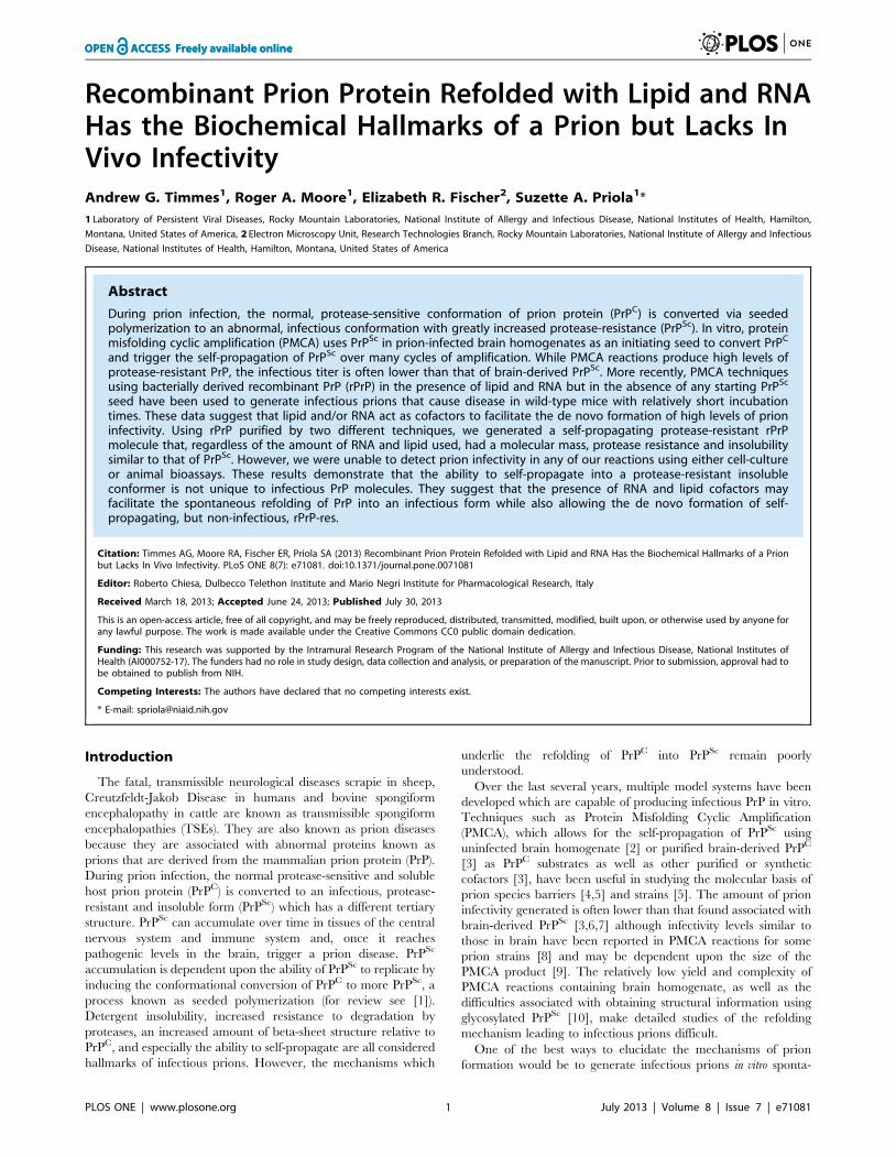
Recombinant Prion Protein Refolded with Lipid and RNAHas the Biochemical Hallmarks of a Prion but Lacks InVivo InfectivityAndrew G. Timmes1, Roger A. Moore1, Elizabeth R. Fischer2, Suzette A. Priola1*
1 Laboratory of Persistent Viral Diseases, Rocky Mountain Laboratories, National Institute of Allergy and Infectious Disease, National Institutes of Health, Hamilton,
Montana, United States of America, 2 Electron Microscopy Unit, Research Technologies Branch, Rocky Mountain Laboratories, National Institute of Allergy and Infectious
Disease, National Institutes of Health, Hamilton, Montana, United States of America
Abstract
During prion infection, the normal, protease-sensitive conformation of prion protein (PrPC) is converted via seededpolymerization to an abnormal, infectious conformation with greatly increased protease-resistance (PrPSc). In vitro, proteinmisfolding cyclic amplification (PMCA) uses PrPSc in prion-infected brain homogenates as an initiating seed to convert PrPC
and trigger the self-propagation of PrPSc over many cycles of amplification. While PMCA reactions produce high levels ofprotease-resistant PrP, the infectious titer is often lower than that of brain-derived PrPSc. More recently, PMCA techniquesusing bacterially derived recombinant PrP (rPrP) in the presence of lipid and RNA but in the absence of any starting PrPSc
seed have been used to generate infectious prions that cause disease in wild-type mice with relatively short incubationtimes. These data suggest that lipid and/or RNA act as cofactors to facilitate the de novo formation of high levels of prioninfectivity. Using rPrP purified by two different techniques, we generated a self-propagating protease-resistant rPrPmolecule that, regardless of the amount of RNA and lipid used, had a molecular mass, protease resistance and insolubilitysimilar to that of PrPSc. However, we were unable to detect prion infectivity in any of our reactions using either cell-cultureor animal bioassays. These results demonstrate that the ability to self-propagate into a protease-resistant insolubleconformer is not unique to infectious PrP molecules. They suggest that the presence of RNA and lipid cofactors mayfacilitate the spontaneous refolding of PrP into an infectious form while also allowing the de novo formation of self-propagating, but non-infectious, rPrP-res.
Citation: Timmes AG, Moore RA, Fischer ER, Priola SA (2013) Recombinant Prion Protein Refolded with Lipid and RNA Has the Biochemical Hallmarks of a Prionbut Lacks In Vivo Infectivity. PLoS ONE 8(7): e71081. doi:10.1371/journal.pone.0071081
Editor: Roberto Chiesa, Dulbecco Telethon Institute and Mario Negri Institute for Pharmacological Research, Italy
Received March 18, 2013; Accepted June 24, 2013; Published July 30, 2013
This is an open-access article, free of all copyright, and may be freely reproduced, distributed, transmitted, modified, built upon, or otherwise used by anyone forany lawful purpose. The work is made available under the Creative Commons CC0 public domain dedication.
Funding: This research was supported by the Intramural Research Program of the National Institute of Allergy and Infectious Disease, National Institutes ofHealth (AI000752-17). The funders had no role in study design, data collection and analysis, or preparation of the manuscript. Prior to submission, approval had tobe obtained to publish from NIH.
Competing Interests: The authors have declared that no competing interests exist.
* E-mail: [email protected]
Introduction
The fatal, transmissible neurological diseases scrapie in sheep,
Creutzfeldt-Jakob Disease in humans and bovine spongiform
encephalopathy in cattle are known as transmissible spongiform
encephalopathies (TSEs). They are also known as prion diseases
because they are associated with abnormal proteins known as
prions that are derived from the mammalian prion protein (PrP).
During prion infection, the normal protease-sensitive and soluble
host prion protein (PrPC) is converted to an infectious, protease-
resistant and insoluble form (PrPSc) which has a different tertiary
structure. PrPSc can accumulate over time in tissues of the central
nervous system and immune system and, once it reaches
pathogenic levels in the brain, trigger a prion disease. PrPSc
accumulation is dependent upon the ability of PrPSc to replicate by
inducing the conformational conversion of PrPC to more PrPSc, a
process known as seeded polymerization (for review see [1]).
Detergent insolubility, increased resistance to degradation by
proteases, an increased amount of beta-sheet structure relative to
PrPC, and especially the ability to self-propagate are all considered
hallmarks of infectious prions. However, the mechanisms which
underlie the refolding of PrPC into PrPSc remain poorly
understood.
Over the last several years, multiple model systems have been
developed which are capable of producing infectious PrP in vitro.
Techniques such as Protein Misfolding Cyclic Amplification
(PMCA), which allows for the self-propagation of PrPSc using
uninfected brain homogenate [2] or purified brain-derived PrPC
[3] as PrPC substrates as well as other purified or synthetic
cofactors [3], have been useful in studying the molecular basis of
prion species barriers [4,5] and strains [5]. The amount of prion
infectivity generated is often lower than that found associated with
brain-derived PrPSc [3,6,7] although infectivity levels similar to
those in brain have been reported in PMCA reactions for some
prion strains [8] and may be dependent upon the size of the
PMCA product [9]. The relatively low yield and complexity of
PMCA reactions containing brain homogenate, as well as the
difficulties associated with obtaining structural information using
glycosylated PrPSc [10], make detailed studies of the refolding
mechanism leading to infectious prions difficult.
One of the best ways to elucidate the mechanisms of prion
formation would be to generate infectious prions in vitro sponta-
PLOS ONE | www.plosone.org 1 July 2013 | Volume 8 | Issue 7 | e71081
neously, i.e. in the absence of a starting seed of PrPSc, using highly
purified E.coli-derived recombinant PrP (rPrP). In recent years,
multiple examples of prion infectivity spontaneously generated
in vitro using rPrP have been reported [11–17]. However, in most
of these studies, the amount of infectivity generated appears to be
extremely low. For example, recombinant hamster PrP exposed to
heat and bovine serum albumin [12] or used as a substrate in
PMCA [13] yielded low levels of hamster prion infectivity. On first
passage, amyloid fibrils of recombinant mouse or hamster PrP
formed spontaneously can induce clinical disease in transgenic
mice overexpressing mutant PrPC [11], or trigger PrPSc formation
in either hamsters [15] or transgenic mice expressing both wild-
type and anchorless forms of mouse PrPC [16]. Depending upon
the sample being tested, in these latter studies clinical disease is
observed with a high attack rate only upon second [15,16] or third
[16] passage. Other experiments have shown that spontaneously
formed recombinant mouse PrP takes from 470–700 days to
induce disease following inoculation into transgenic mice overex-
pressing wild-type PrPC [17]. The fact that reactions using rPrP
appear to generate very little infectivity suggests that they also
would be difficult to use to study the mechanisms of PrPSc
formation or the structure of infectious prions.
RNA can stimulate the conversion of PrPC into PrPSc in the
absence of a starting PrPSc seed [18] and can facilitate the
spontaneous conversion of PrPC into PrPSc which can then
transmit disease to hamsters [3]. Recently, it has been shown that
unseeded PMCA reactions combining mouse rPrP, mouse liver
RNA and lipid [14] or seeded PMCA reactions containing mouse
rPrP, synthetic RNA and lipid [19] yield a form of protease-
resistant rPrP (rPrP-res) that is able to induce clinical disease in
wild-type mice. The average incubation time of the disease
induced by rPrP-res in these studies was approximately 130 and
200 days, respectively, suggesting that the reactions contained
significant prion infectivity. When seeded with rPrP-res, PMCA
reactions containing rPrP and phospholipid, but no RNA, also
produce infectious rPrP-res capable of infecting wild-type mice but
only after an incubation time of almost 400 days [20]. Taken
together, these data suggest that RNA and lipid are important
cofactors in facilitating the spontaneous conversion of rPrP into a
highly infectious form.
In the present study, we have used unseeded PMCA reactions
containing recombinant mouse PrP, mouse liver RNA, and lipid to
make rPrP-res. The rPrP-res generated was insoluble with an N-
terminal truncation similar to that of brain-derived PrPSc.
However, it was unable to induce either sub-clinical or clinical
disease in long-term animal bioassays and was incapable of
inducing the formation of PrPSc in cells susceptible to prion
infection. Furthermore, neither variations in cofactor concentra-
tion nor the use of two different preparations of rPrP yielded rPrP-
res capable of inducing PrPSc formation in cells. Our data are
consistent with the hypothesis that PrP is able to assume multiple
protease-resistant structures and suggest that the ability to self-
propagate into a protease-resistant insoluble conformer is common
to both infectious and non-infectious forms of PrP. Furthermore,
the fact that RNA and lipid cofactors do not necessarily induce
PrP to spontaneously refold into an infectious form suggests that
the de novo generation of infectious PrP may result from a
stochastic event which is only made more likely by the addition of
RNA and lipid.
Materials and Methods
Ethics StatementAll animal experimental protocols were reviewed and approved
by the Rocky Mountain Laboratories Animal Care and Use
Committee (Protocol #2010-64). The Rocky Mountain Labora-
tories are fully accredited by the American Association for
Laboratory Animal Care and this study was carried out in strict
accordance with the recommendations in the Guide for the Care
and Use of Laboratory Animals of the National Institutes of
Health.
HPLC-Mass Spectrometry for Intact ProteinProteins were injected into a reverse phase HPLC (Agilent 1100
series HPLC, Agilent Technologies) with a Zorbax 300SB-C18
(2.1650 mm, 3.5 mM, Agilent Technologies) and introduced into
the mass spectrometer as described [21,22]. Positive ion Electro-
spray Ionization (ESI) mass spectra for intact protein were
obtained with an Agilent 6224 mass spectrometer equipped with
an ESI interface and a time-of-flight (TOF) mass detector (Agilent
Technologies). Mass spectra were analyzed and deconvoluted as
described [23], using an Agilent software MassHunter version
B.04.00 (Agilent Technologies).
Recombinant PrP PurificationThe cloning and expression of recombinant wild-type mouse
PrP has been described previously [16]. Two methods were used
to purify recombinant PrP (rPrP). The first method was a modified
form [24] of the protocol by Zahn et al. [25]. Briefly, approxi-
mately 2 g of bacterial cell pellets equivalent to 250 ml of LB-
Miller growth medium were lysed with BugBuster using lysonase
(EMD Biosciences). Inclusion bodies were isolated by centrifuga-
tion, washed, and then solubilized in denaturing buffer (6 M
guanidine, 100 mM sodium phosphate, 10 mM Tris, pH 8.0)
without reducing agents. The denatured rPrP was again subjected
to centrifugation and then mixed with 15 g of NiNTA resin
(Qiagen) prior to on-column refolding using a linear gradient into
refolding buffer (100 mM sodium phosphate,10 mM Tris,
pH 8.0). Refolded rPrP was further washed in refolding buffer
to remove any residual guanidine prior to elution with 500 mM
imidazole, 100 mM sodium phosphate, 10 mM Tris pH 5.8. The
eluted rPrP was filtered, dialyzed against 10 mM ammonium
acetate, pH 4.5, and stored at 280uC. Prior to use, rPrP was
thawed and dialyzed into water. The mass of rPrP was verified by
electrospray intact mass analysis (ESI-MS) to be 23,061.83 Da
(Expected: 23,063.3 Da; Figure S1A and C).
A second purification also based on the protocol of Zahn et al.
[25], was done in closer accordance to that described by Wang
et al. [14,26]. This purification differed from the first primarily in
the use of beta-mercaptoethanol (BME) during PrP refolding, the
addition of a cation exchange step, and no dialysis of the purified
PrP into 10 mM ammonium acetate, pH 4.5. Briefly, 10 mM
BME was added to the denaturing buffer prior to the on-column
refolding procedure described above. After washing and elution
with 500 mM imidazole, fractions containing rPrP were dialyzed
first against 10 mM sodium phosphate, pH 5.8 and then against
water. A subsequent cation exchange chromatography (CM
Sepharose) step was performed in which rPrP was eluted using a
linear gradient from 0 to 500 mM sodium chloride in 10 mM
sodium phosphate, pH 5.8 buffer. The purified protein was
dialyzed against water and stored at 280uC until needed.
Recombinant PrP purified using this second protocol will be
referred to as rPrP2 throughout the manuscript. The mass of
Non-Infectious Refolded Prion Protein
PLOS ONE | www.plosone.org 2 July 2013 | Volume 8 | Issue 7 | e71081

rPrP2 was verified by ESI-MS to be 23,061.85 Da (Expected:
23,063.3 Da; Figure S1B and D).
For both rPrP and rPrP2, purity was estimated to be at least
99% by SDS-PAGE and silver stain. Approximately 99% of the
protein was monomeric with an intact disulfide bond and no
truncated forms were detected (Figure S1G). Smaller peaks were
present in both preparations that may represent oxidation and/or
sodium and iron adducts (Figure S1E and F). However, both iron
adducts [27] and oxidation [28,29] are known artifacts of ESI-MS
and it is not clear whether these modifications were present in our
rPrP samples prior to analysis.
PMCAThe components of the PMCA reaction were combined
according to Wang, et al., 2010 [14]. Purified rPrP was
centrifuged for 1 hr at 100,0006g and the supernatant containing
soluble rPrP was removed. Recombinant PrP was added to a final
concentration of 5 mg/ml to the synthetic lipid 1-palmitoyl-
2oleoyl-sn-glycero-3-phospho-(19-sn-glycerol) (sodium salt)
(POPG, 4.4 mg/ml final concentration). After a 10 minute
incubation, the rPrP/POPG mixture was diluted into Tris/
NaCl/Triton X-100 to yield a solution with 0.28% Triton X-100,
10 mM Tris-HCl, and 150 mM NaCl. After a 5 minute
incubation, mouse liver extract containing RNA was added to a
final concentration of 30 mg/ml and the solution thoroughly
mixed. Without further incubation, the completed reaction
mixture was aliquoted and frozen at 280uC for use in future
experiments.
For PMCA reactions, the reaction mixture was thawed and
subjected over a 24 hr period to 48 cycles of 30 second sonication
at 37uC followed by a 30 minute incubation without sonication
(one round). The product of the first round of PMCA was used to
seed the second 24 hr PMCA round at a dilution of 1:10 and the
process repeated for twenty rounds (i.e. 20 days). At each round,
reaction products were digested using proteinase K (PK) as in [14]
and analyzed for recombinant, protease-resistant PrP (rPrP-res) by
western blotting using the anti-PrP mouse monoclonal antibody
6D11.
ImmunoblotsSamples were loaded onto either 14% NuPAGE gels or 15%
Tris-HCl Criterion gels (BIO-RAD). The mouse monoclonal anti-
PrP antibodies 6D11 (Covance), POM1 (Prionatus), and 8B4
(Santa Cruz Biotechnology) were used at concentrations of 0.4,
0.5, and 0.2 mg/mL, respectively and diluted in Enhanced
Chemiluminescence (ECL) Advance Blocker/Diluent (GE Health-
care) in 10 mM Tris pH 8.0, 150 mM NaCl, 0.05% Tween-20.
The secondary antibody sheep anti-mouse IgG conjugated with
horseradish peroxidase (GE Healthcare) was used at a dilution of
1:100,000 for 6D11, 1:40,000 for POM1, and 1:10,000 for 8B4 in
the same diluent as the primary antibody. Detection was with ECL
Advance (GE Healthcare) according to the manufacturer’s
instructions. The rabbit polyclonal anti-PrP antibody R20 [30]
was used at a 1:5000 dilution in conjunction with a donkey anti-
rabbit IgG secondary antibody conjugated to horseradish perox-
idase (GE Healthcare) diluted at 1:20,000. All blots using R20
were developed using ECL Prime (GE Healthcare).
The detection limit of our immunoblot procedure for PrP was
determined by comparison to different dilutions of PrP loaded
onto the same gel as the samples being analyzed. Depending upon
the experiment, the PrP used for the dilution series was either
recombinant PrP, PrPSc derived from the brain or spleen of a
mouse positive for clinical scrapie, or PrPC derived from tissue
culture cells. In general, the limit of detection of our immunoblot
procedure was sufficient to detect PrP at a level equivalent to 1%
of the PrPC in the cell, 1% of the PrPSc in the brain of a clinically
ill mouse, or 10% of the PrPSc in the spleen of a mouse with
clinical scrapie.
ImmunohistochemistrySagittal mouse brain sections were fixed and stained as
described previously [7] except that the sections were incubated
for 2 hours in the human monoclonal D13 anti-PrP antibody and
biotinylated anti-human IgG antibody was used as the secondary
antibody.
Transmission Electron Microscopy (TEM)Samples of the rPrP-res PMCA product inoculated into mice
were digested with 100 U/mL Benzonase (Novagen) for one hour
at 37uC and then incubated with 10 mg/mL PK (Roche) for 30
minutes at 37uC. Pefabloc (Roche) was added to a final
concentration of 4 mM, and the samples were chilled on ice for
5 minutes. Digested samples were transferred to polycarbonate
TLA100.3 tubes and spun at 100,000 rpm for one hour at 4uC.
The supernatants were decanted and discarded and the pellets
resuspended in 167 mL deionized water by cuphorn sonication in a
Misonix S3000 sonicator at a power setting of 1.0 for one minute.
The resuspended pellets were then centrifuged at the same speed,
time, and temperature as above. The second supernatant was also
decanted and discarded and the final pellet was resuspended in
30 mL deionized water with sonication. A 5 mL volume of the
suspension was applied to freshly glow discharged carbon coated
copper grids and allowed to settle for fifteen minutes. Excess fluid
was removed by wicking with filter paper and grids were
negatively stained by applying 5 mL 1% uranyl acetate in distilled
water for one minute, before final wicking. Specimens were viewed
at 120 kV on a Tecnai BT Spirit transmission electron microscope
(FEI) at a nominal magnification of 49,0006. Digital images were
acquired with a Hammamatsu XR-100 side mount digital camera
system (Advanced Microscopy Techniques).
Samples of extracted RNA were left untreated or digested for 60
minutes at 37uC either with Benzonase alone (250 U/mL in
0.16PBS pH 7.2, 1 mM MgCl2) or Benzonase plus a-amylase
(Sigma, 1:500 dilution) and amyloglucosidase (Sigma, 1:500
dilution). The reactions were then filtered through 0.02 mm
Anotop filters (Whatman). The final RNA samples, which were 10
times more concentrated than those used in the PMCA reactions,
were applied to grids without any ultracentrifugation steps and
stained as above.
Immunoelectron MicroscopySpecimens were applied to freshly glow discharged carbon
coated nickel grids as described above. After wicking excess
sample, subsequent steps were performed by placing grids
specimen-side down on 20 mL droplets on a PELCOH PTFE
immunostaining pad. After blocking in 3% bovine serum albumin
(BSA), 0.1% Tween-20 in 10 mM Tris pH 8.0 for 10 minutes,
specimens were incubated with the anti-PrP mouse monoclonal
antibody 6D11 diluted 1:100 in dilution buffer (1% BSA, 0.1%
Tween-20 10 mM Tris/pH 8.0) or with dilution buffer alone for
one hour. Grids were washed 3 times for 5 minutes with dilution
buffer and incubated for one hour with a sheep anti-mouse
antibody conjugated to 10 nm colloidal gold (BBInternational)
diluted 1:25 in dilution buffer. Grids were rinsed two times for 5
minutes each with dilution buffer followed by three 5 minute rinses
with distilled water. The grids were then stained with uranyl
acetate and imaged as described above.
Non-Infectious Refolded Prion Protein
PLOS ONE | www.plosone.org 3 July 2013 | Volume 8 | Issue 7 | e71081

Cell CultureTwo cell lines, SN56 derived from septal neurons [31] and
CF10+MoPrP, were inoculated with 22L mouse scrapie brain
homogenate, rPrP-res or rPrP2-res using the protocol described in
Greil et al. [32]. CF10+MoPrP cells are CF10 mouse PrP
knockout cells [32] which have been modified to express wild-
type mouse PrP [33]. Following inoculation, cells were either
analyzed for acute PrP-res formation [34] at passage 0 or passaged
at a dilution of 1:10 into 6-well plates. Up to passage 10, the
remaining 9/10 of the cells that had not been passaged were
analyzed for PrPSc. Cells were lysed with 200 mL lysis buffer
(1 mM Tris-HCl, pH 7.4, 140 mM NaCl, 5 mM EDTA, 0.5%
sodium deoxycholate, 0.5% Triton X-100) and the lysate
centrifuged at 13,000 rpm for 5 minutes at room temperature.
At Passage 10, cells were collected similarly except that all cells
from a 75 cm2 flask were used. All cell lysate supernatants were
stored at 220uC until analyzed.
To assay for PrPSc, cell lysates were thawed and digested with
25 mg/ml PK at 37uC for one hour. Protease digestion was halted
by the addition of Pefabloc (Roche) to a final concentration of
2 mM. The protease digested samples were centrifuged at
100,000 rpm for one hour at 4uC. The resultant pellet was
resuspended in 20 mL SDS-PAGE sample buffer (65 mM Tris-
HCl, pH 6.8, 5% SDS, 3% BME, 10% glycerol, 0.0001%
bromophenol blue) and boiled for ten minutes. Unless otherwise
noted, for all cell samples the entire sample was loaded into a
single lane of an SDS-PAGE gel for analysis by immunoblotting.
Animal ProceduresFemale C57Bl/10 mice or female CD-1 mice from Jackson
Laboratories were used for all bioassays. For infection with rPrP-
res, a single positive reaction tube was amplified by seeding a new
reaction tube with a 1:10 dilution and then pooling multiple
reaction tubes from the same PMCA round. The conversion
efficiency of rPrP into rPrP-res for these reactions was 0.3–3%
(data not shown). Insoluble rPrP in the PMCA product was
concentrated by differential centrifugation as previously described
[14,26]. Samples of inoculum were subjected to PK digestion and
the amount of rPrP-res was determined using a standard dilution
series derived from a known concentration of rPrP.
All inoculations were performed while the mice were under
isoflurane anesthesia. For infection with mouse 22L scrapie, a 10%
stock 22L brain homogenate was diluted 1:10 in PBS/10%FBS
and 30 mL inoculated intracerebrally (IC). The IC titer of the 22L
stock in C57Bl/10 mice was 26108.8 infectious units/g of brain.
For rPrP-res, both CD-1 mice and C57Bl/10 mice were
inoculated IC with 30 mL of undiluted inoculum which contained
,2 ng of rPrP-res. C57Bl/10 mice were also inoculated IC with
serial ten-fold dilutions of rPrP-res diluted into inoculation buffer
(10 mM sodium phosphate pH 7.2, 139 mM NaCl, 1 mg/mL
bovine serum albumin) and intraperitoneally (IP) with 30 mL or
orally (PO) with 100 mL of undiluted rPrP-res. Undiluted and
1021 dilutions of the rPrP-res inoculum were stored at 280uC for
351 days between the inoculation of C57Bl/10 mice and CD-1
mice. Mice were monitored regularly for clinical disease. At either
clinical signs of scrapie or at different time points post-inoculation,
mice were euthanized by isofluorane overdose and spleens and
brains removed for analysis of PrPSc by immunoblot as previously
described [35].
Results
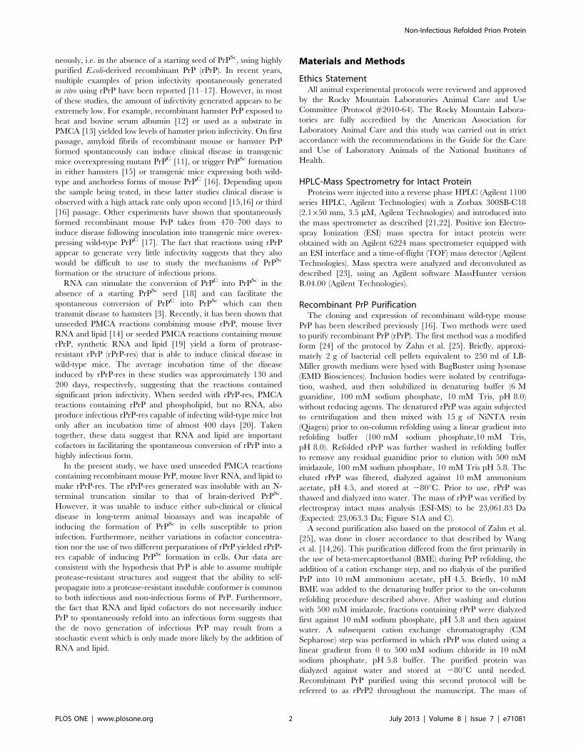
Serial Propagation of Recombinant PrP using PMCARecombinant mouse PrP (rPrP) purified by the method of
Atarashi et al. [24] was subjected to PMCA in the presence of
POPG lipid and mouse liver RNA as described in the Materials
and Methods. Beginning at Round 8 and continuing through
Round 20, protease-resistant rPrP (rPrP-res) was detected near the
top of the gel (Figure 1A) in 10 out of 40 reaction tubes (25%),
suggesting the presence of large SDS and protease-resistant PrP
aggregates. The de novo emergence of a protease-resistant
fragment of 16 kDa was first visible in the ten positive tubes by
round 15 (Figure 1A) and continued to be detectable through
round 19. The significantly lower rPrP-res signal in the twentieth
round (Figure 1A, lane 25) is an example of the tube to tube
variation due to variable delivery of sonication energy that is often
seen when using PMCA [36]. By comparing the intensity of the
16 kDa band to a standard dilution series of undigested rPrP
(Figure 1A, lanes 1–4), we estimated that approximately 0.3–3% of
the total rPrP in the reaction was converted to rPrP-res. Thus,
while the reaction conditions used produced rPrP-res capable of
self-seeding, the reaction itself was inefficient.
One hallmark of PrPSc derived from prion-infected animals is its
high protease resistance [37]. In order to determine the protease
resistance of rPrP-res, two independent samples were digested with
concentrations of PK up to 400 mg/mL. The reactions were then
analyzed by western blot using the anti-PrP mouse monoclonal
antibody 6D11 which recognizes amino acid residues 93–109 in
PrP. As shown in Figure 1B, even after digestion with up to
400 mg/mL of PK the 16 kDa rPrP-res was still detectable. Thus,
at least some of the 16 kDa rPrP-res generated using the PMCA
reaction has a PK resistance similar to that of brain-derived PrPSc
which can be resistant to digestion with 100 mg/ml PK [37].
The Most Protease-resistant Core of rPrP-res ContainsOnly the C-terminus of PrP
The amount of 16 kDa rPrP-res product present in the reaction
decreased with increasing PK concentrations (Figure 1B). This
suggested that either some of the 16 kDa fragment was highly PK-
resistant or that digestion with PK was removing the 6D11
antibody epitope at residues 93–109. To determine if other
protease-resistant forms of abnormal rPrP were present in the
product, immunoblots were also developed with the anti-PrP
antibodies 8B4 (N-terminal residues 37–44), POM1 (multiple
residues between 140–212, see [38]), and R20 (C-terminal residues
218–231). The 16 kDa rPrP-res fragment reacted with both
POM1 and R20 but not with 8B4 (Figure 1C), indicating that it
was truncated at the N-terminus and contained PrP sequence from
approximately 93–230. This truncation of rPrP-res at the N-
terminus by PK is similar to that generated following PK digestion
of PrPSc derived from scrapie-infected brain [39]. By contrast, the
high molecular weight PK and SDS-resistant PrP aggregates
(Figure 1A and B) did not stain with either POM1 or R20,
suggesting that they were not simply aggregates of the 16 kDa PrP
protein but rather a PK resistant species missing part of the C-
terminus.
The C-terminal antibody R20 detected not only the 16 kDa
band but also an approximately 13 kDa rPrP-res fragment not
recognized by any of the other antibodies used (Figure 1C).
Following digestion with increasing concentrations of PK, the
13 kDa fragment increased in intensity as the 16 kDa fragment
decreased in intensity, suggesting that it was derived from the
16 kDa fragment (Figure 1D). The decrease in intensity of the
16 kDa band with increasing PK concentrations (Figure 1B)
Non-Infectious Refolded Prion Protein
PLOS ONE | www.plosone.org 4 July 2013 | Volume 8 | Issue 7 | e71081
therefore coincided with the loss of the 6D11 antibody epitope.
Thus, our data suggest that rPrP-res has a highly protease-resistant
core which does not include the N-terminus but does contain the
far C-terminus of PrP.
rPrP-res is Detergent InsolubleAnother characteristic feature of disease-associated PrPSc is
detergent insolubility. In order to determine if rPrP-res was also
detergent insoluble, PMCA reaction mixtures that had or had not
Figure 1. Generation of rPrP-res with a protease-resistant C-terminal core in PMCA reactions containing RNA and lipid. (A) Theproducts of twenty rounds of serial PMCA were PK digested and assayed by immunoblot using the anti-PrP mouse monoclonal antibody 6D11. Thenumber of PMCA rounds is indicated at the top of the gel. Lanes 1–4 represent a standard curve of 1:100 to 1:2700 dilutions of the non-PK digested,unsonicated rPrP substrate mixture (No sonic). Based on the standard curve, 0.3–3% of the input rPrP was converted to rPrP-res. Lane numbers areshown at the bottom of the gel. For all panels, the arrow on the left indicates rPrP, the open arrowhead on the right indicates rPrP-res, and molecularmass markers in kDa are indicated on the right. (B) Two independent preparations of rPrP-res (A and B) were digested with increasing concentrationsof PK and assayed by immunoblot using the 6D11 antibody. The non-PK digested, unsonicated rPrP substrate mixture (No sonic) was diluted 1:100prior to loading. All other samples were loaded without dilution. The asterisk to the right indicates the location of a cross-reactive proteinase K band.(C) rPrP-res was PK-digested and assayed by immunoblot with three anti-PrP antibodies: 8B4 (residues 37–44), POM1 (multiple residues between140–212, see [38]), and R20 (residues 218–231). Unless otherwise indicated, all samples were loaded without dilution. No sonic = non-PK digested,unsonicated rPrP substrate mixture. (D) rPrP-res was digested with varying concentrations of PK and assayed by immunoblot using the anti-PrPantibody R20. The rPrP PK-minus control sample (No sonic) was diluted 1:100 prior to loading while all PK-digested samples were loaded undiluted.The closed arrowhead indicates the 13 kDa rPrP-res fragment.doi:10.1371/journal.pone.0071081.g001
Non-Infectious Refolded Prion Protein
PLOS ONE | www.plosone.org 5 July 2013 | Volume 8 | Issue 7 | e71081

been sonicated were centrifuged at 100,0006g for 1 hr at 4uC[40]. While rPrP alone remained soluble and in the supernatant
(data not shown), approximately 40% of the rPrP in PMCA
reactions that had not been sonicated and thus contained no rPrP-
res was found in the pellet (Figure 2, lanes 2–6). By contrast, in
PMCA reactions that had been sonicated, rPrP-res was found
exclusively in the pellet (Figure 2, compare lanes 10 and 13). Our
data show that rPrP experiences a significant loss of solubility due
to associations with other components of the substrate mixture and
are consistent with a recent report showing that rPrP becomes
insoluble following incubation with POPG [20]. However, the fact
that all of the rPrP-res generated in the PMCA reaction was found
in the pellet suggested that the change in rPrP solubility following
sonication was most likely linked to the refolding and oligomer-
ization of rPrP. Thus, similar to PrPSc, rPrP-res is a detergent
insoluble aggregate.
Lack of Detectable Prion Infectivity and PrPSc in MiceInoculated with rPrP-res Generated in vitro
Protease-resistant PrP generated in vitro in the absence of PrPSc
can have no infectivity [41] or levels of infectivity that appear to be
relatively low [12,13,16] or relatively high [14]. In order to assess
the infectivity of rPrP-res generated in our PMCA reactions, rPrP-
res product from a positive reaction tube was amplified,
concentrated by centrifugation, resuspended in PBS/1% BSA,
and then inoculated either undiluted or in serial ten-fold dilutions
intracerebrally (IC) into C57Bl/10 mice. Undiluted rPrP-res was
also inoculated into C57Bl/10 mice intraperitoneally (IP) and
orally (PO) and into CD-1 mice IC.
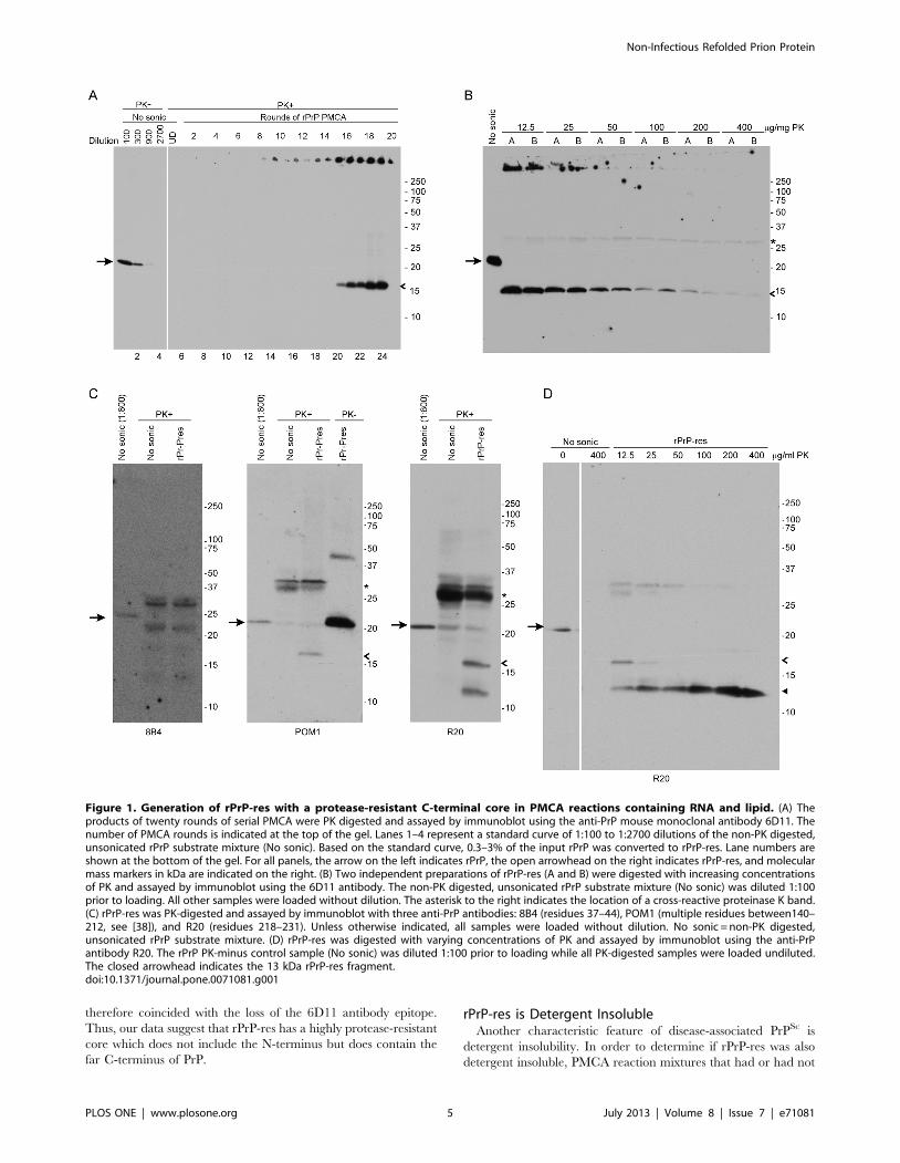
C57Bl/10 mice inoculated IC with 22L mouse scrapie develop
clinical scrapie by approximately 150 days post-inoculation (dpi)
(Table 1). By contrast, no clinical signs of prion disease were
observed up to 626 dpi in C57Bl/10 mice inoculated with rPrP-res
by any route (Table 1). Similarly, no clinical signs of prion
infection were observed in IC inoculated CD-1 mice up to 367 dpi
(Table 1). The lack of clinical disease suggested either that animals
inoculated with rPrP-res were subclinically infected or that no
infectious rPrP-res was present in the inoculum.
Animals subclinically infected with prions can accumulate PrPSc
in the brain even in the absence of clinical signs [42]. Therefore,
we assayed for low levels of PrPSc in rPrP-res inoculated C57BL/
10 and CD-1 mice using immunoblotting. Brains and spleens were
collected from rPrP-res inoculated C57Bl/10 mice at 200, 365,
and 626 dpi and assayed for PrPSc by western blot. No PrPSc was
detected in the brain of any mouse inoculated by any route with
rPrP-res (Figure 3A, B and E). Brains from CD-1 mice inoculated
IC with rPrP-res were also negative at 367 dpi (Figure S2A). In
contrast, PrPSc from the brains of mice clinically ill with 22L
mouse scrapie was detectable using 1% of the material used for the
rPrP-res brain homogenate samples (Figure 3A, B and E). These
results suggest that if rPrP-res inoculated animals had accumulated
PrPSc equivalent to 1% of the amount found in a mouse scrapie-
infected brain, it would have been detected using our immunoblot
protocol.
Figure 2. rPrP-res is detergent insoluble. Unsonicated (No sonic)or sonicated (rPrP-res) PMCA substrate mixtures were subjected toultracentrifugation for one hour at 100,0006g and the resultingsupernatants (SN) and pellets were assayed by immunoblot with theanti-PrP mouse monoclonal antibody 6D11 either with (+) or without(2) PK digestion. Samples were loaded undiluted (UD) or diluted 1:10and 1:100 as indicated. Molecular mass markers (kDa) are shown on theright. rPrP is indicated by the arrow and the 16 kDa protease-resistantband by an open arrowhead. The band at ,66 kDa is bovine serumalbumin which was added to the samples during methanol precipita-tion. The asterisk indicates the location of a cross-reactive proteinase Kband. Lane numbers are shown at the bottom of the gel. Total = nocentrifugation.doi:10.1371/journal.pone.0071081.g002
Table 1. No evidence of clinical or subclinical infectionfollowing inoculation of rPrP-res into mice with differentgenetic backgrounds.
PrPSc
InoculumMousestrain Route Brain Spleen Clinicale DPIf
22La C57Bl/10 IC + + + (6/6) 154611
rPrP-res C57Bl/10b IC – – 2(0/8) .626
rPrP-res C57Bl/10c IP – – 2(0/8) .626
rPrP-res C57Bl/10c PO – – 2(0/8) .626
rPrP-res CD-1d IC – – 2(0/8) .367
aMice inoculated with a 1:10 dilution of a 22L mouse scrapie 10% brainhomogenate. These mice were not inoculated in the same experiment as therPrP-res inoculated mice.bBrains and spleens of two mice tested negative for PrPSc at 200, 365, and626 dpi.cBrains and spleens of two mice tested negative for PrPSc at 365 dpi.dBrains and spleens of three mice tested negative for PrPSc at 367 dpi.eNumbers in parentheses represent the number of scrapie positive mice overthe total number of mice inoculated.fdays post-inoculation (DPI). Numbers represent either average incubation time+ SD or days post-inoculation when the experiment was terminated.doi:10.1371/journal.pone.0071081.t001
Non-Infectious Refolded Prion Protein
PLOS ONE | www.plosone.org 6 July 2013 | Volume 8 | Issue 7 | e71081
PrPSc can often be detected in the spleens of prion-infected
animals before it can be detected in brain [43]. Faint bands were
detected in the spleens of rPrP-res inoculated C57Bl/10 mice at
365 and 626 dpi (Figure 3C and F). However, these bands were
not PrP-specific but rather the result of cross reactivity of the
secondary antibody with IgG (Figure 3D). Similar bands were seen
in mice inoculated with control mixtures that had not been
subjected to PMCA (Figure 3 and Figure S2B). Spleens from CD-1
mice inoculated IC with rPrP-res were also negative at 367dpi
(Figure S2B and S2C). By contrast, PrPSc from the spleens of mice
clinically ill with 22L mouse scrapie was detectable using just 10%
of the material used for the rPrP-res spleen samples (Figure 3A, C
and F). These results show that if rPrP-res inoculated animals had
accumulated PrPSc in the spleen equivalent to 10% of the amount
found in a mouse with clinical scrapie, it would have been detected
using our immunoblot protocol.
Figure 3. C57Bl/10 mice inoculated with rPrP-res do not accumulate detectable PrPSc up to 626 days post inoculation. Brain andspleen samples from C57Bl/10 mice inoculated with rPrP-res, 22L mouse scrapie, vehicle alone (Neg), or a mixture of unsonicated rPrP, POPG, andRNA in inoculation buffer (No sonic) were homogenized, PK digested, and assayed by immunoblot using the anti-PrP mouse monoclonal antibody6D11. (A) Brain and spleen, 200 dpi. (B) Brain, 365 dpi. (C) Spleen, 365 dpi. (D) Spleen, 365 dpi, secondary antibody only. Samples assayed areidentical to those in panel C. The first lane shows the reactivity of the secondary antibody with purified mouse IgG heavy and light chains. (E) Brain,626 dpi. (F) Spleen, 626 dpi. For all panels, tissue samples were loaded undiluted (UD) unless otherwise noted. A standard dilution curve of 22Lmouse brain homogenate containing PrPSc was loaded on each gel as indicated and used to estimate the limit of detection of PrP as detailed in theMaterials and Methods. A, B, and C represent individual mice assayed at each timepoint. IC = intracerebral; IP = intraperitoneal; PO = per ora. Molecularmass markers (kDa) are indicated on the right and the asterisk indicates the location of a cross-reactive proteinase K band.doi:10.1371/journal.pone.0071081.g003
Non-Infectious Refolded Prion Protein
PLOS ONE | www.plosone.org 7 July 2013 | Volume 8 | Issue 7 | e71081
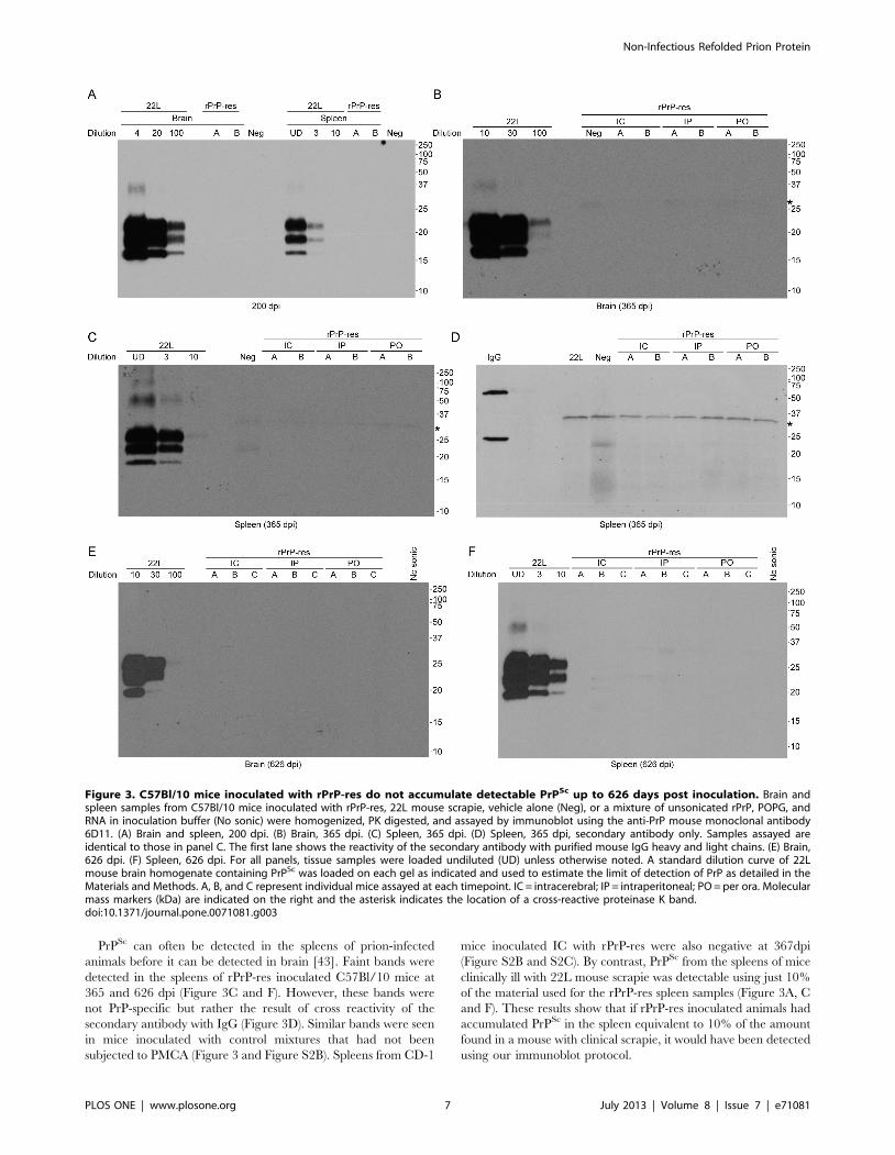
Immunohistochemical analysis was also used to assay for PrPSc
accumulation in rPrP-res inoculated mice. Sagittal brain sections
of C57Bl/10 or CD-1 mice (Figure 4 and Figure S3) inoculated IC
with rPrP-res were stained with the anti-PrP antibody D13. In the
thalamus (Figure 4 and Figure S3) and all other regions of the
brain (data not shown), the level of PrP staining in rPrP-res
inoculated mice was identical to that of mice inoculated IC with
control mixtures that had not been subjected to PMCA. By
contrast, PrPSc was easily detectable in 22L scrapie-infected
C57Bl/10 mice (Figure 4 and Figure S3). Furthermore, standard
H&E staining did not detect the spongiosis commonly associated
with prion disease (data not shown). Thus, using two different
mouse strains and up to three different inoculation routes, we were
unable to detect any sign of clinical or subclinical prion infection in
mice inoculated with rPrP-res. These data strongly suggest that the
rPrP-res generated in our PMCA reactions was not infectious.
rPrP-res does not Induce the Conversion of PrPC to PrPSc
in Scrapie Susceptible Cell LinesOur data demonstrate that, although rPrP-res could self-
propagate in vitro, it was unable to induce PrPSc formation and
clinical disease in mice. This suggested that rPrP-res was unable to
convert fully glycosylated and membrane anchored mammalian
PrPC. Alternatively, it was possible that rPrP-res induced the
formation of PrPSc only transiently. Transient PrPSc formation,
also known as acute PrPSc formation, has been shown to occur
after cells are exposed to prions but prior to passage [34,44].
Importantly, acute PrPSc formation can be induced even by strains
of mouse scrapie which do not infect susceptible cell lines [34]. In
order to determine if rPrP-res could induce the conversion of
mammalian PrPC into PrPSc either acutely or persistently, the
scrapie susceptible septal neuron cell line SN56 [31,45] was plated
at different densities and exposed to either 15 ng of rPrP-res, a 1%
brain homogenate from mice infected with 22L scrapie, or cell-
culture medium alone. After 96 hours (Pass 0) and at selected
passages post-infection, lysates from inoculated cell lines were PK
digested followed by immunoblotting with the anti-PrP antibody
6D11. No PrPSc was detected in SN56 cells inoculated with rPrP-
res or cell culture medium acutely (Figure 5A, Pass 0), at early
passage (Figure 5A, Pass 1 and 2), or at late passage (Figure 5B,
Pass 13 and 15). By contrast, SN56 cells inoculated on the same
day with 22L scrapie-infected mouse brain homogenate induced
PrPSc formation at passage 0, 1 and 2 (Figure 5A). Thus, the SN56
cells used could support PrPSc formation and mouse prion
infection.
As a further test of the ability of rPrP-res to induce PrPSc
formation, we used a cell line that does not normally express PrP
(CF10 cells [32]) which had been stably transfected with a plasmid
over-expressing wild-type mouse PrP (CF10+MoPrP cells). These
cells are susceptible to scrapie infection [33]. As with the SN56 cell
line, inoculation of rPrP-res onto CF10+MoPrP cells resulted in no
detectable PrPSc either acutely (Figure S4A, Pass 0) or persistently
(Figure S4B, Pass 6 and 7). These data strongly suggested that the
rPrP-res product generated in our PMCA reactions in the
presence of lipid and RNA was unable to convert fully glycosylated
and membrane anchored PrPC to PrPSc.
Different Amounts of RNA and POPG do not alter theAbility of rPrP-res to Convert rPrP or PrPC
It was possible that generation of rPrP-res capable of inducing
the conversion of PrPC to PrPSc was dependent upon the
concentration of POPG and/or RNA in the PMCA reaction.
We therefore used rPrP-res to seed PMCA reactions with different
ratios of rPrP to lipid or RNA and assayed whether or not the
newly made rPrP-res would convert PrPC to PrPSc in cells. PMCA
reactions seeded with rPrP-res generated protease-resistant
16 kDa rPrP-res regardless of the concentration of POPG or
RNA used (Figure 6A). Thus, changing the concentration of
POPG or RNA in the PMCA reaction did not affect the ability of
rPrP-res to self-propagate in vitro. These results are consistent
with a recent study demonstrating that complete removal of RNA
or POPG does not necessarily prevent propagation of rPrP-res in
the presence of a starting seed of rPrP-res [46].
Figure 4. Lack of spongiform change and PrPSc in C57Bl/10mice inoculated with rPrP-res. Sagittal sections from C57Bl/10 miceinoculated with 22L scrapie, rPrP-res, or the unsonicated rPrP mixture(No sonic) stained with D13 anti-PrP antibody. A representative regionof the thalamus is shown. Mice inoculated with 22L showed clearspongiform change and PrPSc deposition (brown stain). No spongiformchange or PrPSc was detected in any region of the brain from miceinoculated with rPrP-res. Scale bar = 100 mm.doi:10.1371/journal.pone.0071081.g004
Non-Infectious Refolded Prion Protein
PLOS ONE | www.plosone.org 8 July 2013 | Volume 8 | Issue 7 | e71081
In order to determine if changes in the ratio of rPrP to POPG
and RNA would lead to the formation of rPrP-res that could
induce PrPSc formation in cells, the rPrP-res reaction products
shown in Figure 6A were concentrated and approximately 15 ng
were inoculated in duplicate onto SN56 cells. No cell-derived
PrPSc was detected at any passage following inoculation of the
different rPrP-res reaction products (Figure 6B-E), although the
rPrP-res inoculum was sometimes visible in inoculated cells lysed
without passaging (e.g. Figure 6C, Passage 0). Our results show
that even though rPrP-res could be generated in vitro using
varying concentrations of lipid and RNA, none of these conditions
yielded infectious prions that could stimulate detectable PrPSc
formation in cells.
The Ultrastructure of rPrP-res is Fibrillar and not GranularA study by Piro et al. has suggested that infectious prions
generated from recombinant PrP in the presence of POPG and
RNA are amorphous aggregates and small, spherical aggregates
but are not fibrillar [47]. In order to determine if the rPrP-res
product generated in our PMCA reactions had a similar
ultrastructure, rPrP-res was digested with nuclease and protease,
concentrated by ultracentrifugation, and analyzed by transmission
electron microscopy. The rPrP-res samples tested contained
clusters of fibrils approximately 12 nm wide with a length of up
to 500 nm (Figure 7A and B). Similar in width and size to prion
fibrils from purified brain-derived PrPSc [48,49], these fibrils were
not present in unsonicated PMCA reactions (Figure 7C). Im-
munogold labeling using the anti-PrP antibody 6D11 demonstrat-
ed that the fibrils were positive for PrP (Figure 7F) while samples
labeled only with the gold-conjugated secondary antibody
(Figure 7G) were negative.
The rPrP-res PMCA products generated in this study also
contained large numbers of aggregates approximately 25 nm in
diameter (Figure 7A and B), similar in size to the 26 nm spherical
aggregates previously reported as being associated with infectious
rPrP-res [47]. However, these structures were also present in
unsonicated PMCA reactions which did not contain rPrP-res
(Figure 7C) as well as in mouse liver RNA preparations alone
(Figure 7D) indicating that they were not specific to the rPrP
PMCA reaction. The ,25 nm diameter and rosette organization
of these structures matches that of glycogen granules, which are
known to coprecipitate with RNA during phenol-chloroform
extraction [50,51]. Digestion of the glycogen in our liver RNA
samples with amylase and amyloglucosidase removed these
granular structures (Figure 7E), confirming that they were
glycogen. Furthermore, immunogold staining demonstrated that
the glycogen granules were negative for PrP (Figure 7F, arrow).
Thus, in contrast to previous work where the structures assigned to
infectious rPrP-res were ,26 nm aggregates [47] or ,2 nm
spheres [46], the only ultrastructural component unique to the
non-infectious rPrP-res generated in our PMCA reactions were
PrP-positive fibrils.
Recombinant PrP Purified by Two Different Protocolsdoes not form Infectious rPrP-res
Unlike the previous study of Wang et al. [14], we were unable
to generate high levels of infectious rPrP-res in PMCA reactions
containing POPG and RNA. However, the recombinant mouse
PrP used as a substrate in our study was purified using a slightly
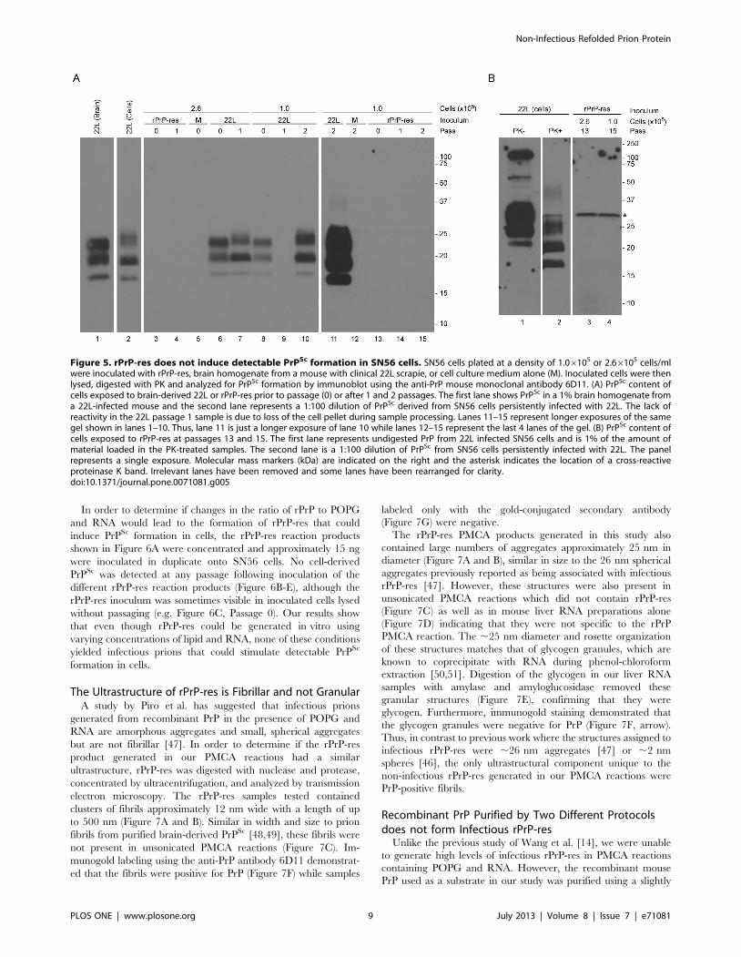
Figure 5. rPrP-res does not induce detectable PrPSc formation in SN56 cells. SN56 cells plated at a density of 1.06105 or 2.66105 cells/mlwere inoculated with rPrP-res, brain homogenate from a mouse with clinical 22L scrapie, or cell culture medium alone (M). Inoculated cells were thenlysed, digested with PK and analyzed for PrPSc formation by immunoblot using the anti-PrP mouse monoclonal antibody 6D11. (A) PrPSc content ofcells exposed to brain-derived 22L or rPrP-res prior to passage (0) or after 1 and 2 passages. The first lane shows PrPSc in a 1% brain homogenate froma 22L-infected mouse and the second lane represents a 1:100 dilution of PrPSc derived from SN56 cells persistently infected with 22L. The lack ofreactivity in the 22L passage 1 sample is due to loss of the cell pellet during sample processing. Lanes 11–15 represent longer exposures of the samegel shown in lanes 1–10. Thus, lane 11 is just a longer exposure of lane 10 while lanes 12–15 represent the last 4 lanes of the gel. (B) PrPSc content ofcells exposed to rPrP-res at passages 13 and 15. The first lane represents undigested PrP from 22L infected SN56 cells and is 1% of the amount ofmaterial loaded in the PK-treated samples. The second lane is a 1:100 dilution of PrPSc from SN56 cells persistently infected with 22L. The panelrepresents a single exposure. Molecular mass markers (kDa) are indicated on the right and the asterisk indicates the location of a cross-reactiveproteinase K band. Irrelevant lanes have been removed and some lanes have been rearranged for clarity.doi:10.1371/journal.pone.0071081.g005
Non-Infectious Refolded Prion Protein
PLOS ONE | www.plosone.org 9 July 2013 | Volume 8 | Issue 7 | e71081

different technique. In particular, our purification did not involve
the use of BME during PrP refolding, the use of a cation exchange
step, or the dialysis of purified PrP into 10 mM sodium phosphate,
pH 5.8 (see Materials and Methods). While rPrP produced using
our purification method has been shown to generate very low
levels of prion infectivity [16], it was possible that its structure was
incompatible with conversion to the highly infectious rPrP-res
reported previously [14]. We therefore repeated our PMCA
reactions using rPrP that had been prepared as closely as possible
to the method used in Wang et al. [14] (designated rPrP2) and
compared it to PMCA reactions using rPrP prepared according to
our original protocol.
Of the 24 PMCA reactions containing rPrP2, POPG and RNA
which were carried for 20 rounds, two were positive for a protease-
resistant product which we designated as rPrP-res2. One of these
samples, which was first positive at round 11, is shown in
Figure 8A. Consistent with our previous results, 18 of 24 reactions
using the rPrP substrate prepared by our original purification
method were also positive for a 16 kDa rPrP-res product by round
20 (data not shown). The 16 kDa rPrP-res2 product was PK-
resistant and could be truncated to the same 13 kDa protease-
resistant core (Figure 8B) described for rPrP-res in our initial
experiments (Figure 1). Thus, both rPrP and rPrP2 were capable
of generating self-propagating rPrP-res in PMCA reactions
containing POPG and RNA.
As a measure of potential prion infectivity, we analyzed the
ability of newly generated rPrP-res and rPrP-res2 to induce
formation of PrPSc in cells. The mouse scrapie strain 22L, rPrP-res
(120 ng) or rPrP-res2 (7 ng and 90 ng) were inoculated onto SN56
cells as described previously. SN56 cells inoculated with 22L
scrapie-infected mouse brain homogenate induced PrPSc forma-
tion at all passes tested (Figure 8C) demonstrating that the cells
were susceptible to mouse scrapie infection. By contrast, no PrPSc
was detected in SN56 cells inoculated with cell culture medium
(Figure 8C, Mock) or with rPrP-res and 7 ng of rPrP-res2 either
acutely (Figure 8D, Pass 0) or at early (Figure 8D) and late
Figure 6. Varying RNA and POPG concentration does not lead to rPrP-res that can induce PrPSc formation in SN56 cells. (A) rPrPPMCA reactions seeded with rPrP-res and serially propagated in the presence of 30 mg/ml of RNA and 3 different concentrations of POPG lipid (POPGlanes) or in the presence of 4.4 mg/ml of POPG and 3 different concentrations of mouse liver RNA (RNA lanes) were digested with PK and assayed induplicate by immunoblot. (B) SN56 cells inoculated with cell culture medium alone (Mock) or rPrP-res made in the presence of 30 mg/ml RNA plus2.2 or 6.7 mg/ml of POPG lipid. (C) SN56 cells inoculated with cell culture medium alone (Mock) or rPrP-res made in the presence of 4.4 mg/ml POPGplus 15 or 45 mg/ml of RNA. (D) SN56 cells inoculated with rPrP-res (Passage 6). The concentration in mg/ml of the RNA and POPG used is indicated inthe parentheses. Lanes labeled 16 indicate PMCA products generated using the original POPG and RNA concentrations of 4.4 mg/ml and 30 mg/ml,respectively. (E) SN56 cells inoculated with rPrP-res (Passage 10). Labeling is as in panel D. The first lane is a 1:100 dilution of undigested PrP fromSN56 cells. Based on a standard dilution curve of PrPSc derived from SN56 cells persistently infected with 22L (first 3–4 lanes in panels B-D), the limit ofdetection of the immunoblot for PrP is equal to ,1–3% of the total cell equivalents loaded. For all panels, blots were developed using the anti-PrPmouse monoclonal antibody 6D11. Molecular mass markers (kDa) are indicated on the right of each panel. Irrelevant lanes have been cropped andsome lanes have been rearranged for clarity, but each immunoblot panel derives from a single film exposure.doi:10.1371/journal.pone.0071081.g006
Non-Infectious Refolded Prion Protein
PLOS ONE | www.plosone.org 10 July 2013 | Volume 8 | Issue 7 | e71081

Figure 7. The ultrastructure of rPrP-res is fibrillar and not granular. Transmission electron microscopy by negative stain techniques was usedto examine rPrP-res (A, B), unsonicated PMCA substrate (C), mouse liver extract alone (D), and mouse liver extract treated with amylase andamyloglucosidase (E). (F) Fibrils of rPrP-res were immune-labelled with the anti-PrP antibody 6D11 and gold-conjugated anti-mouse IgG secondaryantibody, prior to negative staining. The glycogen granule in the upper left of the panel is not immunogenic, while the nearby fibrils are specificallylabelled. The inset is magnified 26. (G) rPrP-res stained with secondary antibody only. Scale bars = 100 nm.doi:10.1371/journal.pone.0071081.g007
Non-Infectious Refolded Prion Protein
PLOS ONE | www.plosone.org 11 July 2013 | Volume 8 | Issue 7 | e71081
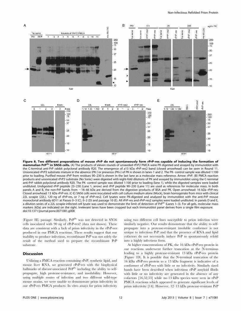
(Figure 8E) passage. Similarly, PrPSc was not detected in SN56
cells inoculated with 90 ng of rPrP-res2 (data not shown). These
data are consistent with a lack of prion infectivity in the rPrP-res
produced in our PMCA reactions. These results suggest that our
inability to produce infectious, recombinant PrP was not solely the
result of the method used to prepare the recombinant PrP
substrate.
Discussion
Utilizing a PMCA reaction containing rPrP, synthetic lipid, and
mouse liver RNA, we generated rPrP-res with the biophysical
hallmarks of disease-associated PrPSc including the ability to self-
propagate, high protease-resistance, and insolubility. However,
using multiple routes of infection and two different wild-type
mouse strains, we were unable to demonstrate prion infectivity in
our rPrP-res PMCA products. In vitro assays for prion infectivity
using two different cell lines susceptible to prion infection were
similarly negative. Our results demonstrate that the ability to self-
propagate into a protease-resistant insoluble conformer is not
unique to infectious PrP and that the presence of RNA and lipid
cofactors do not necessarily induce PrP to spontaneously refold
into a highly infectious form.
At higher concentrations of PK, the 16 kDa rPrP-res protein in
our reactions underwent further truncation at the N-terminus
leading to a highly protease-resistant 13 kDa rPrP-res protein
(Figure 1D). It is possible that the N-terminal truncation of the
16 kDa rPrP-res protein to a 13 kDa fragment is indicative of a
conformer of rPrP-res with little or no infectivity. Similarly sized
bands have been described when infectious rPrP amyloid fibrils
with little or no infectivity are generated in the absence of any
cofactors [16,52,53] while no 13 kDa species were seen in rPrP
PMCA reactions which appeared to generate significant levels of
prion infectivity [14]. However, 12–13 kDa protease-resistant PrP
Figure 8. Two different preparations of mouse rPrP do not spontaneously form rPrP-res capable of inducing the formation ofmammalian PrPSc in SN56 cells. (A) The products of eleven rounds of unseeded rPrP2 PMCA were PK digested and assayed by immunoblot withthe C-terminal anti-PrP rabbit polyclonal antibody R20. The emergence of a13 kDa rPrP-res2 band (closed arrowhead) can be seen in Round 11.Unsonicated rPrP2 substrate mixture in the absence (PK-) or presence (PK+) of PK is shown in lanes 1 and 2. The PK- control sample was diluted 1:100prior to loading. Purified mouse rPrP from residues 90–230 is shown in the last lane as a molecular mass reference. Arrow: rPrP. (B) PMCA reactionproducts and unsonicated rPrP2 substrate (No Sonic) were digested with eight-fold dilutions of PK and assayed by immunoblot using the C-terminalanti-PrP rabbit polyclonal antibody R20. The PK- control sample was diluted 1:100 prior to loading (lane 1), while the digested samples were loadedundiluted. Undigested rPrP peptide 23–230 (Lane 1, arrow) and rPrP peptide 90–230 (Lane 11) are used as references for molecular mass. In bothpanels A and B, the non-PrP bands from ,18–66 kDa are derived from the digestion products of BSA and PK. Open arrowhead: 16 kDa rPrP-res;Closed arrowhead: 13 kDa rPrP-res. (C-E) SN56 cells were inoculated with cell culture medium alone (Mock), brain homogenate from mice with clinical22L scrapie (22L), 120 ng of rPrP-res, or 7 ng of rPrP-res2. Cell lysates were PK-digested and analyzed by immunoblot with the anti-PrP mousemonoclonal antibody 6D11 at Passes 0–3 (C), 0–2 (D) and passage 10 (E). All rPrP-res and rPrP-res2 samples were loaded undiluted. In panels D and E,a dilution series of a 22L scrapie-infected cell lysate was used to demonstrate the limit of detection of PrPSc (Lanes 1–3). For all gels, molecular massmarkers (kDa) are indicated on the right. Irrelevant lanes have been cropped but each immunoblot panel derives from a single film exposure.doi:10.1371/journal.pone.0071081.g008
Non-Infectious Refolded Prion Protein
PLOS ONE | www.plosone.org 12 July 2013 | Volume 8 | Issue 7 | e71081

isoforms have been identified in different types of human prion
disease [54,55] suggesting that they may play a role in prion
pathogenesis. At present, the relevance of these lower molecular
weight species to the generation of prion infectivity in vivo or
in vitro is unknown.
A second rPrP-res species identified in our PMCA reactions was
represented by high molecular weight aggregates that were both
PK and SDS-resistant (Fig. 1A). These aggregates did not react
with the POM1 and R20 antibodies (Fig. 1C and D) indicating
that they were not simply aggregates of the 16 kDa PrP protein
but rather a PK-resistant PrP species missing a portion of the C-
terminus. They always preceded the appearance of the 16 kDa
band by several rounds of amplification and could represent a
different species of self-propagating rPrP-res in our PMCA
reactions. PrPSc aggregate size has been correlated with the
specific infectivity of prions and larger PrPSc aggregates tend to
have a lower specific infectivity than smaller aggregates [56].
Thus, it is possible that the rPrP-res aggregates in our reactions are
too large to trigger PrPSc formation in vitro or prion infection
in vivo. Alternatively, they may interfere and/or compete with the
propagation of infectious rPrP-res in vitro.
The rPrP substrate used might also influence the formation of
infectious rPrP-res. For this reason, we tested two different rPrP
substrates that we prepared according to protocols that generated
relatively low [16] or relatively high [14,26] levels of rPrP-res
infectivity. However, even though both preparations were .99%
pure and contained , 99% monomeric PrP with an intact
disulfide bond (Figure S1C, D and G), neither generated infectious
rPrP-res in the PMCA reaction. Some of the substrate appeared to
be modified, possibly as a result of oxidation and/or adduct
formation (Figure S1E and F). However, these modifications could
also be artifacts of the ESI-MS analysis [28,29]. Oxidation of PrP
has been reported to be both beneficial [57–59] and detrimental
[60] to abnormal PrP formation. We cannot discount the
possibility that both of our rPrP substrates contained a population
of misfolded or oxidized PrP molecules that either could not refold
into an infectious conformation or that inhibited infectious rPrP-
res formation. However, no data are available on how modifica-
tions to rPrP might influence formation of prion infectivity
in vitro. Further study will be needed to understand the potential
impact of different recombinant PrP populations on the generation
of prion infectivity.
It has been suggested that amyloid forms of rPrP-res are
inherently less infectious than non-fibrillar forms [46,47]. The fact
that PrP-positive fibrils were present in our non-infectious rPrP-res
PMCA products (Figure 7) but absent in infectious rPrP-res
[46,47] is consistent with this hypothesis. However, it is possible
that differences in rPrP-res sample preparation, specifically the use
of lower centrifugal force and/or the incomplete removal of the
supernatant, may account for the lack of rPrP-res fibrils in
previous studies [46,47]. We were only able to detect rPrP-res
fibrils following concentration of our PMCA reaction products via
ultracentrifugation and resuspension of the resultant pellet in a
small volume (see Materials and Methods). Moreover, it is unlikely
that fibrillar structure alone is indicative of less infectious prions. In
vivo studies have clearly shown that the presence of PrP fibrils does
not necessarily correlate with low levels of prion infectivity. A
transgenic mouse model of prion infection where PrPSc is
deposited entirely as amyloid has levels of infectivity greater than
or equal to those found in a wild-type mouse model where PrPSc is
not deposited as amyloid [61–63].
Small spherical aggregates of ,26 nm have been reported in
preparations of highly infectious rPrP-res and are thought to be
representative of the ultrastructure of the infectious particle [47].
We found similarly sized spherical aggregates of ,26 nm, as well
as larger clusters of these aggregates, in our rPrP-res preparations
(Figure 7). However, since there was no infectivity associated with
the rPrP-res generated in our PMCA reactions (Table 1) and since
our spherical aggregates did not contain PrP (Figure 7F), the
particles we observed could not be related to prion infectivity.
Based on digestion with amylase and amyloglucosidase (Figure 7E),
we identified these structures as glycogen granules present as
contaminants in the mouse liver RNA used in the PMCA reactions
(Figure 7D). The identification of these aggregates as glycogen
contaminants derived from the purified RNA used as a cofactor in
the PMCA reaction explains the absence of these structures in a
recent study using infectious rPrP-res propagated in the absence of
RNA [46]. In that study, the predominant ultrastructural
components associated with infectious rPrP-res were small, 2 nM
spheres [46]. Our data suggest that, unless specifically removed,
glycogen granules unrelated to prion infectivity will be present in
any PMCA reaction using non-synthetic RNA as a cofactor and
may confound the ultrastructural analysis of infectious rPrP-res.
Finally, although we did not observe a direct interaction between
glycogen and rPrP (Figure 7F), under certain conditions glycogen
can associate with rPrP and accelerate its refolding into a structure
rich in b-sheet [64]. Thus, its presence could conceivably have an
impact on the generation of infectious rPrP-res in vitro.
The de novo generation of prions with apparently high
infectivity from bacterially-derived rPrP has only been accom-
plished in the presence of RNA and lipid cofactors [14]. Our
results indicate that the presence of these cofactors does not
necessarily restrict the refolding reaction to produce an infectious
form of rPrP. Rather, they suggest that the de novo generation of
infectious rPrP-res may have a stochastic element capable of
yielding multiple conformations, only some of which may be
infectious. In the absence of a template of bona fide infectious
prions to guide the conversion process, an unseeded PMCA
reaction may generate rPrP-res variants which differ unpredictably
between reactions. In comparison with PMCA reactions using
brain-derived PrPC, the lack of N-linked glycosylation and the GPI
anchor in rPrP could even exacerbate the generation of a greater
repertoire of rPrP-res conformations by removing the structural
constraints imposed by these complex modifications.
Competition between different serially propagating rPrP-res
conformations would likely favor those that replicate most
efficiently in the PMCA reaction. As our study demonstrates,
however, this does not necessarily correlate with a conformation
capable of triggering disease. This may be because different
selective pressures are present in vivo that eventually favor
conformations of PrP which are not only able to self-propagate
and spread through an organism but are also capable of spread
between individuals. Many proteins including alpha-synuclein
[65], amyloid b protein [66,67], and tau [68] all have the prion-
like ability to propagate amyloid in vitro and can even spread from
cell to cell in some transgenic mouse models. However, they are
not associated with the ability to transmit disease between
individuals. Our data show that prion protein is also capable of
self-propagation in the absence of infectivity and suggest that the
ability of protease-resistant prion protein to replicate its own
conformation is not necessarily linked to the generation of prion
infectivity.
Supporting Information
Figure S1 Analysis of recombinant PrP samples bymass spectrometry. Purified rPrP (left panels) and rPrP2 (right
panels) were analyzed by ESI-MS and by silver stain. (A,B) Mass
Non-Infectious Refolded Prion Protein
PLOS ONE | www.plosone.org 13 July 2013 | Volume 8 | Issue 7 | e71081

spectra and (C-D) deconvoluted spectra show calculated masses of
23061.85 (rPrP) and 23061.83 (rPrP2). The expected mass of rPrP
is 23,063 Da. (E) Deconvolution of ESI-MS spectra for rPrP and
rPrP2 (F). Peaks are labeled by molecular mass, with the smaller
peaks consistent with modifications that occurred either during
purification or the subsequent analysis. The tables list the observed
mass and percentage of total area represented by each peak. (G)
Silver stain of rPrP (left) and rPrP2 (right) after loading 100 ng of
each protein. Consistent with the ESI-MS, SDS-PAGE shows no
evidence of higher molecular weight oligomers or lower molecular
weight PrP fragments. Samples were run on the same gel and the
white space in between represents the deletion of irrelevant lanes.
The analyses are consistent with $99% sample purity for each of
the proteins used in this study. Molecular mass markers are shown
on the left.
(TIF)
Figure S2 CD-1 mice inoculated with rPrP-res do notaccumulate detectable PrPSc up to 367 days postinoculation. Brain and spleen samples from CD-1 mice
inoculated with rPrP-res or the non-PK digested, unsonicated
rPrP substrate mixture (No sonic) were homogenized, PK digested,
and assayed by immunoblot using the anti-PrP mouse monoclonal
antibody 6D11. (A) Brain, 367 dpi. The first lane shows the level
of PrPSc present in a 1:100 dilution of brain homogenate from a
C57Bl/10 mouse inoculated with 22L scrapie. (B) Spleen, 367 dpi.
(C) Spleen, 367 dpi, secondary antibody only. Samples assayed are
identical to those in panel B. For all panels, tissue samples were
loaded undiluted unless otherwise noted. A standard curve of
undiluted (UD) or diluted 22L mouse spleen homogenate
containing PrPSc was loaded on the gels in B and C and used to
estimate the detection limit of the immunoblot for PrPSc as
detailed in the Materials and Methods. A, B, and C represent
individual mice assayed at each time point. Molecular mass
markers (kDa) are indicated on the right.
(TIF)
Figure S3 Lack of spongiform change and PrPSc in CD-1mice inoculated with rPrP-res. Sagittal sections from CD-1
mice inoculated with rPrP-res or the unsonicated rPrP substrate
mixture (No sonic) stained with the anti-PrP D13 antibody. A
representative region of the thalamus is shown. For comparison,
the upper panel is a sagittal section of the thalamus from a C57Bl/
10 mouse clinically ill with 22L demonstrating clear spongiform
change and PrPSc deposition (brown stain). No spongiform change
or PrPSc was detected in any region of the brain from mice
inoculated with rPrP-res. Scale bar = 100 mm.
(TIF)
Figure S4 rPrP-res does not induce detectable PrPSc
formation in CF10+MoPrP cells. CF10+MoPrP cells were
inoculated with rPrP-res, brain homogenate from mice with
clinical 22L scrapie, or cell culture medium alone (Mock) and
analyzed at early (A) and late (B) passages. Cell derived PrPSc was
observed only in 22L inoculated cells. All samples were digested
with PK except where noted. Samples that were not treated with
PK are equivalent to 1% of the PK-treated samples. Molecular
mass markers (kDa) are indicated on the right of each panel. A
cross-reacting proteinase K band can be seen can be seen in panel
A (asterisk). Irrelevant lanes have been removed and some lanes
have been rearranged for clarity, but each immunoblot panel
derives from a single film exposure.
(TIF)
Acknowledgments
The authors thank Drs. Byron Caughey and Leonard Evans for critical
evaluation of the manuscript. We also thank the Biochemistry Core
Facility, NIH/NHLBI for intact protein mass analysis as well as the
Research Technologies Branch, NIAID, NIH for ESI-MS analysis, Dr.
Brent Race for assistance with procedures involving animals, Jeffrey
Severson for animal caretaking, and Anita Mora and Austin Athman for
providing technical assistance in preparation of the figures.
Author Contributions
Conceived and designed the experiments: SAP AGT RAM. Performed the
experiments: AGT RAM ERF. Analyzed the data: SAP AGT RAM.
Contributed reagents/materials/analysis tools: RAM ERF. Wrote the
paper: SAP AGT RAM.
References
1. Priola SA, Vorberg I (2006) Molecular aspects of disease pathogenesis in the
transmissible spongiform encephalopathies. Mol Biotechnol 33: 71–88.
2. Castilla J, Saa P, Hetz C, Soto C (2005) In vitro generation of infectious scrapie
prions. Cell 121: 195–206.
3. Deleault NR, Harris BT, Rees JR, Supattapone S (2007) Formation of native
prions from minimal components in vitro. Proc Natl Acad Sci U S A 104: 9741–
9746.
4. Castilla J, Gonzalez-Romero D, Saa P, Morales R, De Castro J, et al. (2008)
Crossing the species barrier by PrP(Sc) replication in vitro generates unique
infectious prions. Cell 134: 757–768.
5. Green KM, Castilla J, Seward TS, Napier DL, Jewell JE, et al. (2008)
Accelerated high fidelity prion amplification within and across prion species
barriers. PLoS Pathog 4: e1000139.
6. Bieschke J, Weber P, Sarafoff N, Beekes M, Giese A, et al. (2004) Autocatalytic
self-propagation of misfolded prion protein. Proc Natl Acad Sci U S A 101:
12207–12211.
7. Klingeborn M, Race B, Meade-White KD, Chesebro B (2011) Lower specific
infectivity of protease-resistant prion protein generated in cell-free reactions.
Proc Natl Acad Sci U S A 108: E1244–E1253.
8. Shikiya RA, Bartz JC (2011) In vitro generation of high-titer prions. J Virol 85:
13439–13442.
9. Weber P, Giese A, Piening N, Mitteregger G, Thomzig A, et al. (2006) Cell-free
formation of misfolded prion protein with authentic prion infectivity. Proc Natl
Acad Sci U S A 103: 15818–15823.
10. Smirnovas V, Baron GS, Offerdahl DK, Raymond GJ, Caughey B, et al. (2011)
Structural organization of brain-derived mammalian prions examined by
hydrogen-deuterium exchange. Nat Struct Mol Biol 18: 504–506.
11. Legname G, Baskakov IV, Nguyen HO, Riesner D, Cohen FE, et al. (2004)
Synthetic mammalian prions. Science 305: 673–676.
12. Makarava N, Kovacs GG, Bocharova O, Savtchenko R, Alexeeva I, et al. (2010)
Recombinant prion protein induces a new transmissible prion disease in wild-type animals. Acta Neuropathol 119: 177–187.
13. Kim JI, Cali I, Surewicz K, Kong Q, Raymond GJ, et al. (2010) Mammalianprions generated from bacterially expressed prion protein in the absence of any
mammalian cofactors. J Biol Chem 285: 14083–14087.
14. Wang F, Wang X, Yuan CG, Ma J (2010) Generating a prion with bacteriallyexpressed recombinant prion protein. Science 327: 1132–1135.
15. Makarava N, Kovacs GG, Savtchenko R, Alexeeva I, Ostapchenko VG, et al.
(2012) A new mechanism for transmissible prion diseases. J Neurosci 32: 7345–7355.
16. Raymond GJ, Race B, Hollister JR, Offerdahl DK, Moore RA, et al. (2012)
Isolation of novel synthetic prion strains by amplification in transgenic micecoexpressing wild-type and anchorless prion proteins. J Virol 86: 11763–11778.
17. Colby DW, Giles K, Legname G, Wille H, Baskakov IV, et al. (2009) Design andconstruction of diverse mammalian prion strains. Proc Natl Acad Sci U S A 106:
20417–20422.
18. Deleault NR, Lucassen RW, Supattapone S (2003) RNA molecules stimulateprion protein conversion. Nature 425: 717–720.
19. Wang F, Zhang Z, Wang X, Li J, Zha L, et al. (2012) Genetic informational
RNA is not required for recombinant prion infectivity. J Virol 86: 1874–1876.20. Deleault NR, Piro JR, Walsh DJ, Wang F, Ma J, et al. (2012) Isolation of
phosphatidylethanolamine as a solitary cofactor for prion formation in the
absence of nucleic acids. Proc Natl Acad Sci U S A 109: 8546–8551.21. Taggart C, Cervantes-Laurean D, Kim G, McElvaney NG, Wehr N, et al.
(2000) Oxidation of either methionine 351 or methionine 358 in alpha 1-
antitrypsin causes loss of anti-neutrophil elastase activity. J Biol Chem 275:27258–27265.
22. Apffel A, Fischer S, Goldberg G, Goodley PC, Kuhlmann FE (1995) Enhancedsensitivity for peptide mapping with electrospray liquid chromatography-mass
Non-Infectious Refolded Prion Protein
PLOS ONE | www.plosone.org 14 July 2013 | Volume 8 | Issue 7 | e71081
spectrometry in the presence of signal suppression due to trifluoroacetic acid-
containing mobile phases. J Chromatogr A 712: 177–190.
23. Stevens LA, Levine RL, Gochuico BR, Moss J (2009) ADP-ribosylation of
human defensin HNP-1 results in the replacement of the modified arginine with
the noncoded amino acid ornithine. Proc Natl Acad Sci U S A 106: 19796–
19800.
24. Atarashi R, Moore RA, Sim VL, Hughson AG, Dorward DW, et al. (2007)
Ultrasensitive detection of scrapie prion protein using seeded conversion of
recombinant prion protein. Nat Methods 4: 645–650.
25. Zahn R, von SC, Wuthrich K (1997) Human prion proteins expressed in
Escherichia coli and purified by high-affinity column refolding. FEBS Lett 417:
400–404.
26. Wang F, Wang X, Ma J (2011) Conversion of bacterially expressed recombinant
prion protein. Methods 53: 208–213.
27. Levine RL (2006) Fixation of nitrogen in an electrospray mass spectrometer.
Rapid Commun Mass Spectrom 20: 1828–1830.
28. Chen M, Cook KD (2007) Oxidation artifacts in the electrospray mass
spectrometry of Abeta Peptide. Anal Chem 79: 2031–2036.
29. Morand K, Talbo G, Mann M (1993) Oxidation of peptides during electrospray
ionization. Rapid Commun Mass Spectrom 7: 738–743.
30. Caughey B, Raymond GJ, Ernst D, Race RE (1991) N-terminal truncation of
the scrapie-associated form of PrP by lysosomal protease(s): implications
regarding the site of conversion of PrP to the protease-resistant state. J Virol
65: 6597–6603.
31. Hammond DN, Lee HJ, Tonsgard JH, Wainer BH (1990) Development and
characterization of clonal cell lines derived from septal cholinergic neurons.
Brain Res 512: 190–200.
32. Greil CS, Vorberg IM, Ward AE, Meade-White KD, Harris DA, et al. (2008)
Acute cellular uptake of abnormal prion protein is cell type and scrapie-strain
independent. Virology 379: 284–293.
33. McNally KL, Ward AE, Priola SA (2009) Cells expressing anchorless prion
protein are resistant to scrapie infection. J Virol 83: 4469–4475.
34. Vorberg I, Raines A, Priola SA (2004) Acute formation of protease-resistant
prion protein does not always lead to persistent scrapie infection in vitro. J Biol
Chem 279: 29218–29225.
35. Leblanc P, Hasenkrug K, Ward A, Myers L, Messer RJ, et al. (2012) Co-
infection with the friend retrovirus and mouse scrapie does not alter prion
disease pathogenesis in susceptible mice. PLoS One 7: e30872.
36. Atarashi R, Wilham JM, Christensen L, Hughson AG, Moore RA, et al. (2008)
Simplified ultrasensitive prion detection by recombinant PrP conversion with
shaking. Nat Methods 5: 211–212.
37. McKinley MP, Bolton DC, Prusiner SB (1983) A protease-resistant protein is a
structural component of the scrapie prion. Cell 35: 57–62.
38. Baral PK, Wieland B, Swayampakula M, Polymenidou M, Rahman MH, et al.
(2012) Structural studies on the folded domain of the human prion protein
bound to the Fab fragment of the antibody POM1. Acta Crystallogr D Biol
Crystallogr 68: 1501–1512.
39. Oesch B, Westaway D, Walchli M, McKinley MP, Kent SB, et al. (1985) A
cellular gene encodes scrapie PrP 27–30 protein. Cell 40: 735–746.
40. Hjelmeland LM, Chrambach A (1984) Solubilization of functional membrane
proteins. Methods Enzymol 104: 305–318.
41. Hill AF, Antoniou M, Collinge J (1999) Protease-resistant prion protein
produced in vitro lacks detectable infectivity. J Gen Virol 80 (Pt 1): 11–14.
42. Race R, Meade-White K, Raines A, Raymond GJ, Caughey B, et al. (2002)
Subclinical scrapie infection in a resistant species: persistence, replication, and
adaptation of infectivity during four passages. J Infect Dis 186 Suppl 2: S166–
S170.
43. Race RE, Ernst D (1992) Detection of proteinase K-resistant prion protein and
infectivity in mouse spleen by 2 weeks after scrapie agent inoculation. J Gen
Virol 73 (Pt 12): 3319–3323.
44. Vorberg I, Priola SA (2002) Molecular basis of scrapie strain glycoform
variation. J Biol Chem 277: 36775–36781.
45. Baron GS, Magalhaes AC, Prado MA, Caughey B (2006) Mouse-adapted
scrapie infection of SN56 cells: greater efficiency with microsome-associated
versus purified PrP-res. J Virol 80: 2106–2117.
46. Deleault NR, Walsh DJ, Piro JR, Wang F, Wang X, et al. (2012) Cofactor
molecules maintain infectious conformation and restrict strain properties inpurified prions. Proc Natl Acad Sci U S A 109: E1938–E1946.
47. Piro JR, Wang F, Walsh DJ, Rees JR, Ma J, et al. (2011) Seeding specificity and
ultrastructural characteristics of infectious recombinant prions. Biochemistry 50:7111–7116.
48. Merz PA, Somerville RA, Wisniewski HM, Iqbal K (1981) Abnormal fibrils fromscrapie-infected brain. Acta Neuropathol 54: 63–74.
49. Prusiner SB, McKinley MP, Bowman KA, Bolton DC, Bendheim PE, et al.
(1983) Scrapie prions aggregate to form amyloid-like birefringent rods. Cell 35:349–358.
50. Drochmans P (1962) [Morphology of glycogen. Electron microscopic study ofthe negative stains of particulate glycogen]. J Ultrastruct Res 6: 141–163.
51. Calder PC (1991) Glycogen structure and biogenesis. Int J Biochem 23: 1335–1352.
52. Bocharova OV, Breydo L, Salnikov VV, Gill AC, Baskakov IV (2005) Synthetic
prions generated in vitro are similar to a newly identified subpopulation of PrPSc
from sporadic Creutzfeldt-Jakob Disease. Protein Sci 14: 1222–1232.
53. Bocharova OV, Makarava N, Breydo L, Anderson M, Salnikov VV, et al. (2006)Annealing prion protein amyloid fibrils at high temperature results in extension
of a proteinase K-resistant core. J Biol Chem 281: 2373–2379.
54. Zou WQ, Capellari S, Parchi P, Sy MS, Gambetti P, et al. (2003) Identificationof novel proteinase K-resistant C-terminal fragments of PrP in Creutzfeldt-Jakob
disease. J Biol Chem 278: 40429–40436.55. Satoh K, Muramoto T, Tanaka T, Kitamoto N, Ironside JW, et al. (2003)
Association of an 11–12 kDa protease-resistant prion protein fragment withsubtypes of dura graft-associated Creutzfeldt-Jakob disease and other prion
diseases. J Gen Virol 84: 2885–2893.
56. Silveira JR, Raymond GJ, Hughson AG, Race RE, Sim VL, et al. (2005) Themost infectious prion protein particles. Nature 437: 257–261.
57. Wolschner C, Giese A, Kretzschmar HA, Huber R, Moroder L, et al. (2009)Design of anti- and pro-aggregation variants to assess the effects of methionine
oxidation in human prion protein. Proc Natl Acad Sci U S A 106: 7756–7761.
58. Colombo G, Meli M, Morra G, Gabizon R, Gasset M (2009) Methioninesulfoxides on prion protein Helix-3 switch on the alpha-fold destabilization
required for conversion. PLoS One 4: e4296.59. Younan ND, Nadal RC, Davies P, Brown DR, Viles JH (2012) Methionine
oxidation perturbs the structural core of the prion protein and suggests a genericmisfolding pathway. J Biol Chem 287: 28263–28275.
60. Breydo L, Bocharova OV, Makarava N, Salnikov VV, Anderson M, et al. (2005)
Methionine oxidation interferes with conversion of the prion protein into thefibrillar proteinase K-resistant conformation. Biochemistry 44: 15534–15543.
61. Chesebro B, Trifilo M, Race R, Meade-White K, Teng C,et al. (2005)Anchorless prion protein results in infectious amyloid disease without clinical
scrapie. Science 308: 1435–1439.
62. Chesebro B, Race B, Meade-White K, LaCasse R, Race R, et al. (2010) Fataltransmissible amyloid encephalopathy: a new type of prion disease associated
with lack of prion protein membrane anchoring. PLoS Pathog 6: e1000800.63. Sim VL, Caughey B (2009) Ultrastructures and strain comparison of under-
glycosylated scrapie prion fibrils. Neurobiol Aging 30: 2031–2042.64. Panza G, Stohr J, Birkmann E, Riesner D, Willbold D, et al. (2008) Aggregation
and amyloid fibril formation of the prion protein is accelerated in the presence of
glycogen. Rejuvenation Res 11: 365–369.65. Luk KC, Kehm VM, Zhang B, O’Brien P, Trojanowski JQ, et al. (2012)
Intracerebral inoculation of pathological alpha-synuclein initiates a rapidlyprogressive neurodegenerative alpha-synucleinopathy in mice. J Exp Med 209:
975–986.
66. Kane MD, Lipinski WJ, Callahan MJ, Bian F, Durham RA, et al. (2000)Evidence for seeding of beta -amyloid by intracerebral infusion of Alzheimer
brain extracts in beta -amyloid precursor protein-transgenic mice. J Neurosci 20:3606–3611.
67. Meyer-Luehmann M, Coomaraswamy J, Bolmont T, Kaeser S, Schaefer C, et
al. (2006) Exogenous induction of cerebral beta-amyloidogenesis is governed byagent and host. Science 313: 1781–1784.
68. Clavaguera F, Bolmont T, Crowther RA, Abramowski D, Frank S, et al. (2009)Transmission and spreading of tauopathy in transgenic mouse brain. Nat Cell
Biol 11: 909–913.
Non-Infectious Refolded Prion Protein
PLOS ONE | www.plosone.org 15 July 2013 | Volume 8 | Issue 7 | e71081