Embed Size (px)
Citation preview
BASIC RESEARCH
Rat testicular impairment induced by electromagneticradiation from a conventional cellular telephone andthe protective effects of the antioxidants vitamins Cand EMona Abdullah Al-Damegh
Department of Biology, College of Science and Arts, Onaizah, Qassim University, Kingdom of Saudi Arabia.
OBJECTIVE: The aim of this study was to investigate the possible effects of electromagnetic radiation fromconventional cellular phone use on the oxidant and antioxidant status in rat blood and testicular tissue anddetermine the possible protective role of vitamins C and E in preventing the detrimental effects of electromagneticradiation on the testes.
MATERIALS AND METHODS: The treatment groups were exposed to an electromagnetic field, electromagnetic fieldplus vitamin C (40 mg/kg/day) or electromagnetic field plus vitamin E (2.7 mg/kg/day). All groups were exposed tothe same electromagnetic frequency for 15, 30, and 60 min daily for two weeks.
RESULTS: There was a significant increase in the diameter of the seminiferous tubules with a disorganizedseminiferous tubule sperm cycle interruption in the electromagnetism-exposed group. The serum and testiculartissue conjugated diene, lipid hydroperoxide, and catalase activities increased 3-fold, whereas the total serum andtesticular tissue glutathione and glutathione peroxidase levels decreased 3-5 fold in the electromagnetism-exposedanimals.
CONCLUSION: Our results indicate that the adverse effect of the generated electromagnetic frequency had anegative impact on testicular architecture and enzymatic activity. This finding also indicated the possible role ofvitamins C and E in mitigating the oxidative stress imposed on the testes and restoring normality to the testes.
KEYWORDS: Electromagnetic Radiation; Testes; Infertility; Vitamins C and E.
Al-Damegh MA. Rat testicular impairment induced by electromagnetic radiation from a conventional cellular telephone and the protective effects ofthe antioxidants vitamins C and E. Clinics. 2012;67(7):785-792.
Received for publication on January 30, 2012; First review completed on February 16, 2012; Accepted for publication on March 7, 2012
E-mail: [email protected]
Tel.: 063 610651-1024
INTRODUCTION
Radiofrequency (RF) fields are part of the electromagneticspectrum and are generally defined as covering the range offrequencies from 100 kHz to 300 GHz. Mobile phonescommunicate by transmitting radiofrequency waves, whichare electromagnetic fields and, unlike ionizing radiation(e.g., X-rays and gamma rays) cannot break chemical bondsor cause ionization in the human body.
Given the large number of mobile phone users, investi-gating, understanding, and monitoring any potential publichealth impacts of mobile phone use are important. To date,the adverse health effects of mobile phone use are debatable.Partly in response to public concerns, a number of research
studies on the potential health effects of electromagneticfields (EMFs) have been performed in Europe, NorthAmerica, and elsewhere. Studies to assess the potentiallong-term effects of mobile phone use are ongoing. Theexact mechanism of the EMF interactions is not wellunderstood, although a few studies have suggested theinvolvement of lipid peroxidation and free radical forma-tion (1), as well as biochemically induced oxidative stress.
There have been many scientific studies warning aboutthe adverse biological effects of this type of electromagneticradiation (EMR) on the health of humans, animals, andbirds (2-4). Although the radio waves of cellular phones donot have enough energy to cause the ionization of atomsand molecules, the recent concerns over long-term exposureto the electromagnetic radiation emitted by mobile phonesshould be taken more seriously given the growing trendtoward deterioration of the male germ line (spermatogen-esis and sperm maturation) (5). A high-quality, more recentepidemiological animal study was conducted by theTechnical Working Group (TWG) on the health effects ofEMFs (6).
Copyright � 2012 CLINICS – This is an Open Access article distributed underthe terms of the Creative Commons Attribution Non-Commercial License (http://creativecommons.org/licenses/by-nc/3.0/) which permits unrestricted non-commercial use, distribution, and reproduction in any medium, provided theoriginal work is properly cited.
No potential conflict of interest was reported.
CLINICS 2012;67(7):785-792 DOI:10.6061/clinics/2012(07)14
785

Electromagnetic radiation is one of the environmentaltoxicants that are capable of compromising male fertility byinducing a state of oxidative stress in the testes.
Consequently, there is an urgent need to identifyantioxidants that can supplement the tissue’s own antiox-idant strategies to rescue the testes from the consequences ofa ROS attack. Therefore, this study has been performed toclarify the activities of some free radical scavenger enzymesin the blood and testicular tissue and the possible protectiverole of vitamins C and E after mobile phone-inducedtesticular impairment.
MATERIALS AND METHODS
AnimalsThe study population comprised 120 male Wister albino
rats (body weight, 200¡20 grams) that were obtainedfrom the Experimental Animal Center of the College ofPharmacy, King Saud University in Riyadh, Saudi Arabia.The procedures used for animal care and housing werein accordance with the U.S. Department of Agriculturethrough the Animal Welfare Act (7USC 2131) of 1985 andthe Animal Welfare Standards incorporated in 9 CFR Part 3,1991. The rats were maintained under standard laboratoryconditions in an air-conditioned room, where the tempera-ture was maintained at 25 C with constant humidity (40-50%), and kept on a 12/12-hour light/dark cycle throughoutthe experiment. They were provided with standard foodpellets and water ad libitum. They were randomly dividedinto two main groups, the control and exposure groups, asfollows:
Control groupsControl group (without stress and with the EMR exposure
device turned off) (n = 10)Standard food pellets + vitamin C (40 mg/kg/day)-
treated group (n = 10)Standard food pellets + vitamin E (2.7 mg/kg/day)-
treated group (n = 10)
Experimental EMR-exposed groupsEMR-exposed (n = 30)EMR-exposed + vitamin C (40 mg/kg/day)-treated group
(n = 30)EMR-exposed + vitamin E (2.7 mg/kg/day)-treated
group (n = 30)Every ten rats were placed in a separate cage to avoid the
stress of isolation or overcrowding. The duration of theexposure was 15, 30, and 60 min/day for 14 consecutivedays for each of the six groups. At the end of eachexperimental period, ten animals from each group weresacrificed under mild diethyl ether anesthesia. Bloodsamples were taken from the retro-orbital venous plexusand centrifuged at 700 g for 30 min. The clear, non-hemolyzed supernatant sera were quickly removed andkept at 220 C for further biochemical analysis. In addition,the testes were homogenized in an isotonic solution andkept at 220 C for further biochemical investigationsconducted to estimate the anti-oxidant parameters.
Whole-body exposure to electromagnetic radiationIn the EMR exposure room, there were no other metal or
ferromagnetic materials around the clean benches that wouldchange the structure of the electromagnetic field. The use of
any EMR-emitting device (such as an extra cellular phone,centrifuge, fluorescent light ballasts, and computers) was notallowed so that the EMR generated by any of this equipmentwould not interfere with the experimental environment (7).The groups were separated from each other, with the controlgroup isolated far from the source of the EMR. The EMR-exposed animal group was taken to the exposure room andthen exposed to the EMR emitted by a commercially availableGSM cellular phone (900/1800/1900 MHz, 2-W peak power,average power density of 0.02 mW/cm2 at a specificabsorption rate of approximately 0.9 W/kg). The distancebetween the phone and the rats was 50 cm. The rats wereplaced in Plexiglas cages with drilled ventilation holes of cmØ that were attached with a mobile phone hand set (8). Thecontrol animals were exposed to a mobile phone without abattery in similar cages for the same period in a separate butsimilar room.
Biochemical assays to determine oxidative stressSeveral oxidation-related analytes were assayed to inves-
tigate a range of oxidative changes potentially associatedwith EMR exposure. To estimate the different oxidativestress parameters and the anti-oxidant enzymes, a portion ofthe testes (0.25 g) was freeze-clamped, crushed at liquidnitrogen temperature, ice-cooled, homogenized in 2.5 ml ofphosphate buffer (pH 7.4), and then centrifuged at 30,000 gfor 15 min at 4 C. The supernatant was collected andpreserved at 220 C until use. Total glutathione (GSH;CS0260 Sigma-Aldrich, St. Louis, MO, USA), glutathioneperoxidase (c-GPx; CGP1-1KT Sigma-Aldrich), and catalase(CAT; Cat100-1KT Sigma-Aldrich) levels were measured. Inthe course of our investigation of radiation-induced oxida-tion, we measured two indexes of peroxidation, namelyconjugated dienes (CD; performed according to the methodof Girotti et al. (9)) and hydroperoxides (LIP Di; Peroxi-Detect kit [PD1, Sigma-Aldrich]).
Conjugated diene determinationConjugated dienes (CD) were assayed as a measure of
early lipid oxidation, and thiobarbituric acid-reactive sub-stances (TBARS) were assayed as an indicator of lateraldehyde formation. The conjugated diene concentrationswere determined with a modified assay (9).
HistopathologyTestis tissues were submitted for histological and mor-
phologic examinations using conventional methods andwere stained with hematoxylin and eosin.
Statistical analysisThe data were analyzed using analysis of variance
(ANOVA; 30), followed by LSD analysis to evaluate thevariations between groups and for multiple comparisonsamong different groups. The results are expressed as themeans¡SD. Values of p.0.05 were considered statisticallyinsignificant, whereas values of p,0.05 were consideredstatistically significant.
RESULTS
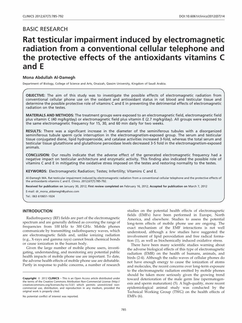
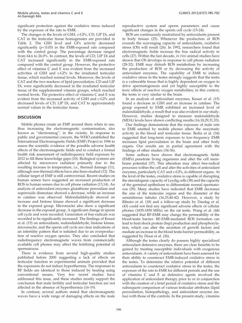
Histopathological alterationsThe testis sections in the three control groups revealed
that the seminiferous tubules consist of several layers ofepithelial cells (Figure 1). The cuboidal spermatogonia cells
Mobile phone, testes and vitamins C and EAl-Damegh MA
CLINICS 2012;67(7):785-792
786
have clear cytoplasm and rounded nuclei, which may showdivision. Sertoli cells are large epithelial cells that have avesicular nucleus with a large nucleolus. Next to thespermatogonia cells is a zone of spermatocytes, the nucleiof which are usually in mitotic division. A large number ofsmall cells, the spermatids, are seen external to thespermatocytes. The spermatids then further develop intospermatozoa. The latter usually lie in groups with theirheads projecting between the deeper cells and are connectedwith one of the Sertoli cells of the lining epithelium.
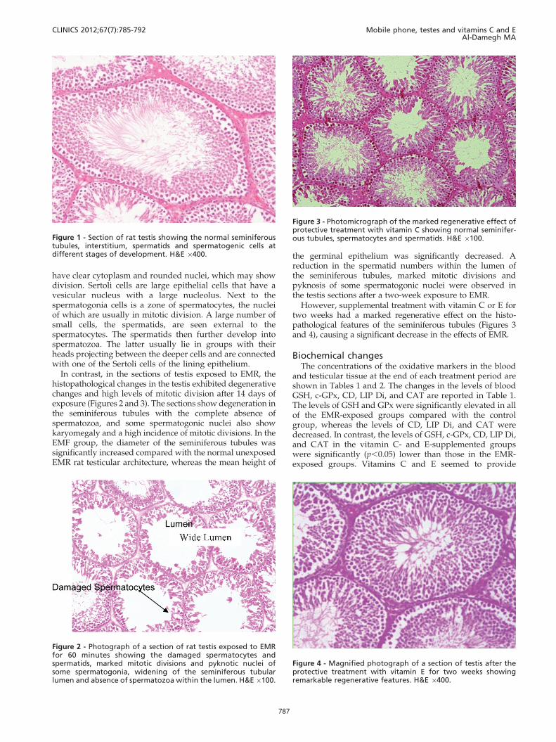
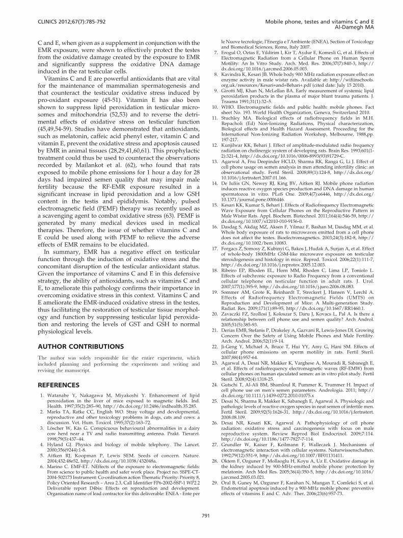
In contrast, in the sections of testis exposed to EMR, thehistopathological changes in the testis exhibited degenerativechanges and high levels of mitotic division after 14 days ofexposure (Figures 2 and 3). The sections show degeneration inthe seminiferous tubules with the complete absence ofspermatozoa, and some spermatogonic nuclei also showkaryomegaly and a high incidence of mitotic divisions. In theEMF group, the diameter of the seminiferous tubules wassignificantly increased compared with the normal unexposedEMR rat testicular architecture, whereas the mean height of
the germinal epithelium was significantly decreased. Areduction in the spermatid numbers within the lumen ofthe seminiferous tubules, marked mitotic divisions andpyknosis of some spermatogonic nuclei were observed inthe testis sections after a two-week exposure to EMR.
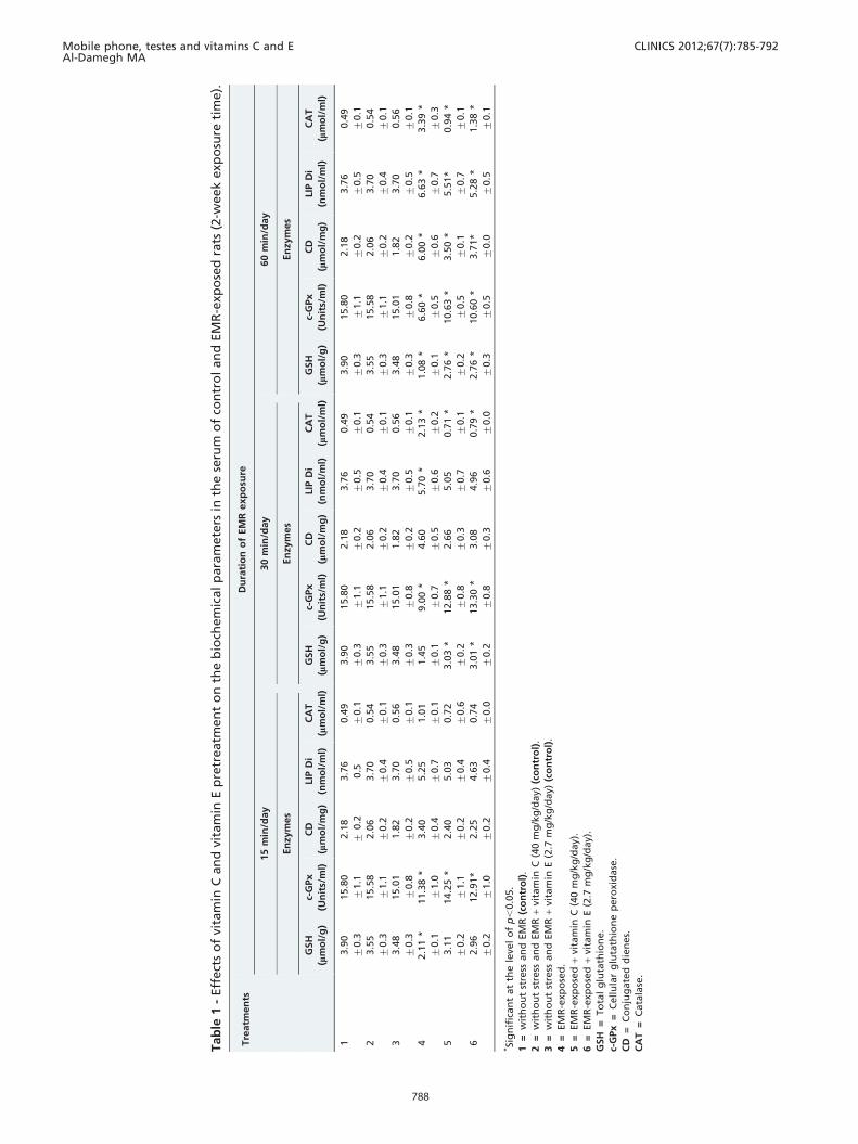
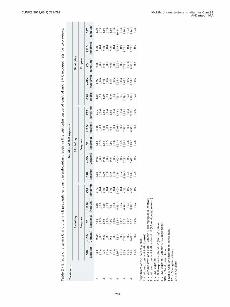
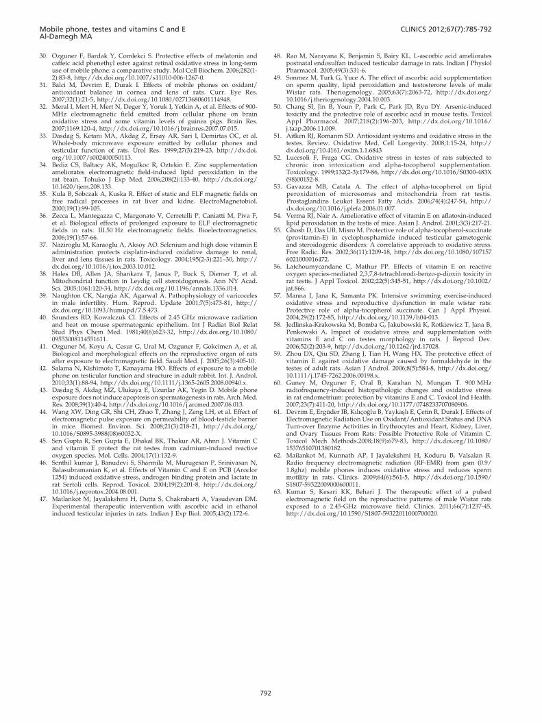
However, supplemental treatment with vitamin C or E fortwo weeks had a marked regenerative effect on the histo-pathological features of the seminiferous tubules (Figures 3and 4), causing a significant decrease in the effects of EMR.
Biochemical changesThe concentrations of the oxidative markers in the blood
and testicular tissue at the end of each treatment period areshown in Tables 1 and 2. The changes in the levels of bloodGSH, c-GPx, CD, LIP Di, and CAT are reported in Table 1.The levels of GSH and GPx were significantly elevated in allof the EMR-exposed groups compared with the controlgroup, whereas the levels of CD, LIP Di, and CAT weredecreased. In contrast, the levels of GSH, c-GPx, CD, LIP Di,and CAT in the vitamin C- and E-supplemented groupswere significantly (p,0.05) lower than those in the EMR-exposed groups. Vitamins C and E seemed to provide
Figure 1 - Section of rat testis showing the normal seminiferoustubules, interstitium, spermatids and spermatogenic cells atdifferent stages of development. H&E 6400.
Figure 2 - Photograph of a section of rat testis exposed to EMRfor 60 minutes showing the damaged spermatocytes andspermatids, marked mitotic divisions and pyknotic nuclei ofsome spermatogonia, widening of the seminiferous tubularlumen and absence of spermatozoa within the lumen. H&E6100.
Figure 3 - Photomicrograph of the marked regenerative effect ofprotective treatment with vitamin C showing normal seminifer-ous tubules, spermatocytes and spermatids. H&E 6100.
Figure 4 - Magnified photograph of a section of testis after theprotective treatment with vitamin E for two weeks showingremarkable regenerative features. H&E 6400.
CLINICS 2012;67(7):785-792 Mobile phone, testes and vitamins C and EAl-Damegh MA
787
Tab
le1
-Eff
ect
so
fvi
tam
inC
an
dvi
tam
inE
pre
treatm
en
to
nth
eb
ioch
em
ical
para
mete
rsin
the
seru
mo
fco
ntr
ol
an
dEM
R-e
xpo
sed
rats
(2-w
eek
exp
osu
reti
me).
Tre
atm
en
tsD
ura
tio
no
fEM
Rexp
osu
re
15
min
/day
30
min
/day
60
min
/day
En
zym
es
En
zym
es
En
zym
es
GSH
(mm
ol/
g)
c-G
Px
(Un
its/
ml)
CD
(mm
ol/
mg
)
LIP
Di
(nm
ol/
ml)
CA
T
(mm
ol/
ml)
GSH
(mm
ol/
g)
c-G
Px
(Un
its/
ml)
CD
(mm
ol/
mg
)
LIP
Di
(nm
ol/
ml)
CA
T
(mm
ol/
ml)
GSH
(mm
ol/
g)
c-G
Px
(Un
its/
ml)
CD
(mm
ol/
mg
)
LIP
Di
(nm
ol/
ml)
CA
T
(mm
ol/
ml)
13.9
0
¡0.3
15.8
0
¡1.1
2.1
8
¡0.2
3.7
6
0.5
0.4
9
¡0.1
3.9
0
¡0.3
15.8
0
¡1.1
2.1
8
¡0.2
3.7
6
¡0.5
0.4
9
¡0.1
3.9
0
¡0.3
15.8
0
¡1.1
2.1
8
¡0.2
3.7
6
¡0.5
0.4
9
¡0.1
23.5
5
¡0.3
15.5
8
¡1.1
2.0
6
¡0.2
3.7
0
¡0.4
0.5
4
¡0.1
3.5
5
¡0.3
15.5
8
¡1.1
2.0
6
¡0.2
3.7
0
¡0.4
0.5
4
¡0.1
3.5
5
¡0.3
15.5
8
¡1.1
2.0
6
¡0.2
3.7
0
¡0.4
0.5
4
¡0.1
33.4
8
¡0.3
15.0
1
¡0.8
1.8
2
¡0.2
3.7
0
¡0.5
0.5
6
¡0.1
3.4
8
¡0.3
15.0
1
¡0.8
1.8
2
¡0.2
3.7
0
¡0.5
0.5
6
¡0.1
3.4
8
¡0.3
15.0
1
¡0.8
1.8
2
¡0.2
3.7
0
¡0.5
0.5
6
¡0.1
42.1
1*
¡0.1
11.3
8*
¡1.0
3.4
0
¡0.4
5.2
5
¡0.7
1.0
1
¡0.1
1.4
5
¡0.1
9.0
0*
¡0.7
4.6
0
¡0.5
5.7
0*
¡0.6
2.1
3*
¡0.2
1.0
8*
¡0.1
6.6
0*
¡0.5
6.0
0*
¡0.6
6.6
3*
¡0.7
3.3
9*
¡0.3
53.1
1
¡0.2
14.2
5*
¡1.1
2.4
0
¡0.2
5.0
3
¡0.4
0.7
2
¡0.6
3.0
3*
¡0.2
12.8
8*
¡0.8
2.6
6
¡0.3
5.0
5
¡0.7
0.7
1*
¡0.1
2.7
6*
¡0.2
10.6
3*
¡0.5
3.5
0*
¡0.1
5.5
1*
¡0.7
0.9
4*
¡0.1
62.9
6
¡0.2
12.9
1*
¡1.0
2.2
5
¡0.2
4.6
3
¡0.4
0.7
4
¡0.0
3.0
1*
¡0.2
13.3
0*
¡0.8
3.0
8
¡0.3
4.9
6
¡0.6
0.7
9*
¡0.0
2.7
6*
¡0.3
10.6
0*
¡0.5
3.7
1*
¡0.0
5.2
8*
¡0.5
1.3
8*
¡0.1
*Si
gn
ific
an
tat
the
leve
lo
fp
,0.0
5.
1=
wit
ho
ut
stre
ssan
dEM
R(c
on
tro
l).
2=
wit
ho
ut
stre
ssan
dEM
R+
vita
min
C(4
0m
g/k
g/d
ay)
(co
ntr
ol)
.
3=
wit
ho
ut
stre
ssan
dEM
R+
vita
min
E(2
.7m
g/k
g/d
ay)
(co
ntr
ol)
.
4=
EM
R-e
xpo
sed
.
5=
EM
R-e
xpo
sed
+vi
tam
inC
(40
mg
/kg
/day)
.
6=
EM
R-e
xpo
sed
+vi
tam
inE
(2.7
mg
/kg
/day)
.
GSH
=To
tal
glu
tath
ion
e.
c-G
Px
=C
ell
ula
rg
luta
thio
ne
pero
xid
ase
.
CD
=C
on
jug
ate
dd
ien
es.
CA
T=
Cata
lase
.
Mobile phone, testes and vitamins C and EAl-Damegh MA
CLINICS 2012;67(7):785-792
788
Tab
le2
-Eff
ect
so
fvi
tam
inC
an
dvi
tam
inE
pre
treatm
en
to
nth
ean
tio
xid
an
tle
vels
inth
ete
stic
ula
rti
ssu
eo
fco
ntr
ol
an
dEM
Rexp
ose
dra
tsfo
rtw
ow
eeks.
Tre
atm
en
tsD
ura
tio
no
fEM
Rexp
osu
re
15
min
/day
30
min
/day
60
min
/day
En
zym
es
En
zym
es
En
zym
es
GSH
(mm
ol/
g)
c-G
Px
(Un
its/
ml)
CD
(mm
ol/
mg
)
LIP
Di
(nm
ol/
ml)
CA
T
(mm
ol/
ml)
GSH
(mm
ol/
g)
c-G
Px
(Un
its/
ml)
CD
(mm
ol/
mg
)
LIP
Di
(nm
ol/
ml)
CA
T
(mm
ol/
ml)
GSH
(mm
ol/
g)
c-G
Px
(Un
its/
ml)
CD
(mm
ol/
mg
)
LIP
Di
(nm
ol/
ml)
CA
T
(mm
ol/
ml)
14.5
8
¡0.4
9.0
0
¡0.6
4.5
8
¡0.5
5.3
8
¡0.4
3.7
0
¡0.4
4.5
8
¡0.4
9.0
0
¡0.6
4.5
8
¡0.5
5.3
8
¡0.4
3.7
0
¡0.4
4.5
8
¡0.4
9.0
0
¡0.6
4.5
8
¡0.5
5.3
8
¡0.4
3.7
0
¡0.4
24.2
0
¡0.4
9.4
3
¡0.6
5.6
1
¡0.5
4.5
5
¡0.4
3.8
6
¡0.4
4.2
0
¡0.4
9.4
3
¡0.6
5.6
1
¡0.5
4.5
5
¡0.4
3.8
6
¡0.4
4.2
0
¡0.4
9.4
3
¡0.6
5.6
1
¡0.5
4.5
5
¡0.4
3.8
6
¡0.4
34.4
9
¡0.4
9.5
1
¡0.5
4.9
0
¡0.6
4.3
1
¡0.4
3.8
6
¡0.4
4.4
9
¡0.4
9.5
1
¡0.5
4.9
0
¡0.6
4.3
1
¡0.4
3.8
6
¡0.4
4.4
9
¡0.4
9.5
1
¡0.5
4.9
0
¡0.6
4.3
1
¡0.4
3.8
6
¡0.4
42.9
6*
¡0.3
5.8
1*
¡0.5
6.3
3*
¡0.7
8.9
3*
¡0.7
5.4
0*
¡0.4
1.7
1*
¡0.2
4.4
0*
¡0.5
9.2
1*
¡0.5
12.0
3*
¡0.7
7.4
8*
¡0.4
1.0
3*
¡0.3
2.3
4*
¡0.4
12.2
0*
¡0.6
15.1
8*
¡0.9
10.3
4*
¡0.4
53.5
9
¡0.4
7.7
5*
¡0.5
5.2
5
¡0.7
6.3
6*
¡0.5
4.2
5
¡0.5
4.1
9*
¡0.5
7.2
8*
¡0.6
6.6
8*
¡0.6
7.5
6*
¡0.4
4.6
5*
¡0.4
2.7
9*
¡0.4
7.6
6*
¡0.5
7.7
2*
¡0.
8
8.7
4*
¡0.4
5.9
6*
¡0.5
63.8
1
¡0.5
9.0
8*
¡0.6
5.2
0
¡0.6
5.8
0*
¡0.7
4.3
6
¡0.6
3.9
1*
¡0.4
7.6
8*
¡0.5
5.7
5*
¡0.6
6.9
5*
¡0.4
5.2
1*
¡0.5
3.3
6*
¡0.5
7.3
4*
¡0.6
6.9
4*
¡0.7
7.7
8*
¡0.5
5.7
4*
¡0.6
*Si
gn
ific
an
tat
the
leve
lo
fp
,0.0
5.
1=
wit
ho
ut
stre
ssan
dEM
R(c
on
tro
l).
2=
wit
ho
ut
stre
ssan
dEM
R+
vita
min
C(4
0m
g/k
g/d
ay)
(co
ntr
ol)
.
3=
wit
ho
ut
stre
ssan
dEM
R+
vita
min
E(2
.7m
g/k
g/d
ay)
(co
ntr
ol)
.
4=
EM
R-e
xpo
sed
.
5=
EM
R-e
xpo
sed
+vi
tam
inC
(40
mg
/kg
/day)
.
6=
EM
R-e
xpo
sed
+vi
tam
inE
(2.7
mg
/kg
/day)
.
GSH
=To
tal
glu
tath
ion
e.
c-G
Px
=C
ell
ula
rg
luta
thio
ne
pero
xid
ase
.
CD
=C
on
jug
ate
dd
ien
es.
CA
T=
Cata
lase
.
CLINICS 2012;67(7):785-792 Mobile phone, testes and vitamins C and EAl-Damegh MA
789
significant protection against the oxidative stress inducedby the exposure of the rats to EMR.
The changes in the levels of GSH, c-GPx, CD, LIP Di, andCAT in the testicular tissue homogenates are provided inTable 2. The GSH level and GPx activity decreasedsignificantly (p,0.05) in the EMR-exposed rats comparedwith the control group. The percentage decrease rangedfrom 64.6 to 26.0%. In contrast, the levels of CD, LIP Di andCAT increased significantly in the EMR-exposed ratscompared with the control group. However, the protectiveeffect of vitamins C and E was evident from the increasedactivities of GSH and c-GPx in the irradiated testiculartissue, which reached normal levels. Moreover, the levels ofCAT and the two markers of lipid peroxidation, CD and LIPDi, were significantly decreased in the irradiated testiculartissue of the supplemented vitamin groups, which reachednormal levels. The protective effect of vitamins C and E wasevident from the increased activity of GSH and c-GPx anddecreased levels of CD, LIP Di, and CAT to approximatelynormal values in the testicular tissue.
DISCUSSION
Mobile phones create an EMF around them when in use,thus increasing the electromagnetic contamination, alsoknown as ‘‘electrosmog’’, in the vicinity. In response topublic and governmental concern, the WHO established theInternational Electromagnetic Fields (EMF) Project in 1996 toassess the scientific evidence of the possible adverse healtheffects of the electromagnetic fields and to conduct a formalhealth risk assessment of radiofrequency field exposure by2012 to fill these knowledge gaps (10). Biological systems areaffected by microwave radiation primarily due to theresulting increase in temperature, i.e., thermal damage (11),although non-thermal effects have also been studied (12). Thecellular target of EMF is still controversial. Recent studies onhuman semen have suggested an increased production ofROS in human semen due to cell phone radiation (13,14). Ananalysis of antioxidant enzymes glutathione peroxidase andsuperoxide dismutase showed a decrease, while an increasein catalase was observed. Malondialdehyde showed anincrease and histone kinase showed a significant decreasein the exposed group. Micronuclei also show a significantdecrease in the exposed group. A significant change in spermcell cycle and were recorded. Generation of free radicals wasrecorded to be significantly increased. The findings of Kesariet al. (15) on antioxidants, malondialdehyde, histone kinase,micronuclei, and the sperm cell cycle are clear indications ofan infertility pattern that is initiated due to an overproduc-tion of reactive oxygen species. They also concluded thatradiofrequency electromagnetic waves from commerciallyavailable cell phones may affect the fertilizing potential ofspermatozoa.
There is evidence from several high-quality studiespublished before 2000 suggesting a lack of effects ontesticular function in experimental animals provided thatthe exposures do not induce hyperthermia. The responses toRF fields are identical to those induced by heating usingconventional means. Very few recent studies haveaddressed this issue, and these studies mostly support theconclusion that male fertility and testicular function are notaffected in the absence of hyperthermia (16-19).
In contrast, several authors stated that electromagneticwaves have a wide range of damaging effects on the male
reproductive system and sperm parameters and causesignificant changes in the sperm cell cycle (15-24).
ROS are continuously neutralized by antioxidants presentin body tissues (25). Whenever the production of ROSexceeds the scavenging capacity of antioxidants, oxidativestress (OS) will result (26). In 1992, researchers found thatelectromagnetic fields increase the free radical activity incells (27). Within the last decade, in vivo animal studies haveshown that OS develops in response to cell phone radiation(28-32). EMR may disturb ROS metabolism by increasingthe production of ROS or by decreasing the activity ofantioxidant enzymes. The capability of EMR to induceoxidative stress in the testes strongly suggests that the testisis a vulnerable tissue that is highly dependent on oxygen todrive spermatogenesis and yet highly susceptible to thetoxic effects of reactive oxygen metabolites; in this context,the testis is very similar to the brain.
In an analysis of antioxidant enzymes, Kesari et al. (15)found a decrease in GSH and an increase in catalase. Thegroup exposed to EMR exhibited an increased level ofmalondialdehyde, a result that was also evident in our study.However, studies designed to measure malonaldehyde(MDA) levels have shown conflicting results (16,18,29,31,33).
Our findings demonstrate that the exposure of male ratsto EMR emitted by mobile phones alters the enzymaticactivity in the blood and testicular tissue. Bediz et al. (34)suggested that long-term exposure to low-frequency EMRincreases lipid peroxidation in the brain and other bodyorgans. Our results are in partial agreement with thefindings of other studies (34-36).
Some authors have shown that electromagnetic fields(EMFs) penetrate living organisms and alter the cell mem-brane potential (37). This alteration may affect free-radicalprocesses within the cell and alter the activities of antioxidantenzymes, particularly CAT and c-GPx, in different organs. Atthe level of the testes, oxidative stress is capable of disruptingthe steroidogenic capacity of Leydig cells (38) and the capacityof the germinal epithelium to differentiate normal spermato-zoa (39). Many studies have indicated that EMR decreasesthe size of the testicular organs and the diameter of theseminiferous tubules (16,33,40-42). However, a study byRibeiro et al. (18) and a follow-up study by Dasdag et al.(43) could not find any significant adverse effects of cellularphones (1835-1850 MHz) on the rat testis. Wang et al. (44)suggested that RF-EMR may change the permeability of theblood-testis barrier. RF-EMR-mediated ROS formation canlead to heat shock protein (hsp) production and phosphoryla-tion, which can alter the secretion of growth factors andmediate an increase in the blood-testis barrier permeability, assuggested by Desai et al. (26).
Although the testes clearly do possess highly specializedantioxidant defensive enzymes, there are clear benefits to begained by treating susceptible individuals with exogenousantioxidants. A variety of antioxidants have been assessed fortheir ability to counteract EMR-induced oxidative stress inthe testes. To determine the relative potential of differentantioxidants to counteract oxidative stress in the testes, theexposure of the rats to EMR for different periods and the useof vitamins C and E as defensive agents involved theapplication of antioxidant therapy prior to or in conjunctionwith the creation of a brief period of oxidative stress and thesubsequent comparison of various testicular attributes (lipidperoxidation, histopathology, and antioxidant enzyme sta-tus) with those of the controls. In the present study, vitamins
Mobile phone, testes and vitamins C and EAl-Damegh MA
CLINICS 2012;67(7):785-792
790
C and E, when given as a supplement in conjunction with theEMR exposure, were shown to effectively protect the testesfrom the oxidative damage created by the exposure to EMRand significantly suppress the oxidative DNA damageinduced in the rat testicular cells.
Vitamins C and E are powerful antioxidants that are vitalfor the maintenance of mammalian spermatogenesis andthat counteract the testicular oxidative stress induced bypro-oxidant exposure (45-51). Vitamin E has also beenshown to suppress lipid peroxidation in testicular micro-somes and mitochondria (52,53) and to reverse the detri-mental effects of oxidative stress on testicular function(45,49,54-59). Studies have demonstrated that antioxidants,such as melatonin, caffeic acid phenyl ester, vitamin C andvitamin E, prevent the oxidative stress and apoptosis causedby EMR in animal tissues (28,29,41,60,61). This prophylactictreatment could thus be used to counteract the observationsrecorded by Mailankot et al. (62), who found that ratsexposed to mobile phone emissions for 1 hour a day for 28days had impaired semen quality that may impair malefertility because the RF-EMR exposure resulted in asignificant increase in lipid peroxidation and a low GSHcontent in the testis and epididymis. Notably, pulsedelectromagnetic field (PEMF) therapy was recently used asa scavenging agent to combat oxidative stress (63). PEMF isgenerated by many medical devices used in medicaltherapies. Therefore, the issue of whether vitamins C andE could be used along with PEMF to relieve the adverseeffects of EMR remains to be elucidated.
In summary, EMR has a negative effect on testicularfunction through the induction of oxidative stress and theconcomitant disruption of the testicular antioxidant status.Given the importance of vitamins C and E in this defensivestrategy, the ability of antioxidants, such as vitamins C andE, to ameliorate this pathology confirms their importance inovercoming oxidative stress in this context. Vitamins C andE ameliorate the EMR-induced oxidative stress in the testes,thus facilitating the restoration of testicular tissue morphol-ogy and function by suppressing testicular lipid peroxida-tion and restoring the levels of GST and GSH to normalphysiological levels.
AUTHOR CONTRIBUTIONS
The author was solely responsible for the entire experiment, which
included planning and performing the experiments and writing and
revising the manuscript.
REFERENCES
1. Watanabe Y, Nakagawa M, Miyakoshi Y. Enhancement of lipidperoxidation in the liver of mice exposed to magnetic fields. Ind.Health. 1997;35(2):285–90, http://dx.doi.org/10.2486/indhealth.35.285.
2. Marks TA, Ratke CC, English WO. Stray voltage and developmental,reproductive and other toxicology problems in dogs, cats and cows: adiscussion. Vet. Hum. Toxicol. 1995;37(2):163–72.
3. Loscher W, Kas G. Conspicuous behavioural abnormalities in a dairycow herd near a TV and radio transmitting antenna. Prakt. Tierarzt.1998;79(5):437–44.
4. Hyland GJ. Physics and biology of mobile telephony. The Lancet.2000;356(9244):1-8.
5. Aitken RJ, Koopman P, Lewis SEM. Seeds of concern. Nature.2004;432:48e52, http://dx.doi.org/10.1038/432048a.
6. Marino C. EMF-ET. NEffects of the exposure to electromagnetic fields:From science to public health and safer work place. Project no. SSPE-CT-2004-502173 Instrument: Co-ordination action Thematic Priority: Priority 8,Policy Oriented Research – Area 2.3, Call Identifier FP6-2002-SSP-1 WP2.2Deliverable report D4bis: Effects on reproduction and development.Organisation name of lead contractor for this deliverable: ENEA - Ente per
le Nuove tecnologie, l’Energia e l’Ambiente (ENEA), Section of Toxicologyand Biomedical Sciences, Roma, Italy 2007.
7. Erogul O, Oztas E, Yildirim I, Kir T, Aydur E, Komesli G, et al. Effects ofElectromagnetic Radiation from a Cellular Phone on Human SpermMotility: An In Vitro Study. Arch. Med. Res. 2006;37(7):840-3, http://dx.doi.org/10.1016/j.arcmed.2006.05.003.
8. Kavindra K, Kesari JB. Whole body 900 MHz radiation exposure effect onenzyme activity in male wistar rats. Available at: http://wifiinschools.org.uk/resources/Kesari+and+Behari+.pdf (cited date: July 15 2010).
9. Girotti MJ, Khan N, McLellan BA. Early measurement of systemic lipidperoxidation products in the plasma of major blunt trauma patients. J.Trauma 1991;31(1):32–5.
10. WHO. Electromagnetic fields and public health: mobile phones. Factsheet No. 193. World Health Organization, Geneva, Switzerland 2010.
11. Stuchley MA. Biological effects of radiofrequency fields in M.H.Repacholi (Ed) Non-Ionizing Radiations, Physical characterization,Biological effects and Health Hazard Assessment. Proceeding for theInternational Non-Ionizing Radiation Workshop, Melbourne, 1988,pp.197-217.
12. Kunjilwar KK, Behari J. Effect of amplitude-modulated radio frequencyradiation on cholinergic system of developing rats. Brain Res. 1993;601(1-2):321-4, http://dx.doi.org/10.1016/0006-8993(93)91729-C.
13. Agarwal A, Fnu Deepinder HCLD, Sharma RK, Ranga G, Li J. Effect ofcell phone usage on semen analysis in men attending infertility clinic: anobservational study. Fertil Steril. 2008;89(1):124-8, http://dx.doi.org/10.1016/j.fertnstert.2007.01.166.
14. De Iuliis GN, Newey RJ, King BV, Aitken RJ. Mobile phone radiationinduces reactive oxygen species production and DNA damage in humanspermatozoa in vitro. PLoS One. 2009;4(7):e6446, http://dx.doi.org/10.1371/journal.pone.0006446.
15. Kesari KK, Kumar S, Behari J. Effects of Radiofrequency ElectromagneticWave Exposure from Cellular Phones on the Reproductive Pattern inMale Wistar Rats. Appl. Biochem. Biotechnol. 2011;164(4):546-59, http://dx.doi.org/10.1007/s12010-010-9156-0.
16. Dasdag S, Akdag MZ, Aksen F, Yilmaz F, Bashan M, Dasdag MM, et al.Whole body exposure of rats to microwaves emitted from a cell phonedoes not affect the testes. Bioelectromagnetics. 2003;24(3):182-8, http://dx.doi.org/10.1002/bem.10083.
17. Forgacs Z, Somosy Z, Kubinyi G, Bakos J, Hudak A, Surjan A, et al. Effectof whole-body 1800MHz GSM-like microwave exposure on testicularsteroidogenesis and histology in mice. Reprod. Toxicol. 2006;22(1):111-7,http://dx.doi.org/10.1016/j.reprotox.2005.12.003.
18. Ribeiro EP, Rhoden EL, Horn MM, Rhoden C, Lima LP, Toniolo L.Effects of subchronic exposure to Radio Frequency from a conventionalcellular telephone on testicular function in adult rats. J. Urol.2007;177(1):395-9, http://dx.doi.org/10.1016/j.juro.2006.08.083.
19. Sommer AM, Grote K, Reinhardt T, Streckert J, Hansen V, Lerchl A.Effects of Radiofrequency Electromagnetic Fields (UMTS) onReproduction and Development of Mice: A Multi-generation Study.Radiat. Res. 2009;171(1):89-95, http://dx.doi.org/10.1667/RR1460.1.
20. Zavaczki FZ, Szollosi J, Koloszar S, Daru J, Kovacs L, Pal A. Is there arelationship between cell phone use and semen quality? Arch Androl.2005;51(5):385-93.
21. Derias EMB, Stefanis P, Drakeley A, Gazvani R, Lewis-Jones DI. GrowingConcern Over the Safety of Using Mobile Phones and Male Fertility.Arch. Androl. 2006;52(1):9-14.
22. Ji-Geng Y, Michael A, Bruce T, Hui YY, Amy G, Hani SM. Effects ofcellular phone emissions on sperm motility in rats. Fertil Steril.2007;88(4):957-64.
23. Agarwal A, Desai NR, Makker K, Varghese A, Mouradi R, Sabanegh E,et al. Effects of radiofrequency electromagnetic waves (RF-EMW) fromcellular phones on human ejaculated semen: an in vitro pilot study. FertilSteril. 2008;92(4):1318-25.
24. Gutschi T, Al-Ali BM, Shamloul R, Pummer K, Trummer H. Impact ofcell phone use on men’s semen parameters. Andrologia. 2011; http://dx.doi.org/10.1111/j.1439-0272.2010.01075.x
25. Desai N, Sharma R, Makker K, Sabanegh E, Agarwal A. Physiologic andpathologic levels of reactive oxygen species in neat semen of infertile men.Fertil Steril. 2009;92(5):1626-31, http://dx.doi.org/10.1016/j.fertnstert.2008.08.109.
26. Desai NR, Kesari KK, Agarwal A. Pathophysiology of cell phoneradiation: oxidative stress and carcinogenesis with focus on malereproductive system. Review Reprod Biol Endocrinol. 2009;7:114.http://dx.doi.org/10.1186/1477-7827-7-114.
27. Grundler W, Kaiser F, Keilmann F, Walleczek J. Mechanisms ofelectromagnetic interaction with cellular systems. Naturwissenschaften.1992;79(12):551-9, http://dx.doi.org/10.1007/BF01131411.
28. Oktem F, Ozguner F, Mollaoglu H, Koyu A, Uz E. Oxidative damage inthe kidney induced by 900-MHz-emitted mobile phone: protection bymelatonin. Arch Med Res. 2005;36(4):350-5, http://dx.doi.org/10.1016/j.arcmed.2005.03.021.
29. Oral B, Guney M, Ozguner F, Karahan N, Mungan T, Comlekci S, et al.Endometrial apoptosis induced by a 900-MHz mobile phone: preventiveeffects of vitamins E and C. Adv. Ther. 2006;23(6):957-73.
CLINICS 2012;67(7):785-792 Mobile phone, testes and vitamins C and EAl-Damegh MA
791
30. Ozguner F, Bardak Y, Comlekci S. Protective effects of melatonin andcaffeic acid phenethyl ester against retinal oxidative stress in long-termuse of mobile phone: a comparative study. Mol Cell Biochem. 2006;282(1-2):83-8, http://dx.doi.org/10.1007/s11010-006-1267-0.
31. Balci M, Devrim E, Durak I. Effects of mobile phones on oxidant/antioxidant balance in cornea and lens of rats. Curr. Eye Res.2007;32(1):21-5, http://dx.doi.org/10.1080/02713680601114948.
32. Meral I, Mert H, Mert N, Deger Y, Yoruk I, Yetkin A, et al. Effects of 900-MHz electromagnetic field emitted from cellular phone on brainoxidative stress and some vitamin levels of guinea pigs. Brain Res.2007;1169:120-4, http://dx.doi.org/10.1016/j.brainres.2007.07.015.
33. Dasdag S, Ketani MA, Akdag Z, Ersay AR, Sari I, Demirtas OC, et al.Whole-body microwave exposure emitted by cellular phones andtesticular function of rats. Urol Res. 1999;27(3):219-23, http://dx.doi.org/10.1007/s002400050113.
34. Bediz CS, Baltacy AK, Mogulkoc R, Oztekin E. Zinc supplementationameliorates electromagnetic field-induced lipid peroxidation in therat brain. Tohuko J Exp Med. 2006;208(2):133-40, http://dx.doi.org/10.1620/tjem.208.133.
35. Kula B, Sobczak A, Kuska R. Effect of static and ELF magnetic fields onfree radical processes in rat liver and kidne. ElectroMagnetobiol.2000;19(1):99-105.
36. Zecca L, Mantegazza C, Margonato V, Cerretelli P, Caniatti M, Piva F,et al. Biological effects of prolonged exposure to ELF electromagneticfields in rats: III.50 Hz electromagnetic fields. Bioelectromagnetics.2006;19(1):57-66.
37. Naziroglu M, Karaoglu A, Aksoy AO. Selenium and high dose vitamin Eadministration protects cisplatin-induced oxidative damage to renal,liver and lens tissues in rats. Toxicology. 2004;195(2-3):221–30, http://dx.doi.org/10.1016/j.tox.2003.10.012.
38. Hales DB, Allen JA, Shankara T, Janus P, Buck S, Diemer T, et al.Mitochondrial function in Leydig cell steroidogenesis. Ann NY Acad.Sci. 2005;1061:120-34, http://dx.doi.org/10.1196/annals.1336.014.
39. Naughton CK, Nangia AK, Agarwal A. Pathophysiology of varicocelesin male infertility. Hum. Reprod. Update 2001;7(5):473-81, http://dx.doi.org/10.1093/humupd/7.5.473.
40. Saunders RD, Kowalczuk CI. Effects of 2.45 GHz microwave radiationand heat on mouse spermatogenic epithelium. Int J Radiat Biol RelatStud Phys Chem Med. 1981;40(6):623-32, http://dx.doi.org/10.1080/09553008114551611.
41. Ozguner M, Koyu A, Cesur G, Ural M, Ozguner F, Gokcimen A, et al.Biological and morphological effects on the reproductive organ of ratsafter exposure to electromagnetic field. Saudi Med. J. 2005;26(3):405-10.
42. Salama N, Kishimoto T, Kanayama HO. Effects of exposure to a mobilephone on testicular function and structure in adult rabbit. Int. J. Androl.2010;33(1):88-94, http://dx.doi.org/10.1111/j.1365-2605.2008.00940.x.
43. Dasdag S, Akdag MZ, Ulukaya E, Uzunlar AK, Yegin D. Mobile phoneexposure does not induce apoptosis on spermatogenesis in rats. Arch. Med.Res. 2008;39(1):40-4, http://dx.doi.org/10.1016/j.arcmed.2007.06.013.
44. Wang XW, Ding GR, Shi CH, Zhao T, Zhang J, Zeng LH, et al. Effect ofelectromagnetic pulse exposure on permeability of blood-testicle barrierin mice. Biomed. Environ. Sci. 2008;21(3):218-21, http://dx.doi.org/10.1016/S0895-3988(08)60032-X.
45. Sen Gupta R, Sen Gupta E, Dhakal BK, Thakur AR, Ahnn J. Vitamin Cand vitamin E protect the rat testes from cadmium-induced reactiveoxygen species. Mol. Cells. 2004;17(1):132-9.
46. Senthil kumar J, Banudevi S, Sharmila M, Murugesan P, Srinivasan N,Balasubramanian K, et al. Effects of Vitamin C and E on PCB (Aroclor1254) induced oxidative stress, androgen binding protein and lactate inrat Sertoli cells. Reprod. Toxicol. 2004;19(2):201-8, http://dx.doi.org/10.1016/j.reprotox.2004.08.001.
47. Mailankot M, Jayalakshmi H, Dutta S, Chakrabarti A, Vasudevan DM.Experimental therapeutic intervention with ascorbic acid in ethanolinduced testicular injuries in rats. Indian J Exp Biol. 2005;43(2):172-6.
48. Rao M, Narayana K, Benjamin S, Bairy KL. L-ascorbic acid amelioratespostnatal endosulfan induced testicular damage in rats. Indian J PhysiolPharmacol. 2005;49(3):331-6.
49. Sonmez M, Turk G, Yuce A. The effect of ascorbic acid supplementationon sperm quality, lipid peroxidation and testosterone levels of maleWistar rats. Theriogenology. 2005;63(7):2063-72, http://dx.doi.org/10.1016/j.theriogenology.2004.10.003.
50. Chang SI, Jin B, Youn P, Park C, Park JD, Ryu DY. Arsenic-inducedtoxicity and the protective role of ascorbic acid in mouse testis. ToxicolAppl Pharmacol. 2007;218(2):196-203, http://dx.doi.org/10.1016/j.taap.2006.11.009.
51. Aitken RJ, Romanm SD. Antioxidant systems and oxidative stress in thetestes. Review. Oxidative Med. Cell Longevity. 2008;1:15-24, http://dx.doi.org/10.4161/oxim.1.1.6843
52. Lucesoli F, Fraga CG. Oxidative stress in testes of rats subjected tochronic iron intoxication and alpha-tocopherol supplementation.Toxicology. 1999;132(2-3):179-86, http://dx.doi.org/10.1016/S0300-483X(98)00152-8.
53. Gavazza MB, Catala A. The effect of alpha-tocopherol on lipidperoxidation of microsomes and mitochondria from rat testis.Prostaglandins Leukot Essent Fatty Acids. 2006;74(4):247-54, http://dx.doi.org/10.1016/j.plefa.2006.01.007.
54. Verma RJ, Nair A. Ameliorative effect of vitamin E on aflatoxin-inducedlipid peroxidation in the testis of mice. Asian J. Androl. 2001;3(3):217-21.
55. Ghosh D, Das UB, Misro M. Protective role of alpha-tocopherol-succinate(provitamin-E) in cyclophosphamide induced testicular gametogenicand steroidogenic disorders: A correlative approach to oxidative stress.Free Radic. Res. 2002;36(11):1209-18, http://dx.doi.org/10.1080/1071576021000016472.
56. Latchoumycandane C, Mathur PP. Effects of vitamin E on reactiveoxygen species-mediated 2,3,7,8-tetrachlorodi-benzo-p-dioxin toxicity inrat testis. J Appl Toxicol. 2002;22(5):345-51, http://dx.doi.org/10.1002/jat.866.
57. Manna I, Jana K, Samanta PK. Intensive swimming exercise-inducedoxidative stress and reproductive dysfunction in male wistar rats:Protective role of alpha-tocopherol succinate. Can J Appl Physiol.2004;29(2):172-85, http://dx.doi.org/10.1139/h04-013.
58. Jedlinska-Krakowska M, Bomba G, Jakubowski K, Rotkiewicz T, Jana B,Penkowski A. Impact of oxidative stress and supplementation withvitamins E and C on testes morphology in rats. J Reprod Dev.2006;52(2):203-9, http://dx.doi.org/10.1262/jrd.17028.
59. Zhou DX, Qiu SD, Zhang J, Tian H, Wang HX. The protective effect ofvitamin E against oxidative damage caused by formaldehyde in thetestes of adult rats. Asian J Androl. 2006;8(5):584-8, http://dx.doi.org/10.1111/j.1745-7262.2006.00198.x.
60. Guney M, Ozguner F, Oral B, Karahan N, Mungan T. 900 MHzradiofrequency-induced histopathologic changes and oxidative stressin rat endometrium: protection by vitamins E and C. Toxicol Ind Health.2007;23(7):411-20, http://dx.doi.org/10.1177/0748233707080906.
61. Devrim E, Erguder IB, Kılıcoglu B, Yaykaslı E, Cetin R, Durak J. Effects ofElectromagnetic Radiation Use on Oxidant/Antioxidant Status and DNATurn-over Enzyme Activities in Erythrocytes and Heart, Kidney, Liver,and Ovary Tissues From Rats: Possible Protective Role of Vitamin C.Toxicol Mech Methods.2008;18(9):679-83, http://dx.doi.org/10.1080/15376510701380182.
62. Mailankot M, Kunnath AP, I Jayalekshmi H, Koduru B, Valsalan R.Radio frequency electromagnetic radiation (RF-EMR) from gsm (0.9/1.8ghz) mobile phones induces oxidative stress and reduces spermmotility in rats. Clinics. 2009;64(6):561-5, http://dx.doi.org/10.1590/S1807-59322009000600011.
63. Kumar S, Kesari KK, Behari J. The therapeutic effect of a pulsedelectromagnetic field on the reproductive patterns of male Wistar ratsexposed to a 2.45-GHz microwave field. Clinics. 2011;66(7):1237-45,http://dx.doi.org/10.1590/S1807-59322011000700020.
Mobile phone, testes and vitamins C and EAl-Damegh MA
CLINICS 2012;67(7):785-792
792


![Isolated Testicular Tuberculosis Mimicking Testicular ... involvement, but testicular involvement is an unusual clinical condition [3]. In this report, a case with isolated testicular](https://img.dokumen.tips/doc/110x75/5f3d57bf74280d66ef795ba2/isolated-testicular-tuberculosis-mimicking-testicular-involvement-but-testicular.jpg)