Embed Size (px)
Citation preview
Radiation Safety
University of Nevada Reno
EH&S
Revised February 2015
Compiled by M. Jo
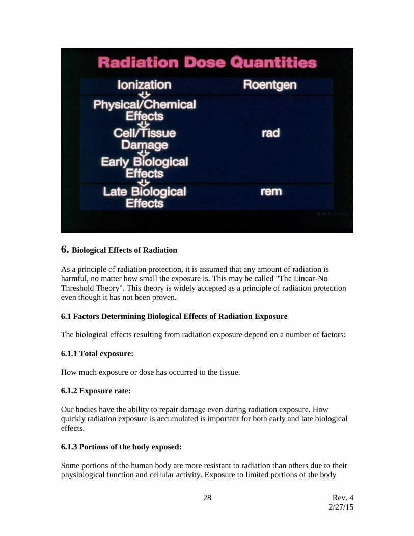
Copyright (c) 2015, Environmental Health & Safety, All Rights Reserved. Some graphs in this document are the property of the ENRG, Inc; used by permission.

Rev. 4
2/27/15
2
Rev. 4
2/27/15
3
Table of Contents
1. Radiation Safety Program Organization and Responsibilities
2. Atoms, Radioactivity, Radioactive Decay, and Radiation
2.1 Atomic Structure
2.2 Radioactive Decay and Half-life
2.3 Types of Radiation
3. Radiation Interactions with Matter
3.1 Alpha Radiation interactions with matter
3.2 Beta Radiation interactions with matter
3.3 Gamma and X-ray interactions with matter
3.1 Neutron interactions with matter
4. Radiation Detection Equipment
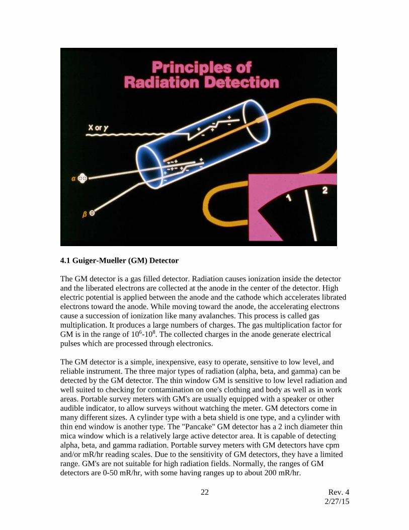
4.1 Guiger-Muller (GM) Detector
4.2 Ion Chamber
4.3 Gas Proportional Counter
4.4 Scintillation Detector
4.5 Solid State/Semiconductor Detectors
4.6 Personal Monitoring Devices
5. Units of Radioactivity and Radiation Measurement
5.1 Radioactivity
5.2 Radiation exposure
5.3 Absorbed dose
5.4 Dose equivalent
5.5 Effective dose equivalent
6. Biological Effects of Radiation
6.1 Factors Determining Biological Effects of Radiation Exposure
6.2 Cell sensitivity to radiation
6.3 Radiation effects on live cells
6.4 Damaged cell repair mechanism
6.5Acute and chronic exposure
6.6 Database of biological effects of radiation exposure
7. Radiation Exposure Limits
8. External Radiation Protection
8.1 Time, distance, and shielding
8.2 Security of radioactive material
8.3 Amount of radioactive material
9. Internal Radiation Protection, Contamination Control and monitoring
9.1 Contamination
9.2 Internal dosimetry
10. Emergency Procedures
10.1 Personnel decontamination 10.2 Minor spills and contamination
10.3 Major spills
Rev. 4
2/27/15
4
1. Radiation Safety Program Organization and Responsibilities
1.1 INTRODUCTION
The University of Nevada Reno (UNR) is committed to provide faculty, staff, and
students protection from the hazards associated with the use of radiation in academics
and research. UNR, as licensee for the possession and use of radiation sources at the
University, recognizes its responsibility to establish appropriate policies and procedures
for the safe use of radiation sources. UNR has established a Radiation Safety Committee
to develop and implement Radiation Safety policies and procedures, established the
EH&S Department to manage all safety functions including radiation safety, and
appointed a Radiation Safety Officer (RSO) to manage day-to-day radiation safety
program operation. UNR’s Radiation Safety Manual describes the organization and
responsibilities outlined in UNR's comprehensive Radiation Safety Program, including
the radiation services available to each user of a radiation source. The Manual was
prepared to be consistent with all applicable Federal and State regulations.
Radionuclides used in research, industry, education and medicine are valuable assets
which can benefit mankind if used properly. They can, however, present hazards because
of their ability to irradiate and contaminate humans and our environment. As a
consequence, persons who use radiation sources must understand the various types of
radiation hazards and adhere to regulations and standard practices designed to ensure
their safe use.
Radiation sources at UNR are regulated by the State Radiological Health Section in
accordance with the provisions of NAC 459.320 through 374.
The radiation protection standard is based on the Linear No-threshold Theory, even
though it is not a scientifically proven theory. As a result, it is assumed that there is no
safe amount of radiation and that any radiation exposure has a certain risk even though it
may be small. Therefore, it is the responsibility of all radiation users to avoid all
unnecessary and avoidable radiation exposure to himself/herself and others. This is called
“as low as reasonably achievable” or ALARA principle.
1.2 ORGANIZATION and RESPONSIBILITY
1.2.1 Radiation Safety Committee (RSC)
Membership
The RSC is appointed by UNR management. It advises the UNR management on all
matters relating to radiation safety. The committee includes the Radiation Safety Officer
(RSO); a representative of management; and members who represent broad areas or
divisions of UNR which are likely to use radiation sources, and is thus a mechanism for
dissemination of information to the various possible users. The RSO is a permanent
member of the RSC and of all of its subcommittees.
Rev. 4
2/27/15
5
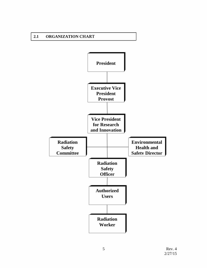
2.1 ORGANIZATION CHART
Radiation
Safety
Committee
Environmental
Health and
Safety Director
Radiation
Worker
Authorized
Users
Radiation
Safety
Officer
Vice President
for Research
and Innovation
Executive Vice
President
Provost
President
Rev. 4
2/27/15
6
Committee Responsibilities
The RSC establishes appropriate policies and procedures to ensure control of the
procurement and use of byproduct materials, completion of safety evaluations of
proposed uses and users, and the overall development and implementation of the
radiation safety program. Any new user must be approved by RSC before first use.
The committee is responsible for assuring that an adequate safety program is developed.
The RSC may delegate its authority to various persons and subcommittees with specific
expertise in areas under their purview.
The committee delegates its authority to the RSO to review and approve/disapprove
requests of a routine nature relating to the use of radiation sources.
The committee meets as often as necessary, but not less than quarterly.
1.2.2 EH&S
The EH&S Department consists of four components of safety programs. They are
Radiation Safety, Industrial Hygiene/Occupational Safety, Hazardous Waste Operations
(Hazardous Material Office), and Safety Training Coordination. The comprehensive
EH&S program is managed by a Director who reports directly to the Vice President for
Research. Supervisors of the four functional units are directly responsible for their
operations. Technical services and training offered to the campus are internally
complementary and solutions to many problems in laboratory and workplace
environments result from a coordinated team approache.
The EH&S Department provides the following services
Receipt & issue of radioactive source
Personnel monitoring
Radiation and radioactivity monitoring
Radiation instrument calibration
Waste pick-up and disposal
Transportation and shipping assistance
Emergency assistance
Radiation safety training
1.2.3 Radiation Safety Officer
The Radiation Safety Officer shall be the committee's authorized representative regarding
measures concerning radiation safety within the jurisdiction of UNR's radioactive
material license.
Rev. 4
2/27/15
7
The RSO in addition to administering and directing the day-to-day operations of the
Radiation Safety Program, reviews all applications to use radiation sources and advises
the RSC.
1.2.4 Authorized User
An individual is designated an Authorized User by the RSC after careful consideration of
his/her training and experience relative to radiation sources. He/she must apply and
receive written authorization before conducting any activity involving radioactive
material. The Authorized User shall have the primary responsibility for insuring the safe
use of the radiation source and compliance with applicable rules and regulations. He/She
must also insure that any person acting under his/her supervision is trained in accordance
with requirements of the university policy and is aware of the radiation hazards
associated with the activity of the materials in use. Application forms, procedures, and
additional requirements are specified in the EH&S webpage and in the Radiation Safety
Manual. Authorized user permits are for 5 years based on evaluation of the radiation
safety record by the Radiation Safety Office. The user must reapply for the new user
permit before the current permit expires.
1.2.5 Radiation worker (Persons working under the supervision of an Authorized
User)
Persons working under an Authorized User must follow the policies and procedures of
UNR and the laboratory. They must use radiation sources only under the supervision of
the Authorized User and in the manner specified in the application for authorization to
use such source(s). Before working with radiation sources, any user must have received
radiation safety training in accordance with UNR requirements.
2. Atoms, Radioactivity, Radioactive Decay, and Radiation
2.1 Atomic Structure
There are hundreds of thousands of different materials we know which appear to have
little in common. But if we dismantle these materials, we would find various
combinations of different atoms. There are a little over one hundred different types of
atoms. If we could take atoms apart we would find combinations of only three kinds of
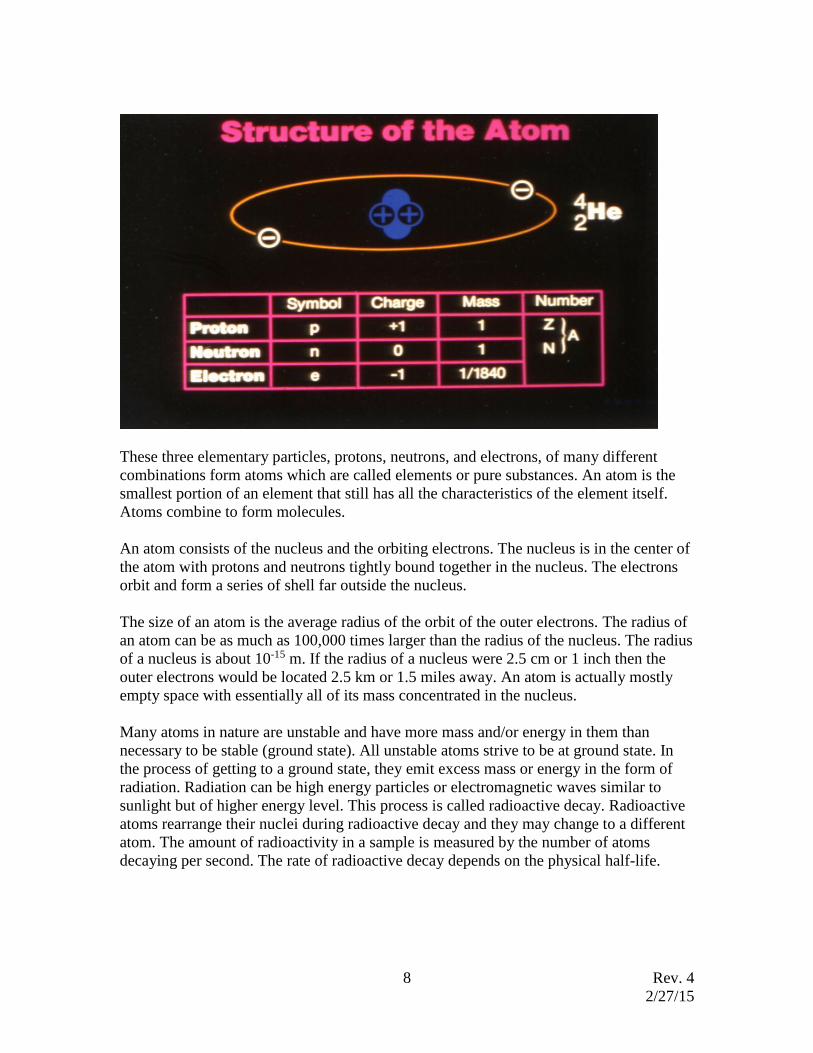
elementary particles. The three basic particles are called proton, neutron, and electron.
Proton: Protons are an elementary particle with a single positive charge of
1.6022x10⁻¹⁹ Coulomb and a rest mass of 1.6726x10⁻²⁷ kg.
Neutron: Neutrons are an elementary particle with no electric charge and a rest
mass of 1.6749x10⁻²⁷ kg.
Electron: Electrons are an elementary particle with a single negative charge of -
1.6022x10⁻¹⁹ Coulomb and a rest mass of 9.10946x10⁻³¹
Rev. 4
2/27/15
8
These three elementary particles, protons, neutrons, and electrons, of many different
combinations form atoms which are called elements or pure substances. An atom is the
smallest portion of an element that still has all the characteristics of the element itself.
Atoms combine to form molecules.
An atom consists of the nucleus and the orbiting electrons. The nucleus is in the center of
the atom with protons and neutrons tightly bound together in the nucleus. The electrons
orbit and form a series of shell far outside the nucleus.
The size of an atom is the average radius of the orbit of the outer electrons. The radius of
an atom can be as much as 100,000 times larger than the radius of the nucleus. The radius
of a nucleus is about 10-15 m. If the radius of a nucleus were 2.5 cm or 1 inch then the
outer electrons would be located 2.5 km or 1.5 miles away. An atom is actually mostly
empty space with essentially all of its mass concentrated in the nucleus.
Many atoms in nature are unstable and have more mass and/or energy in them than
necessary to be stable (ground state). All unstable atoms strive to be at ground state. In
the process of getting to a ground state, they emit excess mass or energy in the form of
radiation. Radiation can be high energy particles or electromagnetic waves similar to
sunlight but of higher energy level. This process is called radioactive decay. Radioactive
atoms rearrange their nuclei during radioactive decay and they may change to a different
atom. The amount of radioactivity in a sample is measured by the number of atoms
decaying per second. The rate of radioactive decay depends on the physical half-life.
Rev. 4
2/27/15
9
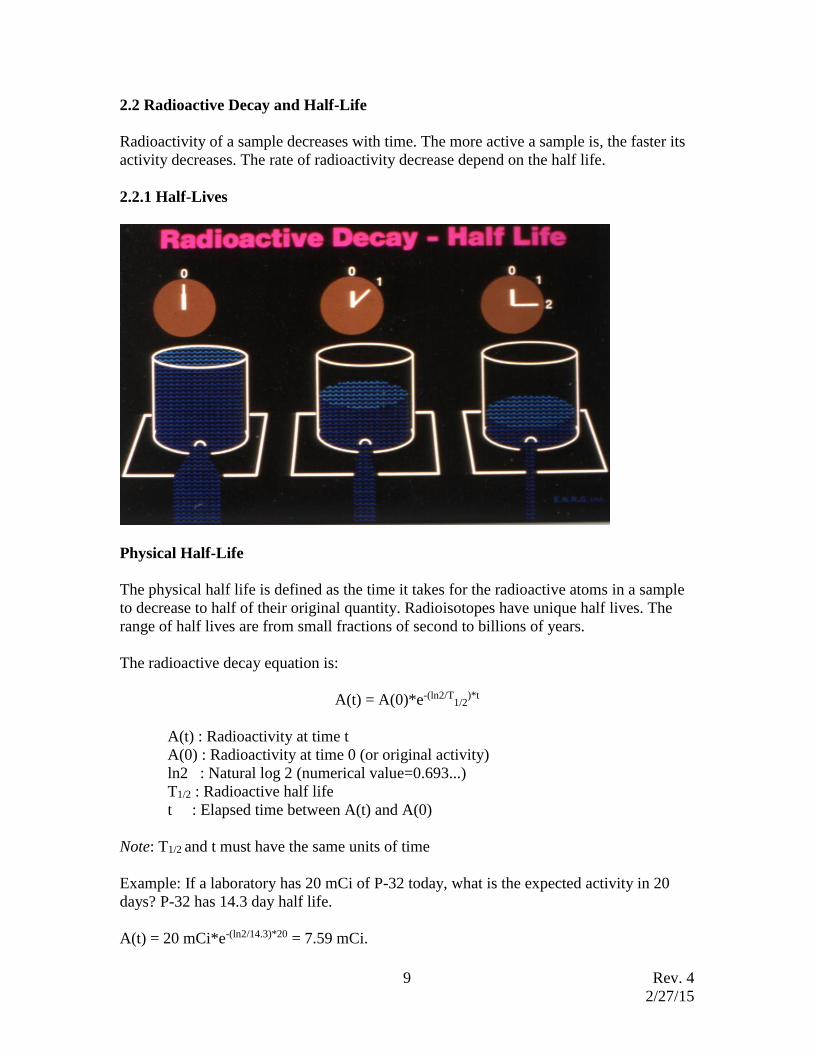
2.2 Radioactive Decay and Half-Life
Radioactivity of a sample decreases with time. The more active a sample is, the faster its
activity decreases. The rate of radioactivity decrease depend on the half life.
2.2.1 Half-Lives
Physical Half-Life
The physical half life is defined as the time it takes for the radioactive atoms in a sample
to decrease to half of their original quantity. Radioisotopes have unique half lives. The
range of half lives are from small fractions of second to billions of years.
The radioactive decay equation is:
A(t) = A(0)*e-(ln2/T1/2
)*t
A(t) : Radioactivity at time t
A(0) : Radioactivity at time 0 (or original activity)
ln2 : Natural log 2 (numerical value=0.693...)
T1/2 : Radioactive half life
t : Elapsed time between A(t) and A(0)
Note: T1/2 and t must have the same units of time
Example: If a laboratory has 20 mCi of P-32 today, what is the expected activity in 20
days? P-32 has 14.3 day half life.
A(t) = 20 mCi*e-(ln2/14.3)*20 = 7.59 mCi.
Rev. 4
2/27/15
10
Biological Half-Life
The biological half-life is the time required for a biological system to eliminate, by
natural process, half the amount of a substance such as radioactive materials or chemicals
that enters the system.
Effective Half-Life
The effective half–life is the net effect of the physical and biological half-life combined
together in removing the radioactive materials from the body. The effective half-life is
always shorter than the physical or biological half-life.
Effective half-life (Teff) = (Tp x Tb)/(Tp+Tb)
Tp: physical half-life
Tb: biological half life
Note: Tp and Tb are in the same unit
Example: H-3 has 12.33 year physical half-life and 12 day biological half life. The
effective half-life of H-3 is:
Teff = ((12.33 y x 365 d/y)(12 d))/((12.33 x 365)+12) = 11.97 d
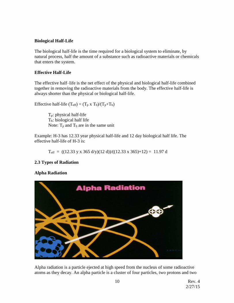
2.3 Types of Radiation
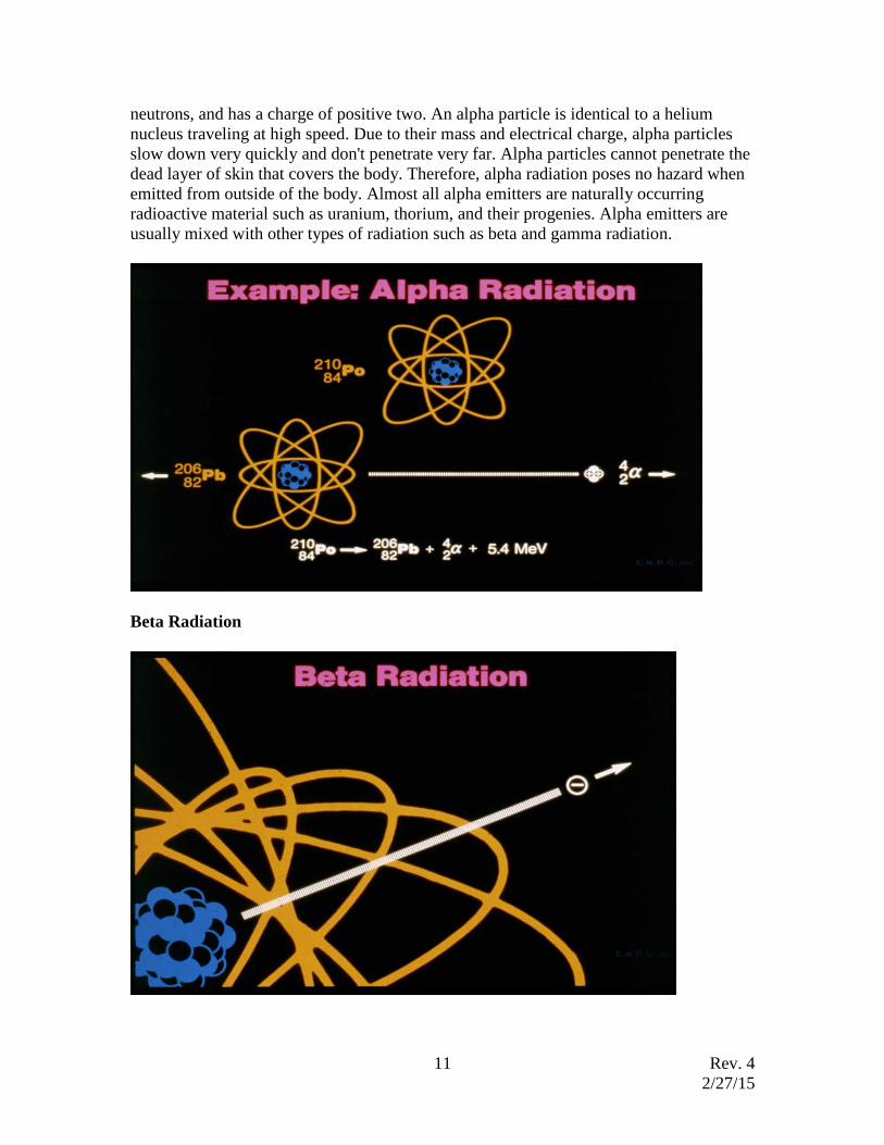
Alpha Radiation
Alpha radiation is a particle ejected at high speed from the nucleus of some radioactive
atoms as they decay. An alpha particle is a cluster of four particles, two protons and two
Rev. 4
2/27/15
11
neutrons, and has a charge of positive two. An alpha particle is identical to a helium
nucleus traveling at high speed. Due to their mass and electrical charge, alpha particles
slow down very quickly and don't penetrate very far. Alpha particles cannot penetrate the
dead layer of skin that covers the body. Therefore, alpha radiation poses no hazard when
emitted from outside of the body. Almost all alpha emitters are naturally occurring
radioactive material such as uranium, thorium, and their progenies. Alpha emitters are
usually mixed with other types of radiation such as beta and gamma radiation.
Beta Radiation
Rev. 4
2/27/15
12
A beta particle is identical to an electron except that it comes from the nucleus, not the
outer shells. The beta particle has a charge of minus one and mass of 1/7347 of an alpha
particle. For this reason, beta particles can penetrate much farther than alpha radiation.
Energetic beta particles can penetrate the dead outer layer of skin and cause damage to
live tissue. Although beta particles are more penetrating than alpha particles they cannot
penetrate to internal organs. Beta particles emitted from external sources close to the
body may damage live cells of the skin and the lens of the eye. Most radioisotopes used
in biological and medical research are beta emitters.
In beta decay, a sub-atomic particle called antineutrino is simultaneously emitted with a
beta particle. The antineutrino has little or no mass and no charge. Available energy
during beta decay is shared between a beta particle and an antineutrino in all possible
ratios. This is the reason why the betas do not have discrete energy, rather the beta
particles have a continuous spectrum from zero to its maximum energy. The antineutrino
has such an extremely low probability of interaction with matter that there is no easy way
to detect it. Normally the average beta energy is considered to be 1/3 of the maximum
beta energy.
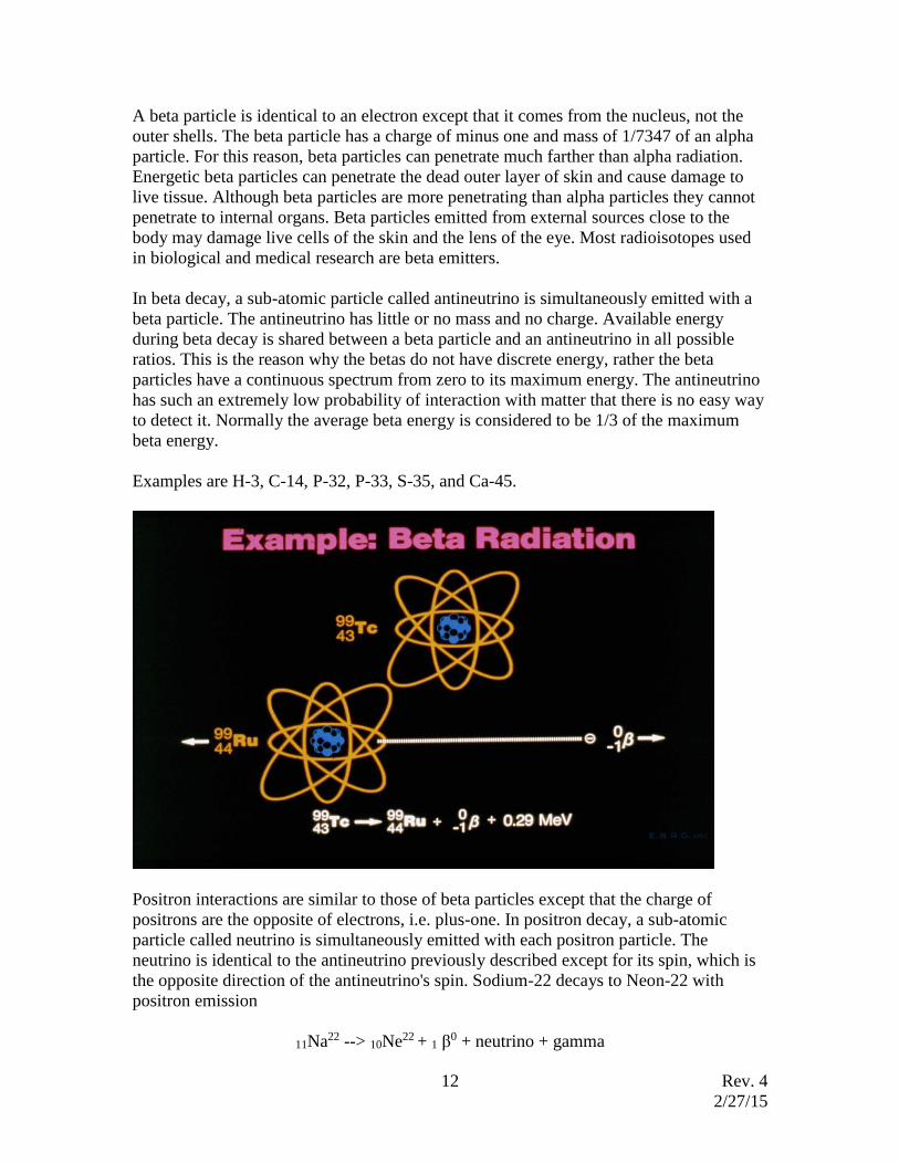
Examples are H-3, C-14, P-32, P-33, S-35, and Ca-45.
Positron interactions are similar to those of beta particles except that the charge of
positrons are the opposite of electrons, i.e. plus-one. In positron decay, a sub-atomic
particle called neutrino is simultaneously emitted with each positron particle. The
neutrino is identical to the antineutrino previously described except for its spin, which is
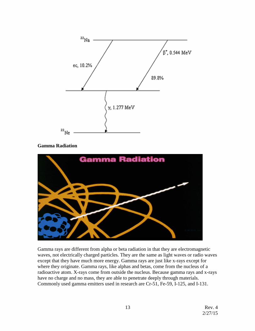
the opposite direction of the antineutrino's spin. Sodium-22 decays to Neon-22 with
positron emission
11Na22 --> 10Ne22 + 1 β0 + neutrino + gamma
Rev. 4
2/27/15
13
Gamma Radiation
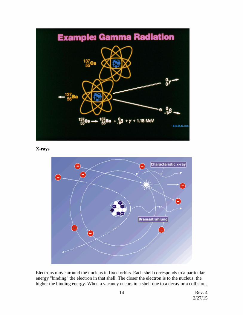
Gamma rays are different from alpha or beta radiation in that they are electromagnetic
waves, not electrically charged particles. They are the same as light waves or radio waves
except that they have much more energy. Gamma rays are just like x-rays except for
where they originate. Gamma rays, like alphas and betas, come from the nucleus of a
radioactive atom. X-rays come from outside the nucleus. Because gamma rays and x-rays
have no charge and no mass, they are able to penetrate deeply through materials.
Commonly used gamma emitters used in research are Cr-51, Fe-59, I-125, and I-131.
Rev. 4
2/27/15
14
X-rays
Electrons move around the nucleus in fixed orbits. Each shell corresponds to a particular
energy "binding" the electron in that shell. The closer the electron is to the nucleus, the
higher the binding energy. When a vacancy occurs in a shell due to a decay or a collision,

Rev. 4
2/27/15
15
a transition can occur where an electron in one shell moves to another. This causes the
emission of an electromagnetic ray or radiation with an energy equal to the difference in
energy between the two shells. These radiations are called characteristic x-rays because
the energy of the ray is characteristic of the type of atom.
The second way in which x-rays are produced is called bremsstrahlung. Whenever
charged particles are accelerated (or decelerated) an electromagnetic ray is emitted.
Electrons are accelerated around the nucleus of atoms because of the attractive force of
the opposite charges. This occurs when electrons reach high velocities, such as in an x-
ray tube, and hit a dense target (high atomic number). This is how x-ray machines
produce x rays. Another way this can happen is if beta emitting radioactive material is
placed close to or contained in a dense material such as lead. Bremsstrahlung is more
likely when the charged particle has little mass, high energy, and when the target is dense.
Neutron Radiation
Neutrons can be emitted during nuclear reactions and, they can be emitted by decay of
certain radionuclides, although this rarely occurs with naturally occurring radionuclides.
Neutrons, because of their large mass and neutral charge, can be absorbed or scattered by
the nucleus of atoms they interact with. When neutrons are absorbed, nuclear reactions,
such as fission, are possible and often result in the emission of secondary radiation. In
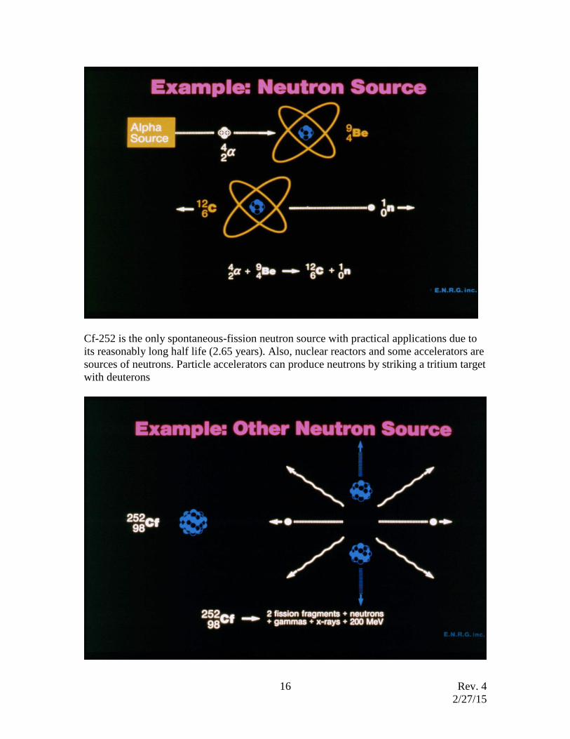
this indirect manner, neutrons cause effects similar to those caused by other radiation. 241Am-Be, 239Pu-Be, and 226Ra-Be are commonly used neutron sources. The alpha
particles from 241Am, 239Pu, and 226Ra hit the target of 9Be to produce an alpha-neutron
reaction (a,n). One Ci of 239Pu-Be alpha source will produce 2.1 x 10⁶ neutrons/second
and 2.6x10⁶ neutrons/second are produced by one Ci of an 241Am-Be alpha source
Rev. 4
2/27/15
16
Cf-252 is the only spontaneous-fission neutron source with practical applications due to
its reasonably long half life (2.65 years). Also, nuclear reactors and some accelerators are
sources of neutrons. Particle accelerators can produce neutrons by striking a tritium target
with deuterons

Rev. 4
2/27/15
17
3. Radiation Interactions with Matter
A significant characteristic of radiation is that radiation can ionize atoms. Most atoms are
electrically neutral, that is they have same number of protons (+ charges) and electrons (-
charges). Radiation has the ability to directly or indirectly cause electrons to be removed
from atoms. This creates a pair of charged particles. One is negative electron; the other is
the remaining atom, now positively charged. This process is called ionization.
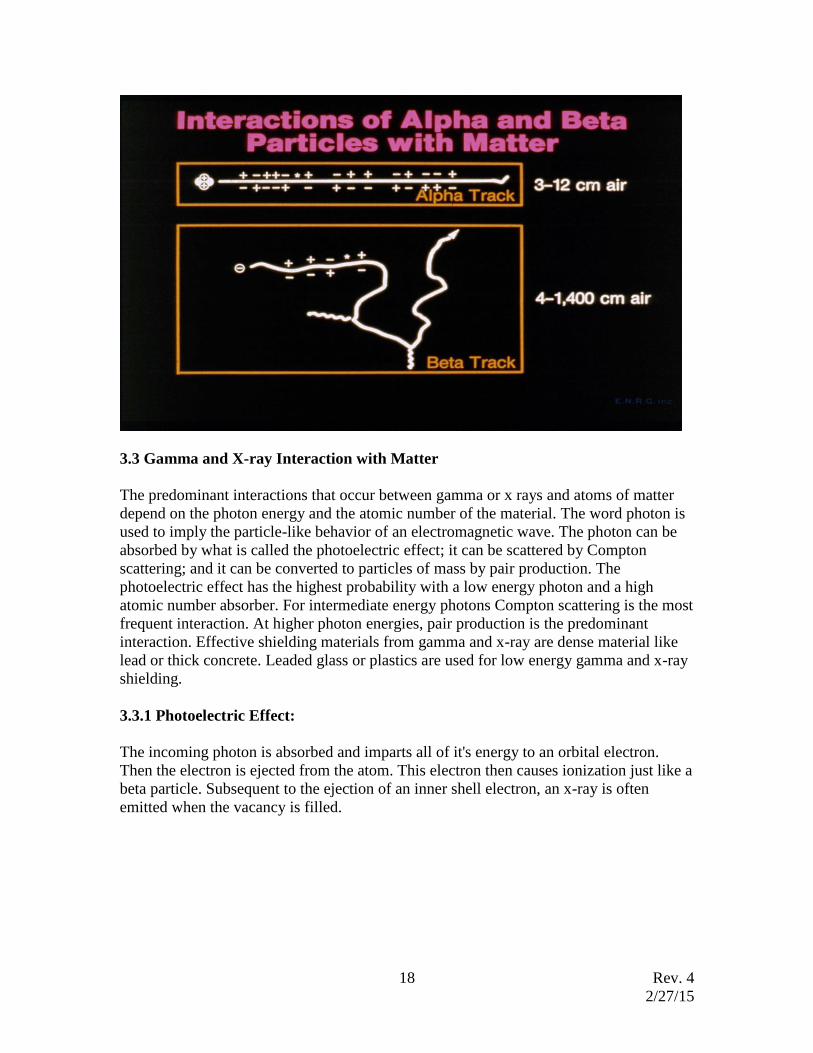
3.1 Alpha Radiation Interaction with Matter
The alpha particle follows a straight, short path in the material it penetrates, causing very
dense ionization and excitation events along the track, in orders of millions per inch.
Most alpha ranges are less than 5-6 cm in air. Alpha particles can be stopped by a sheet
of paper.
3.2 Beta Radiation Interaction with Matter
The beta particle track is very different from the short, straight alpha track. The beta
particle scatters frequently, causing what is often described as a drunken man's path. The
range of a beta particle is considerably greater than an alpha particle. As a rule of thumb,
one MeV beta particle can travel about 10 feet in air or about 0.4 cm in water. The
ionization and excitation events along the path are much less than that due to alpha
particles, yet the amount is still large, thousands to hundreds of thousands per inch.
Additionally, the high energy betas near dense material can generate bremsstrahlung x-
rays which is much more penetrating than the beta particle itself. The production of
bremsstrahlung x rays increases with the atomic number of the target material and beta
energy. Therefore, low Z material such as Lucite and plastic (large hydrogen, atomic
number 1, population) are used as a beta shield.
Rev. 4
2/27/15
18
3.3 Gamma and X-ray Interaction with Matter
The predominant interactions that occur between gamma or x rays and atoms of matter
depend on the photon energy and the atomic number of the material. The word photon is
used to imply the particle-like behavior of an electromagnetic wave. The photon can be
absorbed by what is called the photoelectric effect; it can be scattered by Compton
scattering; and it can be converted to particles of mass by pair production. The
photoelectric effect has the highest probability with a low energy photon and a high
atomic number absorber. For intermediate energy photons Compton scattering is the most
frequent interaction. At higher photon energies, pair production is the predominant
interaction. Effective shielding materials from gamma and x-ray are dense material like
lead or thick concrete. Leaded glass or plastics are used for low energy gamma and x-ray
shielding.
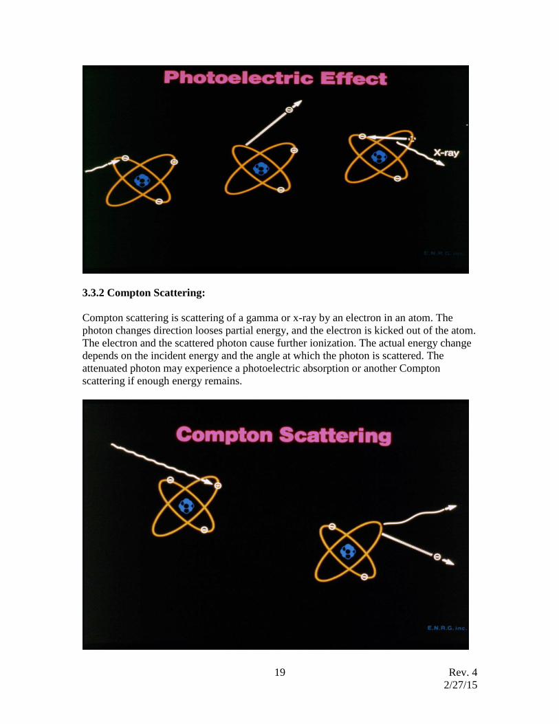
3.3.1 Photoelectric Effect:
The incoming photon is absorbed and imparts all of it's energy to an orbital electron.
Then the electron is ejected from the atom. This electron then causes ionization just like a
beta particle. Subsequent to the ejection of an inner shell electron, an x-ray is often
emitted when the vacancy is filled.
Rev. 4
2/27/15
19
3.3.2 Compton Scattering:
Compton scattering is scattering of a gamma or x-ray by an electron in an atom. The
photon changes direction looses partial energy, and the electron is kicked out of the atom.
The electron and the scattered photon cause further ionization. The actual energy change
depends on the incident energy and the angle at which the photon is scattered. The
attenuated photon may experience a photoelectric absorption or another Compton
scattering if enough energy remains.
Rev. 4
2/27/15
20
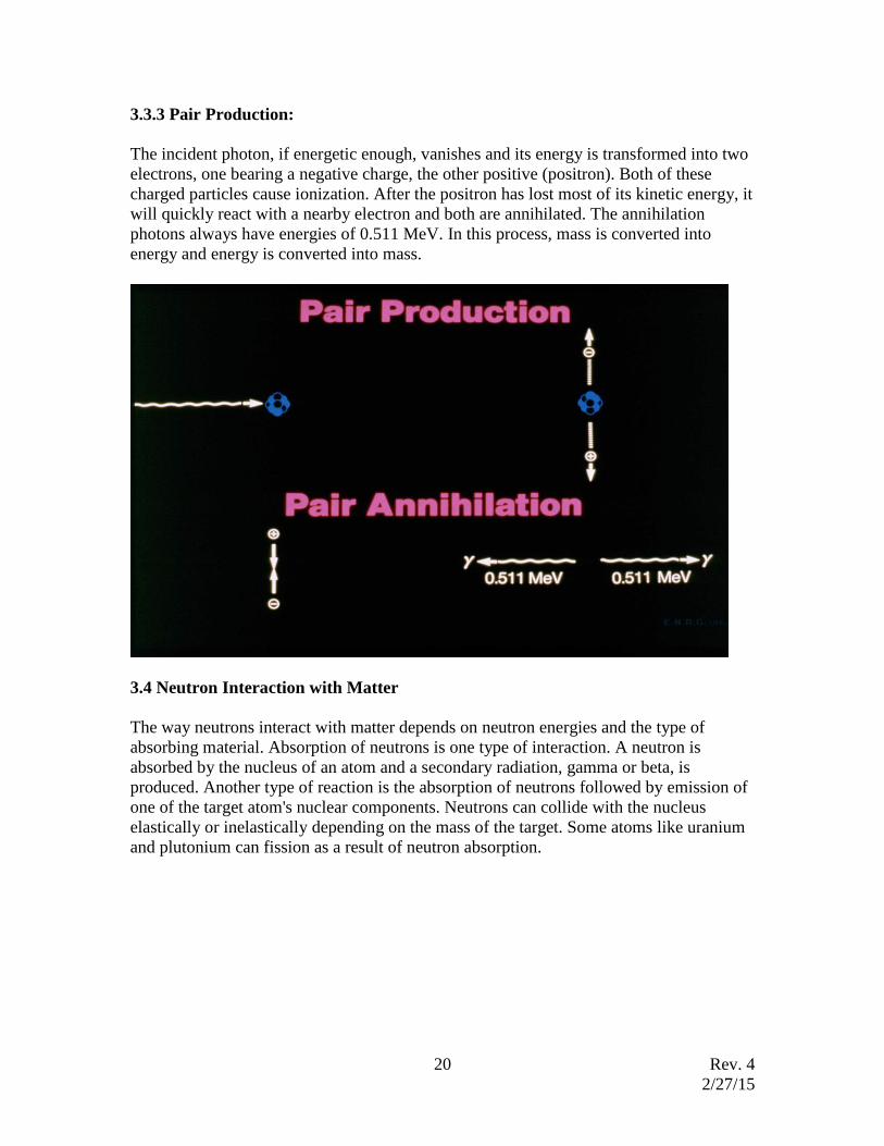
3.3.3 Pair Production:
The incident photon, if energetic enough, vanishes and its energy is transformed into two
electrons, one bearing a negative charge, the other positive (positron). Both of these
charged particles cause ionization. After the positron has lost most of its kinetic energy, it
will quickly react with a nearby electron and both are annihilated. The annihilation
photons always have energies of 0.511 MeV. In this process, mass is converted into
energy and energy is converted into mass.
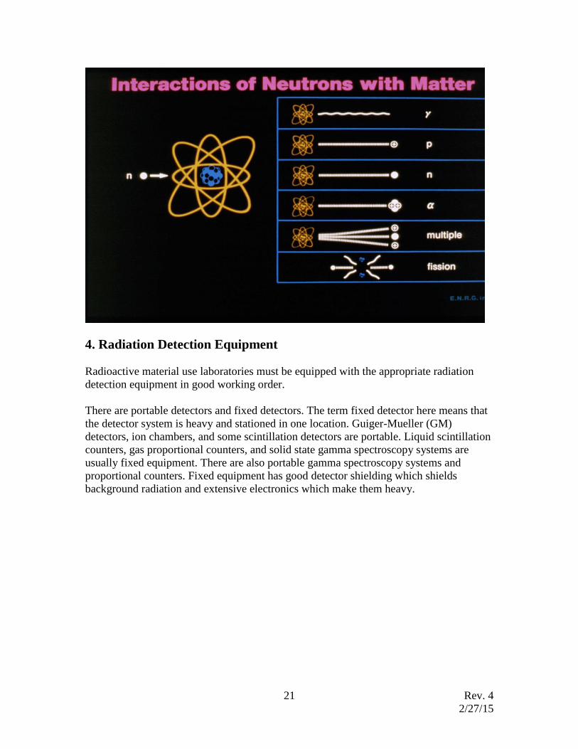
3.4 Neutron Interaction with Matter
The way neutrons interact with matter depends on neutron energies and the type of
absorbing material. Absorption of neutrons is one type of interaction. A neutron is
absorbed by the nucleus of an atom and a secondary radiation, gamma or beta, is
produced. Another type of reaction is the absorption of neutrons followed by emission of
one of the target atom's nuclear components. Neutrons can collide with the nucleus
elastically or inelastically depending on the mass of the target. Some atoms like uranium
and plutonium can fission as a result of neutron absorption.
Rev. 4
2/27/15
21
4. Radiation Detection Equipment
Radioactive material use laboratories must be equipped with the appropriate radiation
detection equipment in good working order.
There are portable detectors and fixed detectors. The term fixed detector here means that
the detector system is heavy and stationed in one location. Guiger-Mueller (GM)
detectors, ion chambers, and some scintillation detectors are portable. Liquid scintillation
counters, gas proportional counters, and solid state gamma spectroscopy systems are
usually fixed equipment. There are also portable gamma spectroscopy systems and
proportional counters. Fixed equipment has good detector shielding which shields
background radiation and extensive electronics which make them heavy.
Rev. 4
2/27/15
22
4.1 Guiger-Mueller (GM) Detector
The GM detector is a gas filled detector. Radiation causes ionization inside the detector
and the liberated electrons are collected at the anode in the center of the detector. High
electric potential is applied between the anode and the cathode which accelerates librated
electrons toward the anode. While moving toward the anode, the accelerating electrons
cause a succession of ionization like many avalanches. This process is called gas
multiplication. It produces a large numbers of charges. The gas multiplication factor for
GM is in the range of 106-108. The collected charges in the anode generate electrical
pulses which are processed through electronics.
The GM detector is a simple, inexpensive, easy to operate, sensitive to low level, and
reliable instrument. The three major types of radiation (alpha, beta, and gamma) can be
detected by the GM detector. The thin window GM is sensitive to low level radiation and
well suited to checking for contamination on one's clothing and body as well as in work
areas. Portable survey meters with GM's are usually equipped with a speaker or other
audible indicator, to allow surveys without watching the meter. GM detectors come in
many different sizes. A cylinder type with a beta shield is one type, and a cylinder with
thin end window is another type. The "Pancake" GM detector has a 2 inch diameter thin
mica window which is a relatively large active detector area. It is capable of detecting
alpha, beta, and gamma radiation. Portable survey meters with GM detectors have cpm
and/or mR/hr reading scales. Due to the sensitivity of GM detectors, they have a limited
range. GM's are not suitable for high radiation fields. Normally, the ranges of GM
detectors are 0-50 mR/hr, with some having ranges up to about 200 mR/hr.
Rev. 4
2/27/15
23
GM detectors with thin end windows can detect almost all commonly used radionuclides
in research laboratories except Hydrogen-3. The liquid scintillation counter is the most
efficient and widely used method to count Hydrogen-3.
4.2 Ion Chamber
An ion chamber is a gas filled detector which is less sensitive to low level radiation fields
than GM detectors. The gas multiplication factor is one (1) for ionization chambers and
millions for GMs. An ion chamber is normally designed to monitor gamma and x rays.
Though there are ion chambers which can detect beta radiation through beta windows,
they are not practical in research laboratories where the quantity of radioactive material
are small and radiation levels are low.
Ion chambers may be useful in areas where significantly higher radiation levels than
background levels exist such as: particle accelerators, x-ray producing machines, and
irradiators.
There are ion chambers which can measure background level. Pressurized ion chambers
have higher sensitivity and can measure radiation levels from microR/hr to R/hr range.
4.3 Gas Proportional Counter
The gas proportional counter (GC) is a gas filled detector. The operating principle is
similar to other gas filled detectors. The gas multiplication factor for GPC is from
hundreds up to a million. The output charge is proportional to the incoming radiation
energy. A GPC uses different operating voltages for alpha and beta radiation counting
therefore GPCs can analyze samples which emit both alpha and beta radiation and
distinguish between alpha and beta radiation. GPCs can detect gamma radiation but
gamma detection efficiency is much lower than alpha and beta detection efficiency. P-10
gas which is 90% argon and 10% methane is commonly used for a GPC. There are also
other gases used in GPCs.
The GPCs are usually fixed instruments which have a heavy detector and sample shield.
Some portable contamination monitors are made out of GPCs.
Many GPCs have automated sample exchangers which can handle tens of samples at a
time. Therefore, use of GPC can save a considerable amount of time if a large number of
samples are to be analyzed.
4.4 Scintillation Detector
There are scintillation detectors for alpha, beta, gamma, and neutron radiation.
Scintillators are made of plastic, organics, or inorganic materials. They can be solid,
liquid, and gas. They can be made in all shapes and sizes. Scintillation detectors can be
used with portable survey meters or fixed equipment. Incoming radiation interacts with a
scintillating material and a portion of or the total energy is transferred to the scintillating
Rev. 4
2/27/15
24
material. The excited scintillating molecules produce light photons during the de-
excitation process. Scintillation detectors may directly count these photons or convert
these photons to electric current via photomultiplier tube and measure the current
produced by the converted electrons. A sodium iodide (NaI) detector is commonly used
for gamma detection and analysis. Due to the high sensitivity, NaI detectors give high
background radiation levels. Detector shielding can reduce background radiation level.
4.4.1 Liquid Scintillation Counter
Liquid scintillation counting (LSC) is widely used for low level beta radiation detection.
The LSC can detect alpha, beta, and gamma radiation with high efficiency. The
sensitivity of LSC is higher than other detectors. The sample is immersed in the
scintillating medium and it is in direct contact with the scintillating medium which makes
the LSC efficient and capable of detecting low radioactivity levels and low energy
radiation. The function of the scintillation medium is to convert the radiation energy into
light photons which can be detected by the scintillation counter. The energy of radiation
is absorbed by the scintillating medium which causes the molecules to become excited.
The excited molecules emit photons and then return to their ground state. The light output
is proportional to the incoming radiation energy.
LSC requires use of a scintillating medium. Sample preparation can be time consuming if
a large number of samples are needed. But a large number of samples can be counted
without attendance because the prepared samples are usually fed automatically. Liquid
scintillation counters are large fixed instruments, not a portable system. The scintillation
solution is expensive to purchase and to dispose of.
Most tritium counting is done by LSC. Due to its low energy, tritium is extremely
difficult to detect.
4.5 Solid State/Semiconductor Detectors
The solid state detectors, such as silicon and germanium detectors, are mainly used for
gamma spectroscopy. The main advantage of solid state detectors is very good energy
resolution. Gamma detection efficiency of germanium is low but the energy resolution of
gamma spectrum by germanium is the best among all detectors. The operating
temperature is very low and requires liquid nitrogen to maintain operating temperature.
Gamma emitting Isotope identification and quantitation are the main uses for these
detectors. The gamma spectroscopy system can be a fixed system or portable system.
Normally they are fixed instruments with a thick and heavy shield around the detector
and sample to reduce background radiation.
4.6 Personal Monitoring Devices
The film badge and thermoluminescent dosimeter (TLD) are widely used for personal
monitoring. The personal dosimeters are exchanged in monthly, bi-monthly, or quarterly.
They should be kept in a dosimeter rack away from any radiation sources when not in use.
Rev. 4
2/27/15
25
The whole body dosimeters should be worn at chest or collar area when in use. The
extremity monitors such as TLD ring should be worn inside of glove with the label facing
towards palm.
The film badge works similar to ordinary photographic film. The film is enclosed in a
light-tight packet and radiation penetrates to expose the film. They are not sensitive to
low energy betas such as Hydrogen-3, Carbon-14 or alpha radiation. If the packet is
damaged or opened then the film badge is invalid due exposure to light.
The TLD is a small crystal. When exposed to radiation, the molecules of the detector
material are raised to metastable states by energy received from radiation. They stay in
the excited states. When these crystals are heated, these molecules return to its ground
states with emission of light photons. The amount of light photons emitted is proportional
to the radiation energy absorbed.
Personal dosimeter may only be used for personal monitoring of the person the dosimeter
is specifically issued to.
5. Units of Radioactivity and Radiation Measurement
Table 1. Units of radioactivity and radiation exposure
Traditional units SI units Relations
Unit Definition Unit Definition
Radioactivity Curie
(Ci)
3.7x10¹⁰
disintegrations
per second
(dps)
Becquerel
(Bq)
1 dps 1 Ci =
3.7x10¹⁰ Bq
Exposure Roentgen
(R)
2.58x10⁻⁴
coulomb per kg
of air
R
Absorbed
dose
rad 100 ergs/g Gray (Gy) 1 joule/kg 100 rad = 1
Gy
Dose
equivalent
rem Rad x quality
factor (Q)
Sievert
(Sv)
Gy x Radiation
weighting
factor (WR)
100 rem = 1
Sv
Table 2. Quality factor (Q) and radiation weighting factors (WR)
Radiation Q WR
X, gamma, beta 1 1
Alpha 20 20
5.1 Radioactivity:
The Becquerel (Bq) and the Curie (Ci) are commonly used as units of radioactivity. The
Bq is defined as one disintegration per second. The Ci is defined as 3.7x10¹⁰
disintegrations per second. Commonly used multiples and sub-multiples of the units are

Rev. 4
2/27/15
26
mega Bq (MBq, million Bq), giga Bq (GBq, billion Bq), millicurie (1/1000 of Ci), and
microcurie (1/1,000,000 of Ci). The half life described in the next section describes how
long the radioactive material might last and the number of Bq or Ci tells how "active" this
material is now.
5.2 Radiation Exposure:
The unit of exposure is the Roentgen (R). The Roentgen is defined by how gamma and x-
rays interact in air. It is defined as the quantity of gamma or x rays which, when
interacting with one kilogram of air, liberate energetic electrons that produce 0.000258
Coulombs of charge by ionization when the electrons are completely stopped.
5.3 Absorbed Dose
The absorbed dose is defined as the energy imparted by radiation per mass of absorbing
material; the material here includes all types of exposed material. The absorbed dose is a
quantity that refers to how much energy is deposited in material by the radiation. The
term "RAD" is derived from the expression "Radiation Absorbed Dose".
The units are: 1 rad = 100 ergs/gram of material,
1 Gy (Gray, SI unit) = 1 Joule/kg of material, and
1 Gy = 100 rad.
Rev. 4
2/27/15
27
5.4 Dose Equivalent
The dose equivalent is obtained by modifying the absorbed dose according to the types of
radiation involved. The dose equivalent is the product of the absorbed dose and the
quality factor (Q) of a given radiation (rad x Q). The quality factor is based on the type
and energy of the radiation causing damage. It is based on the density of ionization along
the radiation path. The quality factors for different types of radiation are: 20 for alpha, 1
for beta, gamma, and x-ray, 10 for neutrons of unknown energy (energy dependent), and
10 for high energy protons.
The dose equivalent units are: Rem (Roentgen Equivalent Man) = rad x Q, ie. 1 rad of
alpha radiation = 20 rem or 0.2 Sv.
The rem (or Sv) is designed to be a unit of biological risk.
5.5 Effective Dose Equivalent (EDE)
Different organs or tissues in the body have varying degree of sensitivity to radiation.
The tissue weighting factor (WT) may be used to estimate risks, Effective Dose
Equivalent (HE), of a whole body exposure when radiation exposure is limited to only a
portion of the body. For example, if a person’s stomach receives 10 rem, the EDE is 1.2
rem (10 rem x 0.12).
Table 3. Tissue weighting factors, WT
Tissue or organ WT, ICRP60
Gonads 0.20
Bone marrow (red) 0.12
Colon 0.12
Lung 0.12
Stomach 0.12
Bladder 0.05
Breast 0.05
Liver 0.05
Esophagus 0.05
Thyroid 0.05
Skin 0.01
Bone surface 0.01
Reminder 0.05
Rev. 4
2/27/15
28
6. Biological Effects of Radiation
As a principle of radiation protection, it is assumed that any amount of radiation is
harmful, no matter how small the exposure is. This may be called "The Linear-No
Threshold Theory". This theory is widely accepted as a principle of radiation protection
even though it has not been proven.
6.1 Factors Determining Biological Effects of Radiation Exposure
The biological effects resulting from radiation exposure depend on a number of factors:
6.1.1 Total exposure:
How much exposure or dose has occurred to the tissue.
6.1.2 Exposure rate:
Our bodies have the ability to repair damage even during radiation exposure. How
quickly radiation exposure is accumulated is important for both early and late biological
effects.
6.1.3 Portions of the body exposed:
Some portions of the human body are more resistant to radiation than others due to their
physiological function and cellular activity. Exposure to limited portions of the body
Rev. 4
2/27/15
29
have less effect than equal exposure to the whole body. A massive dose that would be
fatal if delivered to the whole body might not even cause sickness if delivered to, for
example, only the extremities.
6.1.4 Type of Radiation Received:
The three main types of radiation, alpha, beta, and gamma have different penetrating
abilities. Alpha radiation to external skin is no hazard because it is likely that the outer
(dead) layer of the skin stops all alpha radiation. But if alpha radiation is received
internally the damage to the surrounding tissue is expected to be 20 times more harmful
than the expected damages from beta or gamma radiation. The Quality Factor (or
Radiation Weighting Factor) for alpha is 20.
6.1.5 Biological Factors:
Age, sex, state of health, body size, body weight and other biological factors react
differently to radiation exposure even under identical conditions. Actively dividing cells
have increased sensitivity to radiation exposure.
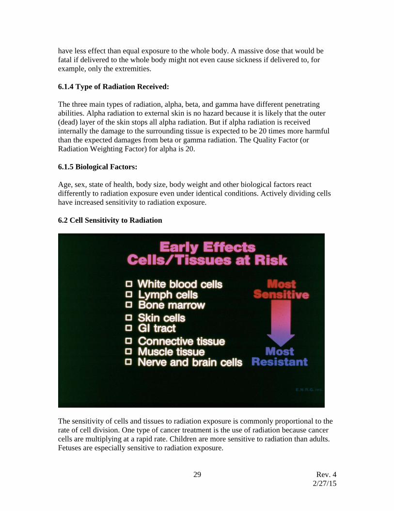
6.2 Cell Sensitivity to Radiation
The sensitivity of cells and tissues to radiation exposure is commonly proportional to the
rate of cell division. One type of cancer treatment is the use of radiation because cancer
cells are multiplying at a rapid rate. Children are more sensitive to radiation than adults.
Fetuses are especially sensitive to radiation exposure.

Rev. 4
2/27/15
30
For an adult, white blood cells are most sensitive due to their rate of cell division. White
blood cells, bone marrow, skin cells, and the gastrointestinal tract lining are very
sensitive. Tendons, ligaments, and other connective tissues are moderately sensitive.
Muscle, nerve cells, and brain cells are the most resistant cells in the body.
6.3 Radiation Effects on Live Cells
Radiation causes ionization that causes physical and chemical effects to the atoms and
cells with which it interacts. Radiation passes through tissue and causes ionization within
the cells of the tissue. The ions produced within the cell are electrically charged and
chemically active. These charged, chemically active ions tend to react quickly with
surrounding atoms and molecules of the cell and alter the cell structure and/or produce
chemically active free radicals.
For an example, water is a primary constituent of a living cell. As a result of ionizing
radiation interaction, the bonds between hydrogen and oxygen may be broken. The
dissociated hydrogen and oxygen from water may not recombine as water molecules but
may recombine in many different combinations between oxygen, hydrogen, and electrons
i.e. H2O2, HO2, OH, e-, etc.
Radiation interaction can happen in any location of a cell such as the DNA or the
chromosome, which if damaged, could be fatal for the cell's survival. If a large enough
cell population damage occurs, radiation effects may be immediate and fatal to the living
organism. The radiation effects may show up in a matter of days, as acute effects, or
years after the exposure, as latent effects.
6.4 Damaged Cell Repair Mechanism
Cells may be damaged by many factors such as life style, chemical exposure, radiation
exposure, etc,. Most cells are capable of repairing damage including damage to the
genetic material if given enough time (rate of exposure). But major damage might not be
repaired and may result in cell death. Ability to repair damaged cells may depend on the
type of chemicals produced by radiation in the cell and/or surrounding the cell.
If the chemicals produced are less active and stay away from genetic material (DNA) or
other vital components necessary for cell survival, the cells would likely be less
susceptible to radiation damage. The rate at which people recover from radiation
exposure is not well known and variations among individuals are great.
6.5 Acute and Chronic Exposure
Acute exposure or an acute dose means the exposure is delivered in a short period of time.
The exact time frame is not well defined but exposures received in hours or days are
considered acute. The acute exposure does not necessarily mean a large and a lethal dose.
It just mean a short time frame. Chronic exposure is exposure spread out through a longer
period of time.
Rev. 4
2/27/15
31
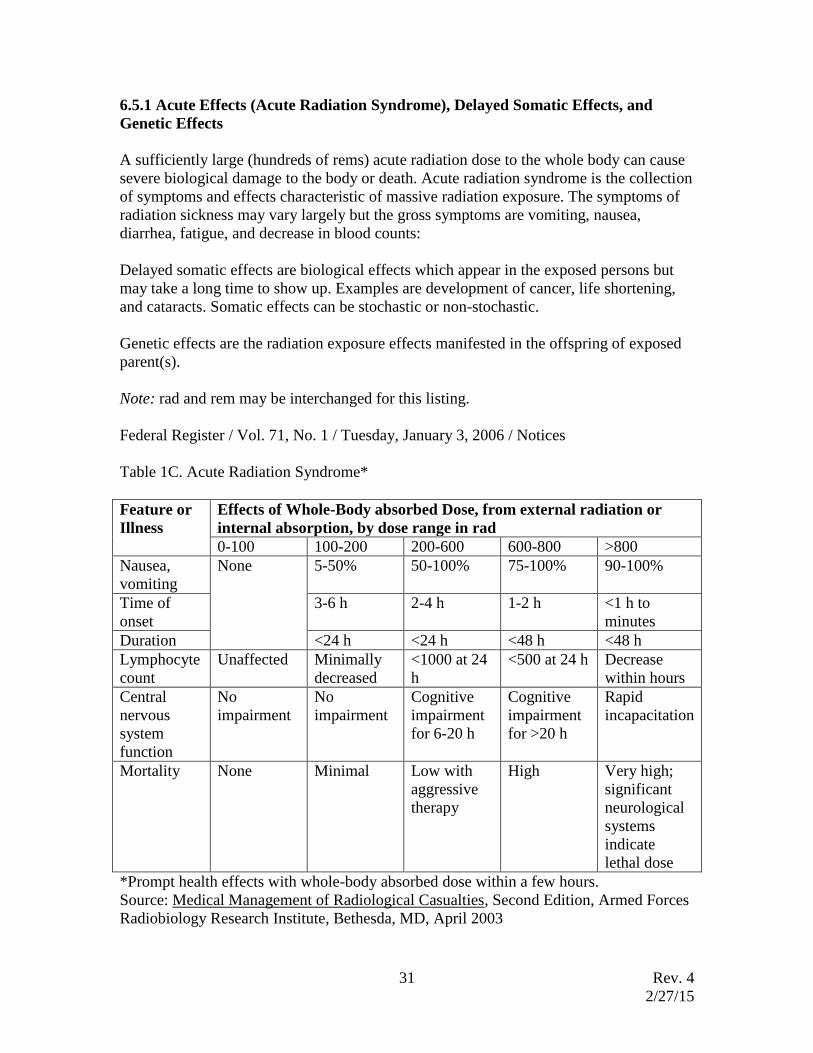
6.5.1 Acute Effects (Acute Radiation Syndrome), Delayed Somatic Effects, and
Genetic Effects
A sufficiently large (hundreds of rems) acute radiation dose to the whole body can cause
severe biological damage to the body or death. Acute radiation syndrome is the collection
of symptoms and effects characteristic of massive radiation exposure. The symptoms of
radiation sickness may vary largely but the gross symptoms are vomiting, nausea,
diarrhea, fatigue, and decrease in blood counts:
Delayed somatic effects are biological effects which appear in the exposed persons but
may take a long time to show up. Examples are development of cancer, life shortening,
and cataracts. Somatic effects can be stochastic or non-stochastic.
Genetic effects are the radiation exposure effects manifested in the offspring of exposed
parent(s).
Note: rad and rem may be interchanged for this listing.
Federal Register / Vol. 71, No. 1 / Tuesday, January 3, 2006 / Notices
Table 1C. Acute Radiation Syndrome*
Feature or
Illness
Effects of Whole-Body absorbed Dose, from external radiation or
internal absorption, by dose range in rad
0-100 100-200 200-600 600-800 >800
Nausea,
vomiting
None 5-50% 50-100% 75-100% 90-100%
Time of
onset
3-6 h 2-4 h 1-2 h <1 h to
minutes
Duration <24 h <24 h <48 h <48 h
Lymphocyte
count
Unaffected Minimally
decreased
<1000 at 24
h
<500 at 24 h Decrease
within hours
Central
nervous
system
function
No
impairment
No
impairment
Cognitive
impairment
for 6-20 h
Cognitive
impairment
for >20 h
Rapid
incapacitation
Mortality None Minimal Low with
aggressive
therapy
High Very high;
significant
neurological
systems
indicate
lethal dose
*Prompt health effects with whole-body absorbed dose within a few hours.
Source: Medical Management of Radiological Casualties, Second Edition, Armed Forces
Radiobiology Research Institute, Bethesda, MD, April 2003

Rev. 4
2/27/15
32
6.6 Database of Biological Effects of Radiation Exposure
6.6.1 Natural Background Radiation Exposure
Energetic radiation from outer space and the sun are continually bombarding us. Soil
contains naturally radioactive material such as potassium, uranium, thorium, and their
progenies. Radon, one progeny of uranium and thorium, contributes the largest natural
radiation exposure to humans. The background radiation exposure may vary with location
due to differing radionuclide concentrations in rock and soil, water, and an increase of
cosmic radiation with altitude. The food we eat, the water we drink, and the air we breath
contains radioactive material. Total average annual effective dose to members of the US
population is estimated to be 320 mrem per year (3.2 mSv/year). Additionally, man made
radiation sources such as medical x rays and nuclear medicine contributes about 300
mrem per year (3 mSv per year) exposure to Americans. The average effective annual
dose equivalent from all sources to members of the US population is 620 mrem (6.2 mSv).
Naturally Occurring Long-lived Radionuclides in Human Body
Isotope 238U
226Ra
228Ra
210Pb
210Po
40K
14C
3H
87Rb
90Sr
Activity,
pCi
26
120
50
600
200
130,000
87,000
27,700
29,000
2,886
The total radioactivity in the body is 277,582 pCi. This is 10,270 radioactive decays per
second (dps) and 887,374,138 (887 million) disintegration per day in the body. Each
radioactive decay produces radiation.
Source: Radiation Protection (pages 56, 370), Shapiro, 1990, Harvard Press.
Natural Radioactivity in a Banana
Bananas are a good source of potassium, a very important nutrient. All natural potassium
contains 0.0117% potassium-40 (40K) which is radioactive potassium. A medium size
banana contains about 451 mg of potassium. The amount of 40K contained in a banana is
0.0528 mg. This is equivalent to 14 dps or 0.00037 uCi. The dose equivalent, if a banana
is eaten, is about 0.01 mrem. Sometimes this is called the banana equivalent dose.
Sources: Food Values of Portion Commonly Used, 16th edition, Bosen and Church.
Chart of Nuclide, F. William Walker et al.
6.6.2 Low Level Radiation Exposure Studies
Background radiation exposes the entire world population. Due to that reason alone, it is
very difficult to determine the effects of low level radiation exposure. More information
is available about high level exposure and its effects. Radiation effects in low level
Rev. 4
2/27/15
33
exposure is extrapolated from the high level exposure. Regulatory Guide 8.29 from the
US Nuclear Regulatory Commission stated that there is a risk of 4 in 10,000 of a 1,000
mrem (0.01 Sv) dose causing a fatal cancer". Because the effects of low level radiation
are not well known, it is assumed that any amount of radiation is harmful, no matter how
small it is. This is not a proven fact but widely accepted.
However, there have been studies which have reported a beneficial effect of low level
radiation exposure. One study was conducted in areas of Yangjiang, China where the
background radiation is 2.64 times higher than the average background level. The group
of population who lived there more than 6 generations was studied and compared to a
similar life style population group with average background radiation level. The period of
study started in 1972 and lasted until 1986. This study examined the cancer mortality data
between 1970 to 1986 and observed over one million person-years in each area of the
high background and the controlled area (CA).
The back ground radiation in the high background area was 547 mrem (5.47 mSv) per
year and 207 mrem (2.07 mSv) per year in the controlled area. The study concluded that
"No increase of cancer mortality has been found in high background radiation area
(HBRA), but on the contrary, there was a tendency for the cancer mortality in HBRA to
be lower than that in CA...It is likely that there may be a dose threshold for cancer
incident, but this remains to be determined by further research."
Dr. Bernard L. Cohen studied lung cancer mortality rates and average radon
concentration in homes in 1601 US counties to "Test the Linear-No threshold Theory...".
Dr. Cohen stated in the paper "... there is a strong tendency for lung cancer rates to
decrease with increasing radon exposure, in sharp contrast to the increase expected from
the theory. There is now a substantial body of evidence indicating that the low level
radiation does indeed stimulate such biological defense mechanisms...". The beneficial
effects of low level radiation are not generally accepted.
6.6.3 Uncertainties associated with the Low Level Exposure
The National Research Council in BEIR V report stated "In this report it is estimated if
100,000 persons of all ages received a whole body dose of 0.1 Gy (10 rad) of gamma
radiation in a single brief exposure, about 800 extra cancer deaths would be expected to
occur during their remaining lifetimes in addition to the nearly 20,000 cancer deaths that
occur in the absence of the radiation. Because the extra cancer deaths would be
indistinguishable from those that occurred naturally, even to obtain a measure of how
many extra deaths occurred is a difficult statistical estimation problem." It is assumed
that radiation is harmful even at low levels but there are high uncertainties associated
with this assumption and it has not been proven.
6.6.4 High Level Radiation Exposure Studies
A lot more data is available in high level radiation exposure studies: A large number of
bomb survivors after world war II in Japan, patients who received a large amount of

Rev. 4
2/27/15
34
medical use radiation during 1930's to 50’s, a few hundred radium dial painters, uranium
minors, and animal studies have been examined. Radiation effects of high level exposure
are reasonably well established.
7. Radiation Exposure Standard (NAC 459.325)
Occupational Dose Limits:
Whole body (Total Effective Dose Equivalent) = 5 rem/year,
Sum of individual organs or tissue = 50 rem/year,
Eye dose (lens of eye) = 15 rem/year,
Skin or any extremity = 50 rem/year,
Dose to embryos of declared pregnant woman* = 0.5 rem for the entire pregnancy,
Minors = 10% of above limits, and
Member of public = 0.1 rem/year.
* Declared Pregnant Woman means a woman who has voluntarily informed her
supervisor, in writing, of her pregnancy and the estimated date of conception. The
declaration remains in effect until the declared woman withdraws the declaration in
writing or is no longer pregnant.
Note: Average Americans receive about 320 mrem per year from natural background and
300 mrem from medical exposures.
8. External Radiation Protection
External radiation can be reduced by limiting the duration of an exposure period,
increasing the distance between the external radiation source and the person, and placing
a shielding material between the external radiation source and the person.
8.1 Time, Distance, and Shielding
Time
Radiation exposure can be reduced by minimizing the time of exposure. Practice runs
without source may help to reduce exposure times when an actual experiment is
performed. If limitation of the stay time in the vicinity of an external radiation source is
not possible due to the required time to perform a given task, then other means of
exposure reduction should be utilized.
Distance
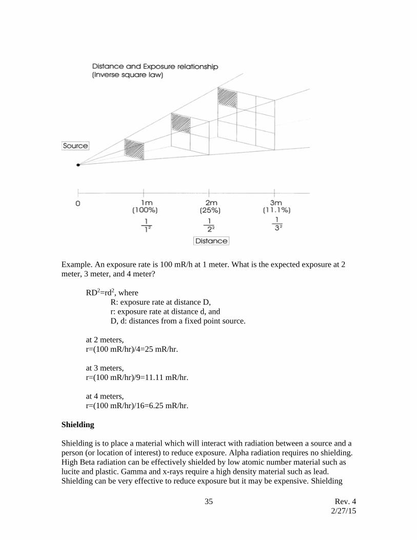
Distance is a simple, inexpensive, and very effective method of dose reduction. The
intensity of radiation from a point source decreases by 1/(distance)2. If a distance between
a person and a source is doubled (x2) then the exposure rate is decreased by 4. This is
called the "Inverse Square Law".
Rev. 4
2/27/15
35
Example. An exposure rate is 100 mR/h at 1 meter. What is the expected exposure at 2
meter, 3 meter, and 4 meter?
RD2=rd2, where
R: exposure rate at distance D,
r: exposure rate at distance d, and
D, d: distances from a fixed point source.
at 2 meters,
r=(100 mR/hr)/4=25 mR/hr.
at 3 meters,
r=(100 mR/hr)/9=11.11 mR/hr.
at 4 meters,
r=(100 mR/hr)/16=6.25 mR/hr.
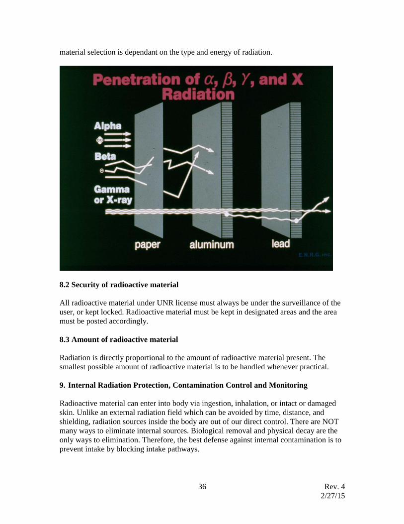
Shielding
Shielding is to place a material which will interact with radiation between a source and a
person (or location of interest) to reduce exposure. Alpha radiation requires no shielding.
High Beta radiation can be effectively shielded by low atomic number material such as
lucite and plastic. Gamma and x-rays require a high density material such as lead.
Shielding can be very effective to reduce exposure but it may be expensive. Shielding
Rev. 4
2/27/15
36
material selection is dependant on the type and energy of radiation.
8.2 Security of radioactive material
All radioactive material under UNR license must always be under the surveillance of the
user, or kept locked. Radioactive material must be kept in designated areas and the area
must be posted accordingly.
8.3 Amount of radioactive material
Radiation is directly proportional to the amount of radioactive material present. The
smallest possible amount of radioactive material is to be handled whenever practical.
9. Internal Radiation Protection, Contamination Control and Monitoring
Radioactive material can enter into body via ingestion, inhalation, or intact or damaged
skin. Unlike an external radiation field which can be avoided by time, distance, and
shielding, radiation sources inside the body are out of our direct control. There are NOT
many ways to eliminate internal sources. Biological removal and physical decay are the
only ways to elimination. Therefore, the best defense against internal contamination is to
prevent intake by blocking intake pathways.

Rev. 4
2/27/15
37
9.1 Contamination
Contamination is presence of radioactive material in any place where it is not necessary.
It may cause unnecessary radiation exposure and/or poses a possible intake pathway.
Contamination Control
The work area where radioactive material is used should be monitored before and after
use of radioactive material. The surrounding area should be monitored periodically. The
monitoring results need to be recorded for future reference. Always work over trays or
work surfaces lined with an absorbent material. Keep and transport radioisotope doubly
contained. Label radioisotope containers with your name, date, radionuclide and its
quantity. No eating, drinking, smoking or application of cosmetics is permitted in a
radioisotope laboratory. Wash hands after handling radioisotope and before doing other
work. Always use rubber or plastic gloves when handling radioisotope. Lab coats and
safety goggles shall be worn in the laboratory and left in the laboratory. They shall not be
used for other work, sent to another area, or released for cleaning until demonstrated to
be free of contamination. No open toe shoes are allowed while handling radioisotope.
Contamination Monitoring
A widely used contamination monitor is thin window GM detector. A GM detector
called "pancake GM" has a wide detector area (2 inch in diameter) with a thin window.
This probe can detect alpha, beta, and x, gamma rays. Most of radiation types used in
research laboratories may be detected by a pancake detector. Its high sensitivity makes it
easy to use in a low level contamination area. All new contamination monitors are
Rev. 4
2/27/15
38
equipped with audible sound which allows the user to focus on the area being monitored.
Normally contamination can be identified by listening to the detector sound. When
monitoring surface area for contamination, the active detector area should be close to the
surface without touching it and move slowly over the monitoring area about an inch per
second. Swipe surveys are another method of monitoring contamination. A filter paper,
filter cloth, or q-tip are used to take wipe samples which are analyzed by an appropriate
analyzer. For example, hydrogen-3 (a weak beta emitter) can be monitored by swipe
surveys. Liquid scintillation counting is a common method to analyze hydrogen-3 swipes.
Decontamination
Contamination may be associated with buildings, equipment, and personnel. If
contamination is found, the contamination should be removed as soon as possible. All
personal who are affected must be notified to avoid the spreading of contamination and to
minimize potential radiation exposure. The Radiation Safety Office can provide
assistance if a problem persists. It is easy to remove contamination in workbench tops if
the work area is covered with plastic backed absorbent pad. Application of a new pad and
discarding the contaminated absorbent pad into a radioactive waste container takes care
of the contamination. This is one of the reasons all radioactive material handling areas
such as bench tops should be covered with plastic backed absorbent pads.
Personnel Decontamination
Personnel contamination such as on clothing, shoes, or a part or whole body should be
approached in such a way as to prevent spreading of contamination and keeping it away
from wounds. Water and mild soap should be used initially. If harsher methods are
needed due to stubborn contaminants, an evaluation should be made to avoid embedding
the contaminant deeper into the skin. If contamination is fixed and not removable, the
contaminated area should be marked accordingly. An evaluation should be made based
on the isotope amount, half life, radiation type, and occupancy of the area, etc, to correct
contamination. It may be practical to wait for decay, or, to remove the contaminated
area/equipment. After decontamination, the result and its effectiveness must be verified
by re-survey.
9.2 Internal Dosimetry (monitoring methods of personnel suspected of having
radioactive material intake)
Bioassays include such tests as radioanalysis of blood, urine, fecal samples, nose swabs
or of sputum. In addition the term bioassay includes whole body or thyroid counts. As a
general principle, bioassays will be required after any incident (e.g., contamination of
personnel or exposure of persons to airborne radioactivity) where the possibility of
internal deposition of radioisotopes exists.
Urine analysis is a commonly used method for monitoring personnel suspected of having
or with high potential of radioactive material intake. Urine sample is collected post
Rev. 4
2/27/15
39
radioactive material use and analyzed by appropriate radiation system. The most
commonly used instrument for urine analysis is Liquid Scintillation Counter.
For personnel who use radioactive iodine, direct measurement of person’s thyroid is a
common bioassay method.
In addition to the above bioassay requirement, bioassays are required for one time tritium
use greater than 10 mCi and use of unbound radioiodine greater than 1 mCi at one time.
Bioassay service is available at any time upon the request of the User. Bioassays may be
arranged by calling the EH&S at 784-4540. Bioassay need to be performed within a few
days from the radionuclide use.
10. Emergency Procedures
Any medical emergency takes priority over a radiological emergency. Radioactive
materials used in research at UNR cannot produce life threatening levels of radiation
under any circumstances.
The objectives for handling radiological emergencies are to assist injured personnel,
minimize the radioactive material entering into human body, prevent the spread of
contamination, and remove the contamination as soon as possible. When approaching
radiological emergencies, it is recommended to apply these objectives using standard
laboratory safety precautions, and a common sense approach because it is not possible to
address all possible emergency scenarios.
Decontamination and/or spill clean-up in a radiation use facility is the responsibility of
the Authorized User of the facility. The EH&S Department will provide assistance where
needed.
All radiological incidents are to be reported to EH&S as soon as practical with the
exception of easily cleaned minor contamination. All incidents must be documented. This
documentation must include the final survey indicating that all contamination has been
removed.
10.1 Personnel decontamination
Contaminated areas of the body need to be identified using appropriate survey methods.
Do not use any decontamination methods which may spread material, increase
penetration into the body, or spread to wounded area.
Loose particles may be removed by gently applying adhesive side of tape to the particles
attached to skin. Most contamination may be removed by running water over the
contaminated area. Use soap or detergent if water by itself doesn’t remove all the
contaminants. Avoid harsh scrubbing which may increase skin penetration. If
contamination persists, stronger decontamination methods may be necessary after first
consulting with the EH&S Department.
Rev. 4
2/27/15
40
10.2 Minor spills or contamination
Most incidents at UNR will likely involve small quantities of radioactivity. If less than 20
microcurie of radioactivity is involved in a spill or contaminations, it is considered a
minor. Commercial cleaning supplies should be adequate. It is recommended to use them
only when other measures such as plain water did not work. The following steps are
recommended;
• Warn others in the lab that a spill or contamination has occurred.
• Fresh new gloves should be worn to protect hands and avoid spread of
contamination.
• Use paper towels or absorbent paper to prevent spread.
• Mark off the contaminated area.
• Do not allow lab personnel to leave the area without first being monitored.
• Secure all contaminated items in sealed containers to prevent spread of
contamination
10.3 Major spills
It is considered a major spill if greater than 20 microcurie of radioactive materials are
spilled or if personnel are contaminated. It is not possible to address all types of accidents.
But the following steps are general guide lines to deal with major accidents:
• Upright or cut off the release of radioactivity from the source if possible.
• Minimize radiation exposure to personnel.
• Minimize contamination from spreading.
• If airborne radioactivity is possible, shut off ventilation, hood, and close
windows if possible.
• Secure all contaminated items to prevent spread of contamination.
• Secure the contaminated area.
• Report incident to EH&S.
• Remain in the general area until EH&S personnel arrive.