Embed Size (px)
Citation preview

Received: 8 November 2017 Revised: 16 December 2017 Accepted: 17 January 2018
RE S EARCH ART I C L E
DOI: 10.1002/jlcr.3610
Ra‐224 labeling of calcium carbonate microparticles forinternal α‐therapy: Preparation, stability, andbiodistribution in mice
Sara Westrøm1,2,3 | Marion Malenge1 | Ida Sofie Jorstad1 | Elisa Napoli1,3,4 |
Øyvind S. Bruland1,3,5 | Tina B. Bønsdorff1 | Roy H. Larsen1
1Oncoinvent AS, Oslo, Norway2Department of Tumor Biology, Institutefor Cancer Research, The NorwegianRadium Hospital,, Oslo UniversityHospital, Oslo, Norway3 Institute of Clinical Medicine, Universityof Oslo, Oslo, Norway4Department of Radiation Biology,Institute for Cancer Research, TheNorwegian Radium Hospital, OsloUniversity Hospital, Oslo, Norway5Department of Oncology, The NorwegianRadium Hospital, Oslo UniversityHospital, Oslo, Norway
CorrespondenceRoy H. Larsen, Oncoinvent AS,Gullhaugveien 7, 0484 Oslo, Norway.Email: [email protected]
Funding informationOncoinvent AS; Oncoinvent AS;Norwegian Research Council, Grant/Award Numbers: 235531 and 237661
- - - - - - - - - - - - - - - - - - - - - - - - - - - - - - - - - - - - - - - -
This is an open access article under the terms of the C
original work is properly cited.
© 2018 Oncoinvent AS. Journal of Labelled Compou
J Label Compd Radiopharm. 2018;1–15.
Internal therapy with α‐emitters should be well suited for micrometastatic dis-
ease. Radium‐224 emits multiple α‐particles through its decay and has a conve-
nient 3.6 days of half‐life. Despite its attractive properties, the use of 224Ra has
been limited to bone‐seeking applications because it cannot be stably bound to
a targeting molecule. Alternative delivery systems for 224Ra are therefore of con-
siderable interest. In this study, calcium carbonate microparticles are proposed
as carriers for 224Ra, designed for local therapy of disseminated cancers in cav-
itary regions, such as peritoneal carcinomatosis. Calcium carbonate microparti-
cles were radiolabeled by precipitation of 224Ra on the particle surface, resulting
in high labeling efficiencies for both 224Ra and daughter 212Pb and retention of
more than 95% of these nuclides for up to 1 week in vitro. The biodistribution
after intraperitoneal administration of the 224Ra‐labeled CaCO3 microparticles
in immunodeficient mice revealed that the radioactivity mainly remained in
the peritoneal cavity. In addition, the systemic distribution of 224Ra was found
to be strongly dependent on the amount of administered microparticles, with a
reduced skeletal uptake of 224Ra with increasing dose. The results altogether
suggest that the 224Ra‐labeled CaCO3 microparticles have promising properties
for use as a localized internal α‐therapy of cavitary cancers.
KEYWORDS
alpha therapy, calcium carbonate, intraperitoneal, microparticles, peritoneal carcinomatosis, radium‐224
1 | INTRODUCTION
The use of internal α‐emitters to treat cancer has attractedsignificant attention during the last decade. Their superiorcytotoxicity and limited range in tissue corresponding toonly a few cell diameters are properties that make themsuitable for treatment of micrometastatic disease.1,2 So far,
- - - - - - - - - - - - - - - - - - - - - - - - - -
reative Commons Attribution Lice
nds and Radiopharmaceuticals Pub
the research efforts have culminated in 1 approved α‐emit-ting radiopharmaceutical, 223Ra‐dichloride (Xofigo®, Bayer),which is used for treatment of patients with skeletal metas-tases from castration‐resistant prostate cancer.3 Radium isan alkaline earth metal and thus chemically similar to cal-cium. This resemblance causes radium to target the bones,and to a larger degree, osteoblastic bone metastases.4 It is
- - - - - - - - - - - - - - - - - - - - - - - - - - - - - - - - - - - - - - - - - - - - - - - - - - - - - - - - - - - - - -nse, which permits use, distribution and reproduction in any medium, provided the
lished by John Wiley & Sons, Ltd.
wileyonlinelibrary.com/journal/jlcr 1
2 WESTRØM ET AL.
this property together with efficient cell kill from α‐particlesthat has made intravenous injection of 223Ra‐dichloride aneffective therapy.5 The bone‐seeking property of radiumwas also exploited medically with 224Ra over many years(1950‐1990 and 2000‐2005),6-9 although not in cancer butas palliative treatment of ankylosing spondylitis, a chronicinflammatory rheumatic disease.
Among the radium isotopes, there are 3 that standout to be considered for biomedical applications asinternal α‐emitters: 223Ra (t½ = 11.4 days), 224Ra(t½ = 3.6 days), and 225Ra (t½ = 14.9 days). Ra‐223 and224 are by themselves α‐emitters, whereas 225Ra is a β‐emit-ter. A shared feature with the 3 series is that they decay viamultiple α‐ and β‐emitting progeny with shorter half‐livesthan their respective radium parent and an average of 4emitted α‐particles per complete decay (Figure 1). Thedecay of each series releases a high total energy of 28 to29 MeV, where more than 90% of the energy is associatedwith α‐emissions. This property enables delivery oftherapeutically relevant doses at low administered activitylevels. In addition, the 3 mentioned radium isotopes allhave relatively long half‐lives which allows centralizedproduction and quality control before shipment to theend user, such that challenges relating to production forradionuclides with short half‐lives can be avoided.
Despite their attractive properties for radionuclidetherapy, the use of radium isotopes has so far mainly beenlimited to bone‐seeking applications. One reason for thisis the lack of an appropriate bifunctional chelatingagent for coupling of radium to targeting molecules.
FIGURE 1 The decay chains of 223Ra, 224Ra, and 225Ra including det
There have been a few studies of possible chelatingagents,10,11 but a bifunctional ligand that stably bindsradium with control of daughter nuclides in vivo has yetto be presented. However, 224Ra has been used as a gener-ator nuclide for its daughter 212Pb.12,13 To fully exploit thetherapeutic potential of radium isotopes for other pur-poses than bone targeting, alternative delivery systemsare needed. One proposed strategy is to use nanoparticlesor microparticles as carriers, and radium has been incor-porated into the core of lanthanum phosphate nanoparti-cles,14 loaded into liposomes,15,16 absorbed into the poresof nanozeolites,17,18 and both intrinsically incorporated inthe crystal structure and adsorbed onto the surfaces ofhydroxyapatite nanoparticles and microparticles.19-21
Among these, all particles apart from liposomes are com-posed of inorganic materials. Another inorganic materialthat is promising for different biomedical applications iscalcium carbonate (CaCO3). It is considered as generallyrecognized as safe by the FDA and is widely used as a foodadditive and also in various oral drugs. Due to its low cost,ease of manufacture, and biodegradability, CaCO3
particles have been studied in vitro as potential carriersfor various drugs22-27 and in monkeys and humans forintranasal administration of insulin.28
Internal radiation therapy with radiolabeled particleshas been a treatment option for cancers with intracavitarydissemination, eg, for patients with peritoneal carcinoma-tosis from ovarian carcinoma.29 This condition occurswhen cancer cells from a primary tumor in an adjacentorgan disseminate into the peritoneal cavity and cause
ails on each nuclides' half‐life and main mode of decay
WESTRØM ET AL. 3
micrometastases by adhering to the serosal surfaces of theperitoneal lining. Peritoneal metastases are the most com-mon terminal feature of abdominal cancers and generallyimply a poor prognosis for the patients affected.30 Intra-peritoneal (IP) radionuclide therapy has been evaluatedin clinical trials with suspensions of radioactive nanopar-ticles or microparticles31,32 and, more recently, also asradioimmunotherapy.33-37 An overview of the physicalcharacteristics of the different radionuclides that havebeen utilized is given in Table 1. With particles as carriers,only the β‐emitters 198Au and 32P have been examined inpatients. Of the 2, IP therapy with 32P‐colloid was themost successful.32,38 In a randomized trial, it was reportedto be as effective as adjuvant cisplatin for treatment ofovarian cancer, although late bowel complicationsoccurred more frequently.32 This complication was alsoseen after 198Au‐colloid therapy.29 One possible cause ofthe adverse effects is the emitted electrons, which havea range of several millimeters in tissue and thereforecould irradiate deeper areas of radiation sensitive organsin the abdomen, like the small intestine.
Our long‐term goal is to develop a novel radium com-pound for locoregional treatment of cancers in cavitaryregions. By utilizing the α‐emitting 224Ra, with a penetra-tion depth in tissue of less than 0.1 mm, the aim is todesign a radiotherapeutic microparticle for intracavitaryinjection with highly localized effect on cancer cells resid-ing on serosal surfaces, with minimal normal organ expo-sure. Microparticles were chosen as carriers for 224Ra, as itis possible to select a size that facilitates high retention ofthe particles in the peritoneal cavity.39,40 Consequently,the radiolabeled microparticles will irradiate the areawhere the peritoneal micrometastases are located. In thisstudy, we have investigated the suitability of CaCO3
microparticles as carriers for 224Ra. The complete prepara-tion of the product is described, and the biodistribution in
TABLE 1 Physical characteristics of the radionuclides previously inve224Ra
Radionuclide Half‐Life Decay Mode(s)
32P 14.3 days β90Y 2.7 days β131I 8.0 days β177Lu 6.7 days β198Au 2.7 days β211At 7.2 h α212Pb 10.6 h β + α224Ra 3.6 days 4α + 2β
aFrom ENSDF decay data in MIRD format (http://www.nndc.bnl.gov/mird/). Liste
mice after IP injection of the radiolabeled microparticlesis evaluated.
2 | EXPERIMENTAL
2.1 | Calcium carbonate microparticles
Crystalline CaCO3 microparticles were prepared by a spon-taneous precipitation method based on the protocoldescribed by Volodkin et al.41 Five milligrams of 0.33 MNa2CO3 (Merck, Darmstadt, Germany) solution wasrapidly poured into an equal volume of 0.33 M CaCl2(Merck). First‐generation microparticles were prepared byintense vortexing of the mixture for 30 seconds before theparticle suspension was left for 5 minutes. The precipitatewas collected by using filtration through a 0.45‐ μm nitro-cellulose filter (Whatman, GE Healthcare, UK) in a glassvacuum filtration device, before it was washed with approx-imately 30‐mL ph. Eur water (complying with quality stan-dards of the European Pharmacopeia, VWR, Oslo, Norway)and dried overnight at room temperature. Second‐genera-tion microparticles were prepared by mixing of 0.33 MNa2CO3 and CaCl2 solutions with a magnetic stirrer (BiosanMS3000, Riga, Latvia) before the precipitated particles werecollected by centrifugation. The precipitate was washed inph. Eur water and dried for 1 hour at 180°C (VENTI‐LineDrying oven VL53, VWR). In addition, a batch of CaCO3
microparticles was purchased from PlasmaChem GmbH(Berlin, Germany). A portion of the microparticles weredry sterilized at 180°C for 2 hours. The 3 different particletypes were analyzed by laser diffraction in a Mastersizer3000 (Malvern Instruments Ltd, Worcestershire, UK), andvolume‐based diameters were obtained. The microparticleswere also analyzed for visualization of crystal shape, size,and surface morphology with scanning electron microscopy
stigated clinically for local intraperitoneal therapy compared with
Energya (MeV/Bq‐s) Carrier
0.695 Particles32
0.934 Antibody33
0.573 Antibody34
0.131 Antibody35
0.731 Particles31
6.96 Antibody fragment36
10.27 Antibody37
29.26 n/a
d α, β, γ, and X‐rays for mother nuclides and, if applicable, progeny combined.

4 WESTRØM ET AL.
(SEM) performed at Particle Analytical (Hørsholm,Denmark) with a Leica Stereoscan 360.
TABLE 2 Overview of X‐ and/or γ‐lines in the 224Ra series with
1% or higher abundance
Nuclide 65‐345 keV (Abundance) >345 keV (Abundance)
224Ra 241.0 keV (4.1%)
2.2 | Preparation of 224Ra generator basedon 228Th
The 224Ra generator was prepared by mixing a 228Thsource with an actinide resin and loading it on a column.A source of 228Th in 1 M HNO3 was purchased fromEckert & Ziegler (Braunschweig, Germany), and an acti-nide resin based on the DIPEX® Extractant was acquiredfrom Eichrom Technologies LLC (Lisle, IL) in the formof a pre‐packed cartridge of 2 mL. The material in an acti-nide resin cartridge was extracted, and the resin waspreconditioned with 1 M HCl (Sigma‐Aldrich). A slurryof approximately 0.25‐ mL actinide resin, 0.25‐ mL 1 MHCl, and 0.1‐ mL 228Th in 1 M HNO3 was prepared in avial (4‐mL vial, E‐C sample, Wheaton, Millville, NJ) andincubated with gentle agitation for immobilization of228Th for 4 hours at room temperature and let to rest fora few days. The generator column was prepared in a1‐ mL filtration column (Isolute SPE, Biotage AB,Uppsala, Sweden) by first applying 0.2 mL of inactiveactinide resin, before the portion containing 228Th wasloaded on top. The inactive resin was introduced in thebottom of the column to serve as a catcher layer if 228Thwas released during operation of the generator. Later,the capacity of the generator was increased. A slurryconsisting of 0.4‐ mL actinide resin, 0.5‐ mL 228Th in1 M HNO3, and 0.5‐ mL 1 M HCl was prepared asdescribed above, before it was loaded onto the generatorcolumn. At its maximum capacity, the 224Ra generatorcolumn contained approximately 2‐ MBq 228Th.
220Rn216Po212Pb 74.8 keV (10.3%)
77.1 keV (17.1%)86.8 keV (2.1%)87.4 keV (4.0%)89.8 keV (1.5%)238.6 keV (43.6%)300.1 keV (3.3%)
212Bi 727.3 keV (4.3%)a
212Po208Tl 510.8 keV (8.1%)b
75.0 keV (1.2%)b 583.2 keV (30.5%)b
277.4 keV (2.4%)b 860.6 keV (4.5%)b
2614.5 keV (35.8%)b
The X and γ‐lines are divided into 2 columns, 1 for energies between 65 and345 keV and the other for energies larger than 345 keV. The energies and
abundance were retrieved from www.nndc.bnl.gov/chart.aBranching corrected for 64.1%.bBranching corrected for 35.9%.
2.3 | Extraction of 224Ra
Radium‐224 could be eluted regularly from the generatorcolumn in 1 to 2 mL of 1 M HCl. For further purification,the crude eluate from the generator column was loadeddirectly onto a second actinide resin column. The secondcolumn was washed with 1 M HCl. This eluate was evap-orated to dryness in a closed system. The vial was placedin a heater block (heated to approximately 100°C) andflushed with N2 gas through a Teflon tube inlet and outletin the rubber/Teflon septum on the vial. The acid vaporwas led into a beaker of saturated NaOH by a stream ofN2 gas. The radioactive residue remaining after evapora-tion was dissolved in 0.2 mL or more of 0.1 M HCl. Aradioisotope calibrator (CRC‐25R, Capintec Inc., Ramsey,NJ) was used to measure the total extracted activity in theprocess. Possible breakthrough of 228Th in the final 224Rasolution was examined by sending 2 samples from differ-ent eluates for analysis at Institute for Energy Technology
(Kjeller, Norway). In 1 sample, the 224Ra and 228Th con-tent was determined by radiochemical separationfollowed by α‐spectroscopy. To detect possible ingrowthof 224Ra from 228Th, the second sample was measuredrepeatedly over a period of 40 days with liquid scintilla-tion to determine the total amount of α‐emitters.
2.4 | Radioactivity measurements
Radioactive samples were measured in the window 70 to80 keV on a Cobra II Autogamma counter (PackardInstruments, Downer Grove, IL) or from 65 to 345 keVon a Hidex Automatic Gamma Counter (Hidex, Turku,Finland). From 70 to 80 and 65 to 345 keV, the most abun-dant X‐ and γ‐radiation is from the 224Ra daughter 212Pb,and it is therefore assumed that the counts in thesewindows mainly originate from 212Pb with minimal con-tribution from other nuclides in the series. This can beseen from Table 2, which provides an overview of X‐and γ‐rays from the different nuclides in the 224Ra series.Because 224Ra decay results in modest γ‐emission in anenergy region with more abundant γ from its progeny212Pb, the 224Ra activity was determined indirectly basedon the counts in the 70 to 80‐ keV or 65 to 345‐ keV win-dow. This was carried out by re‐measuring the samplesbetween 1 and 4 days after the first measurement, whenthe initial 212Pb present in the sample had decayed and
WESTRØM ET AL. 5
equilibrium between 224Ra and newly produced 212Pb hadbeen established. A pure source of 224Ra reaches equilib-rium conditions after approximately 2 days.
2.5 | Labeling of CaCO3 microparticleswith 224Ra
Radium‐224‐labeled microparticles were prepared by pre-cipitation of 224Ra2+ on the CaCO3 surfaces. From 10 to200 mg of CaCO3 microparticles were transferred to anEppendorf tube and washed 3 times in 1‐ mL water and2 times with 1‐ mL 0.1 M Na2SO4 (Alfa Aesar, Karlsruhe,Germany). The particles were separated from the washingsolution by centrifugation (2000×g for 30‐180 s,Spectrafuge™ Mini, Labnet Inc.). After wash, the micro-particles were dispersed in either Dulbecco's phosphate‐buffered saline (PBS; Gibco, Life Technologies, Carlsbad,CA) supplemented with 0.5% bovine serum albumin(BSA), a sucrose solution, or 0.9% NaCl (Merck). Thesucrose solution contained 94‐ mg/mL sucrose (SigmaUltra, St. Louis, MO) and 2.1‐mg/mL Na2SO4 (Alfa Aesar)and was pH adjusted to 7.5. The use of a sucrose solutionfor radiolabeling of the microparticles was exploredbecause of the viscosity of the solution. In the case ofusing Dulbecco's PBS with 0.5% BSA, the particle suspen-sion was incubated with orbital rotation on a HulaMixer(Invitrogen, Life Technologies, Carlsbad, CA) for30 minutes at room temperature before continuing theprotocol. A solution of 224Ra in equilibrium with progenyin 0.1 M HCl and 0.5 M NH4OAc (Merck), and pHbetween 5 and 6 was added to the particles together withsmall amounts of SO4
2− and Ba2+ used as co‐precipitants.The volumes of 0.1 M Na2SO4 and 0.07 M BaCl2·2H2O(Merck) solutions corresponded to 0.3% of SO4
2− andBa2+ per mg of particles, respectively. Microparticles inradiolabeling solution were incubated with orbital rota-tion on a HulaMixer for minimum 90 minutes at roomtemperature. After incubation, the particle solution waswashed twice with 1 mL of either sucrose solution or0.9% NaCl, and the wash solutions collected in separatetubes. The radioactivity (counts per minute, CPM) in theparticle suspension (P) and washing solutions (W1 andW2) was measured, and the 212Pb labeling efficiency wasestimated as the percentage of the total activity still boundto the microparticles after the labeling procedure:
%Labeling efficiency ¼ CPM Pð ÞCPM PþW1þW2ð Þ×100
All samples were left to decay for minimum 24 hours atroom temperature, to reach equilibrium between 224Raand 212Pb, before they were re‐measured, and the 224Ralabeling efficiency was calculated with the same equation
as presented above. It is assumed here that equilibriumbetween 224Ra and 212Pb in the samples is reached after24 hours because the samples are expected to have a rela-tively even distribution of the 2 nuclides; ie, equilibriumwill be reached faster than from a pure source of 224Ra.
2.6 | In vitro stability of 224Ra‐labeledCaCO3 microparticles
To determine the retention of 224Ra and 212Pb on themicroparticles after labeling, the particles were incubatedin 1‐ mL sucrose solution at room temperature. After 1, 3,5, and 7 days, the particle suspension was centrifuged and80% of the supernatant was withdrawn into a separatevial. Afterward, if the stability study was to be continuedto a later time point, the particle pellet was dispersed ina new aliquot of sucrose solution and incubated further.The radioactivity in the removed supernatant (S) andparticle suspension (P) was measured, and the estimatedactivity on the particles without supernatant was dividedby the absolute amount of activity in the sample to deter-mine the fraction of 212Pb retained on the particles:
%Retained activity ¼CPM Pð Þ− CPM Sð Þ × 20
80
� �
CPM Pþ Sð Þ × 100:
Two days after the first measurement, when equilib-rium between 224Ra and 212Pb had been reached, thesamples were re‐measured and the retained 224Ra activitywas estimated with the equation given above.
2.7 | Biodistribution and in vivo stabilityof 224Ra‐labeled CaCO3 microparticles
Institutionally bred, 4 to 48 weeks old, healthy femaleAthymic nude Foxnnu mice with body weights in therange of 17.1 to 28.5 g at the start of the experiment wereused. The animals were maintained under pathogen‐freeconditions with food and water supplied ad libitum. Allprocedures and experiments involving animals wereapproved by the National Animal Research Authority(permit ID 6675) and performed in compliance with regu-lations set by the same authority and the EU Directive2010/63/EU on the protection of animals used for scien-tific purposes.
The biodistribution of radioactivity inmice was studiedafter a single IP injection of 0.4‐mL 224Ra‐labeled CaCO3
microparticle suspension. Table 3 gives an overview ofthe different biodistribution experiments performed with224Ra‐labeled CaCO3 microparticles. Biodistributionexperiments with free 224Ra (dissolved RaCl2) were alsoperformed, by administering approximately 3 kBq 224Ra
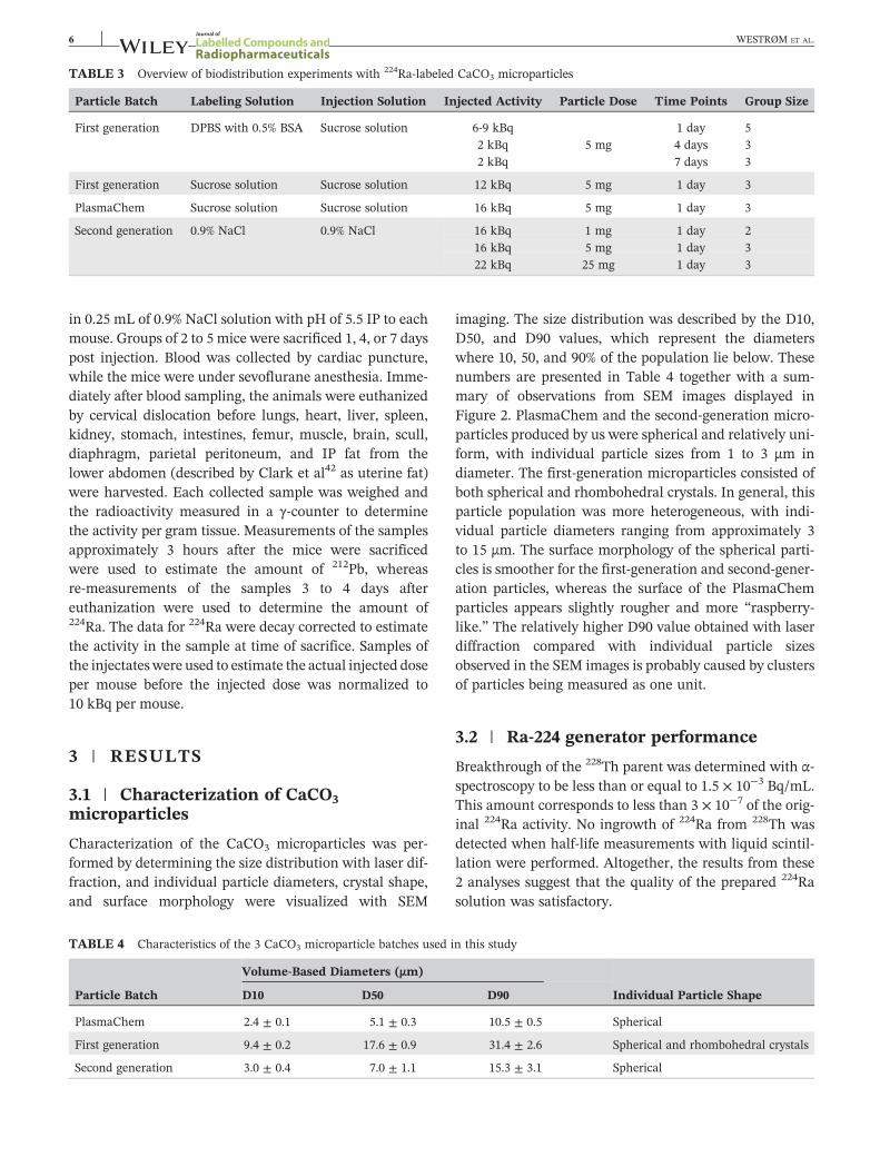
TABLE 3 Overview of biodistribution experiments with 224Ra‐labeled CaCO3 microparticles
Particle Batch Labeling Solution Injection Solution Injected Activity Particle Dose Time Points Group Size
First generation DPBS with 0.5% BSA Sucrose solution 6‐9 kBq 1 day 52 kBq 5 mg 4 days 32 kBq 7 days 3
First generation Sucrose solution Sucrose solution 12 kBq 5 mg 1 day 3
PlasmaChem Sucrose solution Sucrose solution 16 kBq 5 mg 1 day 3
Second generation 0.9% NaCl 0.9% NaCl 16 kBq 1 mg 1 day 216 kBq 5 mg 1 day 322 kBq 25 mg 1 day 3
6 WESTRØM ET AL.
in 0.25 mL of 0.9% NaCl solution with pH of 5.5 IP to eachmouse. Groups of 2 to 5 mice were sacrificed 1, 4, or 7 dayspost injection. Blood was collected by cardiac puncture,while the mice were under sevoflurane anesthesia. Imme-diately after blood sampling, the animals were euthanizedby cervical dislocation before lungs, heart, liver, spleen,kidney, stomach, intestines, femur, muscle, brain, scull,diaphragm, parietal peritoneum, and IP fat from thelower abdomen (described by Clark et al42 as uterine fat)were harvested. Each collected sample was weighed andthe radioactivity measured in a γ‐counter to determinethe activity per gram tissue. Measurements of the samplesapproximately 3 hours after the mice were sacrificedwere used to estimate the amount of 212Pb, whereasre‐measurements of the samples 3 to 4 days aftereuthanization were used to determine the amount of224Ra. The data for 224Ra were decay corrected to estimatethe activity in the sample at time of sacrifice. Samples ofthe injectateswere used to estimate the actual injected doseper mouse before the injected dose was normalized to10 kBq per mouse.
3 | RESULTS
3.1 | Characterization of CaCO3microparticles
Characterization of the CaCO3 microparticles was per-formed by determining the size distribution with laser dif-fraction, and individual particle diameters, crystal shape,and surface morphology were visualized with SEM
TABLE 4 Characteristics of the 3 CaCO3 microparticle batches used i
Particle Batch
Volume‐Based Diameters (μm)
D10 D50
PlasmaChem 2.4 ± 0.1 5.1 ± 0.3
First generation 9.4 ± 0.2 17.6 ± 0.9
Second generation 3.0 ± 0.4 7.0 ± 1.1
imaging. The size distribution was described by the D10,D50, and D90 values, which represent the diameterswhere 10, 50, and 90% of the population lie below. Thesenumbers are presented in Table 4 together with a sum-mary of observations from SEM images displayed inFigure 2. PlasmaChem and the second‐generation micro-particles produced by us were spherical and relatively uni-form, with individual particle sizes from 1 to 3 μm indiameter. The first‐generation microparticles consisted ofboth spherical and rhombohedral crystals. In general, thisparticle population was more heterogeneous, with indi-vidual particle diameters ranging from approximately 3to 15 μm. The surface morphology of the spherical parti-cles is smoother for the first‐generation and second‐gener-ation particles, whereas the surface of the PlasmaChemparticles appears slightly rougher and more “raspberry‐like.” The relatively higher D90 value obtained with laserdiffraction compared with individual particle sizesobserved in the SEM images is probably caused by clustersof particles being measured as one unit.
3.2 | Ra‐224 generator performance
Breakthrough of the 228Th parent was determined with α‐spectroscopy to be less than or equal to 1.5 × 10⁻³ Bq/mL.This amount corresponds to less than 3 × 10⁻⁷ of the orig-inal 224Ra activity. No ingrowth of 224Ra from 228Th wasdetected when half‐life measurements with liquid scintil-lation were performed. Altogether, the results from these2 analyses suggest that the quality of the prepared 224Rasolution was satisfactory.
n this study
Individual Particle ShapeD90
10.5 ± 0.5 Spherical
31.4 ± 2.6 Spherical and rhombohedral crystals
15.3 ± 3.1 Spherical
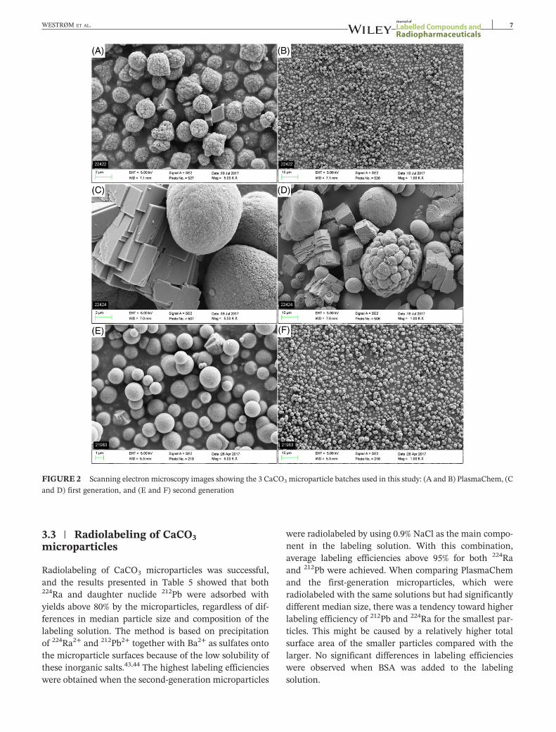
FIGURE 2 Scanning electron microscopy images showing the 3 CaCO3 microparticle batches used in this study: (A and B) PlasmaChem, (C
and D) first generation, and (E and F) second generation
WESTRØM ET AL. 7
3.3 | Radiolabeling of CaCO3microparticles
Radiolabeling of CaCO3 microparticles was successful,and the results presented in Table 5 showed that both224Ra and daughter nuclide 212Pb were adsorbed withyields above 80% by the microparticles, regardless of dif-ferences in median particle size and composition of thelabeling solution. The method is based on precipitationof 224Ra2+ and 212Pb2+ together with Ba2+ as sulfates ontothe microparticle surfaces because of the low solubility ofthese inorganic salts.43,44 The highest labeling efficiencieswere obtained when the second‐generation microparticles
were radiolabeled by using 0.9% NaCl as the main compo-nent in the labeling solution. With this combination,average labeling efficiencies above 95% for both 224Raand 212Pb were achieved. When comparing PlasmaChemand the first‐generation microparticles, which wereradiolabeled with the same solutions but had significantlydifferent median size, there was a tendency toward higherlabeling efficiency of 212Pb and 224Ra for the smallest par-ticles. This might be caused by a relatively higher totalsurface area of the smaller particles compared with thelarger. No significant differences in labeling efficiencieswere observed when BSA was added to the labelingsolution.
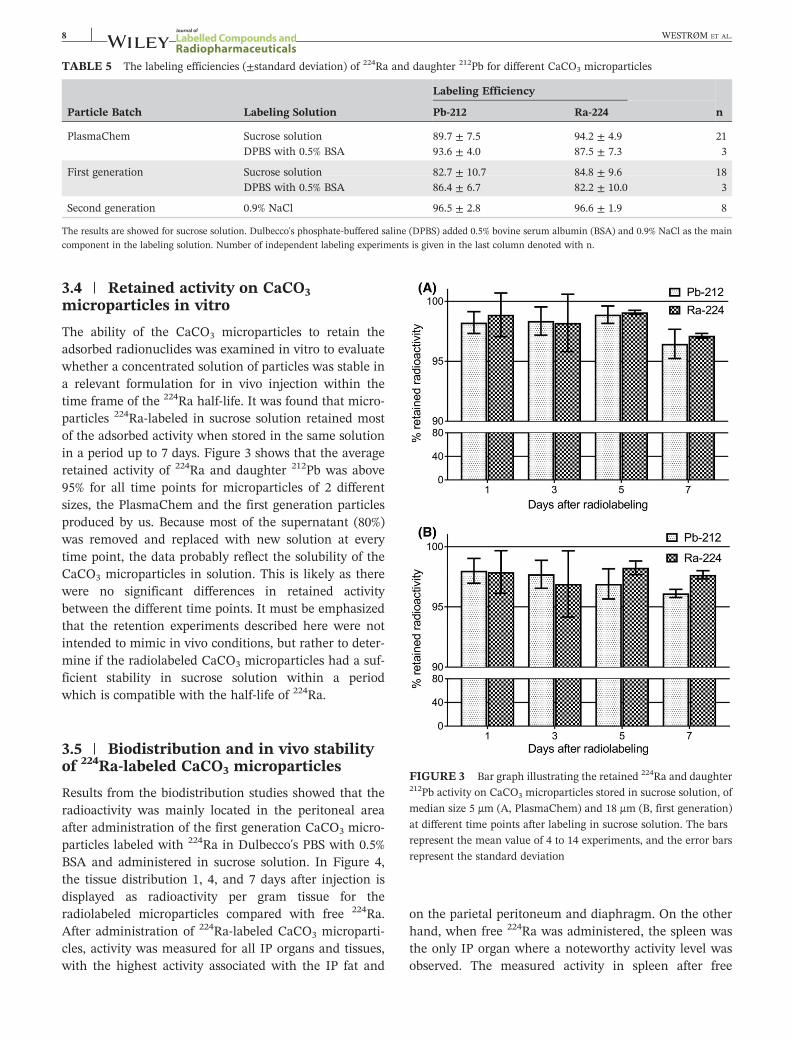
TABLE 5 The labeling efficiencies (±standard deviation) of 224Ra and daughter 212Pb for different CaCO3 microparticles
Particle Batch Labeling Solution
Labeling Efficiency
nPb‐212 Ra‐224
PlasmaChem Sucrose solution 89.7 ± 7.5 94.2 ± 4.9 21DPBS with 0.5% BSA 93.6 ± 4.0 87.5 ± 7.3 3
First generation Sucrose solution 82.7 ± 10.7 84.8 ± 9.6 18DPBS with 0.5% BSA 86.4 ± 6.7 82.2 ± 10.0 3
Second generation 0.9% NaCl 96.5 ± 2.8 96.6 ± 1.9 8
The results are showed for sucrose solution. Dulbecco's phosphate‐buffered saline (DPBS) added 0.5% bovine serum albumin (BSA) and 0.9% NaCl as the maincomponent in the labeling solution. Number of independent labeling experiments is given in the last column denoted with n.
8 WESTRØM ET AL.
3.4 | Retained activity on CaCO3microparticles in vitro
The ability of the CaCO3 microparticles to retain theadsorbed radionuclides was examined in vitro to evaluatewhether a concentrated solution of particles was stable ina relevant formulation for in vivo injection within thetime frame of the 224Ra half‐life. It was found that micro-particles 224Ra‐labeled in sucrose solution retained mostof the adsorbed activity when stored in the same solutionin a period up to 7 days. Figure 3 shows that the averageretained activity of 224Ra and daughter 212Pb was above95% for all time points for microparticles of 2 differentsizes, the PlasmaChem and the first generation particlesproduced by us. Because most of the supernatant (80%)was removed and replaced with new solution at everytime point, the data probably reflect the solubility of theCaCO3 microparticles in solution. This is likely as therewere no significant differences in retained activitybetween the different time points. It must be emphasizedthat the retention experiments described here were notintended to mimic in vivo conditions, but rather to deter-mine if the radiolabeled CaCO3 microparticles had a suf-ficient stability in sucrose solution within a periodwhich is compatible with the half‐life of 224Ra.
FIGURE 3 Bar graph illustrating the retained 224Ra and daughter212Pb activity on CaCO3 microparticles stored in sucrose solution, of
median size 5 μm (A, PlasmaChem) and 18 μm (B, first generation)
at different time points after labeling in sucrose solution. The bars
represent the mean value of 4 to 14 experiments, and the error bars
represent the standard deviation
3.5 | Biodistribution and in vivo stabilityof 224Ra‐labeled CaCO3 microparticles
Results from the biodistribution studies showed that theradioactivity was mainly located in the peritoneal areaafter administration of the first generation CaCO3 micro-particles labeled with 224Ra in Dulbecco's PBS with 0.5%BSA and administered in sucrose solution. In Figure 4,the tissue distribution 1, 4, and 7 days after injection isdisplayed as radioactivity per gram tissue for theradiolabeled microparticles compared with free 224Ra.After administration of 224Ra‐labeled CaCO3 microparti-cles, activity was measured for all IP organs and tissues,with the highest activity associated with the IP fat and
on the parietal peritoneum and diaphragm. On the otherhand, when free 224Ra was administered, the spleen wasthe only IP organ where a noteworthy activity level wasobserved. The measured activity in spleen after free
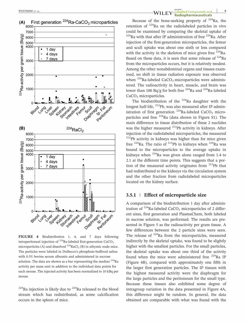
FIGURE 4 Biodistribution 1, 4, and 7 days following
intraperitoneal injection of 224Ra‐labeled first‐generation CaCO3
microparticles (A) and dissolved 224RaCl2 (B) in athymic nude mice.
The particles were labeled in Dulbecco's phosphate‐buffered saline
with 0.5% bovine serum albumin and administered in sucrose
solution. The data are shown as a bar representing the median 224Ra
activity per mass unit in addition to the individual data points for
each mouse. The injected activity has been normalized to 10 kBq per
mouse
WESTRØM ET AL. 9
224Ra injection is likely due to 224Ra released to the bloodstream which has redistributed, as some calcificationoccurs in the spleen of mice.
Because of the bone‐seeking property of 224Ra, theretention of 224Ra on the radiolabeled particles in vivocould be examined by comparing the skeletal uptake of224Ra with that after IP administration of free 224Ra. Afterinjection of the first‐generation microparticles, the femurand scull uptake was about one sixth or less comparedwith the activity in the skeleton of mice given free 224Ra.Based on these data, it is seen that some release of 224Rafrom the microparticles occurs, but it is relatively modest.Among the other nonabdominal organs and tissues exam-ined, no shift in tissue radiation exposure was observedwhen 224Ra‐labeled CaCO3‐microparticles were adminis-tered. The radioactivity in heart, muscle, and brain waslower than 100 Bq/g for both free 224Ra and 224Ra‐labeledCaCO3 microparticles.
The biodistribution of the 224Ra daughter with thelongest half‐life, 212Pb, was also measured after IP admin-istration of first generation 224Ra‐labeled CaCO3 micro-particles and free 224Ra (data shown in Figure S1). Themain difference in tissue distribution of these 2 nuclideswas the higher measured 212Pb activity in kidneys. Afterinjection of the radiolabeled microparticles, the measured212Pb activity in kidneys was higher than for mice givenfree 224Ra. The ratio of 212Pb in kidneys when 224Ra wasbound to the microparticles to the average uptake inkidneys when 224Ra was given alone ranged from 1.4 to2.1 at the different time points. This suggests that a por-tion of the measured activity originates from 212Pb thathad redistributed to the kidneys via the circulation systemand the other fraction from radiolabeled microparticleslocated on the kidney surface.
3.5.1 | Effect of microparticle size
A comparison of the biodistribution 1 day after adminis-tration of 224Ra‐labeled CaCO3 microparticles of 2 differ-ent sizes, first generation and PlasmaChem, both labeledin sucrose solution, was performed. The results are pre-sented in Figure 5 as the radioactivity per gram tissue. Afew differences between the 2 particle sizes were seen.The release of 224Ra from the microparticles, measuredindirectly by the skeletal uptake, was found to be slightlyhigher with the smallest particles. For the small particles,the skeletal uptake was about one third of the activityfound when the mice were administered free 224Ra IP(Figure 4B), compared with approximately one fifth inthe larger first generation particles. The IP tissues withthe highest measured activity were the diaphragm forthe large particles and the peritoneum for the small type.Because these tissues also exhibited some degree ofintragroup variation in the data presented in Figure 4A,this difference might be random. In general, the dataobtained are comparable with what was found with the

FIGURE 5 Comparison of biodistribution 1 day after
intraperitoneal injection of 224Ra‐labeled CaCO3 microparticles
with small (PlasmaChem) and large (first generation) median
diameter in athymic nude mice. The particles were labeled and
administered in sucrose solution. The data are shown as a bar
representing the median 224Ra activity per mass unit in addition to
the individual data points for each mouse. The injected activity has
been normalized to 10 kBq per mouse
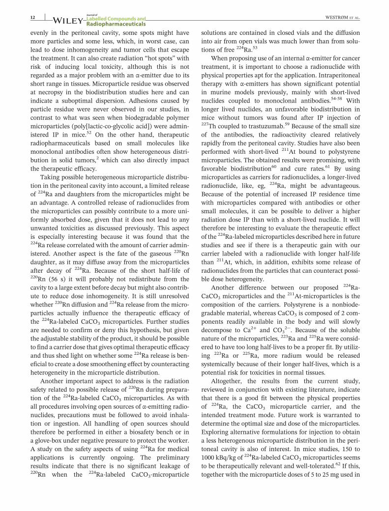
FIGURE 6 Biodistribution in athymic nude mice 1 day following
intraperitoneal administration of 1, 5, and 25 mg of 224Ra‐labeled
second generation CaCO3 microparticles. The particles were labeled
and administered in 0.9% NaCl. The data are shown as a bar
representing the median 224Ra activity per mass unit in addition to
the individual data points for each mouse. The injected activity has
been normalized to 10 kBq per mouse
10 WESTRØM ET AL.
first‐generation microparticles labeled with Dulbecco'sPBS with 0.5% BSA (Figure 4A), apart from a notablyhigher measured activity in the liver and spleen with thefirst‐generation particles labeled in sucrose solution.Because the high activity in these organs was not observedwith PlasmaChem and to a lesser extent when the first‐generation microparticles were labeled by using BSA, itis not likely to be a consequence of the absence of BSAin the labeling solution. One possible explanation forthe variability in the measured activity in the spleencan be incomplete removal of the surrounding fat. Gener-ally, high activity levels were measured in the IP fat afteradministration of the radiolabeled microparticles. Thepresence of fat associated with the spleen could thereforegreatly influence the measured activity and cause thehigh levels.
3.5.2 | Effect of amount of microparticles
The effect of amount of carrier was investigated by com-paring the biodistribution 1 day after administration of1, 5, and 25 mg of second‐generation microparticles224Ra‐labeled and administered in 0.9% NaCl. The resultsare mainly similar to the previously presentedbiodistribution results, but with 1 striking difference. Itis seen in Figure 6 that the skeletal uptake, and therebyalso the 224Ra release, is strongly correlated to the amountof microparticles administered. The level of 224Ra in the
femurs of mice given 1‐ mg 224Ra‐labeled CaCO3 micro-particles was only 30% less than after IP injection of free224Ra, showing a very high release. On the contrary, therelease was almost negligible after injection of 25‐ mg224Ra‐labeled CaCO3 microparticles. The skeletal uptakewas only 4% of what was found in the skeleton of mice1 day after administration of free 224Ra. The intermediateamount, 5 mg, also exhibited intermediate stability withalmost identical skeletal uptake after 1 day as when5‐ mg PlasmaChem microparticles labeled and adminis-tered in sucrose solution were used. This observation isconsistent with the PlasmaChem and second‐generationparticles being of the approximate same size, as the previ-ous results showed that 224Ra release from the particleswas somewhat dependent on size.
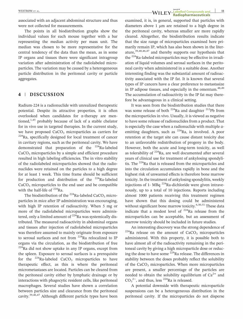
3.5.3 | Microparticle distribution in theperitoneal cavity
In most of the mice, particulate residue was observed atnecropsy. The residue was found regardless of the micro-particle type and amount of particles and also indepen-dent of the different labeling and injection solutions thatwere used. The amount of residue increased with increas-ing amount of particles administered. When 1 and 5‐ mgparticles were administered, the residue was relativelysmall, below 3 mm in diameter, and was always foundon the anterior parietal peritoneum. After administrationof 25‐ mg particles, the residue was larger and severalpieces were observed. It was also always located on theanterior parietal peritoneum but in addition found morespread out in the entire peritoneal cavity. All observedresidues were either completely free‐floating or loosely
WESTRØM ET AL. 11
associated with an adjacent abdominal structure and thuswere not collected for measurements.
The points in all biodistribution graphs show theindividual values for each mouse together with a barrepresenting the median activity per mass unit. Themedian was chosen to be more representative for thecentral tendency of the data than the mean, as in someIP organs and tissues there were significant intragroupvariation after administration of the radiolabeled micro-particles. The variation may be caused by a heterogeneousparticle distribution in the peritoneal cavity or particleaggregates.
4 | DISCUSSION
Radium‐224 is a radionuclide with unrealized therapeuticpotential. Despite its attractive properties, it is oftenoverlooked when candidates for α‐therapy are men-tioned,1,45 probably because of lack of a stable chelatorfor in vivo use in targeted therapies. In the current study,we have proposed CaCO3 microparticles as carriers for224Ra, specifically designed for local treatment of cancerin cavitary regions, such as the peritoneal cavity. We havedemonstrated that preparation of the 224Ra‐labeledCaCO3 microparticles by a simple and efficient procedureresulted in high labeling efficiencies. The in vitro stabilityof the radiolabeled microparticles showed that the radio-nuclides were retained on the particles to a high degreefor at least 1 week. This time frame should be sufficientfor production and distribution of the 224Ra‐labeledCaCO3 microparticles to the end user and be compatiblewith the half‐life of 224Ra.
The biodistribution of the 224Ra‐labeled CaCO3 micro-particles in mice after IP administration was encouraging,with high IP retention of radioactivity. When 5 mg ormore of the radiolabeled microparticles were adminis-tered, only a limited amount of 224Ra was systemically dis-tributed. The measured radioactivity in abdominal organsand tissues after injection of radiolabeled microparticleswas therefore assumed to mainly originate from exposureto serosal surfaces and not from 224Ra relocalized to IPorgans via the circulation, as the biodistribution of free224Ra did not show uptake in any IP organs, except fromthe spleen. Exposure to serosal surfaces is a prerequisitefor the 224Ra‐labeled CaCO3 microparticles to havetherapeutic effect, as this is where the peritonealmicrometastases are located. Particles can be cleared fromthe peritoneal cavity either by lymphatic drainage or byinteractions with phagocytic resident cells, like peritonealmacrophages. Several studies have shown a correlationbetween particles size and clearance from the peritonealcavity.39,46,47 Although different particle types have been
examined, it is, in general, supported that particles withdiameters above 1 μm are retained to a high degree inthe peritoneal cavity, whereas smaller are more rapidlycleared. Altogether, the biodistribution results indicatethat the size range of microparticles examined here pri-marily remain IP, which has also been shown in the liter-ature,39,40,46,47 and thereby supports our hypothesis thatthe 224Ra‐labeled microparticles may be effective in irradi-ation of liquid volumes and serosal surfaces in the perito-neal cavity when administered in a suitable dose. Anotherinteresting finding was the substantial amount of radioac-tivity associated with the IP fat. It is known that severaltypes of IP cancers have a clear preference to metastasizein IP adipose tissues, and especially in the omentum.48,49
The accumulation of radioactivity in the IP fat may there-fore be advantageous in a clinical setting.
It was seen from the biodistribution studies that therewas some release of both 224Ra and daughter 212Pb fromthe microparticles in vivo. Usually, it is viewed as negativeto have some release of radionuclides from a product. Thatis especially the case when a radionuclide with multiple α‐emitting daughters, such as 224Ra, is involved. A poorretention at the target site can cause distant toxicity dueto an unfavorable redistribution of progeny in the body.However, both the acute and long‐term toxicity, as wellas tolerability of 224Ra, are well documented after severalyears of clinical use for treatment of ankylosing spondyli-tis. The 224Ra that is released from the microparticles andinto the circulation accumulates rapidly in bone and thehighest risk of unwanted effects is therefore bone marrowtoxicity. In the treatment of ankylosing spondylitis, weeklyinjections of 1‐ MBq 224Ra‐dichloride were given intrave-nously, up to a total of 10 injections. Reports includingalmost 1000 patients receiving this treatment regimenhave shown that this dosing could be administeredwithout significant bone marrow toxicity.6,50,51 These dataindicate that a modest level of 224Ra release from themicroparticles can be acceptable, but an assessment ofmarrow toxicity should be included in future studies.
An interesting discovery was the strong dependence of224Ra release on the amount of CaCO3 microparticlesadministered. With this property, it is possible both tohave almost all of the radioactivity remaining in the peri-toneal cavity by giving a high microparticle dose or reduc-ing the dose to have some 224Ra release. The differences instability between the doses probably reflect the solubilityof the CaCO3 microparticles. When more microparticlesare present, a smaller percentage of the particles areneeded to obtain the solubility equilibrium of Ca2+ andCO3
2−, and thus, less 224Ra is released.A potential downside with therapeutic microparticle
suspensions can be a heterogeneous distribution in theperitoneal cavity. If the microparticles do not disperse
12 WESTRØM ET AL.
evenly in the peritoneal cavity, some spots might havemore particles and some less, which, in worst case, canlead to dose inhomogeneity and tumor cells that escapethe treatment. It can also create radiation “hot spots” withrisk of inducing local toxicity, although this is notregarded as a major problem with an α‐emitter due to itsshort range in tissues. Microparticle residue was observedat necropsy in the biodistribution studies here and canindicate a suboptimal dispersion. Adhesions caused byparticle residue were never observed in our studies, incontrast to what was seen when biodegradable polymermicroparticles (poly[lactic‐co‐glycolic acid]) were admin-istered IP in mice.52 On the other hand, therapeuticradiopharmaceuticals based on small molecules likemonoclonal antibodies often show heterogeneous distri-bution in solid tumors,2 which can also directly impactthe therapeutic efficacy.
Taking possible heterogeneous microparticle distribu-tion in the peritoneal cavity into account, a limited releaseof 224Ra and daughters from the microparticles might bean advantage. A controlled release of radionuclides fromthe microparticles can possibly contribute to a more uni-formly absorbed dose, given that it does not lead to anyunwanted toxicities as discussed previously. This aspectis especially interesting because it was found that the224Ra release correlated with the amount of carrier admin-istered. Another aspect is the fate of the gaseous 220Rndaughter, as it may diffuse away from the microparticlesafter decay of 224Ra. Because of the short half‐life of220Rn (56 s) it will probably not redistribute from thecavity to a large extent before decay but might also contrib-ute to reduce dose inhomogeneity. It is still unresolvedwhether 220Rn diffusion and 224Ra release from the micro-particles actually influence the therapeutic efficacy ofthe 224Ra‐labeled CaCO3 microparticles. Further studiesare needed to confirm or deny this hypothesis, but giventhe adjustable stability of the product, it should be possibleto find a carrier dose that gives optimal therapeutic efficacyand thus shed light on whether some 224Ra release is ben-eficial to create a dose smoothening effect by counteractingheterogeneity in the microparticle distribution.
Another important aspect to address is the radiationsafety related to possible release of 220Rn during prepara-tion of the 224Ra‐labeled CaCO3 microparticles. As withall procedures involving open sources of α‐emitting radio-nuclides, precautions must be followed to avoid inhala-tion or ingestion. All handling of open sources shouldtherefore be performed in either a biosafety bench or ina glove‐box under negative pressure to protect the worker.A study on the safety aspects of using 224Ra for medicalapplications is currently ongoing. The preliminaryresults indicate that there is no significant leakage of220Rn when the 224Ra‐labeled CaCO3‐microparticle
solutions are contained in closed vials and the diffusioninto air from open vials was much lower than from solu-tions of free 224Ra.53
When proposing use of an internal α‐emitter for cancertreatment, it is important to choose a radionuclide withphysical properties apt for the application. Intraperitonealtherapy with α‐emitters has shown significant potentialin murine models previously, mainly with short‐livednuclides coupled to monoclonal antibodies.54-58 Withlonger lived nuclides, an unfavorable biodistribution inmice without tumors was found after IP injection of227Th coupled to trastuzumab.59 Because of the small sizeof the antibodies, the radioactivity cleared relativelyrapidly from the peritoneal cavity. Studies have also beenperformed with short‐lived 211At bound to polystyrenemicroparticles. The obtained results were promising, withfavorable biodistribution60 and cure rates.61 By usingmicroparticles as carriers for radionuclides, a longer‐livedradionuclide, like, eg, 224Ra, might be advantageous.Because of the potential of increased IP residence timewith microparticles compared with antibodies or othersmall molecules, it can be possible to deliver a higherradiation dose IP than with a short‐lived nuclide. It willtherefore be interesting to evaluate the therapeutic effectof the 224Ra‐labeledmicroparticles described here in futurestudies and see if there is a therapeutic gain with ourcarrier labeled with a radionuclide with longer half‐lifethan 211At, which, in addition, exhibits some release ofradionuclides from the particles that can counteract possi-ble dose heterogeneity.
Another difference between our proposed 224Ra‐CaCO3 microparticles and the 211At‐microparticles is thecomposition of the carriers. Polystyrene is a nonbiode-gradable material, whereas CaCO3 is composed of 2 com-ponents readily available in the body and will slowlydecompose to Ca2+ and CO3
2−. Because of the solublenature of the microparticles, 223Ra and 225Ra were consid-ered to have too long half‐lives to be a proper fit. By utiliz-ing 223Ra or 225Ra, more radium would be releasedsystemically because of their longer half‐lives, which is apotential risk for toxicities in normal tissues.
Altogether, the results from the current study,reviewed in conjunction with existing literature, indicatethat there is a good fit between the physical propertiesof 224Ra, the CaCO3 microparticle carrier, and theintended treatment mode. Future work is warranted todetermine the optimal size and dose of the microparticles.Exploring alternative formulations for injection to obtaina less heterogenous microparticle distribution in the peri-toneal cavity is also of interest. In mice studies, 150 to1000 kBq/kg of 224Ra‐labeled CaCO3 microparticles seemsto be therapeutically relevant and well‐tolerated.62 If this,together with the microparticle doses of 5 to 25 mg used in
WESTRØM ET AL. 13
this study, is to be translated into human equivalent dosesaccording to the procedure recommended by the FDA,63
doses of 0.5 to 3 MBq/m2 and 0.6 to 3 g/m2 are obtained.By using an average body surface area of 1.79 m2 foundin a study of adult cancer patients,64 this corresponds to1 to 5.4 MBq 224Ra adsorbed onto 1 to 5.4‐ g CaCO3
microparticles.
5 | CONCLUSIONS
Calcium carbonate microparticles labeled with 224Ra ontheir surfaces were prepared efficiently and with highyields. The retention of 224Ra and daughter 212Pb by themicroparticles in vitro was also high, indicating that theradiolabeled particles may have a shelf‐life which allowssufficient time for centralized production, quality control,and shipment to the end user. The in vivo biodistributionstudies suggest that the 224Ra‐labeled CaCO3 microparti-cles remain in the peritoneal cavity with modest distribu-tion of 224Ra systemically, when administered at arelevant microparticle dose. An especially interestingobservation was that release of 224Ra from the microparti-cles in vivo correlated with the amount of administeredparticles. In conclusion, the α‐emitting microparticleshave properties that could make them a promising newmodality for intracavitary cancer therapy.
ACKNOWLEDGEMENTS
The study was supported by the Norwegian ResearchCouncil (grant numbers 237661 and 235531) and the pri-vate Norwegian company Oncoinvent AS.
DISCLOSURES
SW, ISJ, EN, TBB, and ØSB are employed and own stockin Oncoinvent AS. MM was employed by Oncoinvent ASat the time when her contribution to the research articleoccurred. RHL is chairman of the board of OncoinventAS and a shareholder. Oncoinvent AS holds intellectualproperty rights to the presented technology (patent name:radiotherapeutic particles and suspensions. Patent num-ber: US9539346 B1).
ORCID
Sara Westrøm http://orcid.org/0000-0001-9556-3807
REFERENCES
1. Kim YS, Brechbiel MW. An overview of targeted alpha therapy.Tumour Biol. 2012;33(3):573‐590.
2. Gudkov SV, Shilyagina NY, Vodeneev VA, Zvyagin AV. Targetedradionuclide therapy of human tumors. Int J Mol Sci. 2015;17(1).
3. Kluetz PG, Pierce W, Maher VE, et al. Radium Ra 223 dichlorideinjection: US Food and Drug Administration drug approval sum-mary. Clin Cancer Res. 2014;20(1):9‐14.
4. Sartor O, Hoskin P, Bruland ØS. Targeted radio‐nuclide therapyof skeletal metastases. Cancer Treat Rev. 2013;39(1):18‐26.
5. Nilsson S. Radium‐223 dichloride for the treatment of bone met-astatic castration‐resistant prostate cancer: an evaluation of itssafety. Expert Opin Drug Saf. 2015;14(7):1127‐1136.
6. Koch W. Indication for 224Ra‐therapy in ankylosing spondylitis(Morbus Struempell‐Bechterew‐Marie). In: Müller W, Ebert H,eds. Biological Effects of 224Ra—Benefits and Risks of TherapeuticApplication—Proceedings of the Second Symposium atNeuherberg/München, September 20‐21, 1976. The Hague/Boston: Martinus Nijhoff Medical Division; 1978:21‐29.
7. Wick RR, Nekolla EA, Gaubitz M, Schulte TL. Increased risk ofmyeloid leukaemia in patients with ankylosing spondylitisfollowing treatment with radium‐224. Rheumatology (Oxford).2008;47(6):855‐859.
8. Kommission Pharmakotherapie. Stellungnahme der DeutschenGesellschaft fürRheumatologie zurTherapie der ankylosierendenSpondylitis (AS) mit Radiumchlorid (224SpondylAT®). ZRheumatol. 2001;60:84.
9. Eckert & Ziegler. Eckert & Ziegler stoppt klinische Entwicklungvon SpondylAT®; 2006. Available from: http://www.ezag.com/de/startseite/presse/pressemeldungen/detail/?tx_ttnews%5Btt_news%5D=425&cHash=e69ca5ac3dcd82b7ed0efd648b88c474.Accessed 05 Feb 2017.
10. Henriksen G, Hoff P, Larsen RH. Evaluation of potential chelat-ing agents for radium. Appl Radiat Isot. 2002;56(5):667‐671.
11. Chen X, Ji M, Fisher DR, Wai CM. Ionizable calixarene‐crownethers with high selectivity for radium over light alkaline earthmetal ions. Inorg Chem. 2009;38:5449‐5452.
12. Baidoo KE, Milenic DE, Brechbiel MW. Methodology for label-ing proteins and peptides with lead‐212 (212Pb). Nucl Med Biol.2013;40(5):592‐599.
13. Westrøm S, Generalov R, Bønsdorff TB, Larsen RH. Preparationof 212Pb‐labeled monoclonal antibody using a novel 224Ra‐basedgenerator solution. Nucl Med Biol. 2017;51:1‐9.
14. Rojas JV, Woodward JD, Chen N, Rondinone AJ, Castano CH,Mirzadeh S. Synthesis and characterization of lanthanum phos-phate nanoparticles as carriers for 223Ra and 225Ra for targetedalpha therapy. Nucl Med Biol. 2015;42(7):614‐620.
15. Henriksen G, Schoultz BW, Michaelsen TE, Bruland S, LarsenRH. Sterically stabilized liposomes as a carrier for α‐emittingradium and actinium radionuclides. Nucl Med Biol. 2004;31(4):441‐449.
16. Jonasdottir TJ, Fisher DR, Borrebaek J, Bruland OS, Larsen RH.First in vivo evaluation of liposome‐encapsulated 223Ra as apotential alpha‐particle‐emitting cancer therapeutic agent. Anti-cancer Res. 2006;26(4B):2841‐2848.
17. Piotrowska A, Leszczuk E, Bruchertseifer F, Morgenstern A,Bilewicz A. Functionalized NaA nanozeolites labeled with
14 WESTRØM ET AL.
224,225Ra for targeted alpha therapy. J Nanopart Res. 2013;15(11):2082.
18. Piotrowska A, Meczynska‐Wielgosz S, Majkowska‐Pilip A, et al.Nanozeolite bioconjugates labeled with 223Ra for targeted alphatherapy. Nucl Med Biol. 2017;47:10‐18.
19. Vasiliev A, Severin A, Lapshina E, Chernykh E, Ermolaev S,Kalmykov S. Hydroxyapatite particles as carriers for 223Ra. JRadioanal Nucl Chem. 2017;311(2):1503‐1509.
20. Kozempel J, Vlk M, Málková E, et al. Prospective carriers of223Ra for targeted alpha particle therapy. J Radioanal NuclChem. 2015;304(1):443‐447.
21. Larsen RH, Salberg G, inventors; Algeta AS, assignee. Alpha‐emitting hydroxyapatite particles. US Patent 8,142,758; 2012.
22. Preisig D, Haid D, Varum FJO, et al. Drug loading into porouscalcium carbonate microparticles by solvent evaporation. Eur JPharm Biopharm. 2014;87(3):548‐558.
23. Peng H, Li K, Wang T, et al. Preparation of hierarchical mesopo-rous CaCO3 by a facile binary solvent approach as anticancerdrug carrier for etoposide. Nanoscale Res Lett. 2013;8(1):321.
24. Zhou C, Chen T, Wu C, et al. Aptamer CaCO3 nanostructures: afacile, pH‐responsive, specific platform for targeted anticancertheranostics. Chem Asian J. 2015;10(1):166‐171.
25. Wei W, Ma GH, Hu G, et al. Preparation of hierarchical hollowCaCO3 particles and the application as anticancer drug carrier.J Am Chem Soc. 2008;130(47):15808‐15810.
26. Wu JL, Wang CQ, Zhuo RX, Cheng SX. Multi‐drug delivery sys-tem based on alginate/calcium carbonate hybrid nanoparticlesfor combination chemotherapy. Colloids Surf B Biointerfaces.2014;123:498‐505.
27. Shafiu Kamba A, Ismail M, Tengku Ibrahim TA, Zakaria ZAB. ApH‐sensitive, biobased calcium carbonate aragonite nanocrystalas a novel anticancer delivery system. Biomed Res Int.2013;2013:587451:1‐10.
28. Haruta S, Hanafusa T, Fukase H, Miyajima H, Oki T. An effec-tive absorption behavior of insulin for diabetic treatmentfollowing intranasal delivery using porous spherical calcium car-bonate in monkeys and healthy human volunteers. DiabetesTechnol Ther. 2003;5:1‐9.
29. Rosenshein NB, Leichner PK, Vogelsang G. Radiocolloids in thetreatment of ovarian cancer. Obstet Gynecol Surv. 1979;34(9):708‐720.
30. Coccolini F, Gheza F, Lotti M, et al. Peritoneal carcinomatosis.World J Gastroenterol. 2013;19(41):6979‐6994.
31. Dybicki J, Balchum OJ, Meneely GR. Treatment of pleural andperitoneal effusion with intracavitary colloidal radiogold(Au198). Arch Intern Med. 1959;104(5):802‐815.
32. Vergote IB, Vergote‐De Vos LN, Abeler VM, et al. Randomizedtrial comparing cisplatin with radioactive phosphorus orwhole‐abdomen irradiation as adjuvant treatment of ovariancancer. Cancer. 1992;69(3):741‐749.
33. Verheijen RH, Massuger LF, Benigno BB, et al. Phase III trial ofintraperitoneal therapy with Yttrium‐90‐labeled HMFG1 murinemonoclonal antibody in patients with epithelial ovarian cancerafter a surgically defined complete remission. J Clin Oncol.2006;24(4):571‐578.
34. Stewart JS, Hird V, Snook D, et al. Intraperitonealradioimmunotherapy for ovarian cancer: pharmacokinetics,toxicity, and efficacy of I‐131 labeled monoclonal antibodies.Int J Radiat Oncol Biol Phys. 1989;16(2):405‐413.
35. Alvarez RD, Partridge EE, Khazaeli MB, et al. Intraperitonealradioimmunotherapy of ovarian cancer with 177Lu‐CC49: aphase I/II study. Gynecol Oncol. 1997;65(1):94‐101.
36. Andersson H, Cederkrantz E, Bäck T, et al. Intraperitoneal α‐particle radioimmunotherapy of ovarian cancer patients: phar-macokinetics and dosimetry of 211At‐MX35 F(ab′)2—a phase Istudy. J Nucl Med. 2009;50(7):1153‐1160.
37. Meredith RF, Torgue JJ, Rozgaja TA, et al. Safety and outcomemeasures of first‐in‐human intraperitonealα radioimmunotherapywith 212Pb‐TCMC‐Trastuzumab. Am J Clin Oncol. 2016. https://doi.org/10.1097/COC.0000000000000353
38. Spencer TR, Marks RD, Fenn JO, Jenrette JM, Lutz MH. Intra-peritoneal P‐32 after negative second‐look laparotomy inovarian carcinoma. Cancer. 1989;63(12):2434‐2437.
39. Tsai M, Lu Z, Wang J, Yeh TK, Wientjes MG, Au JLS. Effects ofcarrier on disposition and antitumor activity of intraperitonealPaclitaxel. Pharm Res. 2007;24(9):1691‐1701.
40. Lu Z, Tsai M, Lu D, Wang J, Wientjes MG, Au JLS. Tumor‐penetrating microparticles for intraperitoneal therapy of ovariancancer. J Pharmacol Exp Ther. 2008;327(3):673‐682.
41. Volodkin DV, Petrov AI, Prevot M, Sukhorukov GB. Matrix poly-electrolyte microcapsules: new system for macromoleculeencapsulation. Langmuir. 2004;20(8):3398‐3406.
42. Clark R, Krishnan V, Schoof M, et al. Milky spots promote ovar-ian cancer metastatic colonization of peritoneal adipose inexperimental models. Am J Pathol. 2013;183(2):576‐591.
43. Rumble JR (Ed). Solubility product constants of inorganic salts.In: Handbook of Chemistry and Physics. 98th ed. Boca Raton,FL: CRC Press/Taylor & Francis; 2014.
44. Langmuir D, Riese AC. The thermodynamic properties ofradium. Geochim Cosmochim Acta. 1985;49(7):1593‐1601.
45. Sartor O, Maalouf B, Hauck C, Macklis R. Targeted use of alphaparticles: current status in cancer therapeutics. J Nucl MedRadiat Ther. 2012;3:136‐144.
46. Seymour L, Schacht E, Duncan R. The effect of size of polysty-rene particles on their retention within the rat peritonealcompartment, and on their interaction with rat peritoneal mac-rophages in vitro. Cell Biol Int Rep. 1991;15:277‐286.
47. Mirahmadi N, Babaei MH, Vali AM, Dadashzadeh S. Effect ofliposome size on peritoneal retention and organ distributionafter intraperitoneal injection in mice. Int J Pharm. 2010;383(1‐2):7‐13.
48. Nieman KM, Romero IL, Van Houten B, Lengyel E. Adiposetissue and adipocytes support tumorigenesis and metastasis.Biochim Biophys Acta. 2013;1831(10):1533‐1541.
49. Gerber SA, Rybalko VY, Bigelow CE, et al. Preferential attach-ment of peritoneal tumor metastases to omental immuneaggregates and possible role of a unique vascular microenviron-ment in metastatic survival and growth. Am J Pathol.2006;169(5):1739‐1752.

WESTRØM ET AL. 15
50. Tiepolt C, Grüning T, Franke WG. Renaissance of 224Ra for thetreatment of ankylosing spondylitis: clinical experiences. NuclMed Commun. 2002;23(1):61‐66.
51. Alberding A, Stierle H, Brandt J, Braun J. Wirksamkeit undVerträglichkeit von Radiumchlorid in der Behandlung derankylosierenden Spondylitis–Ergebnisse einer Anwendungsbeo-bachtung. Z Rheumatol. 2006;65(3):245‐251.
52. Kohane DS, Tse JY, Yeo Y, Padera R, Shubina M, Langer R. Bio-degradable polymeric microspheres and nanospheres for drugdelivery in the peritoneum. J Biomed Mater Res A. 2006;77(2):351‐361.
53. Napoli E, Westrøm S, Bønsdorff TB, Bruland ØS, Larsen RH. OP‐022: re‐localization of 212Pb from 224Ra sources due to thoron(220Rn) diffusion. In: Annual Congress of the European Associa-tion of Nuclear Medicine October 21‐25, 2017 Vienna, Austria.Abstracts. Eur J Nucl Med Mol Imaging. 2017;44(2 Supplement):S134‐S135.
54. Gustafsson‐Lutz A, Bäck T, Aneheim E, et al. Therapeutic effi-cacy of α‐radioimmunotherapy with different activity levels ofthe213Bi‐labeled monoclonal antibody MX35 in an ovarian can-cer model. EJNMMI Res. 2017;7(1):38.
55. Kasten BB, Arend RC, Katre AA, et al. B7‐H3‐targeted 212Pbradioimmunotherapy of ovarian cancer in preclinical models.Nucl Med Biol. 2017;47:23‐30.
56. Derrien A, Gouard S, Maurel C, et al. Therapeutic efficacy ofalpha‐RIT using a 213Bi‐anti‐hCD138 antibody in a mouse modelof ovarian peritoneal carcinomatosis. Front Med (Lausanne).2015;2.
57. Elgqvist J, Andersson H, Bäck T, et al. Therapeutic efficacy andtumor dose estimations in radioimmunotherapy of intraperito-neally growing OVCAR‐3 cells in nude mice with 211At‐labeledmonoclonal antibody MX35. J Nucl Med. 2005;46(11):1907‐1915.
58. Milenic DE, Garmestani K, Brady ED, et al. α‐Particleradioimmunotherapy of disseminated peritoneal disease usinga 212Pb‐labeled radioimmunoconjugate targeting HER2. CancerBiother Radiopharm. 2005;20(5):557‐568.
59. Heyerdahl H, Abbas N, Sponheim K, Mollatt C, Bruland Ø,Dahle J. Targeted alpha therapy with 227Th‐trastuzumab of
intraperitoneal ovarian cancer in nude mice. Curr Radiopharm.2013;6(2):106‐116.
60. Vergote I, Larsen RH, De Vos L, et al. Distribution of intraperi-toneally injected microspheres labeled with the α‐emitterastatine (211At) compared with phosphorus (32P) and yttrium(90Y) colloids in mice. Gynecol Oncol. 1992;47(3):358‐365.
61. Vergote I, Larsen RH, de Vos L, et al. Therapeutic efficacy of theα‐emitter 211At bound on microspheres compared with 90Y and32P colloids in a murine intraperitoneal tumor model. GynecolOncol. 1992;47(3):366‐372.
62. Westrøm S, Bønsdorff TB, Bruland ØS, Larsen RH. Therapeuticeffect of α‐emitting 224Ra‐labeled calcium carbonate microparti-cles in mice with intraperitoneal ovarian cancer. Transl Oncol.2018; In Press;11(2):259‐267.
63. US Food and Drug Administration. Guidance for industry esti-mating the maximum safe starting dose in initial clinical trialsfor therapeutics in adult healthy volunteers. U.S. Departmentof Health and Human Services, Center for Drug Evaluationand Research (CDER); 2005. Available from: https://www.fda.gov/downloads/drugs/guidances/ucm078932.pdf.
64. Sacco JJ, Botten J, Macbeth F, Bagust A, Clark P. The averagebody surface area of adult cancer patients in the UK: amulticentre retrospective study. PLoS One. 2010;5:e8933.
SUPPORTING INFORMATION
Additional Supporting Information may be found onlinein the supporting information tab for this article.
How to cite this article: Westrøm S, Malenge M,Jorstad IS, et al. Ra‐224 labeling of calciumcarbonate microparticles for internal α‐therapy:Preparation, stability, and biodistribution in mice. JLabel Compd Radiopharm. 2018;1–15. https://doi.org/10.1002/jlcr.3610