Embed Size (px)
DESCRIPTION
Quantify vs. Quantitate Both are used, but quantify appears to be the preferred choice: - PowerPoint PPT Presentation
Citation preview

Quantify vs. QuantitateBoth are used, but quantify appears to be the preferred choice:-“quantitate is a needless variant of quantify, newly popular with social scientists, whose word choice should never be treated as a strong recommendation” (Bryan A. Garner in The Oxford Dictionary of American Style and Usage)-Quantify is recognized by 5 online libraries, but quantitate by only two.-Quantify is recognized by Microsoft products, but quantitate is not.

Quantify vs. QuantitateBoth are used, but quantify appears to be the preferred choice:-“quantitate is a needless variant of quantify, newly popular with social scientists, whose word choice should never be treated as a strong recommendation” (Bryan A. Garner in The Oxford Dictionary of American Style and Usage)-Quantify is recognized by 5 online libraries, but quantitate by only two.-Quantify is recognized by Microsoft products, but quantitate is not.
Use QUANTIFY in your writing.
“Showed” vs. “Show”Both are used in science writing, although the use of past tense is traditional. “Show” has become popular with the advent of “glamour journals” such as Cell, Science, etc.
Likewise, article titles have evolved from statements declaring the nature of the study (e.g., “Molecular Structure of Nucleic Acids,” “Mutations of bacteria from virus sensitivity to virus resistance”) to statements declaring the specific results of the study (e.g., “Apoptosis Initiated When BH3 Ligands Engage Multiple Bcl-2 Homologs, Not Bax or Bak” (current issue of Science), “Kinetochore Orientation during Meiosis Is Controlled by Aurora B and the Monopolin Complex” (current issue of Cell)).
Affect: Ordinarily a verb, but can also be used as a noun, although this usage is rare and limited to psychology (Graham McVicker).

NIH:1. Grant applications are assigned to appropriate NIH Institutes for review and to a specific “Study Section” (http://www.csr.nih.gov/Roster_proto/sectionI.asp) within a few weeks of their receipt.2. Appropriate reviewers (typically academic scientists) from the particular study section that the grant application is assigned to are chosen to critique the grant application (typically 3 reviewers/proposal).3. Reviewers submit a preliminary written critique and provide a preliminary score (on a 1-5 scale; 1 being best, 5 being worst) to NIH for each proposal they are asked to review (typically 5-10) to identify the best 50% of the proposals. The weaker 50% of proposals are said to be “unscored” and are not considered further for funding.4. All study section reviewers meet as a group to discuss the better half of the proposals. Following this brief discussion (~5-15 minutes/proposal) all of the reviewers present are asked to score the proposal (again, on a 1-5 scale).5. The primary reviewers are asked to appropriately revise their critiques in response to the discussion and to submit final copies that will ultimately be provided to the author of the proposal, along with a summary critique written by an NIH “program officer” who is also present during the critique session. A score (pretty much meaningless) and percentile ranking (important in determining funding) will also accompany the critiques.6. Funding decisions are made at a subsequent Advisory Council meeting. The Advisory Council committee, composed of scientists and members of the public, considers the conclusions of the scientific review, further evaluates the proposal's merit, and also reviews the proposal's relevance to NIH priorities. An application must receive council approval to be eligible for funding.
How long does all of this take?
16. Grant Applications
How are Grant Applications reviewed?

NIH:1. Grant applications are assigned to appropriate NIH Institutes for review and to a specific “Study Section” (http://www.csr.nih.gov/Roster_proto/sectionI.asp) within a few weeks of their receipt.2. Appropriate reviewers (typically academic scientists) from the particular study section that the grant application is assigned to are chosen to critique the grant application (typically 3 reviewers/proposal).3. Reviewers submit a preliminary written critique and provide a preliminary score (on a 1-5 scale; 1 being best, 5 being worst) to NIH for each proposal they are asked to review (typically 5-10) to identify the best 50% of the proposals. The weaker 50% of proposals are said to be “unscored” and are not considered further for funding.4. All study section reviewers meet as a group to discuss the better half of the proposals. Following this brief discussion (~5-15 minutes/proposal) all of the reviewers present are asked to score the proposal (again, on a 1-5 scale).5. The primary reviewers are asked to appropriately revise their critiques in response to the discussion and to submit final copies that will ultimately be provided to the author of the proposal, along with a summary critique written by an NIH “program officer” who is also present during the critique session. A score (pretty much meaningless) and percentile ranking (important in determining funding) will also accompany the critiques.6. Funding decisions are made at a subsequent Advisory Council meeting. The Advisory Council committee, composed of scientists and members of the public, considers the conclusions of the scientific review, further evaluates the proposal's merit, and also reviews the proposal's relevance to NIH priorities. An application must receive council approval to be eligible for funding.
How long does all of this take? About 9 months!
16. Grant Applications
How are Grant Applications reviewed?

How are grant applications organized?
A. TITLEOften restricted in length (NIH: 81 characters, including spaces and punctuation).In general, you should avoid using abbreviations and jargon, other than generally accepted terms (DNA, mRNA, etc.).Include words that convey what you want to do and why it is important:Protein Ubiquitination in Cell Models of Disease.Genetics of New Synaptic Components and Their Functions.
B. SPECIFIC AIMSThe purpose of the specific aims is to describe, in a single page or less, what the proposed research is intended to accomplish. Preparation of a research proposal usually begins here, although the abstract will come ahead of the specific aims section.
The specific aims, and the abstract are the two most important parts of a grant application. These two sections will be the only parts of the proposal that some reviewers will read.
The specific aims should ideally include:1. The general goal and significance of the research (one to three sentences).2. A theoretical model.3. The hypothesis or hypotheses to be tested.
This is nearly essential for successful funding! Make sure that you have a real hypothesis.

Not this:The central hypothesis is to show that components of automobile exhaust accelerate degradation of statuary in Washington, D.C.

Not this:The central hypothesis is to show that components of automobile exhaust accelerate degradation of statuary in Washington, D.C.
This:The central hypothesis is that components of automobile exhaust accelerate degradation of statuary in Washington, D.C.

Not this:The central hypothesis is to show that components of automobile exhaust accelerate degradation of statuary in Washington, D.C.
This:The central hypothesis is that components of automobile exhaust accelerate degradation of statuary in Washington, D.C.4. The tests of the hypothesis or hypotheses. These should be listed numerically (typically 2-5 tests (aims)/proposal) and should be repeated verbatim as individual subheadings in the Research Design and Methods section of the proposal.

An example of what not to do:Alzheimers Disease (AD) is a dementing disorder of unknown etiology. The diagnosis of “presumed” or “probable” AD is made through clinical diagnosis, in recognition that AD can only be definitively diagnosed histopathologically. Characteristically, memory is initially impaired, followed by visuo-spatial deficits, and, finally, involvement of all cognitive functions.
We hope to address a number of Specific Aims by the completion of this project:
1. Is there selective involvement of a particular component or class of cells in the visual system of AD patients? If so, can this be related to the pathophysiology of AD in the rest of the brain? If there is a predilection for loss of a class of ganglion cells in AD, this may yield insight to the reasons for predominant degeneration of large neurons in other areas of the brain.2 Can visual testing be used, in conjunction with present neurological and psychometric evaluations, as a screening procedure to identify AD?3. Can visual testing or histopathological assessments of the visual system be used to identify subtypes of AD? If so, this might provide insights leading to possible management and treatment strategies for AD.4. We will gain insights into both anatomical and functional AD subgroups through correlative histopathological and clinical assessments of the visual system in the age-matched controls (normals) used in this study.5. Significant new data relevant to the effect of age on the visual system will be gathered.
Good!
Not useful
No hypothesisNo theory
Weak!
Could be combined as a hypothesis and test
Subtypes of AD?
Depends on outcome of Aim 3
Aim 5 is not really an aim

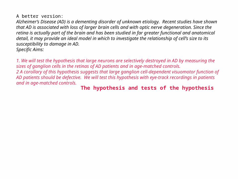
A better version:Alzheimer’s Disease (AD) is a dementing disorder of unknown etiology. Recent studies have shown that AD is associated with loss of larger brain cells and with optic nerve degeneration. Since the retina is actually part of the brain and has been studied in far greater functional and anatomical detail, it may provide an ideal model in which to investigate the relationship of cell’s size to its susceptibility to damage in AD.Specific Aims:
1. We will test the hypothesis that large neurons are selectively destroyed in AD by measuring the sizes of ganglion cells in the retinas of AD patients and in age-matched controls.2 A corollary of this hypothesis suggests that large ganglion cell-dependent visuomotor function of AD patients should be defective. We will test this hypothesis with eye-track recordings in patients and in age-matched controls.
Problem and significance

A better version:Alzheimer’s Disease (AD) is a dementing disorder of unknown etiology. Recent studies have shown that AD is associated with loss of larger brain cells and with optic nerve degeneration. Since the retina is actually part of the brain and has been studied in far greater functional and anatomical detail, it may provide an ideal model in which to investigate the relationship of cell’s size to its susceptibility to damage in AD.Specific Aims:
1. We will test the hypothesis that large neurons are selectively destroyed in AD by measuring the sizes of ganglion cells in the retinas of AD patients and in age-matched controls.2 A corollary of this hypothesis suggests that large ganglion cell-dependent visuomotor function of AD patients should be defective. We will test this hypothesis with eye-track recordings in patients and in age-matched controls.
A theoretical model
A better version:Alzheimer’s Disease (AD) is a dementing disorder of unknown etiology. Recent studies have shown that AD is associated with loss of larger brain cells and with optic nerve degeneration. Since the retina is actually part of the brain and has been studied in far greater functional and anatomical detail, it may provide an ideal model in which to investigate the relationship of cell’s size to its susceptibility to damage in AD.Specific Aims:
1. We will test the hypothesis that large neurons are selectively destroyed in AD by measuring the sizes of ganglion cells in the retinas of AD patients and in age-matched controls.2 A corollary of this hypothesis suggests that large ganglion cell-dependent visuomotor function of AD patients should be defective. We will test this hypothesis with eye-track recordings in patients and in age-matched controls.
The hypothesis and tests of the hypothesis

A better version:Alzheimer’s Disease (AD) is a dementing disorder of unknown etiology. Recent studies have shown that AD is associated with loss of larger brain cells and with optic nerve degeneration. Since the retina is actually part of the brain and has been studied in far greater functional and anatomical detail, it may provide an ideal model in which to investigate the relationship of cell’s size to its susceptibility to damage in AD.Specific Aims:
1. We will test the hypothesis that large neurons are selectively destroyed in AD by measuring the sizes of ganglion cells in the retinas of AD patients and in age-matched controls.2 A corollary of this hypothesis suggests that large ganglion cell-dependent visuomotor function of AD patients should be defective. We will test this hypothesis with eye-track recordings in patients and in age-matched controls.
C. ABSTRACTThe abstract should describe succinctly (~1/2 page) every major aspect of the proposed project except the budget.
The abstract should ideally include:1. A brief background of the project.2. The hypothesis or hypotheses to be tested.3. The tests of the hypothesis or hypotheses.4. The significance of the proposed research.
The hypothesis and tests of the hypothesis

Example:Communication between neurons relies on the release of neurotransmitters from pre-synaptic nerve terminals. Release is triggered by increases in intracellular [Ca2+] and is mediated by the fusion of transmitter-filled synaptic vesicles with the plasma membrane. The molecular mechanism that couples Ca2+ to exocytosis is not known. Synaptotagmin is a Ca2+ binding protein that has been proposed to function as a Ca2+ sensor that triggers release. We propose to examine the function of this protein, primarily on its two putative Ca2+-sensing domains, C2A and C2B. The structure and Ca2+-binding properties of the C2A-domain have been previously studied in detail. However, little is known concerning the function of the C2B-domain. We hypothesize that C2B is a Ca2+-sensing module that plays a critical role in Ca2+-triggered exocytosis. Support for this hypothesis is provided by preliminary data indicating that C2B must bind Ca2+, change conformation and oligomerize in order for docked synaptic vesicles to fuse in response to stimulation in vivo. To determine how C2B functions in exocytosis, three Specific Aims are proposed. (1) A series of biochemical studies will examine the kinetics of Ca2+-C2B interactions, the Ca2+ requirements for synaptotagmin oligomerization, and the role of C2B in the facilitation of SNARE complex assembly. (2) Time resolved amperometry, as well as genetic manipulations of Drosophila, will be used to address the role of C2B in fusion pore dynamics and in excitation-secretion coupling. A critical preliminary finding is that synaptotagmin regulates the opening and dilation kinetics of fusion pores, placing synaptotagmin at the final stages of the fusion reaction that is mediated by SNAREs. This finding supports the hypothesis that synaptotagmins and SNAREs are constituents of Ca2+-regulated exocytotic fusion pores. (3) Biochemical studies will be carried out to determine the subunit stoichiometry of this complex, its morphology, and the interfaces that mediate its assembly. The ability of the release machinery to respond to Ca2+ is subject to modulation and is likely to comprise an important locus for synaptic plasticity. Thus, a better understanding of the release process will provide insights into novel modes of synaptic plasticity and should ultimately provide targets for the treatment of diseases in which synaptic transmission is impaired.

Example:Communication between neurons relies on the release of neurotransmitters from pre-synaptic nerve terminals. Release is triggered by increases in intracellular [Ca2+] and is mediated by the fusion of transmitter-filled synaptic vesicles with the plasma membrane. The molecular mechanism that couples Ca2+ to exocytosis is not known. Synaptotagmin is a Ca2+ binding protein that has been proposed to function as a Ca2+ sensor that triggers release. We propose to examine the function of this protein, primarily on its two putative Ca2+-sensing domains, C2A and C2B. The structure and Ca2+-binding properties of the C2A-domain have been previously studied in detail. However, little is known concerning the function of the C2B-domain. We hypothesize that C2B is a Ca2+-sensing module that plays a critical role in Ca2+-triggered exocytosis. Support for this hypothesis is provided by preliminary data indicating that C2B must bind Ca2+, change conformation and oligomerize in order for docked synaptic vesicles to fuse in response to stimulation in vivo. To determine how C2B functions in exocytosis, three Specific Aims are proposed. (1) A series of biochemical studies will examine the kinetics of Ca2+-C2B interactions, the Ca2+ requirements for synaptotagmin oligomerization, and the role of C2B in the facilitation of SNARE complex assembly. (2) Time resolved amperometry, as well as genetic manipulations of Drosophila, will be used to address the role of C2B in fusion pore dynamics and in excitation-secretion coupling. A critical preliminary finding is that synaptotagmin regulates the opening and dilation kinetics of fusion pores, placing synaptotagmin at the final stages of the fusion reaction that is mediated by SNAREs. This finding supports the hypothesis that synaptotagmins and SNAREs are constituents of Ca2+-regulated exocytotic fusion pores. (3) Biochemical studies will be carried out to determine the subunit stoichiometry of this complex, its morphology, and the interfaces that mediate its assembly. The ability of the release machinery to respond to Ca2+ is subject to modulation and is likely to comprise an important locus for synaptic plasticity. Thus, a better understanding of the release process will provide insights into novel modes of synaptic plasticity and should ultimately provide targets for the treatment of diseases in which synaptic transmission is impaired.

Example:Communication between neurons relies on the release of neurotransmitters from pre-synaptic nerve terminals. Release is triggered by increases in intracellular [Ca2+] and is mediated by the fusion of transmitter-filled synaptic vesicles with the plasma membrane. The molecular mechanism that couples Ca2+ to exocytosis is not known. Synaptotagmin is a Ca2+ binding protein that has been proposed to function as a Ca2+ sensor that triggers release. We propose to examine the function of this protein, primarily on its two putative Ca2+-sensing domains, C2A and C2B. The structure and Ca2+-binding properties of the C2A-domain have been previously studied in detail. However, little is known concerning the function of the C2B-domain. We hypothesize that C2B is a Ca2+-sensing module that plays a critical role in Ca2+-triggered exocytosis. Support for this hypothesis is provided by preliminary data indicating that C2B must bind Ca2+, change conformation and oligomerize in order for docked synaptic vesicles to fuse in response to stimulation in vivo. To determine how C2B functions in exocytosis, three Specific Aims are proposed. (1) A series of biochemical studies will examine the kinetics of Ca2+-C2B interactions, the Ca2+ requirements for synaptotagmin oligomerization, and the role of C2B in the facilitation of SNARE complex assembly. (2) Time resolved amperometry, as well as genetic manipulations of Drosophila, will be used to address the role of C2B in fusion pore dynamics and in excitation-secretion coupling. A critical preliminary finding is that synaptotagmin regulates the opening and dilation kinetics of fusion pores, placing synaptotagmin at the final stages of the fusion reaction that is mediated by SNAREs. This finding supports the hypothesis that synaptotagmins and SNAREs are constituents of Ca2+-regulated exocytotic fusion pores. (3) Biochemical studies will be carried out to determine the subunit stoichiometry of this complex, its morphology, and the interfaces that mediate its assembly. The ability of the release machinery to respond to Ca2+ is subject to modulation and is likely to comprise an important locus for synaptic plasticity. Thus, a better understanding of the release process will provide insights into novel modes of synaptic plasticity and should ultimately provide targets for the treatment of diseases in which synaptic transmission is impaired.

Example:Communication between neurons relies on the release of neurotransmitters from pre-synaptic nerve terminals. Release is triggered by increases in intracellular [Ca2+] and is mediated by the fusion of transmitter-filled synaptic vesicles with the plasma membrane. The molecular mechanism that couples Ca2+ to exocytosis is not known. Synaptotagmin is a Ca2+ binding protein that has been proposed to function as a Ca2+ sensor that triggers release. We propose to examine the function of this protein, primarily on its two putative Ca2+-sensing domains, C2A and C2B. The structure and Ca2+-binding properties of the C2A-domain have been previously studied in detail. However, little is known concerning the function of the C2B-domain. We hypothesize that C2B is a Ca2+-sensing module that plays a critical role in Ca2+-triggered exocytosis. Support for this hypothesis is provided by preliminary data indicating that C2B must bind Ca2+, change conformation and oligomerize in order for docked synaptic vesicles to fuse in response to stimulation in vivo. To determine how C2B functions in exocytosis, three Specific Aims are proposed. (1) A series of biochemical studies will examine the kinetics of Ca2+-C2B interactions, the Ca2+ requirements for synaptotagmin oligomerization, and the role of C2B in the facilitation of SNARE complex assembly. (2) Time resolved amperometry, as well as genetic manipulations of Drosophila, will be used to address the role of C2B in fusion pore dynamics and in excitation-secretion coupling. A critical preliminary finding is that synaptotagmin regulates the opening and dilation kinetics of fusion pores, placing synaptotagmin at the final stages of the fusion reaction that is mediated by SNAREs. This finding supports the hypothesis that synaptotagmins and SNAREs are constituents of Ca2+-regulated exocytotic fusion pores. (3) Biochemical studies will be carried out to determine the subunit stoichiometry of this complex, its morphology, and the interfaces that mediate its assembly. The ability of the release machinery to respond to Ca2+ is subject to modulation and is likely to comprise an important locus for synaptic plasticity. Thus, a better understanding of the release process will provide insights into novel modes of synaptic plasticity and should ultimately provide targets for the treatment of diseases in which synaptic transmission is impaired.

Example:Communication between neurons relies on the release of neurotransmitters from pre-synaptic nerve terminals. Release is triggered by increases in intracellular [Ca2+] and is mediated by the fusion of transmitter-filled synaptic vesicles with the plasma membrane. The molecular mechanism that couples Ca2+ to exocytosis is not known. Synaptotagmin is a Ca2+ binding protein that has been proposed to function as a Ca2+ sensor that triggers release. We propose to examine the function of this protein, primarily on its two putative Ca2+-sensing domains, C2A and C2B. The structure and Ca2+-binding properties of the C2A-domain have been previously studied in detail. However, little is known concerning the function of the C2B-domain. We hypothesize that C2B is a Ca2+-sensing module that plays a critical role in Ca2+-triggered exocytosis. Support for this hypothesis is provided by preliminary data indicating that C2B must bind Ca2+, change conformation and oligomerize in order for docked synaptic vesicles to fuse in response to stimulation in vivo. To determine how C2B functions in exocytosis, three Specific Aims are proposed. (1) A series of biochemical studies will examine the kinetics of Ca2+-C2B interactions, the Ca2+ requirements for synaptotagmin oligomerization, and the role of C2B in the facilitation of SNARE complex assembly. (2) Time resolved amperometry, as well as genetic manipulations of Drosophila, will be used to address the role of C2B in fusion pore dynamics and in excitation-secretion coupling. A critical preliminary finding is that synaptotagmin regulates the opening and dilation kinetics of fusion pores, placing synaptotagmin at the final stages of the fusion reaction that is mediated by SNAREs. This finding supports the hypothesis that synaptotagmins and SNAREs are constituents of Ca2+-regulated exocytotic fusion pores. (3) Biochemical studies will be carried out to determine the subunit stoichiometry of this complex, its morphology, and the interfaces that mediate its assembly. The ability of the release machinery to respond to Ca2+ is subject to modulation and is likely to comprise an important locus for synaptic plasticity. Thus, a better understanding of the release process will provide insights into novel modes of synaptic plasticity and should ultimately provide targets for the treatment of diseases in which synaptic transmission is impaired.

C. BACKGROUND AND SIGNIFICANCEThe Specific Aims section of the proposal provides the reader with a general idea concerning the scope of the project, the technology involved, the logic of the experimental approach, and the hypothesis to be tested. However, these ideas were presented as unsupported statements, so a major goal of the Background and Significance section is to provide this support through expansion and judicious reference to the literature. Because the primary reviewers may not be familiar with the area of investigation, or the technology being used, it is also important to address these matters. However, the purpose IS NOT to be comprehensive: the purpose IS to present citations that build a solid foundation for your proposal.
This section of the proposal needs to establish the following matters:1. The project is important. It relates to significant human disease or to a significant deficit in our knowledge of an important biologic process: the results of the hypothesis tests will have a predictable impact on theory and/or ultimately lead to improvement of the human condition. 2. The project is interesting. The subject can be related to a general theoretical model that is the subject of widespread interest; important areas within the model that are unproven, controversial, or ambiguous are addressed.3. There is a high probability of success. Specific hypothesis to be tested can be identified as part of a theoretical model; tests of the hypotheses are feasible, definitive, and within the range of the PI’s apparent expertise.

Some suggestions:-Begin this section with the importance of the project.-Briefly describe what is known.-Briefly describe what is not known.-Be critical of earlier work in the field (but not meanspirited).-Briefly describe what needs to be done.-Briefly emphasize how your results will meet the stated need.-Cite reviews sparingly-cite your reviewers (if not contrived).-Often helpful to include a limited number of figures/tables summarizing complex information, or describing the models to be tested.-Write this section last.
D. PRELIMINARY STUDIESThe purpose of the Preliminary Studies section is to describe (your) prior work that is relevant to the proposed project.
This section of the proposal needs to establish the following matters:1. That you are fully competent in all of the procedures proposed (obviously, this is not a concern for a graduate student oral exam proposal, as a major point of the research plan is to expose the student to novel training opportunities).2. To establish beyond a reasonable doubt that the proposed studies are logical and feasible.
This section should include substantial data (figures and/or Tables) that directly addresses the points listed above. The more preliminary data you have, the better your chances of success.
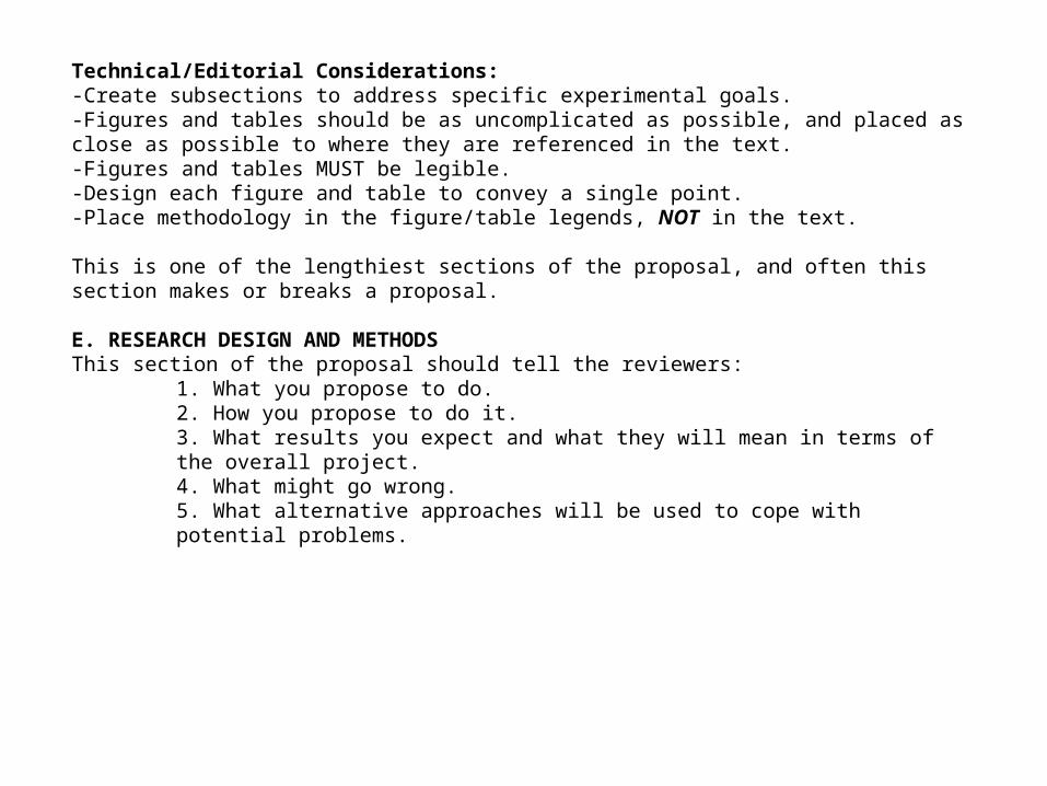
Technical/Editorial Considerations:-Create subsections to address specific experimental goals.-Figures and tables should be as uncomplicated as possible, and placed as close as possible to where they are referenced in the text.-Figures and tables MUST be legible.-Design each figure and table to convey a single point.-Place methodology in the figure/table legends, NOT in the text.
This is one of the lengthiest sections of the proposal, and often this section makes or breaks a proposal.
E. RESEARCH DESIGN AND METHODSThis section of the proposal should tell the reviewers:
1. What you propose to do.2. How you propose to do it.3. What results you expect and what they will mean in terms of the overall project.4. What might go wrong.5. What alternative approaches will be used to cope with potential problems.
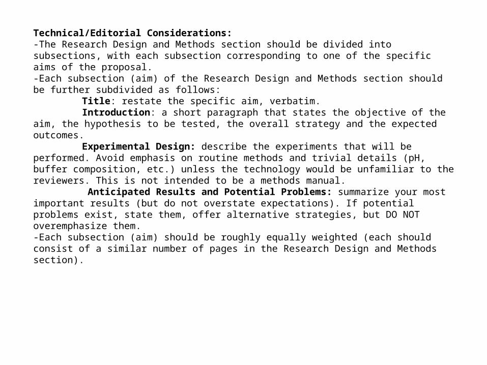
Technical/Editorial Considerations:-The Research Design and Methods section should be divided into subsections, with each subsection corresponding to one of the specific aims of the proposal.-Each subsection (aim) of the Research Design and Methods section should be further subdivided as follows:
Title: restate the specific aim, verbatim.Introduction: a short paragraph that states the objective of the aim, the hypothesis to
be tested, the overall strategy and the expected outcomes.Experimental Design: describe the experiments that will be performed. Avoid
emphasis on routine methods and trivial details (pH, buffer composition, etc.) unless the technology would be unfamiliar to the reviewers. This is not intended to be a methods manual.
Anticipated Results and Potential Problems: summarize your most important results (but do not overstate expectations). If potential problems exist, state them, offer alternative strategies, but DO NOT overemphasize them.-Each subsection (aim) should be roughly equally weighted (each should consist of a similar number of pages in the Research Design and Methods section).
![New Method to Quantitate Clonogenic Tumor Cells in the ......[CANCER RESEARCH 43, 5451-5455, November 1983] New Method to Quantitate Clonogenic Tumor Cells in the Blood Circulation](https://img.dokumen.tips/doc/110x75/6068d20ce566193e3e18220a/new-method-to-quantitate-clonogenic-tumor-cells-in-the-cancer-research.jpg)


![Value Proposition - Quantify POWER [ENG]](https://img.dokumen.tips/doc/110x75/6156ff56a097e25c764fe364/value-proposition-quantify-power-eng.jpg)