Embed Size (px)
Citation preview

QUALITY ASSURANCE IN LPCC BREAST CANCER SCREENING PROGRAM
-‐ TECHNICAL IMAGE QUALITY AND RADIATION DOSES -‐
LPCC-‐BCSP António Ferro de Carvalho Physicist Coordinator

LIGA PORTUGUESA CONTRA O CANCRO (LPCC)
The Portuguese Cancer League is a non-‐profit, non-‐governmental organizaOon, found in 1941 to give
n support to oncological paOents,
n promote cancer prevenOon
n promote research and training in oncology.

LPCC – Breast Cancer Screening Program
BCSP/LPCC is beeing carried out in cooperaOon and support of
n Ministry of Health
n Regional Health AdministraOons (ARS)
n Local Health Centers.

Breast Cancer Screening Program (LPCC/BCSP)
BCSP started in Portugal as a pilot-‐test in Center Region in 1986.
In 1989-‐1991 BCS acOviOes were integrated in the Europe Against Cancer, joining other Pilot Projects (Portugal-‐Central Region, Spain-‐Navarra, France-‐Strasbourg, Greece-‐ Athenas, Ireland-‐Dublin, Italy-‐Florence, Ormylia-‐Greece)

The European Breast Screening Network
AffiliaAon in 2000

LPCC/BCSP Breast Cancer Screening Program
¤ In 1997 and 1999, the North and South Regional Branches of LPCC, started similar screening programs.
¤ In 2000 the LPCC created a “NaAonal BCS Program” joining the three regional Programs, under a NaOonal CoordinaOon Office, with similar methodologies, mechanisms of quality control and monitoring and informaOon.

LPCC – BCSP Coverage
May 2013
NaAonal Program LPCC
CounOes integrated: 217 (83%)
DetecOon rate/1000 women (1st round) 4,4
Period 1990/2012
Women integrated 756.478
Mamography tests: 2.247.860
Women assessed: 93.373
Hospital Referral: 10.655

LPCC/BCSP Screening Process
a) Use of 28 screening units + 3 assessment centers b) Target populaOon: women aged 45-‐69 years c) Screening interval: 2 years d) Double view mammogram per breast e) Personal invitaOon (call leher) f) Double blind reading of mammograms; g) Women with posiOve screening test are invited for a
imagiological and clinical assessment in dedicated BCSP faciliOes (non-‐hospital environment);

LPCC – BCSP Process
h) Women needing final diagnosis and treatment are referred to hospitals;
i) Epidemiological monitorizaOon and evaluaOon is the fundamental guiding and unifying discipline throughout the screening
j) Physical and technical mammography Quality Control is carried out systemaOcally in all screening and assessment units.

QUALITY ASSURANCE (QA)
QA is a prerequisite for a successful screening project
QA aspects
organizaOonal medical
technical

Quality Control (QC)
QC of Physical and Technical Aspects is a key acOvity in the screening program, with the following objecAves: 1. Assure that images have the best possible diagnosOc
informaOon, necessary to detect smaller lesions.
2. The image quality is stable with respect to informaOon content.
3. The breast dose is As Low As Reasonably Achievable (ALARA) for the mammographic informaOon required.

QC acAviAes are fully documented
¤ Technical procedures for QC ¤ Criteria for acceptance, correcOons and suspension ¤ Reports of QC and recommendaOons for correcOons and opOmizaOon
¤ Technical informaOon notes

Technical procedures

Screening Units Acceptance Criteria

Reports

References


Type and frequency of QC
¤ Acceptance tests n New screening units before they start being used
¤ Periodical tests n Weekly n Monthly n Biannual
¤ Extra tests n Aler repairs or maintenance that can affect image quality or radiaOon doses for users.

Acceptance, Biannual and Extra QC
¤ Mammography systems
n Performance of mammography unit n Image technical quality n Standard dose (AGD)
¤ Working staOons n image display n reading environment
Tests are carried out by trained physicists

QC tesAng (DR technology)
¨ supplementary tes-ng
¤ CAE response for local dense area
¤ Dynamic range ¤ Homogeneity response of image detector manufactor tesOng; BCSP/LPCC tesOng

QC tesAng (CR technology)
¨ supplementary tes-ng
¤ Image plate reader n CalibraOon n EffecOveness of image erasure
¤ IPs Response Homogeneity
¤ Inter plate sensiOvity variaOons
¤ RadiaOon exposure of IPs
¤ Image processing n Processing parameters are according to recommendaOons

monthly and weekly QC
¨ Constancy tests are carried out by Radiographic Technicians that analyze the results
¨ M&W tests are saved and transferred to a QC dedicated directory in the Regional Branch of the LPCC/BCSP
¨ M&W tests can be assessed and analyzed on-‐line by physicists

M&W registraAon by screening unit
¨ QC monthly n Constancy of AEC performance
¨ QC weekly n Constancy of exposure, image quality and processing

QC WORKFLOW for image aquisiAon
Acceptance tests
fail pass CorrecOon of
non-‐conformiOes by supplier
Non-‐conformiOes’ review
pass fail M&W tests
pass fail
Biannual tests EvaluaOon and planning of extra
tests pass fail
recommendaOons
opOmizaOon CorrecOons suspension
Advice for correcOons
recommendaOons
Keep unchanged

QC of Working StaAons
¤ Image display Luminance range Luminance uniformity ResoluOon Contrast visibility Greyscale Display FuncOon (5 events/72 display.year) Display arOfacts
¤ Ambient light

Image display evaluaOon DICOM -‐ GDS
Not acceptable Acceptable

Image display visual cheks every month
• luminance
• Display arOfacts
• ResoluOon
• Geometrical distorOon
• Contrast visibility

QC WORKFLOW for image display
Acceptance tests
fail pass CorrecOon of
non-‐conformiOes by supplier
Non-‐conformiOes’ review
pass fail Every month
pass fail
Annual tests
EvaluaOon and planning of extra
tests pass fail
recommendaOons
opOmizaOon CorrecOons suspension
Advice for correcOons
recommendaOons
Keep unchanged
Annual tests performed on-‐
line by maintenance
service

OpAmisaAon Image Quality / RadiaAon Doses
¨ Minimum requirements were fixed to image technical evaluaOon
¤ Threshold limits ¤ SDNR minimum acceptable value ¤ SpaOal resoluOon ¤ Standard doses

RadiaAon Doses 2012 EvaluaAon of LPCC/BCSP
ParOcipaOon of 25 screening units (CR technology) Sample: 532 mammograms with 2180 images
Mean of AGD in 2180 images ………….………............……2,7 mGy/image
AGD for CC images…………………………………………………....2,6 mGy/image
AGD for OB images ………………………………………………..….2,9 mGy/image
AGD per breast (2 images/breast) …………………………..…5,3 mGy LPCC reference (3rd quarOl on sample 50 to 59 mm) ……………. 2,8 mGy
Average breast thickness …………………..….…56 mm

RadiaAon Doses 2012 EvaluaOon of LPCC/BCSP
ParOcipaOon of 25 screening units (CR technology) Sample: 532 mammograms with 2180 images
Mean of AGD in 2180 images (mGy/image)……….…2,7 2,7 (2006;EUREF)
AGD for CC images /mGy/image)………………..………...2,6 2,5 (Ireland;screen/film)
AGD for OB images (mGy/image)…………..................2,9 2,8 (Ireland; screen/film)
AGD per breast (2 images/breast) ……………….………..5,3 mGy LPCC reference (3rd quarOl on sample 55 mm)…… 2,8 mGy 3,5 (2005; UK) Average breast thickness …………….…56 mm

RadiaAon Doses 2013 EvaluaAon of LPCC/BCSP
ParOcipaOon of 3 screening units (DR technology)
Sample: 118 mammograms with 480 images
Mean of AGD in 480 images ……..1,6 (mGy/image)
40% reducOon in relaOon to CR Doses

dose glandular média (DGM) vs esp.mama
0
1
2
3
4
5
6
7
0 10 20 30 40 50 60 70 80 90 100
espessura equiv. mama mm
DG
M
mG
y
CR Fujifim (NRC-U1)DR Fujifilm modo L (UM_COA)Limites EUREF
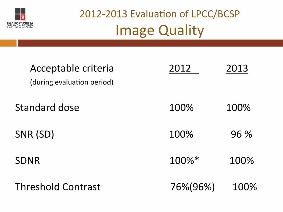
2012-‐2013 EvaluaOon of LPCC/BCSP Image Quality
Acceptable criteria 2012 2013
(during evaluaOon period) Standard dose 100% 100% SNR (SD) 100% 96 % SDNR 100%* 100% Threshold Contrast 76%(96%) 100%


![Kathryn Hughey, MA, LPCC · 3/11/2020 · Kathryn Hughey, MA, LPCC Consent for Counseling and Mandatory Disclosure Statement Degrees and credentials: ... ce }f P ]ac Rgh X I dead](https://img.dokumen.tips/doc/110x75/5fa605ff21f0c9587b5c9758/kathryn-hughey-ma-lpcc-3112020-kathryn-hughey-ma-lpcc-consent-for-counseling.jpg)