Embed Size (px)
Citation preview
1
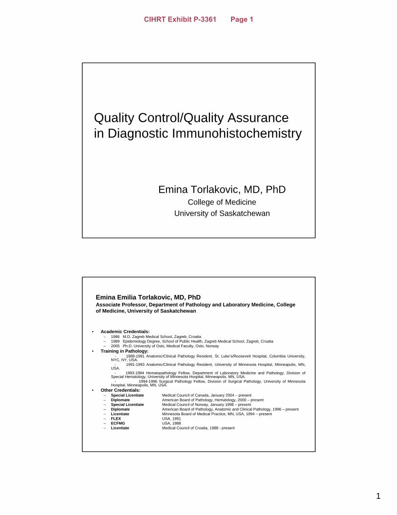
Quality Control/Quality Assurance in Diagnostic Immunohistochemistry
Emina Torlakovic, MD, PhDCollege of Medicine
University of Saskatchewan
Emina Emilia Torlakovic, MD, PhDAssociate Professor, Department of Pathology and Laboratory Medicine, College of Medicine, University of Saskatchewan
• Academic Credentials:– 1986 M.D. Zagreb Medical School, Zagreb, Croatia– 1989 Epidemiology Degree, School of Public Health, Zagreb Medical School, Zagreb, Croatia– 2005 Ph.D. University of Oslo, Medical Faculty, Oslo, Norway
• Training in Pathology:- 1989-1991 Anatomic/Clinical Pathology Resident, St. Luke’s/Roosevelt Hospital, Columbia University,
NYC, NY, USA.- 1991-1993 Anatomic/Clinical Pathology Resident, University of Minnesota Hospital, Minneapolis, MN,
USA. - 1993-1994 Hematopathology Fellow, Department of Laboratory Medicine and Pathology, Division of
Special Hematology, University of Minnesota Hospital, Minneapolis, MN, USA. - 1994-1996 Surgical Pathology Fellow, Division of Surgical Pathology, University of Minnesota
Hospital, Minneapolis, MN, USA.• Other Credentials:
– Special Licentiate Medical Council of Canada, January 2004 – present – Diplomate American Board of Pathology, Hematology, 2000 – present– Special Licentiate Medical Council of Norway, January 1998 – present– Diplomate American Board of Pathology, Anatomic and Clinical Pathology, 1996 – present– Licentiate Minnesota Board of Medical Practice, MN, USA, 1994 – present – FLEX USA, 1991 – ECFMG USA, 1988– Licentiate Medical Council of Croatia, 1988 - present
CIHRT Exhibit P-3361 Page 1
2
Immunohistochemistry: Personal Background
• Director of Immunohistochemistry– 1997-2003 Department of Pathology, The Norwegian Radium Hospital, Oslo,
Norway– 2004-Present Department of Pathology and Laboratory Medicine, College of
Medicine, University of Saskatchewan• NordiQC Core Group
– 1999-2004 Norwegian representative– 2004-Present External Contributor (review articles)
• Canadian Immunohistochemistry Quality Control (cIQc)– 2005-Present
• Chair, CAP National Standards Committee/Immunohistochemistry– 2007-Present
• Leader, European Bone Marrow Working Group Immunohistochemistry Committee (introducing standardization for bone marrow IHC for all European countries)
• Member, ASCO/CAP ER/PR Expert Panel, • Publications:
– 34/64 of my articles in PubMed are searchable under “torlakovic”+”immunohistochemistry”
– Bone Marrow Immunohistochemistry, book published by ASCP Press 2008• Lectures:
– Many invited lectures in USA, Europe, Canada
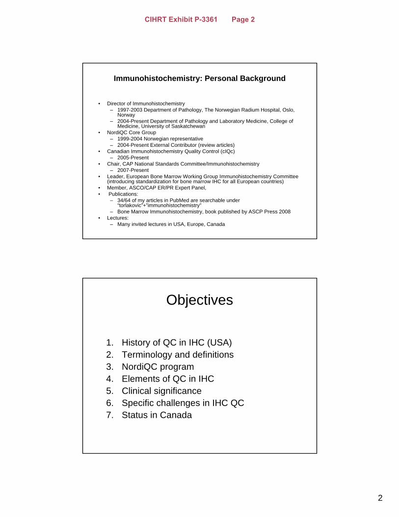
Objectives
1. History of QC in IHC (USA)2. Terminology and definitions3. NordiQC program4. Elements of QC in IHC5. Clinical significance6. Specific challenges in IHC QC7. Status in Canada
CIHRT Exhibit P-3361 Page 2
3
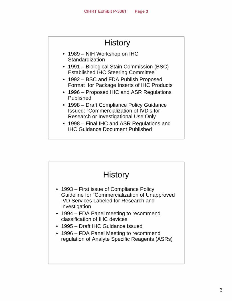
History• 1989 – NIH Workshop on IHC
Standardization• 1991 – Biological Stain Commission (BSC)
Established IHC Steering Committee• 1992 – BSC and FDA Publish Proposed
Format for Package Inserts of IHC Products• 1996 – Proposed IHC and ASR Regulations
Published• 1998 – Draft Compliance Policy Guidance
Issued: “Commercialization of IVD’s for Research or Investigational Use Only
• 1998 – Final IHC and ASR Regulations and IHC Guidance Document Published
History• 1993 – First issue of Compliance Policy
Guideline for “Commercialization of Unapproved IVD Services Labeled for Research and Investigation
• 1994 – FDA Panel meeting to recommend classification of IHC devices
• 1995 – Draft IHC Guidance Issued• 1996 – FDA Panel Meeting to recommend
regulation of Analyte Specific Reagents (ASRs)
CIHRT Exhibit P-3361 Page 3
4
Extralaboratory Quality Assurance (EQA)
• UKNEQAS (1968, 1990) UK• CAP (1949,1961,2003,2006) USA• NordiQC (1999/2003) Scandinavia• cIQc (2006) Canada
• Other regional/provincial programs (Finland, Ontario, BC)
The Role of Medical Laboratories in Patients’ Care
• Dr. J. Butany:“Canada’s medical laboratory system is the
foundation upon which good patient care, diagnosis and treatment rest.”
CIHRT Exhibit P-3361 Page 4
5
What is Immunohistochemistry?• Application of immunoassay in tissue sections.• Immunological localization of the protein of
interest in its natural environment.• Simultaneous evaluation of morphology and
staining of the localized protein provide very complex information.
• The intensity of signal may or may not represent the real quantity of the protein in tissue.
Class I
• Class I IHC tests provide adjunctive diagnostic information not independently reported to clinical physicians.
• They are used after the tumor is diagnosed by other methods and are used only by pathologists.
• E.g. cytokeratin, vimentin, CD45, and other differentiation markers
CIHRT Exhibit P-3361 Page 5
6
Class II• So-called “stand alone” test that are reported
independently of other clinical or laboratory information.
• The results of these tests are used as either predictive or prognostic markers and are often critically relied upon to stratify patients for appropriate therapies.
• The tests are accepted as such after widely accepted valid scientific claims. National and international guidelines for these tests are usually published.
• E.g. hormone receptors in breast cancer.
Classification of IHC Tests• Class I, class II, class III• Qualitative, quantitative• Test - drug combo vs. all other tests
• Panels (undifferentiatiated tumor panel, melanoma panel) in which non-specific tests when used together are considered highly specific vs. single specific test used in appropriate context has high specificity (ALK-1, CD117)
CIHRT Exhibit P-3361 Page 6
7
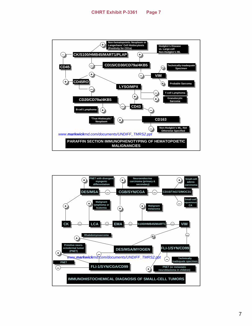
PARAFFIN SECTION IMMUNOPHENOTYPING OF HEMATOPOIETICMALIGNANCIES
CD45CD45
CK/S100/HMB45/MART1/PLAPCK/S100/HMB45/MART1/PLAP
CD15/CD30/CD79a/4KB5CD15/CD30/CD79a/4KB5
VIMVIM
CD45ROCD45ROLYSO/MPXLYSO/MPX
CD20/CD79a/4KB5CD20/CD79a/4KB5
CD43CD43
CD163CD163
----
++
----
++
++
----
----
++
++
--------
++
--------
++
----
++
Non-hematopoietic Neoplasm orLangerhans’ Cell Histiocytosis(Positivity for CD1a)
Non-hematopoietic Neoplasm orLangerhans’ Cell Histiocytosis(Positivity for CD1a) Hodgkin’s Disease
vs. Large-cell Non-Hodgkin’s ML
Hodgkin’s Disease vs. Large-cell Non-Hodgkin’s ML
Technically-inadequateSpecimen
Technically-inadequateSpecimen
Probable SarcomaProbable Sarcoma
T-cell LymphomaT-cell Lymphoma
GranulocyticSarcoma
GranulocyticSarcoma
Non-Hodgkin’s ML, NotOtherwise Specified
Non-Hodgkin’s ML, NotOtherwise Specified
“True Histiocytic”Neoplasm
“True Histiocytic”Neoplasm
++B-cell LymphomaB-cell Lymphoma
www.markwickmd.com/documents/UNDIFF_TMRS2.ppt
CKCK
DES/MSADES/MSA CGB/SYN/CGACGB/SYN/CGA CD15/TAG72/MOC31CD15/TAG72/MOC31
LCALCA EMAEMA S100/HMB45/MART1S100/HMB45/MART1 VIMVIM
FLI-1/SYN/CD99FLI-1/SYN/CD99DES/MSA/MYOGENDES/MSA/MYOGEN
FLI-1/SYN/CGA/CD99FLI-1/SYN/CGA/CD99
IMMUNOHISTOCHEMICAL DIAGNOSIS OF SMALL-CELL TUMORS
++
-- -- -- --
--
--
++
++++
--++
--
-- --
++
--
++ ++
++++
PNET with divergentmyogenic
differentiation
PNET with divergentmyogenic
differentiation
Neuroendocrine carcinoma (primary &
secondary)
Neuroendocrine carcinoma (primary &
secondary)Small-cell
adeno-carcinoma
Small-celladeno-
carcinoma
Small-cellsquamous
CA
Small-cellsquamous
CAMalignant
lymphoma orleukemia
Malignantlymphoma or
leukemia ++ Malignantmelanoma
Malignantmelanoma
Technicallyinadequate specimen
Technicallyinadequate specimen
PNET (or metastaticneuroblastoma in children)
PNET (or metastaticneuroblastoma in children)
RhabdomyosarcomaRhabdomyosarcoma
Primitive neuro-ectodermal tumor
(PNET)
Primitive neuro-ectodermal tumor
(PNET)
PNETPNET
www.markwickmd.com/documents/UNDIFF_TMRS2.ppt
CIHRT Exhibit P-3361 Page 7

8
Class II IHC tests
• Despite the need for finely tuned calibration and quantitative nature of the tests, they are usually reported simply as positive or negative.
• The simplicity of the report masks the true biological and technical complexity of the testing.
• Classical definition given by Galen & Gambino:
Spec = True negatives/True negatives + False positives
• Specificity, sensitivity, and concordance with reference laboratory are usually not reported for IHC tests.
• “Specificity” of IHC reagents must be evaluated in well-defined contexts. Hence, “specificity” is a relative term in this applied clinical setting.
• There is no reason not to report on sensitivity, concordance, and kappa-values in relation to reference laboratory values.
Specificity and Sensitivity of IHC tests
CIHRT Exhibit P-3361 Page 8
9
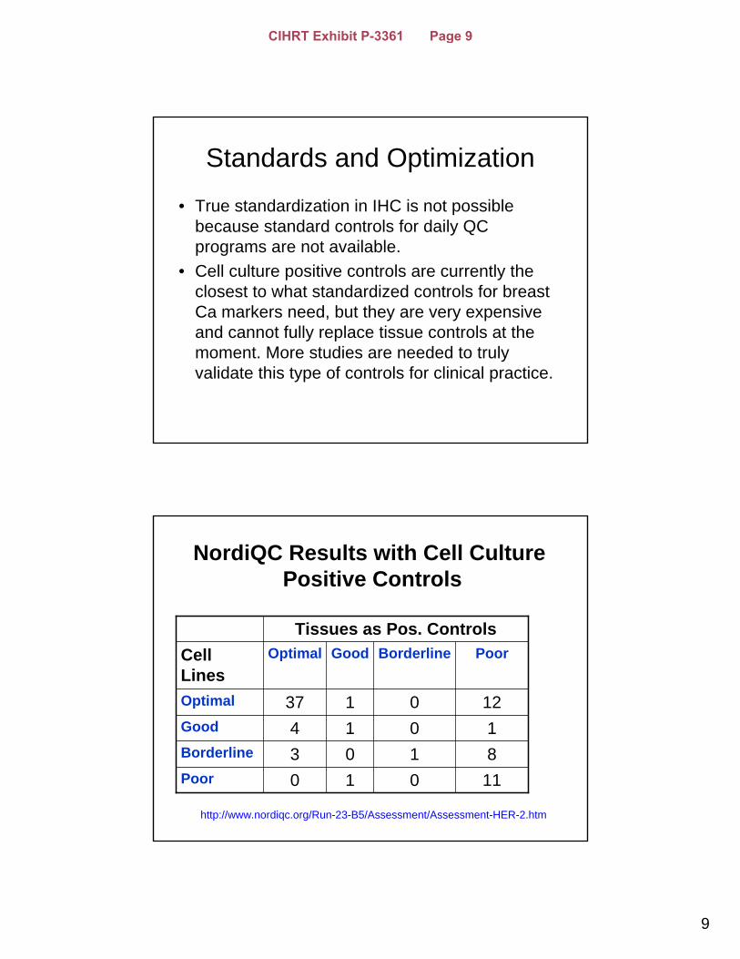
Standards and Optimization
• True standardization in IHC is not possible because standard controls for daily QC programs are not available.
• Cell culture positive controls are currently the closest to what standardized controls for breast Ca markers need, but they are very expensive and cannot fully replace tissue controls at the moment. More studies are needed to truly validate this type of controls for clinical practice.
11010Poor8103Borderline1014Good
120137Optimal
PoorBorderlineGoodOptimalCell Lines
Tissues as Pos. Controls
NordiQC Results with Cell Culture Positive Controls
http://www.nordiqc.org/Run-23-B5/Assessment/Assessment-HER-2.htm
CIHRT Exhibit P-3361 Page 9
10
Use of Cell Lines as Positive Controls: Results and Conclusion
1. An insufficient (false negative) reaction in the breast ductal carcinoma no. 3 in combination with an optimal staining of the cell lines. This was seen in 13/17 cases.
2. A sufficient staining in the histological specimens in combination with an insufficient staining of the cell lines due to impaired morphology of the cell lines, probably as a results of excessive retrieval.
3. These data indicate that histological specimens should be preferred for EQA of HER-2. However, due to potential heterogeneity of tissue material, cell cultures may be valuable as a supplement.
http://www.nordiqc.org/Run-23-B5/Assessment/Assessment-HER-2.htm
Main Conclusions Regarding Standardization
• No standardized positive controls – No standardization.
• Standardization of protocols is meaningless without control standardization.
• Standardization of positive controls also includes agreement or standardization of expected results in control tissues.
• “Standardization” is greatly misused term in this context.
• Standardization is possible only if there are so-called “gold standards” for reference values.
CIHRT Exhibit P-3361 Page 10
11
ER NordiQC Pass Rates
79107Run B5 2008
8473Run B3 2007
7568Run B1 2006
8489Run 13 2005
6777Run 10 2004
4571Run 8 2003
Sufficient Results (%)
Participants (N)NordiQC
http://www.nordiqc.org
VIMENTINA
ED
B C
F
CIHRT Exhibit P-3361 Page 11
12
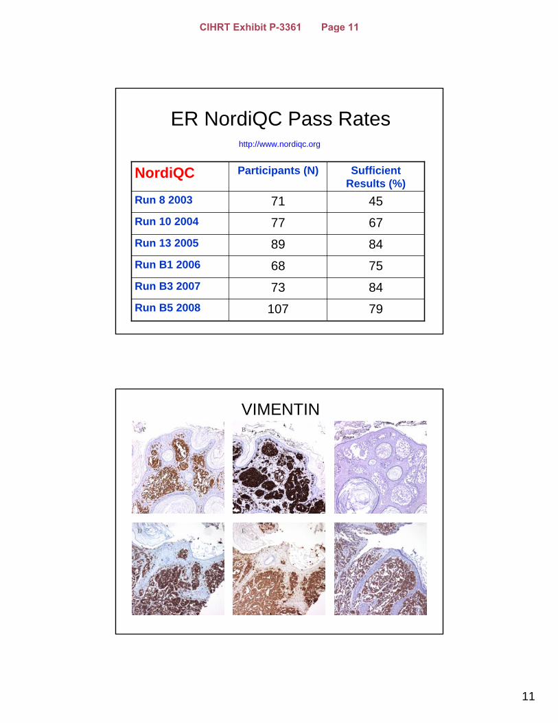
S-100A
ED
CB
F
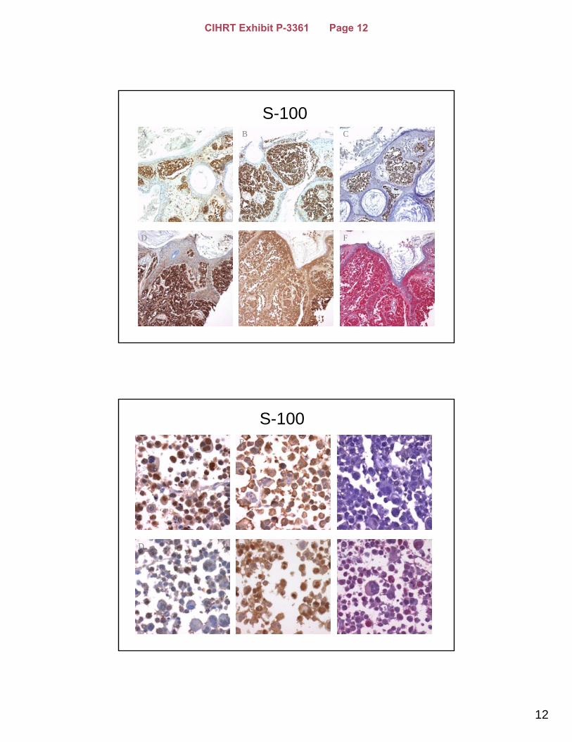
S-100A
FED
CB
CIHRT Exhibit P-3361 Page 12
13
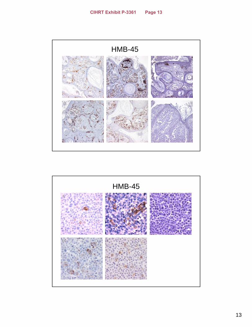
HMB-45A
F
B
E
C
D
HMB-45A
D E
CB
CIHRT Exhibit P-3361 Page 13
14
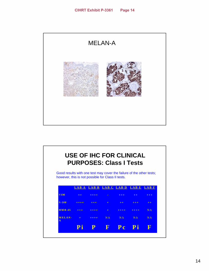
MELAN-A
A B
USE OF IHC FOR CLINICAL PURPOSES: Class I Tests
L A B A L A B B L A B C L A B D L A B E L A B F
V IM + + + + + + - + + + + + + + +
S -1 0 0 + + + + + + + + + + + + + + +
H M B -4 5 + + + + + + + + + + + + + + + + N A
M E L A N -A
+ + + + + N A N A N A N A
P i P F P c P i F
Good results with one test may cover the failure of the other tests; however, this is not possible for Class II tests.
CIHRT Exhibit P-3361 Page 14
15
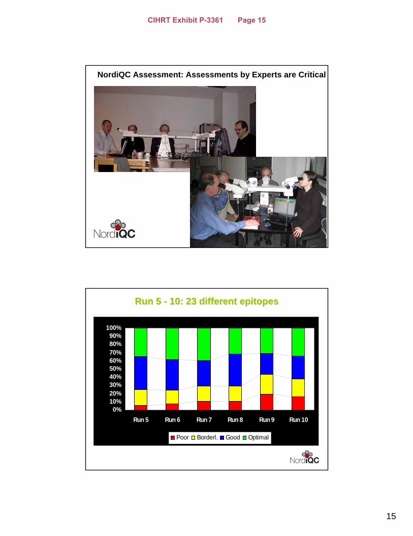
NordiQC Assessment: Assessments by Experts are Critical
30
Run 5 Run 5 -- 10: 23 10: 23 different epitopesdifferent epitopes
0%10%20%30%40%50%60%70%80%90%
100%
Run 5 Run 6 Run 7 Run 8 Run 9 Run 10
Poor Borderl. Good Optimal
CIHRT Exhibit P-3361 Page 15
16
31
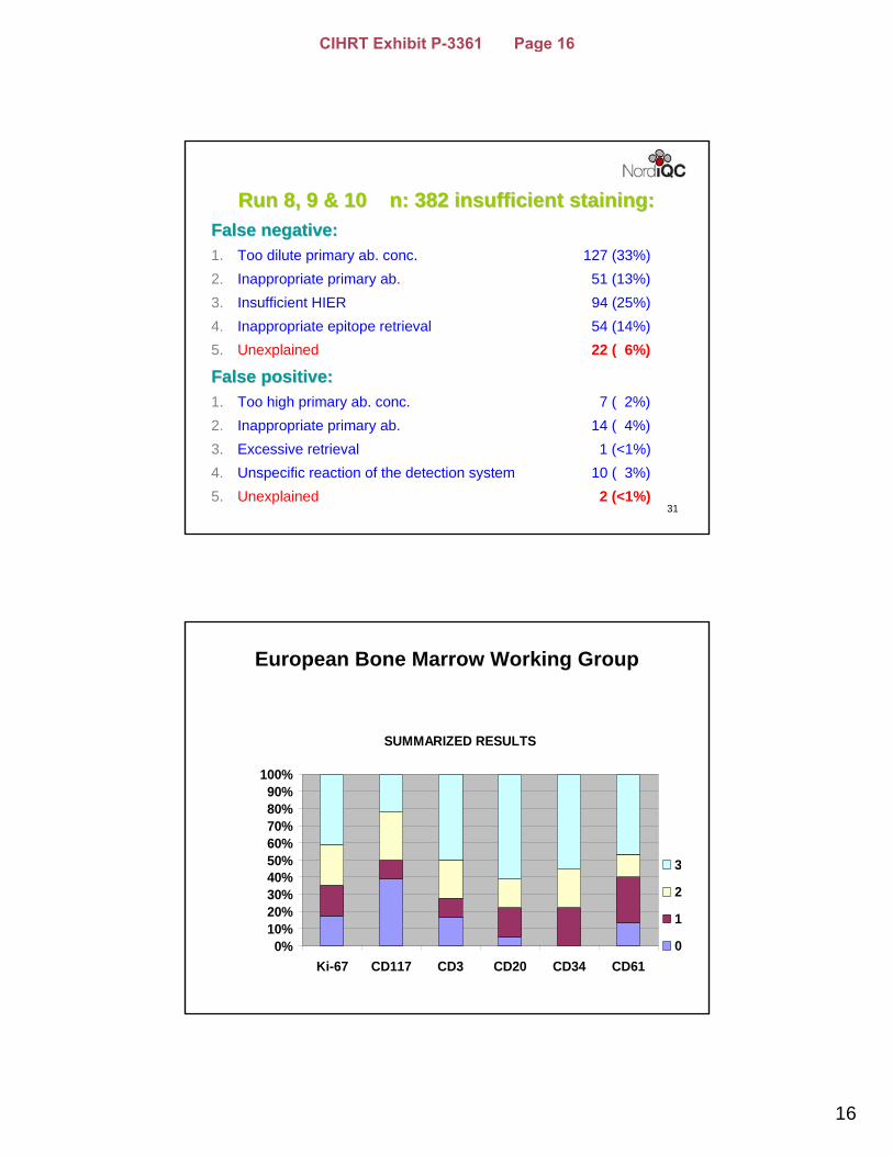
Run 8, 9 & 10 n: 382 insufficient staining:Run 8, 9 & 10 n: 382 insufficient staining:False negative:False negative:1. Too dilute primary ab. conc. 127 (33%) 2. Inappropriate primary ab. 51 (13%)3. Insufficient HIER 94 (25%) 4. Inappropriate epitope retrieval 54 (14%)5. Unexplained 22 ( 6%)
False positive:False positive:1. Too high primary ab. conc. 7 ( 2%)2. Inappropriate primary ab. 14 ( 4%)3. Excessive retrieval 1 (<1%)4. Unspecific reaction of the detection system 10 ( 3%)5. Unexplained 2 (<1%)
SUMMARIZED RESULTS
0%10%20%30%40%50%60%70%80%90%
100%
Ki-67 CD117 CD3 CD20 CD34 CD61
3
2
1
0
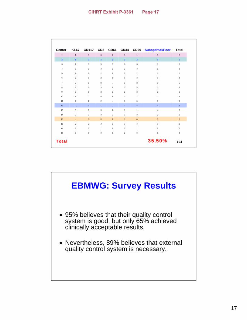
European Bone Marrow Working Group
CIHRT Exhibit P-3361 Page 16
17
10435.50%Total
6132330318
6213313317
6033332216
5501100.15
6233033014
6411130313
5322.10012
5033.22211
6233102310
623203039
603333238
5333.0007
603332326
602322225
613233134
613333313
662122012
651113111
TotalSuboptimal/PoorCD20CD34CD61CD3CD117Ki-67Center
EBMWG: Survey Results
• 95% believes that their quality control system is good, but only 65% achieved clinically acceptable results.
• Nevertheless, 89% believes that external quality control system is necessary.
CIHRT Exhibit P-3361 Page 17
18
What do we want to optimize or standardize?
• METHODS - Not necessarily!
• RESULTS - Obligatory!
How to standardize results?
• Fist step:
• Standardization of what is considered “optimal result”, based on current standard of practice.
• Each laboratory should consider that standardization of tissue processing would make it easier to standardize results.
CIHRT Exhibit P-3361 Page 18
19
37
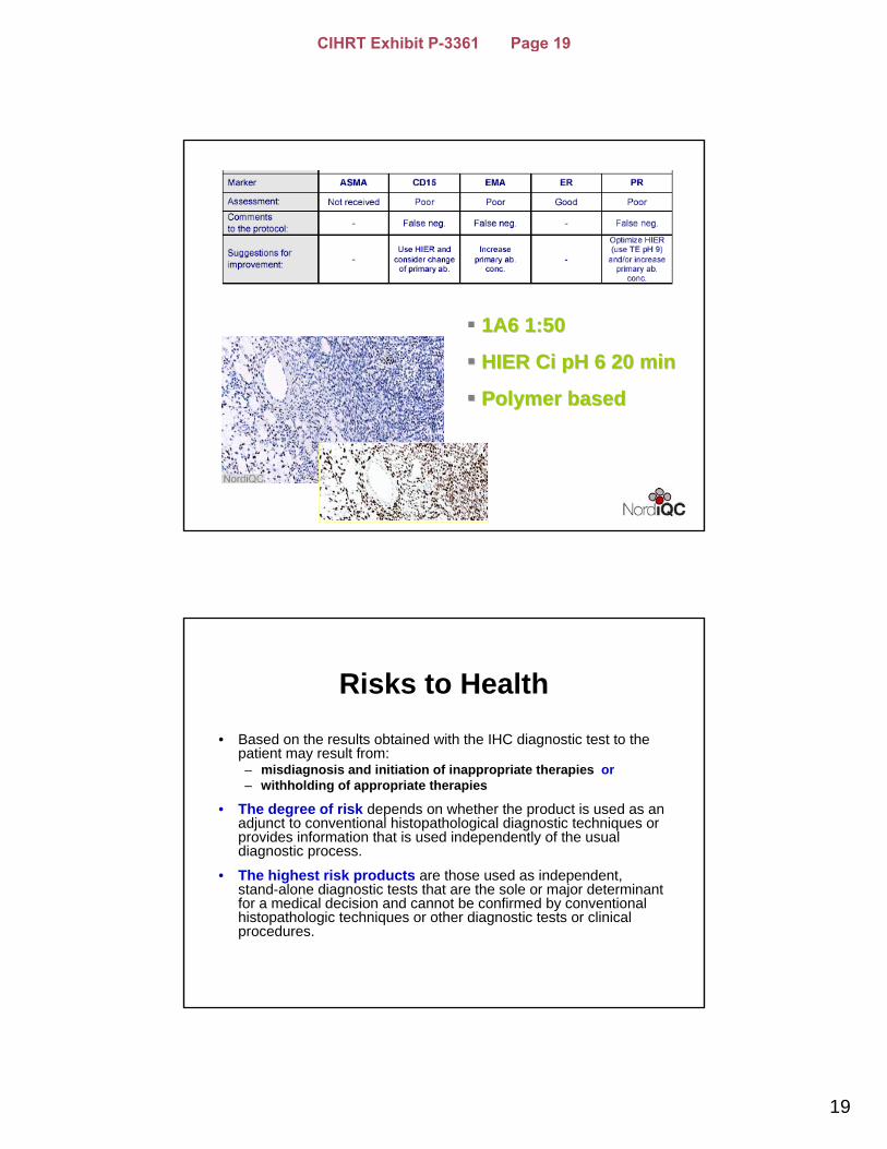
1A6 1:501A6 1:50
HIER HIER Ci Ci pH 6 20 minpH 6 20 min
Polymer Polymer basedbased
Risks to Health
• Based on the results obtained with the IHC diagnostic test to the patient may result from: – misdiagnosis and initiation of inappropriate therapies or– withholding of appropriate therapies
• The degree of risk depends on whether the product is used as an adjunct to conventional histopathological diagnostic techniques or provides information that is used independently of the usual diagnostic process.
• The highest risk products are those used as independent, stand-alone diagnostic tests that are the sole or major determinant for a medical decision and cannot be confirmed by conventional histopathologic techniques or other diagnostic tests or clinicalprocedures.
CIHRT Exhibit P-3361 Page 19
20
FDA is focused on whether this level of regulation is adequate for the protection of
public health
• FDA is aware that variability in IHC results may be introduced at every step:
– Collection and fixation of the specimen,– Automated processing, – Embedding and sectioning, – Staining of the final slide preparation, and – Microscopic interpretation by the pathologist.
FDA counts on:
Ongoing initiatives by professional organizations and manufacturersdirected at ensuring that pre- and postanalytic, as well as analytic procedures, are properly performed.
That there is clear distinction in laboratory practices regarding Class I and Class II tests in regard quality control/quality assurance measures by the laboratories.
CIHRT Exhibit P-3361 Page 20
21
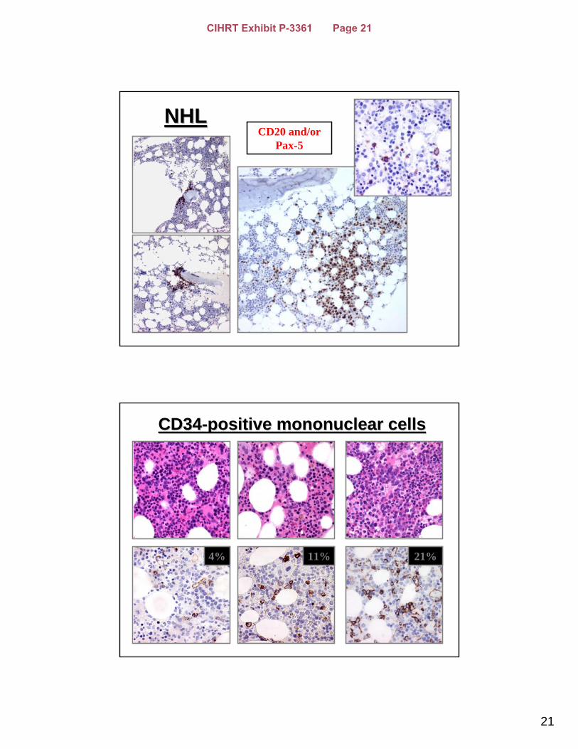
NHLNHLCD20 and/or
Pax-5
CD34CD34--positive positive mononuclear cellsmononuclear cells
4% 21%11%
CIHRT Exhibit P-3361 Page 21
22
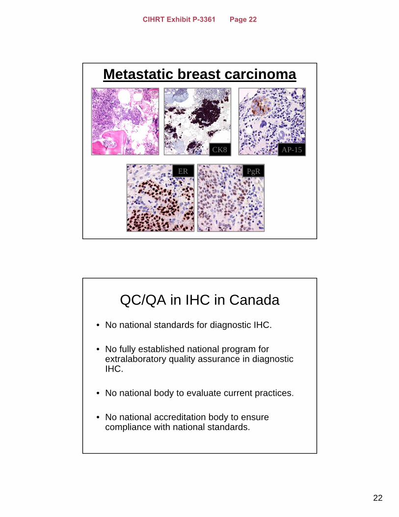
Metastatic breast carcinoma
CK8 AP-15
ER PgR
QC/QA in IHC in Canada• No national standards for diagnostic IHC.
• No fully established national program for extralaboratory quality assurance in diagnostic IHC.
• No national body to evaluate current practices.
• No national accreditation body to ensure compliance with national standards.
CIHRT Exhibit P-3361 Page 22
23
QC/QA in IHC in Canada• No national list of diagnostic laboratories that preform the IHC
testing for patients’ care.– Not able to contact laboratories for surveys.– Not able to determine the extent of problem.– No insight how far we are from standardization.– No information to plan the size or other components of the
national program needed for standardization and EQA.
• Many, if not most Canadian laboratories take participation in programs provided by USA (CAP), Scandinavia (NordiQC), and UK (UKNEQAS). These programs are not the same and they do not provide the same information to the laboratories.
• Recent initiative from the Canadian Association of Pathologists:– National Standards Committee/Immunohistochemistry
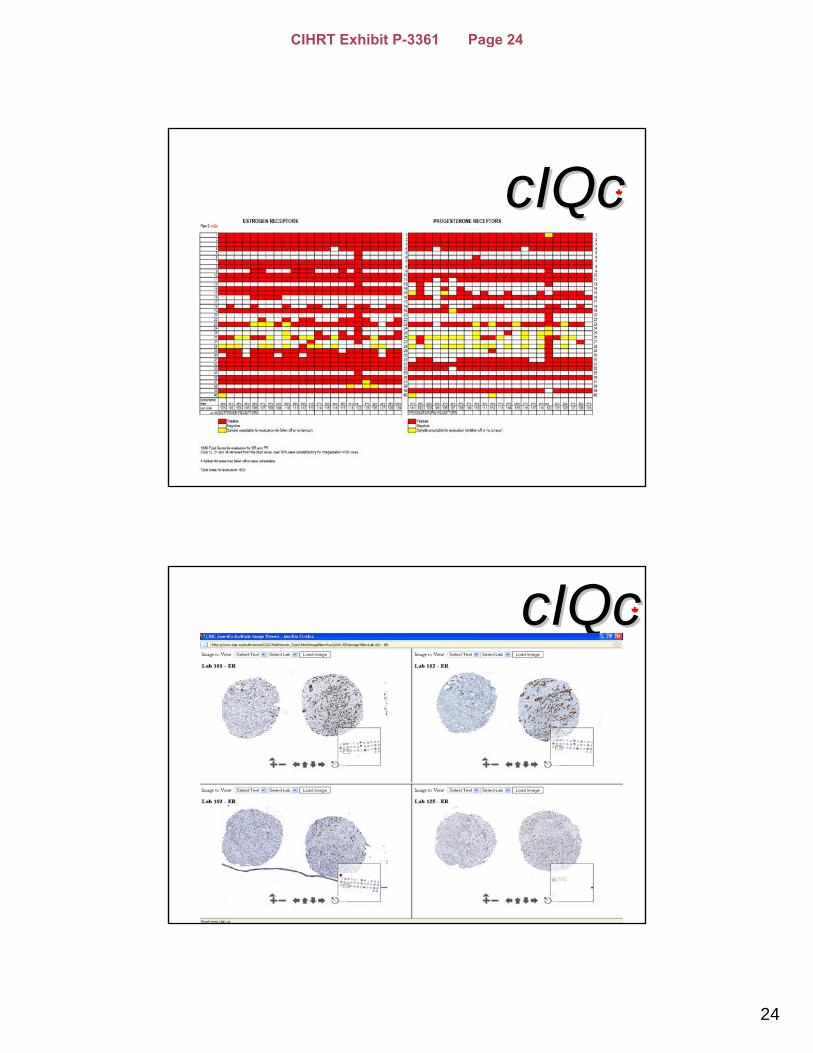
Canadian Immunohistochemistry Quality Control
• www.cIQc.ca• RUN1: Undifferentiated tumor panel• RUN2: ER/PR and HER2/neu• RUN3: ER/PR• 12 labs in RUN1, 18 in RUN2, 23 in RUN3• No funding so far.• Provides extensive feedback to participating laboratories, who can
use this information to improve results immediately. • The program is adequate to fulfil the criteria for mandatory
certification. • The program provides testing material adequate for sensible
statistical analyses currently recommended in new guidelines forclass II tests (e.i. HER2).
cIQccIQc
CIHRT Exhibit P-3361 Page 23
24
cIQccIQc
cIQccIQc
CIHRT Exhibit P-3361 Page 24
25
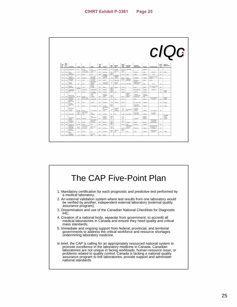
cIQccIQc
The CAP Five-Point Plan1. Mandatory certification for each prognostic and predictive test performed by
a medical laboratory;2. An external validation system where test results from one laboratory would
be verified by another, independent external laboratory (external quality assurance program);
3. Dissemination and use of the Canadian National Checklists for Diagnostic IHC.
4. Creation of a national body, separate from government, to accredit all medical laboratories in Canada and ensure they need quality and critical mass standards;
5. Immediate and ongoing support from federal, provincial, and territorial governments to address the critical workforce and resource shortages undermining laboratory medicine.
In brief, the CAP is calling for an appropriately resourced national system to promote excellence in the laboratory medicine in Canada. Canadian laboratories are not unique in facing workloads, human-resource issue, or problems related to quality control. Canada is lacking a national quality assurance program to link laboratories, provide support and administer national standards.
CIHRT Exhibit P-3361 Page 25

26
CIHRT Exhibit P-3361 Page 26