Embed Size (px)
Citation preview
SHORT COMMUNICATION
Pulling My Gut Out—Simple Tools for Engaging Students inGross Anatomy Lectures
Lap Ki Chan*
Department of Anatomy and Institute of Medical and Health Sciences Education,Li Ka Shing Faculty of Medicine, The University of Hong Kong, Hong Kong SAR, China
A lecture is not necessarily a monologue, promoting only passive learning. If appropriatetechniques are used, a lecture can stimulate active learning too. One such method is dem-onstration, which can engage learners’ attention and increase the interaction between thelecturer and the learners. This article describes two simple and useful tools for demon-stration during gross anatomy lectures. One is an apron for demonstrating midgutrotation and the other is a simple ‘‘human’’ model for demonstrating the relationshipbetween the uterus and the peritoneum. Anat Sci Educ 3: 148–150. © 2010 American Association
of Anatomists.
Key words: anatomy teaching; anatomy models; embryology instructions; gut rotation;uterus topography; pelvic anatomy
INTRODUCTION
Lecturing is still a common practice in medical and healthsciences education, although it is often considered to promotepassive learning and discourage the higher forms of cognitivelearning described in Bloom’s Taxonomy, such as application,analysis, synthesis, and evaluation (Anderson et al., 2001). Infact, Sir Joseph Barcroft, Chair of Physiology at Cambridge,has defined lecturing as ‘‘a process by which information istransferred from the notes of the lecturer to the notes of thestudent without going through the minds of either’’ (Book,1999).
One of the reasons that lecturing has become less popularas a pedagogical strategy is ‘‘lecturalgia,’’ which often resultsfrom the teacher’s lack of knowledge in how to deliver agood lecture (McLaughlin and Mandlin, 2001). But, withappropriate knowledge and skills, we can give stimulatinglectures and our students can become more active learners(Brown and Tomlinson, 1979; McLaughlin and Mandlin,2001; Cantillon, 2003). For example, appropriate questioning
during lectures can trigger more active learning in students(Mazur, 2009). Another way of encouraging active learning isthrough demonstration (Whitman, 1998), which can increasethe interaction between the lecturer and the learners, thusturning the lecture from a monologue into dialogue. It alsohas the advantage of breaking the monotony of a lecture, andregaining the attention of the learners, which is said to lastfor only 15 to 20 min (Stuart and Rutherford, 1978).Described below are two examples of tools that can be usedfor such demonstrations in gross anatomy lectures: an apronwhich is used to demonstrate midgut rotation, and a‘‘human’’ model of the uterus.
DESCRIPTIONS OF THE TOOLS
The Midgut Rotation Apron
The anticlockwise rotation of the midgut in the developingembryo, with the result that the distal limb of the midgutloop comes to lie anterior to the proximal limb, is a difficultvisual concept for learners to grasp. The use of static illustra-tion requires learners to do a lot of mental manipulation. An-imation can certainly help. But an even more effective tool isa special apron that can be made from any commerciallyavailable solid-colored apron (Fig. 1).
The foregut and hindgut are represented by two tubes ofequal length, made of a different color fabric from that of theapron. The tubes are glued onto the upper and lower third ofthe midline of the apron. The midgut is represented by athird tube, with the proximal and distal halves in differentcolors. It is about five times the length of the foregut tube,but most of it is hidden behind the apron, since it can beretracted through two openings cut in the apron, one at the
*Correspondence to: Dr. Lap Ki Chan, Department of Anatomy andInstitute of Medical and Health Sciences Education, Li Ka ShingFaculty of Medicine, The University of Hong Kong, 2/F, William MWMong Block, Faculty of Medicine Building, 21 Sassoon Road,Pokfulam, Hong Kong SAR, China. E-mail: [email protected]
Received 1 March 2010; Accepted 31 March 2010.
Published online 23 April 2010 in Wiley InterScience (www.interscience.wiley.com). DOI 10.1002/ase.154
© 2010 American Association of Anatomists
Anat Sci Educ 3:148–150 (2010) MAY/JUNE 2010 Anatomical Sciences Education
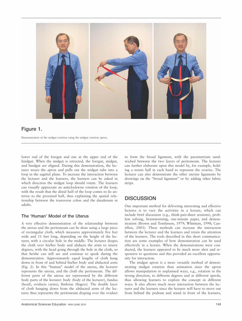
lower end of the foregut and one at the upper end of thehindgut. When the midgut is retracted, the foregut, midgut,and hindgut are aligned. During this demonstration, the lec-turer wears the apron and pulls out the midgut tube into aloop in the sagittal plane. To increase the interaction betweenthe lecturer and the learners, the learners can be asked inwhich direction the midgut loop should rotate. The learnerscan visually appreciate an anticlockwise rotation of the loop,with the result that the distal half of the loop comes to lie an-terior to the proximal half, thus explaining the spatial rela-tionship between the transverse colon and the duodenum inadults.
The ‘Human’ Model of the Uterus
A very effective demonstration of the relationship betweenthe uterus and the peritoneum can be done using a large pieceof rectangular cloth, which measures approximately five feetwide and 11 feet long, depending on the height of the lec-turer, with a circular hole in the middle. The lecturer drapesthe cloth over his/her body and abducts the arms to ninetydegrees, with the head going through the hole in the cloth, sothat he/she can still see and continue to speak during thedemonstration. Approximately equal lengths of cloth hangdown in front of and behind his/her body and abducted arms(Fig. 2). In this ‘‘human’’ model of the uterus, the lecturerrepresents the uterus, and the cloth the peritoneum. The dif-ferent parts of the uterus are represented by the differentbody parts of the lecturer: body (body of the lecturer), fundus(head), oviducts (arms), fimbriae (fingers). The double layerof cloth hanging down from the abducted arms of the lec-turer thus represents the peritoneum draping over the oviduct
to form the broad ligament, with the parametrium sand-wiched between the two layers of peritoneum. The lecturercan further elaborate upon this model by, for example, hold-ing a tennis ball in each hand to represent the ovaries. Thelecturer can also demonstrate the other uterine ligaments bydrawings on the ‘‘broad ligament’’ or by adding other fabricstrips.
DISCUSSION
One important method for delivering interesting and effectivelectures is to vary the activities in a lecture, which caninclude brief discussion (e.g., think-pair-share sessions), prob-lem solving, brainstorming, one-minute paper, and demon-stration (Brown and Tomlinson, 1979; Whitman, 1998; Can-tillon, 2003). These methods can increase the interactionbetween the lecturer and the learners and retain the attentionof the learners. The tools described in this short communica-tion are some examples of how demonstration can be usedeffectively in a lecture. When the demonstrations were con-ducted, the learners appeared to be much more alert and re-sponsive to questions and this provided an excellent opportu-nity for interaction.
The midgut apron is a more versatile method of demon-strating midgut rotation than animation since the apronallows manipulation in unplanned ways, e.g., rotation in thewrong direction, to different degrees and at different speeds,thus allowing learners to explore the concept in differentways. It also allows much more interaction between the lec-turer and the learners since the lecturer will have to move outfrom behind the podium and stand in front of the learners,
Figure 1.
Demonstration of the midgut rotation using the midgut rotation apron.
Anatomical Sciences Education MAY/JUNE 2010 149

bringing him spatially closer to the learners. The learners willfind this to be much more interesting than ‘‘just another slideon the PowerPoint presentation,’’ and watching the lecturersuddenly put on an apron during class will excite their curios-ity and make the lecture more interesting and memorable.The ‘‘human’’ model of the uterus is also highly engaging,and explains the relationship between the uterus and the peri-toneum in the most intuitive way. The learners can even puton the midgut apron and the ‘‘peritoneum’’ themselves toappreciate the anatomical relationships depicted by them,providing kinesthetic and tactile learning as well, akin tothose provided by clay models in learning anatomy (Motoikeet al., 2009; Oh et al., 2009).
Judging from students’ end-of-semester comments, the useof these models and demonstration was very well received. Anumber of students mentioned that demonstrations and themodels have really helped them in their understanding of thematerials. In the case of the midgut apron, students liked itso much that the lecturer was photographed during the dem-onstration with the photos subsequently posted in Facebook.
NOTES ON CONTRIBUTOR
LAP KI CHAN, M.B.B.S. (HK), F.H.K.A.M., F.H.K.C.O.S.(Orthopedics), F.R.C.S. (Edinburgh), Ph.D. (Duke) is an as-sistant dean of Education and associate professor in the Insti-
tute of Medical and Health Sciences Education and theDepartment of Anatomy at the Li Ka Shing Faculty ofMedicine, The University of Hong Kong, Hong Kong SAR,China. He has a background in orthopedics and physicalanthropology.
LITERATURE CITED
Anderson LW, Krathwohl DR, Airasian PW, Cruikshank KA, Mayer RE, Pin-trich PR, Raths J, Wittrock MC. 2001. A Taxonomy for Learning, Teachingand Assessing: A Revision of Bloom’s Taxonomy of Educational Objectives.2nd Ed. New York, NY: Longman. 336 p.
Book CL. 1999. Lecturing. In: Vangelisti AL, Daly JA, Friedrich GW (Editors).Teaching Communication: Theory, Research, and Methods. 2nd Ed. Mahwah,NJ: Lawrence Erlbaum Associates, Inc. p 333–346.
Brown G, Tomlinson D. 1979. How to: Improving lecturing. Med Teach1:128–135.
Cantillon P. 2003. ABC of learning and teaching in medicine: Teaching largegroups. BMJ 326:437–440.
Mazur E. 2009. Farewell, Lecture? Science 323:50–51.
McLaughlin K, Mandin H. 2001. A schematic approach to diagnosing andresolving lecturalgia. Med Educ 35:1135–1142.
Motoike HK, O’Kane RL, Lenchner E, Haspel C. 2009. Clay modeling as amethod to learn human muscles: A community college study. Anat Sci Educ2:19–23.
Oh CS, Kim JY, Choe YH. 2009. Learning of cross-sectional anatomy usingclay models. Anat Sci Educ 2:156–159.
Stuart J, Rutherford RJ. 1978. Medical student concentration during lectures.Lancet 8088:514–516.
Whitman N. 1998. Developing lecture skills. In: Edwards LC, Marier RL (Edi-tors). Clinical Teaching for Medical Residents: Roles, Techniques, and Pro-grams. 1st Ed. New York, NY: Springer Publishing Company. p 75–91.
Figure 2.
Demonstration of the anatomical relationship between the uterus and the peritoneum using the ‘‘human’’ model of uterus.
150 Chan