Embed Size (px)
Citation preview

296 J Cerebrovasc Endovasc Neurosurg
Pseudoaneurysm Formation after Repetitive Suction Thrombectomy Using a Penumbra Suction Catheter
Eun-Oh Jeong, Hyon-Jo Kwon, Seung-Won Choi, Hyeon-Song KohDepartment of Neurosurgery, School of Medicine, Regional Cerebrovascular Center, Research Institute for Brain Sciences, Chungnam National University & Hospital, Daejeon, Korea
With the recent advent of suction catheters, the use of manual aspiration thrombectomy (MAT) for patients with acute ischemic stroke with large vessel occlusion has increased. Although contrast leakage and sub-arachnoid hemorrhage have been reported during MAT procedures, pseu-doaneurysm formation due to vessel injury by suction catheters has not been. We discuss the case of a 60-year-old woman who presented to our emergency room with dysarthria and left-sided weakness. She under-went suction thrombectomy 5 times for acute middle cerebral artery oc-clusion and significant contrast leakage occurred during the procedure. On follow-up angiogram on post-operative day 15, we noticed a pseu-doaneurysm, which was treated with detachable coil embolization. Surgeons who perform suction thrombectomy should keep in mind the possibility of vessel injury that results in the formation of a pseudoaneur-ysm, especially at the branching site or tortuous segments.
J Cerebrovasc Endovasc Neurosurg. 2016 September;18(3):296-301Received : 25 April 2016Revised : 29 August 2016Accepted : 5 September 2016
Correspondence to Hyon-Jo KwonDepartment of Neurosurgery, School of Medici- ne, Regional Cerebrovascular Center, Chungnam National University & Hospital, 282 Munhwa-ro, Jung-gu, Daejeon 35015, Korea
Tel : 82-42-280-8372 Fax : 82-42-280-7364E-mail : [email protected] : http://orcid.org/0000-0002-6516-3914
The 56th Annual Meeting of the Korean Neurosurgical Society, poster presentation.
This is an Open Access article distributed under the terms of the Creative Commons Attribution Non- Commercial License (http://creativecommons.org/li-censes/by-nc/3.0) which permits unrestricted non- commercial use, distribution, and reproduction in any medium, provided the original work is properly cited.Keywords Pseudoaneurysm, Suction, Thrombectomy
Journal of Cerebrovascular and Endovascular NeurosurgerypISSN 2234-8565, eISSN 2287-3139, http://dx.doi.org/10.7461/jcen.2016.18.3.296 Case Report
INTRODUCTION
With the improvement in catheter technology, the
large bore catheter can be advanced into distal tor-
tuous vessels for manual aspiration thrombectomy
(MAT). Using this type of catheter has shown im-
provements in recanalization rates and favorable clin-
ical outcomes as stent retriever thrombectomy.12)17)18)
Although contrast leakage and subarachnoid hemor-
rhage (SAH) have been reported during and after
MAT, pseudoaneurysm formation due to vessel wall
injury has not been.2)6-15)17)18) We therefore report on a
case of significant contrast leakage during MAT in
which we found pseudoaneurysm formation at fol-
low-up angiogram that was treated by detachable coil
embolization. We also discuss the pathogenetic and
therapeutic considerations of our case.
CASE REPORT
A 60-year-old woman arrived at our emergency room
with complaints of dysarthria and left-sided weakness,
which had abruptly started 1 hour and 40 minutes
previously. Magnetic resonance imaging and magnetic
resonance angiography (MRA) showed right proximal
middle cerebral artery (M1) occlusion with partial in-
farction in the middle cerebral artery (MCA) region.
And perfusion weighted image confirmed perfusion
weighted image-diffusion weighted image mismatch.
We decided to perform a thrombectomy with the pa-
tient in a conscious, sedated state, without intravenous
t-PA(tissue plasminogen activator), because we could
perform a thrombectomy immediately. On initial an-
giogram, right mid M1 occlusion was confirmed, and
MAT using an aspiration catheter (Penumbra 5MAX,

EUN-OH JEONG ET AL
Volume 18 · Number 3 · September 2016 297
A
B
C
D
Fig. 1. Digital subtraction angiography (DSA) roadmap images during thrombectomy. (A) Roadmap shows right middle cerebral ar-tery, mid M1 segment occlusion, and the suction catheter tip (Penumbra 5MAX, Penumbra Inc., Alameda, CA, USA) for the first man-ual aspiration thrombectomy (MAT) (arrow). (B) For the second MAT, the roadmap shows more distal M1 occlusion than seen in the angiogram performed after the first MAT and the suction catheter tip (arrow). (C) Roadmap shows right distal M1 segment occlusion after the third MAT and suction catheter tip for the fourth MAT (arrow). (D) Roadmap shows one M2 branch occlusion by migrated thrombus and suction catheter (Penumbra 4MAX) tip for the fifth MAT (arrow).
Penumbra Inc., Alameda, CA, USA) and 50 mL syringe
was performed four times until M1 was recanalized
(Fig. 1A, B, C). Before each suction thrombectomy, the
Penumbra suction catheter was carefully navigated to
the occlusion point while suction catheter loaded with
a Prowler Select Plus (Codman, Raynham, MA, USA)
microcatheter and Synchro (Stryker; Kalamazoo, MI,
USA) microwire coaxially. However, one of the distal
branches (M2) was occluded. We attempted another
MAT using an aspiration catheter with a smaller in-
ner diameter (Penumbra 4MAX) (Fig. 1D). On the fol-
low-up angiogram after the fifth MAT, we noticed re-
canalized M2 and significant extravasation of contrast
at the distal M1 segment (Fig. 2A). We stopped the
procedure because further runs of angiogram show
diffuse vasospasm and no more contrast leakage. On
postoperative computed tomography (CT) of the
brain, diffuse SAH on the right Sylvian fissure and
multiple sulci were noted with accompanying ven-
triculomegaly (Fig. 2B). The patient was conscious, al-
beit drowsy, and we drained her cerebrospinal fluid
via a lumbar drain. She showed improvement of con-
sciousness on the next day, and we continued con-
servative treatment. On the follow-up CT, SAH along
the Sylvian fissure persisted until postoperative day
(POD) 12, when the patient showed deterioration of
consciousness. On the angiogram, vasospasm was
noted on right M2 segments, and chemical angio-
plasty using Nicardipine (Perdipine, Astellas, Shizuoka,
Japan) was performed daily for three days (Fig. 2C).
On the angiogram taken on POD 15, a tiny pseudoa-
neurysm (Fig. 2D, E) was found at the origin of the
small temporal branch of the right M1, and emboliza-
tion using detachable platinum coils was successfully
performed under general anesthesia (Fig. 3A, B). The
location of the pseudoaneurysm was distal to the
original occlusion site and near the segment where
the contrast leakage started. The patient showed im-
provement of consciousness later and was discharged
rehabilitation with only mild weakness while making
grasping motions with her left hand. On the fol-
low-up angiogram obtained on post-embolization day
17, contrast filling in the pseudoaneurysm was not
observed (Fig. 3C). An MRA obtained on post-emboli-
zation day 125 showed no evidence of a remnant sac
(Fig. 3D).

PSEUDOANEURYSM FORMATION AFTER SUCTION THROMBECTOMY
298 J Cerebrovasc Endovasc Neurosurg
A
B
C
D
E
Fig. 2. Digital subtraction angiography (DSA) and three-dimensional reconstruction images, computed tomography (CT) after thrombectomy. (A) DSA shows contrast leakage (arrow) at distal M1 near the fourth MAT point. (B) CT shows diffuse SAH on the right Sylvian fissure and multiple sulci accompanying ventriculomegaly (C) DSA performed on postoperative day (POD) 12 shows mul-tiple vasospasms at M2 and M3 segments (arrows) and no evidence of an aneurysm sac. (D) DSA performed on POD 15 shows an aneurysm sac near the extravasation site (arrow). (E) Three-dimensional reconstruction image of pseudoaneurysm at the origin site of small temporal branch of the right M1 segment (arrow).
DISCUSSION
Aneurysm is classified into true aneurysm and
pseudoaneurysm histologically. True aneurysm refers
to an aneurysm that is formed due to incomplete in-
jury to the vascular wall with existing adventitia. In
contrast, pseudoaneurysm forms a false lumen with
completely disrupted vascular wall. In our case, hem-
atoma formed around vessel injury site, so no abnor-
mal finding observed around vessel injury site in fol-
low-up digital subtraction angiography. As time goes
by hematoma resolved, false lumen formed around
vessel injury site. We observed pseudoaneurysm in
follow up angiogram due to contrast filling in false
lumen.
With the improvement of endovascular techniques
and devices, the demand for endovascular recanaliza-
tion for major intracranial artery occlusion is increasing.
Because of the improvement in catheter engineering,
it is possible to navigate the relatively large bore cath-

EUN-OH JEONG ET AL
Volume 18 · Number 3 · September 2016 299
A
B
C
D
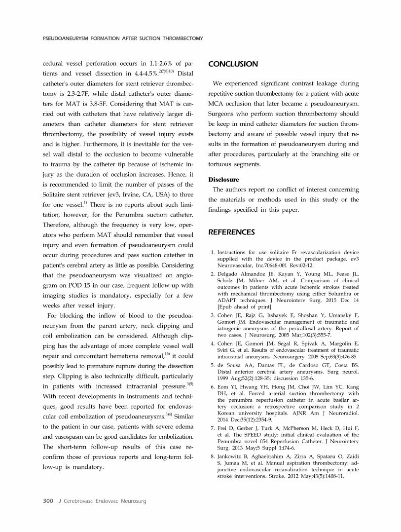
Fig. 3. Angiograms obtained during embolization and the follow-up period. (A) Digital subtraction angiography (DSA) roadmap im-age shows a single microcatheter selected into the pseudoaneurysm sac (arrow). (B) Non-subtracted angiography shows no contrast filling in the pseudoaneurysm after detachable coil embolization (arrow). (C) DSA performed on postoperative (POD) day 17 shows complete occlusion of the pseudoaneurysm (arrow). (D) Magnetic resonance angiography performed on POD 125 shows no evidenceof a remnant sac (arrow).
eter into the tortuous distal cerebral vessels. In reports
comparing MAT using this kind of catheter between
stent retriever, clinical outcome, complication, recanal-
ization rate did not differ significantly.15)17) So as
thrombectomy using stent retriever, MAT became one
of the two current standard of thrombectomy for pa-
tients with acute cerebral artery occlusion.
During and after MAT in patients with acute occlu-
sion, the rates of contrast leakage and SAH have been
reported to be between 0% and 31%.2)6-15)17)18) Intra-pro-
PSEUDOANEURYSM FORMATION AFTER SUCTION THROMBECTOMY
300 J Cerebrovasc Endovasc Neurosurg
cedural vessel perforation occurs in 1.1-2.6% of pa-
tients and vessel dissection in 4.4-4.5%.2)7)8)10) Distal
catheter's outer diameters for stent retriever thrombec-
tomy is 2.3-2.7F, while distal catheter's outer diame-
ters for MAT is 3.8-5F. Considering that MAT is car-
ried out with catheters that have relatively larger di-
ameters than catheter diameters for stent retriever
thrombectomy, the possibility of vessel injury exists
and is higher. Furthermore, it is inevitable for the ves-
sel wall distal to the occlusion to become vulnerable
to trauma by the catheter tip because of ischemic in-
jury as the duration of occlusion increases. Hence, it
is recommended to limit the number of passes of the
Solitaire stent retriever (ev3, Irvine, CA, USA) to three
for one vessel.1) There is no reports about such limi-
tation, however, for the Penumbra suction catheter.
Therefore, although the frequency is very low, oper-
ators who perform MAT should remember that vessel
injury and even formation of pseudoaneurysm could
occur during procedures and pass suction catether in
patient's cerebral artery as little as possible. Considering
that the pseudoaneurysm was visualized on angio-
gram on POD 15 in our case, frequent follow-up with
imaging studies is mandatory, especially for a few
weeks after vessel injury.
For blocking the inflow of blood to the pseudoa-
neurysm from the parent artery, neck clipping and
coil embolization can be considered. Although clip-
ping has the advantage of more complete vessel wall
repair and concomitant hematoma removal,16) it could
possibly lead to premature rupture during the dissection
step. Clipping is also technically difficult, particularly
in patients with increased intracranial pressure.3)5)
With recent developments in instruments and techni-
ques, good results have been reported for endovas-
cular coil embolization of pseudoaneurysms.3)4) Similar
to the patient in our case, patients with severe edema
and vasospasm can be good candidates for embolization.
The short-term follow-up results of this case re-
confirm those of previous reports and long-term fol-
low-up is mandatory.
CONCLUSION
We experienced significant contrast leakage during
repetitive suction thrombectomy for a patient with acute
MCA occlusion that later became a pseudoaneurysm.
Surgeons who perform suction thrombectomy should
be keep in mind catheter diameters for suction throm-
bectomy and aware of possible vessel injury that re-
sults in the formation of pseudoaneurysm during and
after procedures, particularly at the branching site or
tortuous segments.
Disclosure
The authors report no conflict of interest concerning
the materials or methods used in this study or the
findings specified in this paper.
REFERENCES
1. Instructions for use solitaire Fr revascularization device supplied with the device in the product package. ev3 Neurovascular, Inc.70648-001 Rev:02-12.
2. Delgado Almandoz JE, Kayan Y, Young ML, Fease JL, Scholz JM, Milner AM, et al. Comparison of clinical outcomes in patients with acute ischemic strokes treated with mechanical thrombectomy using either Solumbra or ADAPT techniques. J Neurointerv Surg. 2015 Dec 14 [Epub ahead of print]
3. Cohen JE, Rajz G, Itshayek E, Shoshan Y, Umansky F, Gomori JM. Endovascular management of traumatic and iatrogenic aneurysms of the pericallosal artery. Report of two cases. J Neurosurg. 2005 Mar;102(3):555-7.
4. Cohen JE, Gomori JM, Segal R, Spivak A, Margolin E, Sviri G, et al. Results of endovascular treatment of traumatic intracranial aneurysms. Neurosurgery. 2008 Sep;63(3):476-85.
5. de Sousa AA, Dantas FL, de Cardoso GT, Costa BS. Distal anterior cerebral artery aneurysms. Surg neurol. 1999 Aug;52(2):128-35; discussion 135-6.
6. Eom YI, Hwang YH, Hong JM, Choi JW, Lim YC, Kang DH, et al. Forced arterial suction thrombectomy with the penumbra reperfusion catheter in acute basilar ar-tery occlusion: a retrospective comparison study in 2 Korean university hospitals. AJNR Am J Neuroradiol. 2014 Dec;35(12):2354-9.
7. Frei D, Gerber J, Turk A, McPherson M, Heck D, Hui F, et al. The SPEED study: initial clinical evaluation of the Penumbra novel 054 Reperfusion Catheter. J Neurointerv Surg. 2013 May;5 Suppl 1:i74-6.
8. Jankowitz B, Aghaebrahim A, Zirra A, Spataru O, Zaidi S, Jumaa M, et al. Manual aspiration thrombectomy: ad-junctive endovascular recanalization technique in acute stroke interventions. Stroke. 2012 May;43(5):1408-11.

EUN-OH JEONG ET AL
Volume 18 · Number 3 · September 2016 301
9. John S, Hussain MS, Toth G, Bain M, Uchino K, Hui FK. Initial Experience using the 5MAXTM ACE reperfusion cath-eter in Intra-Arterial therapy for acute ischemic stroke. J Cerebrovasc Endovasc Neurosurg. 2014 Dec;16(4):350-7.
10. Kang DH, Hwang YH, Kim YS, Park J, Kwon O, Jung C. Direct thrombus retrieval using the reperfusion cathe-ter of the penumbra system: forced-suction thrombec-tomy in acute ischemic stroke. AJNR Am J Neuroradiol. 2011 Feb;32(2):283-7.
11. Kim SK, Yoon W, Moon SM, Park MS, Jeong GW, Kang HK. Outcomes of manual aspiration thrombectomy for acute ischemic stroke refractory to stent-based thrombectomy. J Neurointerv Surg. 2015 Jul;7(7):473-7.
12. Kowoll A, Weber A, Mpotsaris A, Behme D, Weber W. Direct aspiration first pass technique for the treatment of acute ischemic stroke: initial experience at a European stroke center. J Neurointerv Surg. 2016 Mar;8(3):230-4.
13. Navia P, Larrea JA, Pardo E, Arce A, Martínez-Zabaleta M, Díez-González N, et al. Initial experience using the 3MAX cerebral reperfusion catheter in the endovascular treatment of acute ischemic stroke of distal arteries. J Neurointerv Surg. 2016 Aug;8(8):787-90.
14. Shindo A, Kawanishi M, Kawakita K, Okauchi M, Kawai N, Hayashi N, et al. Treatment of acute cerebral artery occlusion using the Penumbra system: our early experience. Neurol Med Chir (Tokyo). 2014 Jun:54(6):441-9.
15. Son S, Choi DS, Oh MK, Hong J, Kim SK, Kang H, et al. Comparison of Solitaire thrombectomy and Penumbra suction thrombectomy in patients with acute ischemic stroke caused by basilar artery occlusion. J Neurointerv Surg. 2016 Jan;8(1):13-8.
16. Sui M, Mei Q, Sun K. Surgical treatment achieves better outcome in severe traumatic pericallosal aneurysm: case report and literature review. Int J Clin Exp Med. 2015 Feb;8(2):1598-603.
17. Turk AS, Frei D, Fiorella D, Mocco J, Baxter B, Siddiqui A, et al. ADAPT FAST study: a direct aspiration first pass technique for acute stroke thrombectomy. J Neurointerv Surg. 2014 May;6(4):260-4.
18. Turk AS, Spiotta A, Frei D, Mocco J, Baxter B, Fiorella D, et al. Initial clinical experience with the ADAPT tech-nique: a direct aspiration first pass technique for stroke thrombectomy. J Neurointerv Surg. 2014 Apr;6(3):231-7.