Embed Size (px)
Citation preview

Huang et al. Journal of Biomedical Science 2014, 21:44http://www.jbiomedsci.com/content/21/1/44
RESEARCH Open Access
Prospective signs of cleidocranial dysplasia inCebpb deficiencyBoyen Huang1,2, Katsu Takahashi2*, Ernest A Jennings1, Pongthorn Pumtang-on3, Honoka Kiso2, Yumiko Togo2,Kazuyuki Saito2, Manabu Sugai4, Shizuo Akira5, Akira Shimizu4 and Kazuhisa Bessho2
Abstract
Background: Although runt-related transcription factor 2 (RUNX2) has been considered a determinant of cleidocranialdysplasia (CCD), some CCD patients were free of RUNX2 mutations. CCAAT/enhancer-binding protein beta (Cebpb) is akey factor of Runx2 expression and our previous study has reported two CCD signs including hyperdontia and elongatedcoronoid process of the mandible in Cebpb deficient mice. Following that, this work aimed to conduct a case-controlstudy of thoracic, zygomatic and masticatory muscular morphology to propose an association between musculoskeletalphenotypes and deficiency of Cebpb, using a sample of Cebpb-/-, Cebpb+/- and Cebpb+/+ adult mice. Somatic skeletonsand skulls of mice were inspected with soft x-rays and micro-computed tomography (μCT), respectively. Zygomaticinclination was assessed using methods of coordinate geometry and trigonometric function on anatomic landmarksidentified with μCT. Masseter and temporal muscles were collected and weighed. Expression of Cebpb was examinedwith a reverse transcriptase polymerase chain reaction (RT-PCR) technique.
Results: Cebpb-/- mice displayed hypoplastic clavicles, a narrow thoracic cage, and a downward tilted zygomatic arch(p < 0.001). Although Cebpb+/- mice did not show the phenotypes above (p = 0.357), a larger mass percentage oftemporal muscles over masseter muscles was seen in Cebpb+/- littermates (p = 0.012). The mRNA expression of Cebpbwas detected in the clavicle, the zygoma, the temporal muscle and the masseter muscle, respectively.
Conclusions: Prospective signs of CCD were identified in mice with Cebpb deficiency. These could provide anadditional aetiological factor of CCD. Succeeding investigation into interactions among Cebpb, Runx2 andmusculoskeletal development is indicated.
Keywords: Cebpb, Cleidocranial dysplasia, Clavicle, Thoracic cage, Zygomatic arch, Masseter, Temporal muscle
BackgroundCleidocranial dysplasia (CCD) is a congenital skeletal dis-ease typically manifesting cranial deformity, clavicle hypo-plasia and unerupted supernumerary teeth [1-5]. Somehuman CCD cases also displayed hypotrophy of the mas-seter muscle, abnormality of the mandible and/or deform-ation of the zygomatic arch [3-5]. Haploinsufficiency ofthe runt-related transcription factor 2 (RUNX2 in humans,Runx2 in mice) has been considered a determinant ofCCD in humans [6]. A recent study has also identifiedbony and dental defects consistent with signs of humanCCD amongst mice with impaired activity of the Runx2
* Correspondence: [email protected] of Oral and Maxillofacial Surgery, Graduate School of Medicine,Kyoto University, Shogoin-Kawahara-cho 54, Sakyo-ku, Kyoto 606-8507, JapanFull list of author information is available at the end of the article
© 2014 Huang et al.; licensee BioMed CentralCommons Attribution License (http://creativecreproduction in any medium, provided the orDedication waiver (http://creativecommons.orunless otherwise stated.
P1 promoter [7]. Expression of the Runx2 P1 promoter inthe developing skull has been confirmed [8] and its activityis essential to establishing a sufficient number of osteopro-genitor cells for normal skeletogenesis [7]. Although themechanism of CCD-related dental anomalies is not clear,induction of the dentition was associated with fibroblastgrowth factor (FGF) signaling [9] that was mediated byRunx2 during development of teeth [10] and bone [11].Nevertheless, about one-third of CCD patients were freeof RUNX2 mutations [6]. Furthermore, diverse dentalmanifestations resulting from identical mutations of theRUNX2 gene have been observed [1]. These infer potentialinvolvement of other factors on occurrence of CCD.CCAAT/enhancer binding protein beta (CEBPB in
humans, Cebpb in mice) is a transcription factor which
Ltd. This is an Open Access article distributed under the terms of the Creativeommons.org/licenses/by/4.0), which permits unrestricted use, distribution, andiginal work is properly credited. The Creative Commons Public Domaing/publicdomain/zero/1.0/) applies to the data made available in this article,

Table 1 Murine anatomical landmarks used in this study
Landmarks Anatomical positions
LO Anterior notch on frontal process lateralto infraorbital fissure, left side
LP Intersection of parietal, temporal andoccipital bones, left side
RP Intersection of parietal, temporal andoccipital bones, right side
LJ Intersection of zygomatic process of maxillawith zygoma, superior surface, left side
LZ Intersection of zygoma with zygomaticprocess of temporal, superior aspect, left side
LS Joining of squamosal body to zygomaticprocess of squamosal, left side
Huang et al. Journal of Biomedical Science 2014, 21:44 Page 2 of 8http://www.jbiomedsci.com/content/21/1/44
binds to consensus sequences and affects the transcriptionof various genes involved in proliferation and differenti-ation, and found in the liver [12], the mammary gland [13]and the immune system [14,15]. In addition to its rele-vance in tumorigenesis [16] and adipogenesis [17], previ-ous studies have suggested that Cebpb is a key factor ofRunx2 expression in bone formation [18-21]. A reductionin Runx2 expression was accompanied by a decrease inosteogenic potential and shown to be related to ectopicexpression of Cebpb [18]. On the other hand, a functionalsynergism of Runx2 and Cebpb during osteoblast [21] andchondrocyte differentiation [20] has been reported. Sup-pressed differentiation of osteoblasts and delayed chon-drocyte hypertrophy due to a complete deletion of Cebpbis likely to postpone bone formation [19]. This concurswith phenotypes of p20Cebpb (a dominant negative in-hibitor of Cebpb) overexpressing mice, including a re-duced amount of alveolar bone and a lower bone volumefraction of the mandible [22].Recent studies have identified multiple supernumerary
teeth [23], hypoplastic clavicles [20], an open fontanelle[20] and an elongated coronoid process [23] in mice withCebpb deficiency. These phenotypes coincide with signsof CCD in humans [1-5] and consequently a relationshipbetween CEBPB and CCD is suspected [20]. To exploreprospective signs of human CCD in a murine model, thisstudy aimed to conduct a case-control study of thoracic,craniofacial and myological variations, using a sample ofCebpb-/-, Cebpb+/- and Cebpb+/+ mice. A special interestwas to investigate morphology of the zygomatic arch andmass of both masseter and temporal muscles.
MethodsAn appropriate ethics approval has been obtained from theInstitutional Review Board of Kyoto University (ReferenceNumber: Med Kyo 11518). Cebpb+/- and Cebpb-/- micewere generated as described previously [15]. Thirty-fiveadult mice were euthanised with carbon dioxide gas for in-spection, including 5 Cebpb-/- (5 female), 16 Cebpb+/- (10female and 6 male) and 14 Cebpb+/+ (8 female and 6 male)mice. All female mice were sacrificed at the age of12 months, whilst the age of male mice used ranged from4.5 to 13 months. Owing to a high neonatal mortality ofCebpb-/- mice [24], male animals of this genotype were notattainable in this sample.Firstly, skeletons of the female mice were imaged with soft
x-rays (SOFRON; SRO-M50, Sofron X-ray Industry Corpor-ation Ltd., Tokyo, Japan). Both lateral and dorsal-ventral ra-diographs of the experimental animals were evaluated by asingle-blinded examiner who is a qualified veterinarian.Secondly, murine skulls of the 23 female mice were
assessed with a micro-computed tomography (μCT)scanner (SMX-100CT-SV3; Shimadzu, Kyoto, Japan).This technique was applied to identify predetermined
craniofacial landmarks including Landmark LO, LP, RP,LJ, LZ, and LS (Table 1) [23,25,26]. As the external audi-tory meatus of mice is located at a lower level than thatof humans [27], Landmark LP and RP situated slightlysuperior to the external auditory meatus were used toreplace human Porions (the most superior point of ex-ternal auditory meatus visible on a lateral cephalometricradiograph) to establish a horizontal plane (H plane)with Landmark LO in this study. Following this, a jugalplane (J plane: defined by Landmark LJ, LP, and RP), azygomatic plane (Z plane: defined by Landmark LZ, LPand RP), and a squamosal plane (S plane: defined byLandmark LS, LP and RP) were established (Figure 1).Usage of the dihedral angles in the assessment of hu-man zygomatic anatomy has been reported [26]. Thedihedral angles between H plane and J plane, betweenH plane and Z plane, and between H plane and S planewere calculated using a method of coordinate geometryand trigonometric function [28]. The following formu-lae were used.Given the equation of H plane containing Landmark LO
(xLO, yLO, zLO), LP (xLP, yLP, zLP) and RP (xRP, yRP, zRP) wasa1 x + b1 y + c1 z + d1 = 0, the equation of J plane coveringLandmark LJ (xLJ, yLJ, zLJ), LP (xLP, yLP, zLP) and RP (xRP,yRP, zRP) was a2 x + b2 y + c2 z + d2 = 0, and the dihedralangle between H plane and J plane was θJ.Where,
a1 ¼ yLP−yLOð Þ zRP−zLOð Þ− yRP−yLOð Þ zLP−zLOð Þ;b1 ¼ zLP−zLOð Þ xRP−xLOð Þ− zRP−zLOð Þ xLP−xLOð Þ;c1 ¼ xLP−xLOð Þ yRP−yLOð Þ− xRP−xLOð Þ yLP−yLOð Þ;d1 ¼ − a1 xLO þ b1 yLO þ c1 zLOð Þ;a2 ¼ yLP−yLJ
� �zRP−zLJð Þ− yRP−yLJ
� �zLP−zLJð Þ;
b2 ¼ zLP−zLJð Þ xRP−xLJð Þ− zRP−zLJð Þ xLP−xLJð Þ;c2 ¼ xLP−xLJð Þ yRP−yLJ
� �− xRP−xLJð Þ yLP−yLJ
� �;
d2 ¼ − a2 xLJ þ b2 yLJ þ c2 zLJ� �
:

Huang et al. Journal of Biomedical Science 2014, 21:44 Page 3 of 8http://www.jbiomedsci.com/content/21/1/44
Thus,
θJ ¼ arccosa1 a2þ b1 b2þ c1 c2ffiffiffiffiffiffiffiffiffiffiffiffiffiffiffiffiffiffiffiffiffiffiffiffiffiffiffiffiffiffiffiffi
a12 þ b12 þ c12p ffiffiffiffiffiffiffiffiffiffiffiffiffiffiffiffiffiffiffiffiffiffiffiffiffiffiffiffiffiffiffiffi
a22 þ b22 þ c22p
� �
Likewise, the degree of the dihedral angles between Hplane as well as Z plane (θZ) and between H plane aswell as S plane (θS) was determined.Thirdly, masseter and temporal muscles were collected
from 12 male littermates and weighed according to theprocedures reported in an earlier study [29]. The tem-poral/masseter mass percentage was calculated by divid-ing the weight of the left temporal muscle by the weightof the left masseter muscle and then multiplying 100.To identify the expression of Cebpb, mRNA expression
of the target gene (Cebpb) was assessed. Tissues used forthis purpose were collected from the masseter muscle, thetemporal muscle, the zygomatic bone, and the clavicle boneof a Cebpb+/+ mouse, respectively. Standard procedures for
Figure 1 Craniofacial landmarks, established planes and dihedral anghorizontal reference plane (H plane). (a) Landmark LJ, LP and RP formed awas defined as θJ. (b) Landmark LZ, LP and RP formed a zygomatic plane (defined as θZ. (c) Landmark LS, LP and RP formed a squamosal plane (S plaas θS.
the preparation of the reverse transcriptase polymerasechain reaction (RT-PCR) were carried out as suggestedin the literature [30,31]. Primers applied to perform theRT-PCR technique included murine Cebpb sense (5′-ACACGTGTAACTGTCAGCCG-3′) and murine Cebpb anti-sense (5′-GCTCGAAACGGAAAAGGTTC-3′). Specificprimers for murine glyceraldehyde-3-phosphate dehydro-genase (Gapdh) were used to check the internal control[32]. The RT-PCR technique was conducted with a Gen-eAmp PCR System 9700 thermal cycler (Applied Biosys-tems, Foster City, CA, USA). All procedures were carriedout according to the manufacturers’ instructions.Data entry and statistical analysis were conducted with
the IBM SPSS Statistics (version 20.0, IBM Corporation,Somers, NY, USA). An independent samples t-test methodwas used to assess the difference in the degree of ana-tomic dihedral angles between genotypes of Cebpb defi-ciency and the control (Cebpb+/+ mice) [33]. In addition, apaired samples t-test method was applied to examine the
les used in this study. (a-c) Landmark LO, LP and RP formed ajugal plane (J plane). The dihedral angle between J plane and H planeZ plane). The dihedral angle between Z plane and H plane wasne). The dihedral angle between S plane and H plane was defined

Huang et al. Journal of Biomedical Science 2014, 21:44 Page 4 of 8http://www.jbiomedsci.com/content/21/1/44
difference in the temporal/masseter mass percentage be-tween Cebpb+/- and Cebpb+/+ littermates [33]. This pairedmethod has been used to compare dental phenotypes be-tween twins [34]. The level of two-sided significance forall statistical procedures was set at 5%.
ResultsShorter thinner clavicles with a lower radiopacity and a nar-rower thoracic cage were identified exclusively in Cebpb-/-
mice but not Cebpb+/- and/or Cebpb+/+ mice (Figure 2). Anormal morphology of limb joints was seen in radiographsfrom animals of all three genotypes. Of further note, mRNAexpression of Cebpb was found in the clavicle, the zygoma,the temporal muscle and the masseter muscle (Figure 3).The mean measurement of θZ was 16.3 ± 1.1 degree,
13.0 ± 1.1 degree and 13.4 ± 1.0 degree in Cebpb-/-,Cebpb+/- and Cebpb+/+ mice, respectively (Table 2).Those animals with a Cebpb-/- genotype showed a largerdegree of θZ than that were from a Cebpb+/+ (t = 4.964,df = 11, p < 0.001) or a Cebpb+/- genetic background (t =5.429, df = 13, p < 0.001) (Table 2). The latter two geno-types did not differ in the degree of θZ (p = 0.357) (Table 2).The degree of θJ in Cebpb-/-, Cebpb+/- and Cebpb+/+ sub-jects was separately 6.2 ± 1.1 degree, 5.5 ± 0.7 degree and6.3 ± 1.7 degree (p ≥ 0.142) (Table 2). The degree of θSin Cebpb-/-, Cebpb+/- and Cebpb+/+ mice was 3.7 ± 2.2degree, 3.1 ± 2.2 degree and 5.1 ± 3.3 degree, individually(p ≥ 0.148) (Table 2). Figure 4 demonstrates a lateral viewof dry skulls of a Cebpb-/- and a Cebpb+/+ 12-month-oldfemale mice.
Figure 2 Difference in morphology of clavicles and the thoracic cagea Cebpb+/- and a Cebpb-/- 12-month-old mice in this sample. Shorter thinnthoracic cage (red arrow) were exclusively seen in Cebpb-/- mice. Due to ththree mice were taken under the same condition separately. The images wbrightness and/or colour. Scale bar: 1 cm. (n = 23, including 5 Cebpb-/-, 10
The mean weight of the left masseter muscle in Cebpb+/-
and Cebpb+/+ mice was 113.5 ± 23.9 mg and 120.9 ±24.4 mg, respectively (p = 0.362) (Table 3). The averageweight of the left temporal muscle was 36.6 ± 8.8 mg and32.0 ± 10.1 mg separately in Cebpb+/- and Cebpb+/+ sub-jects (p = 0.217) (Table 3). The temporal/masseter masspercentage of Cebpb+/- and Cebpb+/+ littermates was32.3 ± 4.9% and 26.1 ± 6.2%, individually (Table 3). Theformer displayed a larger percentage than the latter(t = 3.841, df = 5, p = 0.012) (Table 3). Figure 5 showsthe difference in the temporal/masseter mass percent-age between Cebpb+/- and Cebpb+/+ mice by age. Themean of the difference of the temporal/masseter mass per-centage between Cebpb+/- and Cebpb+/+ amongst youngadults (less than 6 months of age), adults (6 to 12 monthsof age) and mature adults (more than 12 months of age)was 5.66%, 3.29% and 0.55%, respectively.
DiscussionThis paper reported phenotypes of mice with Cebpb defi-ciency, including hypoplastic clavicles, a narrow thoraciccage, a downward tilted zygomatic arch and a comparativemass change of masseter/temporal muscles. In conjunc-tion with our previous findings such as multiple super-numerary teeth and elongated coronoid process in thesame species of Cebpb deficient mice [23], these indicatedprospective signs of CCD [1-5]. As Cebpb is relevant toexpression of Runx2 [18-21] which has been known a de-terminant of CCD [6], consistency between murine phe-notypes of Cebpb deficiency and human manifestations of
. The radiographic image showed a dorsal-ventral view of a Cebpb+/+,er clavicles with a lower radiopacity (yellow arrow) and a narrowe limited size of a radiograph film, the radiographic images of theere displayed without transformation in dimensions, contrast,Cebpb+/- and 8 Cebpb+/+ female mice).
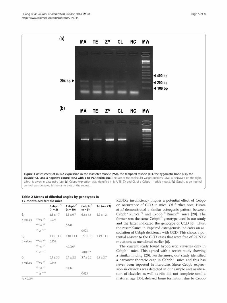
Figure 3 Assessment of mRNA expression in the masseter muscle (MA), the temporal muscle (TE), the zygomatic bone (ZY), theclavicle (CL) and a negative control (NC) with a RT-PCR technique. The size of the molecular weight markers (MW) is displayed on the right,which is given in base pairs (bp). (a) Cebpb expression was identified in MA, TE, ZY and CL of a Cebpb+⁄+ adult mouse. (b) Gapdh, as an internalcontrol, was detected in the same sites of the mouse.
Table 2 Means of dihedral angles by genotypes in12-month-old female mice
Cebpb+/+
(n = 8)Cebpb+/-
(n = 10)Cebpb-/-
(n = 5)All (n = 23)
θJ 6.3 ± 1.7 5.5 ± 0.7 6.2 ± 1.1 5.9 ± 1.2
p values +/+vs +/- 0.227+/- vs -/- 0.142-/- vs +/+ 0.923
θZ 13.4 ± 1.0 13.0 ± 1.1 16.3 ± 1.1 13.9 ± 1.7
p values +/+vs +/- 0.357+/- vs -/- <0.001*-/- vs +/+ <0.001*
θS 5.1 ± 3.3 3.1 ± 2.2 3.7 ± 2.2 3.9 ± 2.7
p values +/+vs +/- 0.148+/- vs -/- 0.432-/- vs +/+ 0.633
*p < 0.001.
Huang et al. Journal of Biomedical Science 2014, 21:44 Page 5 of 8http://www.jbiomedsci.com/content/21/1/44
RUNX2 insufficiency implies a potential effect of Cebpbon occurrence of CCD in mice. Of further note, Hirataet al demonstrated a similar osteogenic pattern betweenCebpb-/-Runx2+/+ and Cebpb+/+Runx2+/- mice [20]. Theformer was the same Cebpb-/- genotype used in our studyand the latter indicated the genotype of CCD [6]. Thus,the resemblance in impaired osteogenesis indicates an as-sociation of Cebpb deficiency with CCD. This shows a po-tential answer to the CCD cases that were free of RUNX2mutations as mentioned earlier [6].The current study found hypoplastic clavicles only in
Cebpb-/- mice. This agreed with a recent study showinga similar finding [20]. Furthermore, our study identifieda narrower thoracic cage in Cebpb-/- mice and this hasnever been reported in literature. Since Cebpb expres-sion in clavicles was detected in our sample and ossifica-tion of clavicles as well as ribs did not complete until amaturer age [35], delayed bone formation due to Cebpb

Figure 4 Difference in tilt angulation of the zygomatic arch. The photographic image showed a lateral view of dry skulls respectivelycollected from a Cebpb+/+ and a Cebpb-/- 12-month-old mice. A downward tilt of the zygomatic arch in the Cebpb-/- mouse was confirmed bythe morphology of the dry skull (yellow arrow). Scale bar: 1 mm. (n = 23, including 5 Cebpb-/-, 10 Cebpb+/- and 8 Cebpb+/+ female mice).
Huang et al. Journal of Biomedical Science 2014, 21:44 Page 6 of 8http://www.jbiomedsci.com/content/21/1/44
deficiency [19] could thereby result in clavicular hypo-plasia and a narrowed ribcage.This study has demonstrated for the first time a down-
ward tilt of the zygomatic arch in Cebpb-/- mice. Thelarger dihedral angle between Z plane and H plane (θZ)identified in Cebpb-/- mice represented that LandmarkLZ was located at a more inferior position in the geno-type. On the other hand, Landmark LJ and LandmarkLS of Cebpb-/- animals were not located at a lower levelcompared to those of Cebpb+/+ and Cebpb+/- mice. Thisindicated that deformation of the zygomatic arch waslimited to the zygoma and not involved with the zygo-matic processes of the maxilla and/or the squamosalbone. This finding resembles the feature of a downwardtilted zygomatic arch in patients sustaining CCD [3,4].Although not observed in our mouse model, a past art-icle has suggested an association of human CCD withzygomatic hypoplasia [4]. This may imply a reason whyCCD patients displayed a downward inclination of thezygomatic arch. Our detection of Cebpb expression inthe zygomatic bone also indicated a potential influenceof Cebpb on zygomatic bone formation. Nevertheless,
Table 3 Means of weight and mass percentage of masseter anlittermates
Cebpb+/+ (n = 6)
Weight of the left masseter muscle (mg) 120.9 ± 24.4
Weight of the left temporal muscle (mg) 32.0 ± 10.1
Temporal/masseter mass percentage (%) 26.1 ± 6.2
*p < 0.05.
functional interactions among masticatory muscles andcraniofacial bones could also contribute to deformity ofthe zygoma [3,4].Moreover, this study has reported for the first time a
larger temporal/masseter mass percentage in Cebpb+/-
mice, which indicated hypotrophy of masseter musclescompared to temporal muscles and/or hypertrophy oftemporal muscles compared to masseter muscles. Thisagreed with a paper which has revealed a volume reduc-tion of masseter muscles in CCD patients [4]. Furuuchiet al suggested a causal relationship between hypoplasticzygomatic arch and hypotrophic masseter muscles ofCCD cases, based on the anatomic connection [4]. How-ever, this would be difficult to justify the phenotype ofmasseter muscles in Cebpb+/- mice, since zygomatic de-formity was not significant in the genotype. On the otherhand, the temporal muscles insert onto the mandibularcoronoid process [36] and elongation of the coronoidprocess in Cebpb+/- mice has been reported by our pre-vious study [23]. Functional activity and muscular devel-opment are likely to reciprocally affect growth of thetemporal muscle and the coronoid process [3,4]. As
d temporal muscles by genotypes in paired male
Cebpb+/- (n = 6) All (n = 12) p value
113.5 ± 23.9 117.2 ± 23.3 0.362
36.6 ± 8.8 34.3 ± 9.4 0.217
32.3 ± 4.9 29.2 ± 6.2 0.012*

Figure 5 Difference in the temporal/masseter mass percentagebetween Cebpb+/- and Cebpb+/+ mice by age. All Cebpb+/- miceshowed a larger temporal/masseter mass percentage than theirCebpb+/+ littermates. The mean of the difference of the temporal/masseter mass percentage between Cebpb+/- and Cebpb+/+
amongst young adults (less than 6 months of age), adults (6 to12 months of age) and senior adults (more than 12 months of age)was 5.66%, 3.29% and 0.55%, respectively. (n = 12, including 6 Cebpb+/-
and 6 Cebpb+/+ male mice).
Huang et al. Journal of Biomedical Science 2014, 21:44 Page 7 of 8http://www.jbiomedsci.com/content/21/1/44
morphological and physiological adaptations of temporalmuscles after masseter myotomy have been reported[37], hypertrophy of the temporal muscle could also re-sult from compensation for the hypotrophic massetermuscles. Figure 5 illustrating a reduced difference in thetemporal/masseter mass percentage between Cebpb+/-
and Cebpb+/+ mice over age might imply that hypotro-phy of masseter muscles and/or hypertrophy of temporalmuscles in Cebpb+/- subjects occurred at an early ageand the difference was compensated and/or correctedfollowing ageing. Cebpb expression detected in masseterand temporal muscles indicated an association of thisgene with both muscles. Of further note, unattainabilityof Cebpb-/- littermates for assessing masticatory muscleswas a limit for our research. Although this was due to ahigh neonatal mortality of Cebpb-/- mice [24], it com-promised the inference of a relationship between abnor-mality of masticatory muscles and the zygomatic arch.Future investigation in the relationships among Cebpb,bone formation the zygomatic arch and development ofmasticatory muscles is required.
ConclusionThis study has reported prospective signs of CCD, includ-ing hypoplastic clavicles, a narrowed thoracic cage, a down-ward tilt of the zygomatic arch and a comparative masschange between masseter as well as temporal muscles, inmice with Cebpb deficiency. The zygomatic deformationwas limited to the zygoma and not involved with the zygo-matic processes of the maxilla and/or the squamosal bone.In addition, the difference in the temporal/masseter mass
percentage between Cebpb deficiency and wild-type micedecreased over age.Cebpb has been demonstrated as a key regulator for
Runx2 which was related to occurrence of most but notall CCD cases. The data presented here, taken togetherwith the authors’ previous study, implicates Cebpb defi-ciency in some CCD-like phenotypes and this contributesto understanding of the genes involved in the disorder.Succeeding investigation into interactions among Cebpb,Runx2 and musculoskeletal development is indicated.
Competing interestsThe authors declare that they have no competing interests.
Authors’ contributionsDesigning research/study: BH, KT, KB. Performing research/study: BH, PP, HK,YT, KS. Contributing important materials/reagents: KT, MS, SA, AS, KB. Datacollection: BH, PP, HK, YT. Data analysis: BH, EJ, PP. Writing paper: BH, EJ. Allauthors read and approved the final manuscript.
AcknowledgementThe publication was supported with a Japanese Society for the Promotion ofScience (JSPS) Postdoctoral Fellowship (P09741) awarded by the JSPS andthe Australian Academy of Science. The authors would like to showappreciation to those staff and students who helped in this project. Inaddition, the paper is indebted to Professor Jen-Shiang Kenny Yu, DrYunlong Kang, Dr Kazumasa Nakao, Dr Noriaki Koyama, Dr Hiroko Tsukamotoand Dr Tomoko Goto for helpful discussions. Special thanks to Dr Mei-lanChen for her assistance in preparation of electronic artwork.
Author details1School of Medicine and Dentistry, James Cook University, Cairns, Australia.2Department of Oral and Maxillofacial Surgery, Graduate School of Medicine,Kyoto University, Shogoin-Kawahara-cho 54, Sakyo-ku, Kyoto 606-8507, Japan.3Setthasiri Animal Hospital, Bangkok, Thailand. 4Translational Research Center,Kyoto University Hospital, Kyoto University, Kyoto, Japan. 5Laboratory of HostDefense, World Premier International Immunology, Frontier Research Center,Osaka University, Osaka, Japan.
Received: 7 April 2014 Accepted: 8 May 2014Published: 13 May 2014
References1. Suda N, Hattori M, Kosaki K, Banshodani A, Kozai K, Tanimoto K, Moriyama K:
Correlation between genotype and supernumerary tooth formation incleidocranial dysplasia. Orthod Craniofac Res 2010, 13:197–202.
2. Chen BH, Chen LY, Jaw TH, Chao MC: Cleidocranial dysplasia: a rare caseassociated with congenital hypothyroidism and severe neonatalhyperbilirubinemia. Kaohsiung J Med Sci 1998, 14:53–57.
3. McNamara CM, O'Riordan BC, Blake M, Sandy JR: Cleidocranial dysplasia:radiological appearances on dental panoramic radiography.Dentomaxillofac Radiol 1999, 28:89–97.
4. Furuuchi T, Kochi S, Sasano T, Iikubo M, Komai S, Igari K: Morphologiccharacteristics of masseter muscle in cleidocranial dysplasia: a report of3 cases. Oral Surg Oral Med Oral Pathol Oral Radiol Endod 2005, 99:185–190.
5. Rizvi S, Raihan H, Rizvi T: Cleidocranial dysplasia - a case report. Biomed Res2006, 17:129–132.
6. Mundlos S, Otto F, Mundlos C, Mulliken JB, Aylsworth AS, Albright S,Lindhout D, Cole WG, Henn W, Knoll JH, Owen MJ, Mertelsmann R, ZabelBU, Olsen BR: Mutations involving the transcription factor CBFA1 causecleidocranial dysplasia. Cell 1997, 89:773–779.
7. Liu JC, Lengner CJ, Gaur T, Lou Y, Hussain S, Jones MD, Borodic B, Colby JL,Steinman HA, van Wijnen AJ, Stein JL, Jones SN, Stein GS, Lian JB: Runx2protein expression utilizes the Runx2 P1 promoter to establishosteoprogenitor cell number for normal bone formation. J Biol Chem2011, 286:30057–30070.

Huang et al. Journal of Biomedical Science 2014, 21:44 Page 8 of 8http://www.jbiomedsci.com/content/21/1/44
8. Ducy P, Zhang R, Geoffroy V, Ridall AL, Karsenty G: Osf2/Cbfa1: atranscriptional activator of osteoblast differentiation. Cell 1997,89:747–754.
9. Gibert Y, Bernard L, Debiais-Thibaud M, Bourrat F, Joly J-S, Pottin K, Meyer A,Retaux S, Stock DW, Jackman WR, Seritrakul P, Begemann G, Laudet V: Formationof oral and pharyngeal dentition in teleosts depends on differentialrecruitment of retinoic acid signaling. FASEB J 2010, 24:3298–3309.
10. Åberg T, Wang X-P, Kim J-H, Yamashiro T, Bei M, Rice R, Ryoo HM, Thesleff I:Runx2 mediates FGF signaling from epithelium to mesenchyme duringtooth morphogenesis. Dev Biol 2004, 270:76–93.
11. Hatch NE, Li Y, Franceschi RT: FGF2 Stimulation of the pyrophosphate-generating enzyme, PC-1, in pre-osteoblast cells is mediated by RUNX2.J Bone Miner Res 2009, 24:652–662.
12. Diehl AM: Roles of CCAAT/enhancer-binding proteins in regulation ofliver regenerative growth. J Biol Chem 1998, 273:30843–30846.
13. Seagroves TN, Krnacik S, Raught B, Gay J, Burgess-Beusse B, Darlington GJ,Rosen JM: C/EBPβ, but not C/EBPα, is essential for ductal morphogenesis,lobuloalveolar proliferation, and functional differentiation in the mousemammary gland. Genes Dev 1998, 12:1917–1928.
14. Poli V: The role of C/EBP isoforms in the control of inflammatory andnative immunity functions. J Biol Chem 1998, 273:29279–29282.
15. Tanaka T, Akira S, Yoshida K, Umemoto M, Yoneda Y, Shirafuji N, Fujiwara H,Suematsu S, Yoshida N, Kishimoto T: Targeted disruption of the NF-IL6gene discloses its essential role in bacteria killing and tumor cytotoxicityby macrophages. Cell 1995, 80:353–361.
16. Kagan BL, Henke RT, Cabal-Manzano R, Stoica GE, Nguyen Q, Wellstein A,Riegel AT: Complex regulation of the fibroblast growth factor-bindingprotein in MDA-MB-468 breast cancer cells by CCAAT/enhancer-bindingprotein β. Cancer Res 2003, 63:1696–1705.
17. Tang Q-Q, Otto TC, Lane MD: CCAAT/enhancer-binding protein β is requiredfor mitotic clonal expansion during adipogenesis. Proc Natl Acad Sci U S A2003, 100:850–855.
18. Wiper-Bergeron N, St-Louis C, Lee JM: CCAAT/enhancer binding protein βabrogates retinoic acid-induced osteoblast differentiation via repressionof Runx2 Transcription. Mol Endocrinol 2007, 21:2124–2135.
19. Tominaga H, Maeda S, Hayashi M, Takeda S, Akira S, Komiya S, Nakamura T,Akiyama H, Imamura T: CCAAT/enhancer-binding protein β promotesosteoblast differentiation by enhancing Runx2 activity with ATF4. MolBiol Cell 2008, 19:5373–5386.
20. Hirata M, Kugimiya F, Fukai A, Saito T, Yano F, Ikeda T, Mabuchi A, SapkotaBR, Akune T, Nishida N, Yoshimura N, Nakagawa T, Tokunaga K, Nakamura K,Chung UI, Kawaguchi H: C/EBPβ and RUNX2 cooperate to degradecartilage with MMP-13 as the target and HIF-2α as the inducer inchondrocytes. Hum Mol Genet 2012, 21:1111–1123.
21. Gutierrez S, Javed A, Tennant DK, van Rees M, Montecino M, Stein GS, SteinJL, Lian JB: CCAAT/enhancer-binding proteins (C/EBP) β and δ activateosteocalcin gene transcription and synergize with Runx2 at the C/EBPelement to regulate bone-specific expression. J Biol Chem 2002,277:1316–1323.
22. Savage T, Bennett T, Huang YF, Kelly PL, Durant NE, Adams DJ, Mina M,Harrison JR: Mandibular phenotype of p20C/EBPβ transgenic mice:reduced alveolar bone mass and site-specific dentin dysplasia. Bone2006, 39:552–564.
23. Huang B, Takahashi K, Sakata-Goto T, Kiso H, Togo Y, Saito K, Tsukamoto H,Sugai M, Akira S, Shimizu A, Bessho K: Phenotypes of CCAAT/enhancer-binding protein beta deficiency: hyperdontia and elongated coronoidprocess. Oral Dis 2013, 19:144–150.
24. Bai T, Tanaka T, Yukawa K, Umesaki N, Matsumoto M, Akira S: Impairedpostnatal development in C/EBP beta deficient mice. J Reprod Dev 2006,52:645–649.
25. Richtsmeier JT, Baxter LL, Reeves RH: Parallels of craniofacialmaldevelopment in down syndrome and Ts65Dn mice. Dev Dyn 2000,217:137–145.
26. Uchida Y, Goto M, Katsuki T, Akiyoshi T: Measurement of the maxilla andzygoma as an aid in installing zygomatic implants. J Oral Maxillofac Surg2001, 59:1193–1198.
27. Cook M: The Anatomy of the Laboratory Mouse. 1st edition. New York:Academic; 1965.
28. Sidebotham TH: The A to Z of Mathematics: A Basic Guide. Hoboken, NJ:Wiley; 2003.
29. Tsai CY, Yang LY, Chen KT, Chiu WC: The influence of masticatoryhypofunction on developing rat craniofacial structure. Int J Oral MaxillofacSurg 2010, 39:593–598.
30. Salingcarnboriboon R, Tsuji K, Komori T, Nakashima K, Ezura Y, Noda M:Runx2 is a target of mechanical unloading to alter osteoblastic activityand bone formation in vivo. Endocrinology 2006, 147:2296–2305.
31. Kawagishi H, Wakoh T, Uno H, Maruyama M, Moriya A, Morikawa S, OkanoH, Sherr CJ, Takagi M, Sugimoto M: Hzf regulates adipogenesis throughtranslational control of C/EBP alpha. EMBO J 2008, 27:1481–1490.
32. Suzuki T, Higgins PJ, Crawford DR: Control selection for RNA quantitation.Biotechniques 2000, 29:332–337.
33. Altman D: Practical Statistics for Medical Research. London: Chapman andHall; 1991.
34. Mihailidis S, Woodroffe SN, Hughes TE, Bockmann MR, Townsend GC:Patterns of asymmetry in primary tooth emergence of Australian twins.Front Oral Biol 2009, 13:110–115.
35. Garamendi PM, Landa MI, Botella MC, Alemán I: Forensic age estimationon digital x-ray images: medial epiphyses of the clavicle and first ribossification in relation to chronological age. J Forensic Sci 2011, 56:S3–S12.
36. Dubrul E: Sicher and Dubrul’s Oral Anatomy. 8th edition. IshiyakuEuroamerica: St. Louis; 1988.
37. Maxwell L, Carlson D, McNamara JJ, Faulkner J: Adaptation of the masseterand temporalis muscles following alteration in length, with or withoutsurgical detachment. Anat Rec 1981, 200:127–137.
doi:10.1186/1423-0127-21-44Cite this article as: Huang et al.: Prospective signs of cleidocranialdysplasia in Cebpb deficiency. Journal of Biomedical Science 2014 21:44.
Submit your next manuscript to BioMed Centraland take full advantage of:
• Convenient online submission
• Thorough peer review
• No space constraints or color figure charges
• Immediate publication on acceptance
• Inclusion in PubMed, CAS, Scopus and Google Scholar
• Research which is freely available for redistribution
Submit your manuscript at www.biomedcentral.com/submit