Embed Size (px)
Citation preview
ISSN: 2161-1173 Anaplastology, an open access journal Reconstructive SurgeryAnaplastology
Open AccessCase Report
Sasidaran et al., Anaplastology 2013, S:1 DOI: 10.4172/2161-1173.S1-003
Keywords: Propeller flaps; Reconstruction
IntroductionHistorically, the principles of surgical management of lower
extremity trauma has progressed from that of amputation during World War I and II to one of extremity salvage by improvements in debridement techniques, fracture fixation (and later bone lengthening/transport) and soft tissue closure via local, regional and free tissue transfers [1]. These advances had seen reductions in mortality, morbidity and length of stay of patients afflicted with such trauma. With the conception of trauma injury severity scores as pioneered by of Gustillo, Anderson and Byrd, traumatic defects of lower limbs were accordingly classified based on severity. High trauma injuries were the Grade III and Grade IV injuries according to Byrd [2]. While he still advocated use of local flaps for Grade III injuries, Grade IV Byrd injuries were indications for free flap surgery. It was also noted that local flaps in higher grades of injuries had higher complications rates as the pedicled for these flaps were in the zone of injury. Pioneering work
by Ger [3], witnessed the transfer of local muscle flaps to reconstruct defects of the lower extremity. These flaps however introduced morbidity of muscle loss, and insufficient reach to the distal third of the leg. Advances in techniques of flap harvest gave birth to perforator flaps through the innovative work by Koshima and Soeda [4]. Several designs and movement of perforator flaps have been designed by various authors amongst which are the keystone flap and the propeller
*Corresponding author: Ramesh Sasidaran, Kuala Lumpur General Hospital and Queen Elizabeth Hospital Kota Kinabalu, 787 Taman Yoon Lian, Jalan Rasah,70300 Seremban, Negeri Sembilan, Malaysia. E-mail: [email protected]
Received May 14, 2013; Accepted June 12, 2013; Published June 20, 2013
Citation: Ramesh S, Mohd Ali MZ, Hj Basiron N, Ajik S (2013) Propeller Flaps in Lower Limb Reconstruction: Case Series. Anaplastology S1: 003. doi:10.4172/2161-1173.S1-003
Copyright: © 2013 Ramesh S, et al. This is an open-access article distributed under the terms of the Creative Commons Attribution License, which permits unrestricted use, distribution, and reproduction in any medium, provided the original author and source are credited.
Propeller Flaps in Lower Limb Reconstruction: Case SeriesRamesh Sasidaran1,2*, Mohd Ali Mat Zain1, Normala Hj Basiron1 and Sergius Ajik2
1Kuala Lumpur General Hospital, Malaysia2Queen Elizabeth Hospital, Kota Kinabalu, Malaysia
AbstractIntroduction: Traditionally in reconstruction of lower limb soft tissue defects, muscle flaps have been the ‘Gold
Standard’ for Gustillo Anderson Grade III B fracture involving upper and middle third defects. Lower third defects were usually reconstructed with free flaps. Evolution in flap surgery has enabled fasciocutaneous, adipofascial and superthin flaps to be harvested for the purpose of reconstruction thereby minimizing morbidity from muscle inclusion into the flap. We present our experience with perforator (propeller) flaps for reconstruction of soft tissue defects in the lower limb.
Materials and methods: Between February 2010 and April 2010, 6 consecutive patients, 34 years (18-50 years old), were referred to our services post trauma to the lower extremity. This case series consists of patients from two separate centers in Malaysia. All wounds were classified as Gustillo Anderson Grade IIIB. All patients were operated under spinal anaesthesia. Perforators were identified and traced to major limb vessel and skin paddle was designed around the perforator. Skin paddle was then rotated into the defect in the lower third of the leg.
Results: All the patients tolerated the procedure well. There was one case of epidermolysis, however the remainder of the flap survived and wound healed completely. Two flaps based on the peroneal artery initially underwent distal congestion but however improved on day three post-operatively. All six flaps survived 100% post inset for middle and distal third leg reconstruction.
Conclusion: Propeller flaps in our experience is a versatile option for reconstruction of lower leg soft tissue defects with associated Grade IIIB open fractures.
Figure 1: Anterior tibial defect with flap design and perforator markings as shown.
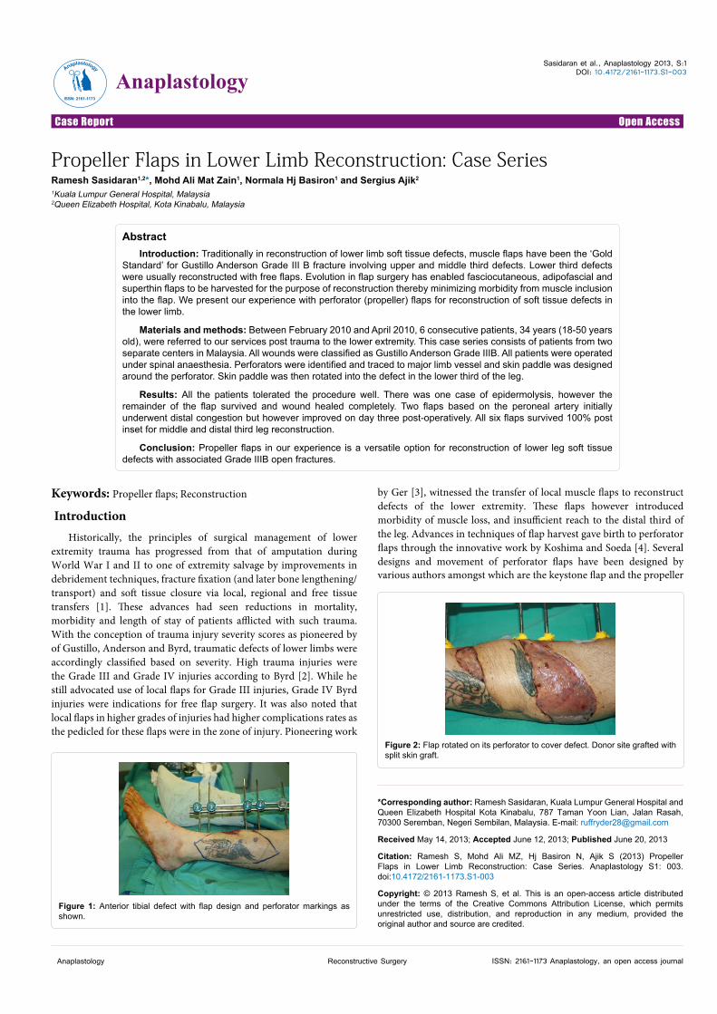
Figure 2: Flap rotated on its perforator to cover defect. Donor site grafted with split skin graft.
AnaplastologyAnaplastology
ISSN: 2161-1173
Citation: Ramesh S, Mohd Ali MZ, Hj Basiron N, Ajik S (2013) Propeller Flaps in Lower Limb Reconstruction: Case Series. Anaplastology S1: 003. doi:10.4172/2161-1173.S1-003
Page 2 of 4
ISSN: 2161-1173 Anaplastology, an open access journal Reconstructive SurgeryAnaplastology
flap. We present a series of cases of lower extremity reconstruction with propeller perforator flaps.
Patients and MethodsBetween February 2010 and December 2012, 6 consecutive
patients, were treated. Average age of patients was 34 years (18-50 years old). Patients were referred to our services post trauma to the lower extremity. All wounds were classified as Gustillo Anderson Grade III B. Perforators were marked and planned preoperatively with an 8 Mhz hand held Doppler device. The proximal limb of the flap is equal to the distal limb of the flap and the wound defect size with the perforator as a pivot point. Perforator vessels were identified by its unidirectional pulsatile flow [5].
Surgical TechniqueAll patients were operated under spinal anaesthesia. For both post
tibial artery and peroneal perforator flaps, the fasciocutaneous flaps were raised from lateral. On confirming the presence of preoperatively
marked perforators, the most suitable perforator with relation to size and closeness to defect was chosen. The medial skin flap was elevated once the perforators of the posterior tibial or peroneal were identified. We did not predetermine the most suitable perforator by clamping [5]. Suitable peforators were chosen based on proximity to the wound edge and size alone. Once identified, the perforators were dissected down to the axial vessels. Once the dissection was completed without inadvertent trauma to the perforators, the proximal and distal skin flaps were now severed. This technique of elevation has been found to be safe in case there is inadvertent trauma to the perforator the flap can simply be replaced and another option considered. The flap is now rotated on its perforator to varying degrees and inset into the defect. Donor site was skin grafted as seen in (Figures 1-12). Perforators to the overlying skin were not found in one patient in the zone of perfusion of the posterior tibial artery and hence we decided to perform a propeller design soleus muscle flap based on its muscular perforator (Figures 7-9).
ResultsSix patients with defects on the leg were operated with this method
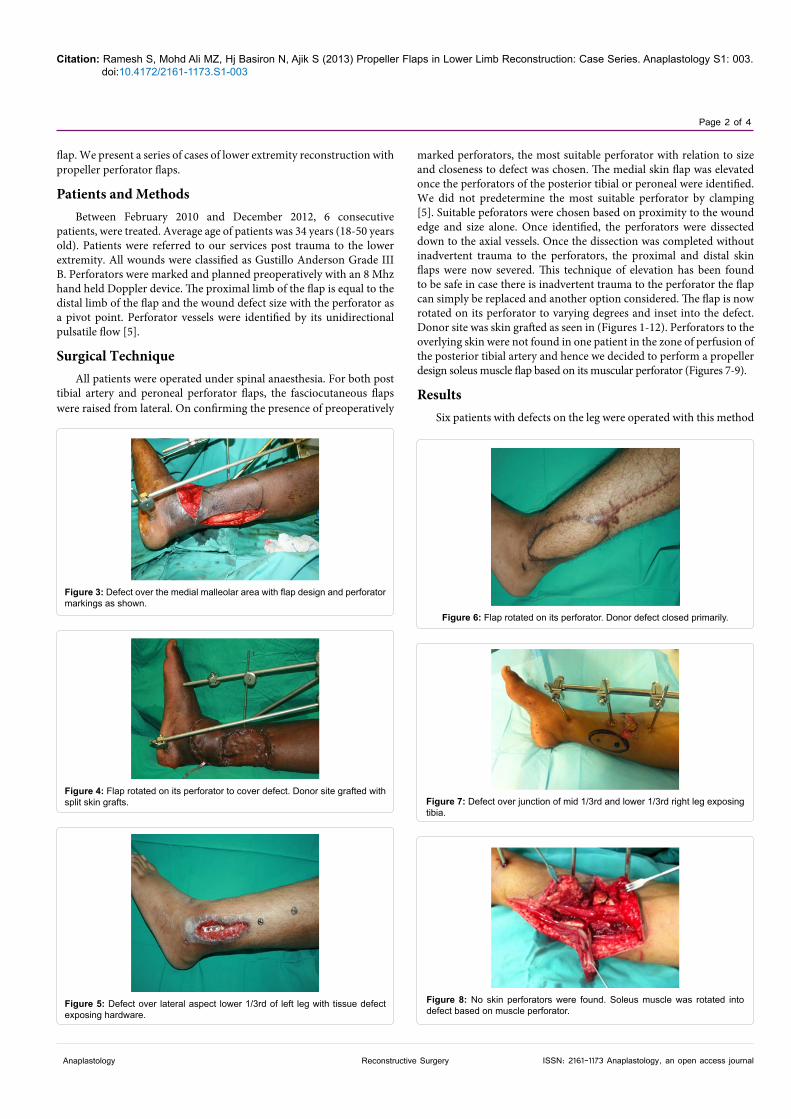
Figure 3: Defect over the medial malleolar area with flap design and perforator markings as shown.
Figure 4: Flap rotated on its perforator to cover defect. Donor site grafted with split skin grafts.
Figure 5: Defect over lateral aspect lower 1/3rd of left leg with tissue defect exposing hardware.
Figure 6: Flap rotated on its perforator. Donor defect closed primarily.
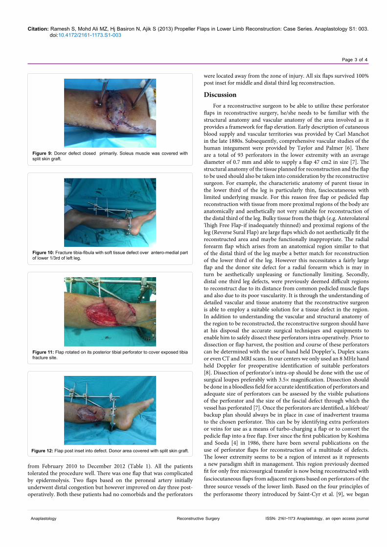
Figure 7: Defect over junction of mid 1/3rd and lower 1/3rd right leg exposing tibia.
Figure 8: No skin perforators were found. Soleus muscle was rotated into defect based on muscle perforator.
Citation: Ramesh S, Mohd Ali MZ, Hj Basiron N, Ajik S (2013) Propeller Flaps in Lower Limb Reconstruction: Case Series. Anaplastology S1: 003. doi:10.4172/2161-1173.S1-003
Page 3 of 4
ISSN: 2161-1173 Anaplastology, an open access journal Reconstructive SurgeryAnaplastology
from February 2010 to December 2012 (Table 1). All the patients tolerated the procedure well. There was one flap that was complicated by epidermolysis. Two flaps based on the peroneal artery initially underwent distal congestion but however improved on day three post-operatively. Both these patients had no comorbids and the perforators
Figure 9: Donor defect closed primarily. Soleus muscle was covered with split skin graft.
Figure 10: Fracture tibia-fibula with soft tissue defect over antero-medial part of lower 1/3rd of left leg.
Figure 11: Flap rotated on its posterior tibial perforator to cover exposed tibia fracture site.
Figure 12: Flap post inset into defect. Donor area covered with split skin graft.
were located away from the zone of injury. All six flaps survived 100% post inset for middle and distal third leg reconstruction.
DiscussionFor a reconstructive surgeon to be able to utilize these perforator
flaps in reconstructive surgery, he/she needs to be familiar with the structural anatomy and vascular anatomy of the area involved as it provides a framework for flap elevation. Early description of cutaneous blood supply and vascular territories was provided by Carl Manchot in the late 1880s. Subsequently, comprehensive vascular studies of the human integument were provided by Taylor and Palmer [6]. There are a total of 93 perforators in the lower extremity with an average diameter of 0.7 mm and able to supply a flap 47 cm2 in size [7]. The structural anatomy of the tissue planned for reconstruction and the flap to be used should also be taken into consideration by the reconstructive surgeon. For example, the characteristic anatomy of parent tissue in the lower third of the leg is particularly thin, fasciocutaneous with limited underlying muscle. For this reason free flap or pedicled flap reconstruction with tissue from more proximal regions of the body are anatomically and aesthetically not very suitable for reconstruction of the distal third of the leg. Bulky tissue from the thigh (e.g. Anterolateral Thigh Free Flap-if inadequately thinned) and proximal regions of the leg (Reverse Sural Flap) are large flaps which do not aesthetically fit the reconstructed area and maybe functionally inappropriate. The radial forearm flap which arises from an anatomical region similar to that of the distal third of the leg maybe a better match for reconstruction of the lower third of the leg. However this necessitates a fairly large flap and the donor site defect for a radial forearm which is may in turn be aesthetically unpleasing or functionally limiting. Secondly, distal one third leg defects, were previously deemed difficult regions to reconstruct due to its distance from common pedicled muscle flaps and also due to its poor vascularity. It is through the understanding of detailed vascular and tissue anatomy that the reconstructive surgeon is able to employ a suitable solution for a tissue defect in the region. In addition to understanding the vascular and structural anatomy of the region to be reconstructed, the reconstructive surgeon should have at his disposal the accurate surgical techniques and equipments to enable him to safely dissect these perforators intra-operatively. Prior to dissection or flap harvest, the position and course of these perforators can be determined with the use of hand held Doppler’s, Duplex scans or even CT and MRI scans. In our centers we only used an 8 MHz hand held Doppler for preoperative identification of suitable perforators [8]. Dissection of perforator’s intra-op should be done with the use of surgical loupes preferably with 3.5× magnification. Dissection should be done in a bloodless field for accurate identification of perforators and adequate size of perforators can be assessed by the visible pulsations of the perforator and the size of the fascial defect through which the vessel has perforated [7]. Once the perforators are identified, a lifeboat/backup plan should always be in place in case of inadvertent trauma to the chosen perforator. This can be by identifying extra perforators or veins for use as a means of turbo-charging a flap or to convert the pedicle flap into a free flap. Ever since the first publication by Koshima and Soeda [4] in 1986, there have been several publications on the use of perforator flaps for reconstruction of a multitude of defects. The lower extremity seems to be a region of interest as it represents a new paradigm shift in management. This region previously deemed fit for only free microsurgical transfer is now being reconstructed with fasciocutaneous flaps from adjacent regions based on perforators of the three source vessels of the lower limb. Based on the four principles of the perforasome theory introduced by Saint-Cyr et al. [9], we began
Citation: Ramesh S, Mohd Ali MZ, Hj Basiron N, Ajik S (2013) Propeller Flaps in Lower Limb Reconstruction: Case Series. Anaplastology S1: 003. doi:10.4172/2161-1173.S1-003
Page 4 of 4
ISSN: 2161-1173 Anaplastology, an open access journal Reconstructive SurgeryAnaplastology
using pedicled perforator flaps for lower extremity reconstruction and found that our patients spent less time under general anaesthesia, less time in ward and had no complications. The reconstructed region was more aesthetically pleasing and the pedicled perforator flaps were a better reconstructive fit for the region. All our cases were post trauma involving middle and distal third of the lower extremity. We’ve had 100% success with propeller flaps for lower third reconstruction so far. We’ve also had 100% success in total with all our propeller flaps. Benefits of the use of perforator flaps are that 1) better aesthetic as the donor is from the adjacent area 2) donor site morbidity is minimized 3) Less time taken for flap harvest 4) Less blood loss comparative toother options 5) Patient only requires spinal anaesthesia as opposed togeneral anaesthesia in free flap surgery 6) Good option in non tertiarycare centers 7) Decreased hospital stay 8) does not require sophisticated monitoring devices post flap transfer. The only drawback is that theraising of these flaps requires a certain amount of skill which in timemost surgeons are able to master.
References
1. Saint-Cyr M, Schaverian MV, Rohrich RJ (2007) Perforator Flaps: History,
Controversies, Physiology, Anatomy and Use in Reconstruction (CME). Plast Reconstr Surg 123: 132e-45e.
2. Byrd HS, Spicer TE, Cierney G III (1985) Management of open tibial fractures.Plast Reconstr Surg 76: 719-730.
3. Ger R (1970) The management of open fracture of the tibia with skin loss. JTrauma 10: 112-121.
4. Koshima I, Soeda S (1989) Inferior epigastric artery skin flap without rectus abdominis muscle. Br J Plast Surg 42: 645-648.
5. Celik N, Wei FC (2003) Technical tips in perforator flap harvest. Clin Plast Surg 30:469-472.
6. Taylor GI , Palmer JH (1987) The vascular territories (angiosomes) of the body: Experimental study and clinical applications. Br J Plast Surg 40: 113-141.
7. Geddes Cr, Tang M, Yang D (2006) Anatomy of integument of the lowerextremity: Perforator flaps. Anatomy, technique and clinical applications. St Louis (MO): QMP: 541-578.
8. Dancey A, Blondeel P (2010) Technical tips for safe perforator vessel Dissection applicable to all perforator flaps. Clin Plastic Surg 37: 593-606.
9. Saint-Cyr M, Wong C, Schaverien M, Mojallal A, Rohrich RJ (2009) The perforasometheory: Vascular Anatomy and Clinical Implications. Plast Reconstr Surg 124: 1529-1544.
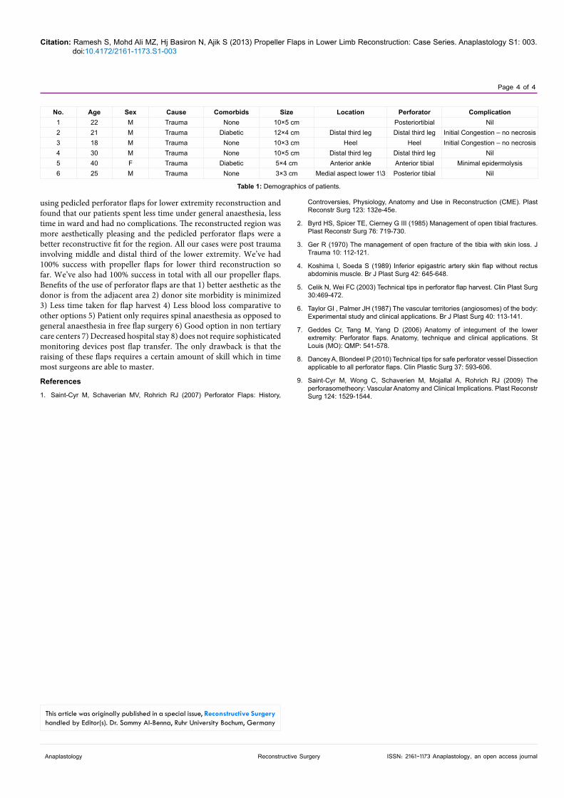
No. Age Sex Cause Comorbids Size Location Perforator Complication1 22 M Trauma None 10×5 cm Posteriortibial Nil2 21 M Trauma Diabetic 12×4 cm Distal third leg Distal third leg Initial Congestion – no necrosis3 18 M Trauma None 10×3 cm Heel Heel Initial Congestion – no necrosis4 30 M Trauma None 10×5 cm Distal third leg Distal third leg Nil5 40 F Trauma Diabetic 5×4 cm Anterior ankle Anterior tibial Minimal epidermolysis6 25 M Trauma None 3×3 cm Medial aspect lower 1\3 Posterior tibial Nil
Table 1: Demographics of patients.
This article was originally published in a special issue, Reconstructive Surgery handled by Editor(s). Dr. Sammy Al-Benna, Ruhr University Bochum, Germany