Embed Size (px)
Citation preview

CHAPTER I
INTRODUCTION
Pregnancy usually lasts 40 weeks or 280 days counted from the first day of last
menstrual period. Pregnancy at term is between 38-42 weeks of gestation and this is the
normal delivery period. However, approximately, 3.4-14% or an average of 10% of
pregnancies lasted until 42 weeks or more. Post term pregnancies especially affect the fetus.
In fact, postterm pregnancy have an influence on fetal development until the death of the
fetus. There is a fetus in gestation 42 weeks or more body weight increased steadily, there is
not increased, there are born weighing less than it should, or die in utero due to lack of
nutrients and oxygen. Postterm pregnancy has a close relationship with mortality, perinatal
morbidity, or macrosomia. 1
However, the actual biologic variation is likely less since the most frequent cause of a
postterm pregnancy diagnosis is inaccurate dating. Risk factors for actual postterm pregnancy
include primiparity, prior postterm pregnancy, male gender of the fetus, and genetic factors.
Perinatal mortality (defined as stillbirths plus early neonatal deaths) at 42 weeks of
gestation is twice that at 40 weeks (4-7 vs 2-3 per 1,000 deliveries, respectively) and
increases 4-fold at 43 weeks and 5- to 7-fold at 44 weeks. These data also demonstrate that,
when calculated per 1000 ongoing pregnancies, fetal and neonatal mortality rates increase
sharply after 40 weeks.
Cotzias et al calculated the risk of stillbirth in ongoing pregnancies for each
gestational age from 35-43 weeks. The risk of stillbirth was 1 in 926 ongoing pregnancies at
40 weeks gestation, 1 in 826 at 41 weeks, 1 in 769 at 42 weeks, and 1 in 633 at 43 weeks.
Uteroplacental insufficiency, asphyxia (with and without meconium), intrauterine infection,
and anencephaly all contribute to excess perinatal deaths, although postterm anencephaly is
essentially nonexistent with modern obstetrical care.
The maternal risks of postterm pregnancy are often underappreciated. These include
an increase in labor dystocia (9-12% vs 2-7% at term), an increase in severe perineal injury
(3rd and 4th degree perineal lacerations) related to macrosomia (3.3% vs 2.6% at term) and
operative vaginal delivery, and a doubling in the rate of cesarean delivery (14% vs 7% at
term). 2
1

CHAPTER II
LITERATURE VIEW OF POST TERM PREGNANCY
II.1 DEFINITION
The international definition of prolonged or post term pregnancy, endorsed by the
American College of Obstetricians and Gynecologists (2004), is 42 completed weeks (294
days) or more from the first day of the last menstrual period. It is important to emphasize the
phrase "42 completed weeks." 1
II.2 INCIDENCE
From their review, Divon and Feldman-Leidner (2008) report that the incidence of
postterm pregnancy ranges from 4 to 19 percent. Using criteria that likely overestimate the
incidence, approximately 6 percent of 4 million infants born in the United States during 2006
were estimated to have been delivered at 42 weeks or more (Martin and colleagues, 2009).
The trend toward fewer births at 42 weeks suggests earlier intervention. Specifically, in 2000,
7.2 percent of births in this country were 42 weeks or beyond, compared with 5.6 percent in
2006.
There are contradictory findings concerning the significance of maternal demographic
factors such as parity, prior postterm birth, socioeconomic class, and age. Olesen and
colleagues (2006) analyzed a variety of risk factors in 3392 participants in the 1998 to
2001.Danish Birth Cohort. They reported that only prepregnancy body mass index (BMI) 25
and nulliparity were significantly associated with prolonged pregnancy. Denison (2008) and
Caughey (2009) and their co-workers also reported similar associations.
The tendency for some mothers to have repeated postterm births suggests that some
prolonged pregnancies are biologically determined. In 27,677 births in Norway, Bakketeig
and Bergsjø (1991) reported that the incidence of a subsequent post term birth increased from
10 to 27 percent if the first birth was postterm. This was increased to 39 percent if there had
been two previous, successive postterm deliveries. Similar results were reported from
Missouri by Kistka and colleagues (2007). And Mogren and colleagues (1999) reported that
prolonged pregnancy recurred across generations in Swedish women. When mother and
2

daughter had a prolonged pregnancy, the risk for the daughter to have a subsequent post term
pregnancy was increased two- to threefold. In another Swedish study, Laursen and associates
(2004) found that maternal, but not paternal, genes influenced prolonged pregnancy. Rare
fetal–placental factors that have been reported as predisposing to postterm pregnancy include
anencephaly, adrenal hypoplasia, and X-linked placental sulfatase deficiency (MacDonald
and Siiteri, 1965; Naeye, 1978; Rabe and colleagues, 1983).
Boyd et al (1988) found an incidence of post term pregnancy of 7.5% when the
diagnosis was based on the menstrual history, 2.6 % when the diagnosis was based on early
ultrasound examination, and 1.1% when the diagnosis was based in concurrent menstrual
history and ultrasound examination.3
II.3 ETIOLOGY
The most common cause of a prolonged pregnancy is an error in the clinical
estimation of the gestational age. Other cause are unknown and are probably associated with
abnormalities in the biochemical and physiological mechanism responsible for initiation of
labor.
One example is the prolongation of pregnancy associated with placental sulfatase
deficiency. This enzyme plays a critical role in in the synthesis of placental esterogens
that are necessary for the development of gap junctions and increased expressions of
oxytocin and prostaglandin reseptors in myometrial cells.
A second example is the prolongation of pregnancy associated with anencephaly. The
lack of development of the fetal hypothalamus negates the production of
corticotropin-relasing hormone and the stimulation of the pituitary-adrenal-placental
axis necessary for the initiation of partutrition.
Third example is caused by decrease in the pregnancy hormone progesterone is
believed that important events endocrine changes in spurring the process of
biomolecular on labor and increases uterine sensitivity to oxytocin, so some authors
suspect that the occurrence of postterm pregnancy is still ongoing due to the influence
of progesterone.
Fourth example oxytocin is physiologically important role in inducing labor and
oxytocin release from neurohipofisis pregnant women who are less advanced in the
pregnancy as one of the factors thought to cause postterm pregnancy.
3

Fifth example is pressure on the cervical ganglion of the plexus Frankenhauser will
evoke uterine contractions. In circumstances where there is no pressure on the plexus,
such as the location of abnormalities, short cord, and the bottom is still high.
Sixth example is hereditary factor. Some authors claim that a mother who experienced
postterm pregnancy have a tendency to give birth through the month in subsequent
pregnancies. Mogren as quoted by Cunningham, states that when a mother
experiencing postterm pregnancy when a girl, then most likely his daughter will
experience a postterm pregnancy.
II.4 DIAGNOSIS
Menstrual History
Some criteria for the diagnosis of postterm pregnancy:
the patient must be convinced by her HPHT
28-day cycle and regular
Not on the pill contracption at least the last 3 months
Further diagnosis is determined by calculating according to formula Naegele. Based
on menstrual history, a person designated as postterm pregnancy possibilities are:
No errors determine the last period and it lasts through the month of pregnancy.
Errors in determining the date of last menstrual period or due to abnormal
menstruation.
Date of last period clearly known, but a delay ovulation.
Antenatal history:
Pregnancy can be expressed as postterm pregnancies obtained when 3 or more of the four
criteria of examination results as follows:
36 weeks have passed since a positive pregnancy test
32 weeks have passed since the first audible fetal heart rate with Doppler
24 weeks have passed since the first fetal movement felt
22 weeks have passed since hearing the fetal heartbeat with a stethoscope Laennec
first.1
4

Ultrasonography examination
Ultrasonographic dating early in pregnancy can improve the reliability of the EDD
(estimated due date). However, it is necessary to understand the margin of error reported
at various times during each trimester. A calculated gestational age by composite
biometry from a sonogram must be considered an estimate and must take into account the
range of possibilities. Measurement of the crownrump length (CRL) at early pregnancy
ultrasound has been shown to give a more accurate estimate of gestational age and so
decrease the incidence of prolonged pregnancy. However, ultrasound has a degree of
error: 7 days up to 20 weeks’ gestation, 14 days between 20 and 30 weeks and 21 days
beyond 30 weeks. It is for these reasons that the National Institute of Clinical Excellence
(NICE) recommends a dating ultrasound examination between 10 and 13 weeks to
estimate the gestation of a pregnancy.5.7
In addition to the CRL, biparietal diameter and femur length, some parameters in
ultrasound examination can also be used such as abdominal circumference, head
circumference, and some formulas that are some of the results of the calculation of the
parameters mentioned above. In contrast, examination shortly after the third trimester can
be used to determine fetal weight, amniotic fluid state, or any state of the placenta is
frequently associated with postterm pregnancy, but it's hard to make sure the age of
pregnancy.
Gestational Age for CRL
Age CRL (cm)
6.1 Weeks: 0.4 cm
7.2 Weeks: 1.0 cm
8.0 Weeks: 1.6 cm
9.2 Weeks: 2.5 cm
9.9 Weeks: 3.0 cm
10.9 Weeks: 4.0 cm
12.1 Weeks: 5.5 cm
13.2 Weeks: 7.0 cm
14.0 Weeks: 8.0 cm
The following formula is an approximation:
Gestational age [weeks of pregnancy] = crown-rump length (cm) + 6.5 4.8
5

Laboratory examination
Levels of lecithin / spingomielin.
When L / P in the amniotic fluid levels are the same, then about 22-28 weeks
gestational age, L = 1.2 P: 28-32 weeks, pregnancy at term on the L / P = 2. This
check can not be used to determine the postterm pregnancy, but only used to
determine whether the fetus is old enough / mature for birth-related action to
prevent errors in termination of pregnancy.
Thromboplastin activity of amniotic fluid (ATCA).
Hatswell successfully mebuktikan that amniotic fluid accelerates blood clotting
time. This activity increases with gestational age 41-42 weeks ATCA range 45-65
seconds, at the age of more than 42 weeks gestation ATCA obtained less than 45
seconds. When obtained ATCA between 42-46 seconds indicates that pregnancy
lasts through time.
Amniotic fluid cytology
Painting with nile blue sulphate can see the fat cells in the amniotic fluid. When
the number of cells containing fat exceeds 10%, then an estimated 36 weeks
gestation and if 50% or more, then the age of 39 weeks' gestation or more.1
II.5 CHANGES ASSOCIATED IN POSTERM PREGNANCY
Placental Changes
The post term placenta shows decrease in diameter and length of the chorionic villi,
fibrinoid necrosis, and accelerated atherosis of the chorionic and decidual vessels. This
changes occur simultaneously with or precede of the hemorragic infracts, which are foci
for calcium deposition and formation of white infracts. Infracts are present in 10-25% of
term and 60-80% of post-term placentas. They are more common at the placental borders.
Deposition of calcium in the post-term placenta reaches up to 10 g of dry tissue weight,
whereas it is only 2-3 g per 100 g in placentas term.
The morphologic changes that occur with placental senescence can be observed by
ultrasound and were originally described by Grannum et al (1979).
There are several grade of placenta:
During the first part of gestation the ultrasonic appearance of the placenta is
homogenous, without echogenic densities, and limited by a smooth chorionic plate
(grade 0 placenta).
6

With proggresion of pregnancy the chorionic plate begins acquire subtle undulation,
and echogenic densities appear randomly dispersed throughout the organ but sparing
its basal layer (grade I placenta).
Near term the indentations in the chorionic plate become more marked, echogenic
densities appear in the basal layer, and commalike densities seem to extend from
chorionic plate into the substance of the placenta (grade II).
Finally, when the pregnancy is at term or post-term the identation in the chorionic
plate become more marked, giving the appearance of cotyledons. This impression is
reinforced by increased of confluency of the comma-like densities that become the
intercotyledonary septations. Also, characteristically, the central portion of the
cotyledons become echo-free (fallour areas), and large irregular densities, capable of
casting acoustic shadows, appear in the substance of the placenta (grade III placenta).4
Amniotic Fluid Changes
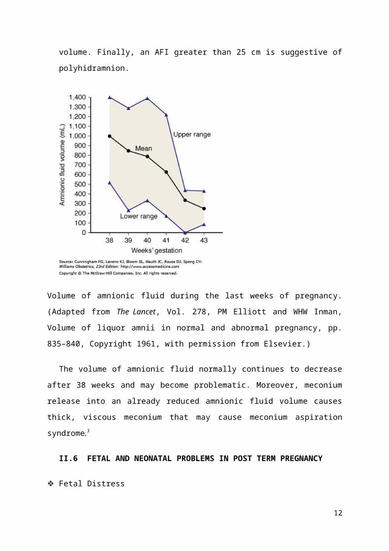
There are quantitative and qualitative changes in the amniotic fluid with prolongation
of pregnancy. The amniotic fluid volume reaches a peak of about 1000 ml at 38 weeks of
gestation and decreases to about 800 ml at 40 weeks. This reduction in volume continous
and the amount of fluid is approximately 480, 250, and 160ml at 42, 43, and 44 weeks,
respectively. An amniotic fluid volume under 400 ml at 40 or more weeks is associated
with fetal complication. The cause of oligohidramnios in prolonged pregnancy seems to
be dismished fetal urine production.
The four-quadrant technique (Phelan et al, 1987) is the most popular method to
evaluate amniotic fluid volume. The four quadrant technique consist of measuring the
vertical diameter of the largest pocket of fluid found in each of the four quadrants of the
uterus. The sum of the results is the amniotic fluid index (AFI). An AFI less than 5cm
indicates oligohidramnios. An AFI between 5 and 10 cm indicates a decreased fluid
volume. An AFI between 10-15 cm is normal. An AFI between 15 and 20 cm indicates
increased fluis volume. Finally, an AFI greater than 25 cm is suggestive of
polyhidramnion.
7

Volume of amnionic fluid during the last weeks of pregnancy. (Adapted from The Lancet,
Vol. 278, PM Elliott and WHW Inman, Volume of liquor amnii in normal and abnormal
pregnancy, pp. 835–840, Copyright 1961, with permission from Elsevier.)
The volume of amnionic fluid normally continues to decrease after 38 weeks and may
become problematic. Moreover, meconium release into an already reduced amnionic fluid
volume causes thick, viscous meconium that may cause meconium aspiration syndrome.3
II.6 FETAL AND NEONATAL PROBLEMS IN POST TERM PREGNANCY
Fetal Distress
The principal reasons for increased risks for postterm fetuses were described by Leveno
and associates (1984). They reported that both antepartum fetal jeopardy and intrapartum
fetal distress were the consequence of cord compression associated with oligohydramnios. In
their analysis of 727 postterm pregnancies, intrapartum fetal distress detected with electronic
monitoring was not associated with late decelerations characteristic of uteroplacental
insufficiency.
Instead, one or more prolonged decelerations such as shown in Figure 37-5 preceded three
fourths of emergency cesarean deliveries for nonreassuring fetal heart rate tracings.
8

In all but two cases, there were also variable decelerations (Fig. 37-6). Another common fetal
heart rate pattern, although not ominous by itself, was the saltatory baseline shown in Figure
37-7.
Saltatory baseline fetal heart rate showing oscillations exceeding 20 bpm and associated with
oligohydramnios in a postterm pregnancy. (This figure was published in American Journal of
Obstetrics & Gynecology, Vol. 150, No. 5, pt. 1, KJ Leveno, JG Quirk, Jr., FG Cunningham,
et al., Prolonged pregnancy. I. Observations concerning the causes of fetal distress, pp. 465–
473, Copyright Elsevier 1984.)
9

These findings are consistent with cord occlusion as the proximate cause of the
nonreassuring tracings. Other correlates found were oligohydramnios and viscous meconium.
Schaffer and colleagues (2005) implicated a nuchal cord in abnormal intrapartum fetal heart
rate patterns, meconium, and compromised newborn condition in prolonged pregnancies.4
Postmaturity Sindrome
The postmature infant presents a unique and characteristic appearance such as shown in
Figure 37-3. Features include wrinkled, patchy, peeling skin; a long, thin body suggesting
wasting; and advanced maturity because the infant is open-eyed, unusually alert, and
appears old and worried. Skin wrinkling can be particularly prominent on the palms and
soles. The nails are typically long. Most such postmature infants are not technically
growth restricted because their birthweight seldom falls below the 10th percentile for
gestational age. On the other hand, severe growth restriction—which logically must have
preceded completion of 42 weeks—may be present.
Postmature infant delivered at 43 weeks' gestation. Thick, viscous meconium coated the
desquamating skin. Note the long, thin appearance and wrinkling of the hands.
(www.accessmedicine.com) 3
10

Based on the degree of placental insufficiency occurs, the sign postmaturitas can be
divided into three stages, namely:
Stage I skin showed loss of vernix caseosa and maceration of the skin is dry, brittle, and
easy to peel.
Stage II of the above symptoms with meconium staining of the skin
Stage III accompanied by yellowish staining of the nails, skin, and umbilical cord.1
Fetal Weight
If there is a large anatomic changes in the placenta, then decreased fetal weight. From
research it appears that Vourherr after 36 weeks gestation srafik average fetal growth leveled
off and looked a decrease after 42 weeks. However, often also the placenta was still able to
function properly so that the weight of the fetus continues to grow in accordance with
increasing gestational age. Zwerdling said that the average fetal weight of more than 3600
grams at 44.5% in postterm pregnancies, while in even-numbered month of pregnancy by
30.6%. The risk of birth to a baby weighing more than 4000 grams at postterm pregnancies
increased by 2-4 times greater than at term pregnancy.(sarwono)
Divon and associates (1998) and Clausson and co-workers (1999) analyzed births
between 1991 and 1995 in the National Swedish Medical Birth Registry. Stillbirths were
more common among growth-restricted infants who were delivered after 42 weeks. Indeed, a
third of postterm stillborn infants were growth restricted.3
11
Mean daily fetal growth during previous week of gestation. (From Jazayeri and co-workers,
1998, with permission.)
Meconium aspiration
Beyond term, the fetus is more likely to have a bowel movement, called meconium,
into the amniotic fluid. If the fetus is stressed, there is a chance it will inhale some of this
meconium stained amniotic fluid, this can cause breathing problems when the baby is
born. The problems occurs more frequently when thick meconium, fetal tachychardia and
absence of FHR accelerations are present.
The further the pregnancy progresses beyond 40 weeks, the more likely it is
that significant amounts of meconium will be present. This is due to increased
uteroplacental insufficiency, which leads to hypoxia in labor and activation of the
vagal system. In addition, the presence of a smaller amount of amniotic fluid
increases the relative concentration of meconium in utero.2.7
II.7 MATERNAL COMPLICATION
The maternal risks due to a prolonged pregnancy are commonly under-
appreciated. Prolonged pregnancy is associated with risks to the mother during labour
12

and delivery whether labour is induced or occurs spontaneously. It leads to anxiety in
the mother due to a perception of danger to her baby. Fetal macrosomia can lead to a
significant increase in prolonged labour, perineal, vaginal and cervical trauma, and
postpartum haemorrhage. There is an increase in the rate of deliveries by caesarean
section which is associated with potential complications such as haemorrhage,
infection and thromboembolism.
Maternal complications of prolonged pregnancy
Macrosomic fetus
Cephalopelvic disproportion
Labour dystocia
Perineal, vaginal and cervical trauma
Delivery by caesarean section
Postpartum haemorrhage
Chorioamnionitis
Anxiety5
II.8 IDENTIFICATION OF PATIENTS WHO NEED TO BE DELIVERED
High risk pregnancies
Patients with high risk pregnancies, especially those with diabetes and
hypertension need to be delivered withoud consideration to the favorability of their
cervix. Expectant management in these cases is not adequate because prolongation of
pregnancy will place their fetuses at additional risk.
Women with favorable cervices
Multiple studies have shown that the risk of caesarean following induction of
labor is directly associated with the status of the cervix. These studies have also
shown that women wit favorable cervices are at low risk for abdominal delivery. Fot
this reason, the majority of investigators are in favor of induction and delivery of
women with favorable cervices who have reached or urpased their EDD.
The classical method for evaluation the cervix is the Bishop score.
13

Score 0 1 2 3
Cervical
dilatation
Closed 1-2 3-4 >= 5
Cervical
effacement (%)
0-30 40-50 60-70 >=80
Fetal Station -3 -2 -1 or 0 +1 or +2
Cervical
consistency
Firm Medium Soft Soft
Cervical
position
Posterior Mid Anterior Anterior
From Bishop EH. Pelvic scoring for elective induction Obstet Gynecology
1964;24;266
Bishop modification by dr Gulardi H Winjosastro SpOG.6
Score 0 1 2
Cervical position Posterior Axial Anterior
Cervical dilatation Closed 1-2 cm >3cm
Cervical consistency Firm Soft Soft
Cervical thickness 3cm 2cm 1cm
Head position - Hodge I-II Hodge II-III
A Bishop score >= 8 is a good index of inducibility, score 6 or more is a favorable cervix to
attempt induction, and score less than 4 is indication to ripening the cervix.
Decreased Amniotic fluid volume
14

The evaluation of amniotic fluid volume is of fundamental importance in prolonged
pregnancies. Chamberlain et al (1984) demonstrated that perinatal mortality increases
dramatically with progressive severity of oligohidramnion. Loveno et al (1984)
demostrated that umbilical cord compression secondary to oligohydramnios is the
most common cause of intrapartum fetal distress in these patients. For this reasons,
women with oligohidramnios need to be delivered.2
Comparison of the prognostic value of various sonographic estimates of amnionic
fluid volume in prolonged pregnancies. Abnormal outcomes include cesarean or
operative vaginal delivery for fetal jeopardy, 5-minute Apgar score of 6 or less,
umbilical arterial blood pH less than 7.1, or admission to the neonatal intensive care
unit. (Adapted from Fischer RL, McDonnell M, Bianculli KW, et al: Amniotic fluid
volume estimation in the postdate pregnancy: A comparison of techniques, Obstetrics
& Gynecology, 1993, vol. 81, no. 5, part 1, pp. 698–704, with permission.)
Regardless of the criteria used to diagnose oligohydramnios in postterm
pregnancies, most investigators have found an increased incidence of "fetal distress"
15

during labor. Clement and co-workers (1987) described six postterm pregnancies in
which amnionic fluid volume diminished abruptly over 24 hours—in one of these, the
fetus died.
Macrosomic fetuses
The velocity of fetal weight gain peaks at approximately 37 weeks. Although
growth velocity slows at that time, most fetuses continue to gain weight. For example,
the percentage of fetuses born in 2006 whose birthweight exceeded 4000 g was 8.5
percent at 37 to 41 weeks and increased to 11.2 percent at 42 weeks or more (Martin
and colleagues, 2009). Intuitively at least, it seems that both maternal and fetal
morbidity associated with macrosomia would be mitigated with timely induction to
preempt further growth. This does not appear to be the case, however, and the
American College of Obstetricians and Gynecologists (2000) has concluded that
current evidence does not support such a practice in women at term with suspected
fetal macrosomia.
The importance of the prenatal estimation of fetal weight in women with
prolonged pregnancis is to determine the approach to delivery. Pasient with estimated
fetal weight more of 4500 grams or more should be causeled to have caesarean
delivery because the possibility of traumatic vaginal delivery is substantial. Caesarean
section should be offered also to women who have previously delivered infants with
similiar or larger birth weight, because prior delivery does not guarantee an easy
delivery of another large baby.3
Fetal growth restriction
A fetal growth abnormality associated with prolonged pregnancy is poor fetal
growth or dysmaturity. Approximately 5-10% of fetuses delivered after their EDD
show wasting of their subcutaneus fat characteristic of intrauterine malnutrition and
are classified as small for gestasional age by neonatal evaluation. Frequently this
fetuses exhibit abnormal FHR patterns before delivery or in the course of labor. The
amount of amniotic fluid is reduced in most of these cases and meconium aspiration is
a common problem. Fetal manutrition is associated with multiple problems during
16

immediate neonatal period including hypoglicemia, hypocalemia, and hyperviscosity
syndrome. 4
II.9 POST TERM PREGNANCY TREATMENT
Antenatal fetal monitoring
In most cases, a healthcare provider will recommend tests on the fetus if the
pregnancy extends beyond the due date. These tests give information about the health
of the fetus and about the risks or benefits of allowing the pregnancy to continue. The
American College of Obstetricians and Gynecologists has stated that it is only
necessary to start antenatal fetal monitoring after 42 weeks (294 days) of gestation,
although many obstetric care providers will start fetal testing at 41 weeks. Many
experts recommend twice weekly testing, including a measurement of amniotic fluid
volume. Testing may include observing the fetus' heart rate using a fetal monitor
(called a nonstress test) or observing the baby's activity with ultrasound (called a
biophysical profile).
Nonstress testing
Nonstress testing is done by monitoring the baby's heart rate with a small
device that is placed on the mother's abdomen. The device uses sound waves
(ultrasound) to measure the baby's heart rate over time, usually for 20 to 30 minutes.
Normally, the baby's baseline heart rate should be between 110 and 160 beats per
minute and should increase above its baseline by at least 15 beats per minute for 15
seconds when the baby moves. The test is considered reassuring (called "reactive") if
two or more fetal heart rate increases are seen within a 20 minute period. Further
testing may be needed if these increases are not observed after monitoring for 40
minutes.
Biophysical profile
A biophysical profile (BPP) score is calculated to assess the fetus' health. It
consists of five components, nonstress testing and ultrasound measurement of four
fetal parameters: fetal body movements, breathing movements, fetal tone (flexion and
extension of an arm, leg, or the spine), and amniotic fluid volume. Each component is
17

scored individually, 2 points if normal and 0 points if not normal. The maximum
possible score is 10. Amniotic fluid volume is an important variable in the BPP
because a low volume (called oligohydramnios) may increase the risk of umbilical
cord compression and may be a sign of changes in the feto-uteroplacental circulation.
Amniotic fluid level can become reduced within a short time period, even a few days.
Contraction stress test
A contraction stress test (CST) can also be done to assess fetal health. It
involves giving an intravenous medication (oxytocin) to the mother to induce uterine
contractions. The fetus' heart rate is monitored in response to the contractions. A fetus
whose heart rate slows down during a CST may require a cesarean delivery.7
Inducing of labor
Once the decision to deliver a patient has been made, the management of the
labor induction depends on the clinical setting, and a brief review of cervical ripening
agents and potential complications of induction of labor is appropriate. As many as
80% of patients who reach 42 weeks' gestation have an unfavorable cervical
examination (ie, Bishop Score < 7). Many options are available for cervical ripening.
The different preparations, indications, contraindications, and multiple dosing regimes
of each require practitioners to familiarize themselves with several of the
preparations.
Currently available chemical preparations include prostaglandin E1 tablets for
oral or vaginal use (misoprostol), prostaglandin E2 gel for intracervical application
(dinoprostone cervical [Prepidil]), and a prostaglandin E2 vaginal insert (dinoprostone
[Cervidil]). Cervidil contains 10 mg of dinoprostone and has a lower constant release
of medication than Prepidil. In addition, this vaginal insert device allows for easier
removal in the event of uterine hyperstimulation.
Many studies have compared the efficacy and risks of various prostaglandin
cervical ripening agents. Rozenburg et al performed a randomized trial comparing
intravaginal misoprostol and dinoprostone vaginal insert in pregnancies at high risk of
18

fetal distress. They found that both methods were equally safe for the induction of
labor and misoprostol was actually more effective.
Another method for ripening the cervix is by mechanical dilation. These
devices may act by a combination of mechanical forces and by causing release of
endogenous prostaglandins. Foley balloon catheters placed in the cervix, extra-
amniotic saline infusions, and laminaria have all been studied and have been shown to
be effective.
Regardless of what method is chosen for cervical ripening, the practitioner
must be aware of the potential hazards surrounding the use of these agents in the
patient with a scarred uterus. In addition, the potential for uterine tachysystole and
subsequent fetal distress requires that care be taken to avoid using too high a dose or
too short a dosing interval in an attempt to get a patient delivered rapidly. Care should
also be taken when using combinations of mechanical and pharmacologic methods of
cervical ripening.
Finally, intrapartum fetal surveillance in an attempt to document fetal
intolerance to labor before it leads to acidosis is critical. Whether continuous fetal
monitoring or intermittent auscultation is used, interpretation of the results by a well-
trained clinician is of paramount importance. If the fetal heart rate tracing is
equivocal, fetal scalp stimulation and/or fetal scalp blood sampling may provide the
reassurance necessary to justify continuing the induction of labor. If the practitioner
cannot find reassurance that the fetus is tolerating labor, cesarean delivery is
recommended. 4
Antepartum management of post term pregnancy.
19

Source: Arias F, Daftary S, et al. Practical Guide to high risk pregnancy and delivery.
Chapter 11: Prolonged pregnancy. Third edition. 2010. Elsevier: India. Page 286.
CHAPTER III
CASE ILUSTRATION
I. IDENTITY
PATIENT
Name : Mrs. IP
Age : 29 yrs
Religion : Islam
Tribe : Betawi
Education : High school
Occupation : Housewife
Address : Depok
20

Sign in hospital: Friday, January 6th, 2012, at 11.45 a.m
II.ANAMNESIS
Autoanamnesis dated, January 6th, 2012, at 11.45 a.m
A. Chief complaint
referenced from a midwife because of reduced amniotic fluid
B. History of Present Illness
Patient admit that she has 10 months pregnancy, first day of last menstrual period : February
28th 2011 ~ 44 weeks. Estimated day of delivery : December 5 th 2011. Patient complain
referenced from a midwife because of reduced amniotic fluid. ANC routinely at Puskesmas,
USG 2 times with first on 18thJuly 2011, the result was 20 weeks pregnancy. The last USG
was on 2 January 2012 and the results was the baby in good condition, head presentation,
reduced amniotic fluid and postmature baby in pregnancy. The patient said there was a little
vaginal discharge 1 hours before admitted with no bloody show . Backacghe about a week
befor admissions and it become more frequently. History of fever and hypertension during
pregnant were denied. Defecate and mixture are no complaints. Patient hasn’t a hole tooth.
Traumatical history, headache, nausea, vomit, and blur vision was denied. Fetal movement
still felt.
C. Menstrual History
Menarche at the age of 15 years, 28 days cycle, regular, duration 7 days, the number of ± 2-3
pads / day, menstrual pain (+)
First day of last menstrual period: February 28th 2011
Estimated day of delivery : December 5th 2011
D. Marital Status
Status married, marriage 2x, first 8 years of marriage and then divorced, second marriage for
3 years and still married untill now.
21

E. History of previous pregnancy
1. Normal delivery, ♀, 12 years old, 1700 gram, delivered by doctor
2. Normal delivery, ♀, 6 years old, 3200 gram, delivered by midwife
3. Normal delivery, ♂, 1.5 years old, 3200 gram, delivered by midwife, history with postterm
pregnancy43 weeks
4. Current pregnancy
F. History of present pregnancy
Early pregnancy : Nausea (+), vomits (-), bleeding (-), hypertension (-)
Later pregnancy : sweeling foot (-), hypertension (-), dyspnea (-).
ANC at Puskesmas monthly.
G. History of contraception
Injection contraception for every 3 months.The pasient stoped to use contraception, at least 5
months before pregnancy.
H. History of Systemic Disease
Heart disease (-), respiratory disease (-), hypertension (-), diabetes mellitus (-)
I. Surgery History
None
J. History of Family Disease
Heart disease (-), respiratory disease (-), hypertension (-), Diabetes Mellitus (-)
K. Habit and Psychosocial History
No smoking, drinking alcohol, drugs and drinking herbal medicine.
22

III. PHYSICAL EXAMINATION
A. General examination
General impression : moderate illness
Degree of consciousness : compos mentis
Vital signs : BP 120/80 mmHg, HR 80 x/m, RR 20x/', T 36.50C
Body weight before present pregnancy: ± 50 kg
Body weight during present pregnancy: 61 kg
Head : Normocephali, black hair, straight, uniform distribution
Eyes : Conjungtiva anemic -/-, sklera not icteric.
Mouth : Not dry, not Cyanosis.
Ears : Normotia, secretions serumen (-/-)
Nose : Normosepta, secretions (-/-).
Throat : Pharynx not hiperemis.
Neck : Enlarged glands (-).
Thorax
Cor : Regular I-II heart sound, murmurs (-), Gallop (-).
Pulmo : Vesicular breath sound, Rh (-/-), Wh (-/-).
Breast : Symmetric, hyperpigmentation on both the areola, retracted nipple (-), mass
(-)
Extremity : warm extremities, swelling -/-
B. Obstetrical Status
23

Abdomen:
Inspection : abdomen enlarged and distended, striae gravidarum (+).
Palpation:
- Leopold I : fungal height 30 cm, a hard, round, ballotable, and nodular body not
easy to move in palpation
- Leopold II :
Left : a small parts of the fetus is palpable
Right : a hard resistant and board like structure
- Leopold III : hard, round, ballotable, moveable, pandular like and nodular body
- Leopold IV : 5/5
- His : irreguler
- Fetal weight estimation(FWE) : 3410 g
Auscultation : 2 punctum maximum, fetal heart sound 142 ppm, regular.
Anogenital:
- Inspection : Vulva/urethra no sign of inflammation, bleeding (-), edema (-), varicose (-)
- Speculum examination : portio livid, ostium opened, fluor (-), fluxus (-)
- VT : firm portio,posterior, cervix dilatation 1 cm, thickness 3 cm, amniotic
membrane (+), head was palpated on H I-II
IV. SUPPORTIVE EXAMINATION
A. Laboratory (January 06th, 2011)
BLOOD Hb : 10 g/dl
24
Ht : 30 vol%
Leukocytes : 10.600 uL
Erythrocyte : 4.52 billions/ uL
Platelets : 292.000 uL
Type of blood : B / +
Spot glucose blood : 80 mg / dl
MCV : 77.0 fl
MCH : 28.0 pg
MCHC: 38.0 g/dl
RDW : 25 %
URINALYSIS
Color : yellow
Clarity : clear
pH : 7.0
Sediments : + 1 epithelial cells
Leukocytes : 6-8 / magnified
view field
Erythrocytes : 1-2 / magnified
view field
Cylinders : -
Crystal : -
Protein : -
Ketones : (-)
Blood : +3
Bilirubin : (-)
Urobilinogen : 1.0
Nitrite :(-)
B. US (January 06th, 2011)
25

Fetus : Alive, Single,
Head presentation
BPD: 9.38 cm
AC: 29.68 cm
FL: 6.96 cm
FWE: 3410 g
Plasenta : right corpus
AFI: 7.53 cm
Aterm
Placenta in right of uterine corpus, does not seem loops of the cord, does not seems major
congenital defect.
26

Assessment : appropriate with aterm pregnancy live, single, head presentation, reduced
amniotic fluid.
C. CTG (January 06th, 2012)
Baseline frequency 140 dpm
Variability 5-20 dpm
Acseleration (+)
Deceleration (-)
Fetus movement (+)
27

His (-)
Assesment : Reassuring
V. RESUME
Patient admit that she has 10 month pregnancy, first day of last menstrual period : February
28th 2011~ 44 weeks. Estimated day of delivery : December 5th 2012. Patient complain
referenced from a midwife because of reduced amniotic fluid. ANC routinely at Puskesmas,
USG 2 times with first on 18thJuly 2011, the result was 20 weeks pregnancy. The last USG
was on 2 January 2012 and the results was the baby in good condition, head presentation,
reduced amniotic fluid and postmature baby in pregnancy. The patient said there was a little
vaginal discharge 1 hours before admitted with no bloody show. Backacghe about a week
befor admissions and it become more frequently. Fetal movement still felt.
In generalist examination are normal.
In Obstetrical examination, we finded:
Abdomen enlarged and distended
Fungal height 30 cm
Irregular contraction
28

Head presentation
Fetal Weight Estimation: 3410g
On auscultation, there is 2 punctum maximum, fetal heart sound 140 ppm
In Anogenital examination we finded no inflammation sign, bleeding, edema,
varicose. Speculum examination : portio livid, ostium opened, fluor (-), fluxus (-);
VT : firm portio, posterior, cervix dilatation 1 cm, thickness 3 cm, amniotic
membrane (+), head was palpated on H I-II.
In USG, placenta in right of uterine corpus, does not seem loops of the cord, does not seems
major congenital defect; appropriate with aterm pregnancy, live, singleton, head presentation,
reduced amniotic fluid..
In CTG, fetus is reassuring
VI. DIAGNOSIS
Maternal :
G4P3 Pregnant 44 weeks, reduced amniotic fluid, immature cervix, not yet inpartu
Fetal : singleton live head presentation
VII. MANAGEMENT
Induction with folley catheter 1x24 hoursobserve progress of labour re-evaluate
after 24 hours
VII. PROGNOSIS
Mother: Dubia ad bonam
Fetus : Dubia ad bonam
29

VIII. Follow Up Result
January 07th 2012 22.00 p.m
S : FC loose spontaneus, fetal movement (+), water brooke (+)
O : General condition: good
Conciousness: compos mentis
BP 120/70 mmHg, HR 88x/’, RR 20x/’, T 36,5oC
The general examination: performed revealed stable ,
Obstetric st. : his 1-2 x/10’/20” , FHR : 142 dpm
Inspection: V/U calm
VT : firm porsio, axial, ø 4 cm, T =1 cm, amniotic membrane (-), the
head was palpated on H I-II
A : latent stage I in delivery G4P3H 44 weeks pregnancy, fetus with singleton live
head presentation
P : obs signs of inpartu, his, FHR/hour
Pervaginam delivery
Acceleration with oksitosin 5 IU/500cc RL start with 8 drops up then 4 drops/30’
untill his 3x/10’
January 07th, 2012 23.00 p.m.
S : contraction (+), fetus movement (+)
O : General condition: good
Conciousness: compos mentis
30
BP 110/70 mmHg, HR 84x/’, RR 20x/’, T 36,5oC
The general examination: other performed revealed stable
Obstetric st. : contraction (+) 3x/10’, FHR : 136 dpm
Inspection: V/U calm
VT : firm porsio, axial, ø 6 cm, T =1 cm, amniotic membrane (-), the
head was palpated on H II-III
A : active stage I in delivery G4P3H 44 mgg fetus singleton live head presentation
P : obs signs of inpartu.
January 8th, 2011 03.00 a.m.
S : straining (+) , fetus movement (+)
O : General condition: good
Conciousness: compos mentis
BP 120/80 mmHg, HR 92x/’, RR 20x/’, T 36,5oC
The general examination: performed revealed stable
Obstetric st. : his 4x/10’/45”, FHR : 142 dpm
Inspection: vulva perineum
VT : firm porsio, axial, ø 10 cm, T =1 cm, amniotic membrane (-), the
head was palpated on H II-III
A : stage II in delivery on G4P3H 44 mgg fetus singleton live head presentation
P : partus pervaginam
FHR/5’
31

January 8th, 2012 04.00 a.m.
Spontaneus delivery, born baby girl with AS 8/9, body weight 3500 grams
meconium aminiotic fluid
Oksitosin 10IU
Complete placenta
Good fundus contraction
IUD post plasenta
Intact perineum, bleeding 400cc
Features of post-maturity syndrome
Wrinkled (sometimes peeling) skin
Meconium-stained skin and nails
32

Long nails
Calcified skull
Little or no vernix
No lanugo
January 9th, 2012 05.30 a.m.
S : pain (-), bleeding (-)
O : General condition: good
Conciousness: compos mentis
BP 100/80 mmHg, HR 86x/’, RR 20x/’, T 36,7oC
The general examination: performed revealed stable
Obstetric st. : fundus uterine height 3 fingers below umbilicus, contraction normal
Inspection: V/U calm, bleeding (-)
A : Puerperium day 1, P 4, spontaneus postpartum + IUD Akseptor
P :
Observe vital sign (blood pressure, pulse, temperature, respiratory rate), bleeding ,
contraction.
Active mobilization
High carbohidrate and high protein diet
Perineum and vulva hygiene
Amoxicillin 3x500 mg
SF 1x1
Asam mefenamat 3x500 mg
33

CHAPTER IV
CASE ANALYSIS
In this patient, Mrs IP 29 years, we diagnosed the patient with G4P3 Pregnant 44
weeks, reduced amniotic fluid, immature cervix, not yet inpartu based upon the anamnesa we
found that she has 44 weeks pregnancy, which is the first day of last menstrual period was on
February 28th 2011 so the estimated day of delivery must be on December 5th 2012. As the
definition from the American College of Obstetricians and Gynecologists (2004),
international definition of prolonged or postterm pregnancy is 42 completed weeks (294
days) or more from the first day of the last menstrual period. She also has 28 day regular
cycle of menstruation and she wasn’t has contraception for 5 months before pregnancy.
Otherwise, to make complete data of post term pregnancy we should have the record of
antenatal examination such as date of test pack a positive pregnancy test, first audible fetal
34

heart rate with Doppler, the first fetal movement felt. Moreover, we need the record of
ultrasound examination between 10 and 13 weeks to estimate the gestation of a pregnancy by
the Crown-rump length (CRL). Then if there is adequate facilities available we can do some
laboratory examination such as levels of lecitin and sphyngomielin, Thromboplastin activity
of amniotic fluid (ATCA), and amniotic fluid cytology. Although this patient didn’t have that
data, we still have diagnose this pregnancy as a post term pregnancy based on anamnesa of
menstrual history because this convinced her last menstrual period, 28-day cycle and regular,
not on the pill contracption at least the last 3 months.
Patient said that she has previous post term pregnancy for 43 weeks before this
pregnancy. As research that the tendency for some mothers to have repeated postterm births
suggests that some prolonged pregnancies are biologically determined. In 27,677 births in
Norway, Bakketeig and Bergsjø (1991) reported that the incidence of a subsequent post term
birth increased from 10 to 27 percent if the first birth was postterm.
On physical examination, we found there was a decrease in fundal height, it could be
caused by the decrease of amniotic fluid in post term pregnancy. This result confirmed by
ultrasonografic and we found that total estimated AFI four quadrant is 7.53 cm. This result
showed reduced in amniotic fluid. In post term pregnancy usually we found the decreased
amniotic fluid volume which is caused by the decrease of placenta function. Chamberlain et
al (1984) demostrated that perinatal mortality increases dramatically with severity if
oligohidramnios. This situation can lead to umbilical cord compression of the fetus. This is
one of the reason that the patient need to be delivered.
In obstetric examination we also found that vaginal touche result is firm portio,
posterior, cervix dilatation 1 cm, thickness 3 cm, amniotic membrane (+), head was palpated
on HI-II So, the bishop score is 2. This result show us unfavorable cervix for this patient. So
to strart the management of delivery we need the cervical ripening before we give induction
and lead the patient to delivery.
The patient born the baby with postmature syndrome: wrinkled (sometimes peeling)
skin, meconium-stained skin and nails, long nails, calcified skull, little or no vernix, no
lanugo.
35

CHAPTER V
CONCLUSION AND SUGGESTION
CONCLUSION
A pregnancy that continues for 42 completed weeks ( 294 days) or more is considered
prolonged or post term pregnancy.
The recurrence risk for post-term pregnancy is 20%.
Early ultrasound estimation of gestational age (using crownrump length (CRL))
reduces the incidence of prolonged pregnancy and reduces induction rates for
prolonged pregnancy.
There is an increased risk of perinatal death with increasing gestational age but the
absolute risk is very low.
36

Present evidence favours routine induction of labour after 41 weeks gestation, as this
reduces perinatal mortality.
The management for delivery of this patient was appropriate to the theory.
SUGGESTION
The health care should advice the patient to have fetal monitoring for pregnancy over
41 weeks about two times a week. Which is consist of monitoring fetal heart rate with
nonstress test and biophusical profile to reduce neonatal mortality in post term
pregnancy.
LITERATURE
1. Prawirohardjo S. Post term pregnancy. Obsetrics. 2009. Second edition. Yayasan Bina
Pustaka Sarwono Prawirohardjo:Jakarta. Page 686-93
2. Aaron B Caughey, MD; Chief Editor: David Chelmow, MD Â. Updated on 25 March
2011. Post term pregnancy. http://emedicine.medscape.com/article/261369-overview
3. Cunningham, Leveno, et al. Chapter 37. Post term Pregnancy.Williams Obstetrics,
23e. 2011. The McGraw-Hill Companies:United States.
4. Arias F, Daftary S, et al. Practical Guide to high risk pregnancy and delivery. Chapter
11: Prolonged pregnancy. Third edition. 2010. Elsevier: India. Page 277-90
37

5. Anand J, Sharmila P, Katharine PS. Prolonged pregnancy. Obstetrics, Gynaecology &
Reproductive Medicine. 2012. Elsevier.
http://www.sciencedirect.com/science/article/pii/S175172140700228X
6. Bishop score modified by Gulardi. Accessed on January 25, 2012. Published on the
website http://puskesmaspalaran.wordpress.com.
7. Norwitz, Errol. Patient information: Post term pregnancy. Updated in 2012.
http://www.uptodate.com/contents/patient-information-postterm-pregnancy.
8. Wikipedia. Crown-rump length. Update in 9 October 2011. www.wikipedia.com
38