Embed Size (px)
Citation preview

Chen et al. Critical Care (2015) 19:159 DOI 10.1186/s13054-015-0882-0
RESEARCH Open Access
Prolonged mechanical ventilation–inducedneuroinflammation affects postoperative memorydysfunction in surgical miceChang Chen†, Zongze Zhang*†, Ting Chen, Mian Peng, Xing Xu and Yanlin Wang
Abstract
Introduction: Patients undergoing surgery frequently develop neuropsychological disturbances, including cognitivedecline or memory impairment, and routine clinical procedures such as mechanical ventilation (MV) may affectacute-phase brain outcome. We aimed to investigate the effect of the prolonged MV on postoperative memorydysfunction in surgical mice.
Methods: Male C57BL/6 mice were randomly divided into the following three groups: (1) The control group(group C) comprised anesthetized, unventilated animals; (2) the surgery group (subgroups S1h, S3h and S6h) wasunventilated animals that underwent surgery under general anesthesia; and (3) the MV group (subgroups MV1h,MV3h and MV6h) was made up of animals under MV for 1 hour, 3 hours or 6 hours after surgery. Separate cohortsof animals were tested for memory function with fear conditioning tests or were killed at 6 hours, 1 day or 3 dayspostsurgery or post-MV to examine levels systemic and hippocampal interleukin (IL)-1β, IL-6 and tumor necrosisfactor α (TNFα), and assessed synaptic structure and microglial activation. Nuclear factor κB (NF-κB) p65, cytochrome c,cleaved caspase-3 and cleaved poly(ADP-ribose) polymerase (PARP) activation were analyzed by Western blotting.
Results: The MV6h group showed increased CD11b-immunopositive cells, synapse degeneration, cytochrome c release,cleaved caspase-3 and cleaved PARP-1 activation after surgery, as well as a decrease in freezing time after surgery. At6 hours and 1 day post-MV, MV6h increased NF-κB activation and levels of systemic and hippocampal IL-1β, IL-6 andTNFα after surgery.
Conclusions: Prolonged MV after surgery further aggravates cognitive decline that may stem from upregulation ofhippocampal IL-1β, IL-6 and TNFα, partially via activation of gliocytes in the surgical mouse hippocampus.
IntroductionMechanical ventilation (MV) is often a lifesaving inter-vention in critically ill patients and is frequently used inpatients under general anesthesia during surgical operation[1]. It is known that the need for MV has been implicatedin the development of delirium [2]. In epidemiologicalstudies, intubation and positive pressure ventilation in-crease the incidence of delirium by up to 74% to 83%compared with 20% to 48% in nonintubated patients [3].Moreover, critical care patients who undergo long-termMV show distinctive neurological impairment, includingmemory and cognitive decline [4].
* Correspondence: [email protected]†Equal contributorsDepartment of Anesthesiology, Zhongnan Hospital, Wuhan University, EastLake Road, Wuhan 430071, Hubei, China
© 2015 Chen et al.; licensee BioMed Central. TCommons Attribution License (http://creativecreproduction in any medium, provided the orDedication waiver (http://creativecommons.orunless otherwise stated.
Many studies have been focused on the lung–brainaxis with the purpose of determining which factors im-plicated in acute lung injury [5] and in its ventilatorymanagement can give rise to the appearance of cognitivealterations [6]. However, we know remarkably little aboutthe mechanisms through which damage to remote organscan reach the brain. There is evidence in ventilated ani-mals that MV triggers hippocampal apoptosis by vagaland dopaminergic pathways [7,8]. During the past fewyears, an increasing amount of evidence has supported theview that the excessive release of proinflammatory cyto-kines, including tumor necrosis factor α (TNFα), interleu-kin (IL)-1β and IL-6, is involved in cognitive impairmentafter surgery [9]. However, the pathogenesis of MV-increased, surgery-induced cognitive impairment is poorlyunderstood, including early neurological effects related to
his is an Open Access article distributed under the terms of the Creativeommons.org/licenses/by/4.0), which permits unrestricted use, distribution, andiginal work is properly credited. The Creative Commons Public Domaing/publicdomain/zero/1.0/) applies to the data made available in this article,

Chen et al. Critical Care (2015) 19:159 Page 2 of 12
MV and the central nervous system (CNS) response tosystemic inflammation.The main objectives of the present study were to investi-
gate the effect of prolonged MV on neuroinflammation ina murine model of MV following orthopedic surgery andto examine the extent to which MV may aggravate acutememory dysfunction. Thus, we measured morphologicalchanges of microglial reactivity induced by MV; the levelsof IL-1β, IL-6 and TNFα in plasma and hippocampus; nu-clear factor κB (NF-κB) p65 expression; and the hallmarkof apoptotic cascades. In addition, the effects of long-termMV on postoperative memory dysfunction in surgicalmice were evaluated.
Materials and methodsEthical approvalThe experiments were performed in accordance with aprotocol approved by the animal use and care committeeof Wuhan University, Hubei, China, and in accordancewith the National Institutes of Health guidelines. Thisstudy was approved by the animal ethics committee atthe Zhongnan Hospital and Research Centre, Hubei,China.
AnimalsNormal male C57BL/6 wild-type mice weighing between20 and 25 g, 6 to 8 weeks of age, were purchased frommedical the experimental animal center of Hubei prov-ince. The animals were housed in individual cages in atemperature-, humidity- and light-controlled room (12-hour light-dark cycle) and were acclimated to these con-ditions for at least 7 days prior to use in experiments.Under aseptic conditions, mice were subjected to an opentibial fracture of the left hind paw with an intramedullaryfixation [10]. Briefly, mice received general anesthesia with2% isoflurane, and analgesia was achieved with buprenor-phine 0.1 mg/kg administered subcutaneously, immedi-ately after anesthetic induction and before surgical insult.A midline incision was performed on the left hind paw,and a 0.38-mm pin was inserted into the intramedullarycanal, the periosteum was stripped and an osteotomy wasperformed. Temperature-controlled, light-emitting diodeshower lights were used to maintain body temperatureat 37 ± 0.5°C. The entire procedure, from induction ofanesthesia to the end of surgery, lasted 12 ± 5 minutes,and then the mice were suspended at a 45° angle, with alight source in the neck area and a homemade metallaryngoscope adjusted to provide the best visualizationof the vocal cords. A 22-gauge venous catheter with aneedle core was inserted 3 mm into the trachea of themice [11]. Then, with the needle core pulled out, thecatheter was connected to the ventilator (TOPO; KentScientific, Torrington, CT, USA). The respiratory ratewas set at 100 breaths/min, and the pressure control
mode was set with a peak inspiratory pressure of 12 to15 cmH2O. The fraction of inspiration oxygen (FiO2)was kept at 0.5. Arterial oxygen saturation was mea-sured noninvasively using a MouseOx pulse oximetrysystem (Starr Life Sciences, Oakmont, PA, USA) duringanesthesia. After surgery, the mice randomized to theMV group received 1.2% isoflurane/oxygen (FiO2 =0.5)for 1 hour, 3 hours or 6 hours, and the S group wasplaced in an anesthetizing chamber flushed with 1.2%isoflurane/oxygen (FiO2 =0.5) for 1 hour, 3 hours or6 hours and did not undergo tracheal intubation.Anesthetic and oxygen concentrations were measuredcontinuously (GE Healthcare, Wauwatosa, WI, USA),and the temperature of the anesthetizing chamber wascontrolled to maintain rat body temperature at 37 ± 0.5°C[12,13]. After MV, the anesthetics were discontinued, andall animals were allowed to recover for 20 minutes in abox flushed with 100% oxygen and then placed in theirhome cages.
Experimental protocolC57BL/6 mice were randomly divided into the followingthree groups: a control (C) group (group C1h, C3h andC6h), a surgery (S) group (groups S1h, S3h and S6h) and aMV group (groups MV1h, MV3h and MV6h). In the con-trol group, mice that did not undergo surgery receivedanesthesia/analgesia alone and were put in the anesthesiachamber with isoflurane for 1 hour, 3 hours or 6 hoursand maintained on spontaneous breathing. In the surgerygroup, surgery consisted of an open tibial fracture withintramedullary fixation in aseptic conditions under generalanesthesia with isoflurane and buprenorphine, and micewere kept on spontaneous breathing and then placed inthe anesthesia chamber with isoflurane for 1 hour, 3 hoursor 6 hours. In the MV group, after the same surgery, 1.2%isoflurane was administered to maintain anesthetic levelsduring the MV 1-hour, 3-hour and 6-hour procedures.Animals were trained 24 hours prior to surgery using afear conditioning (FC) protocol and assessed in their train-ing environment and in a novel context 6 hours, 1 dayand 3 days after treatment. Blood was collected by cardiacpuncture, and the hippocampus was removed at 6 hours,1 day and 3 days postsurgery or post-MV. Plasma andhippocampal IL-1β, IL-6 and TNFα were measured byenzyme-linked immunosorbent assay. Fixed brains werecollected for immunohistochemical for microglial activa-tion using CD11b and for ultrastructure changes of synap-ses by transmission electron microscopy (TEM). Westernblot analysis was performed for NF-κB p65 protein ex-pression, cytochrome c (Cytc) release and cleaved caspase-3 and cleaved poly(ADP-ribose) polymerase (PARP)-1activation.The animals were tagged and randomly allocated to each
group before any treatment or procedure. Researchers

Chen et al. Critical Care (2015) 19:159 Page 3 of 12
were blinded to the group assignment, which was revealedonly after the analysis phase.
Fear conditioning testsFreezing behavior is an indicator of aversive memory thatis measured when subject mice are reexposed to the con-ditional stimulus. For this study, we used a previouslypublished paradigm [14]. The FC paradigm consists of atraining phase prior to surgery and an evaluation phaseafter surgery or MV when memory is assessed. Briefly,1 day prior to surgery or MV, control (n =12), surgery(n =36) and MV (n =36) animals were trained for FC tolearn the task and establish long-term memory. Micewere allowed to familiarize themselves with the surround-ings (context) for 120 seconds, followed by a 20-second,70-dB tone (conditional stimulus) and then a delay of25 seconds. This contextual interval was terminated by anunconditional stimulus, a 0.70-mA electrical foot shockfor 2 milliseconds. The pairs of conditional–unconditionalstimuli were separated by random intervals from 45 to60 seconds, which was the intertraining interval. Theintertraining interval allowed the mice to disengage fromthe process of association before a new set of stimuli wasintroduced. After six pairs of conditional–unconditionalstimuli, the mice learned the association and establishedlong-term memory. After surgery or MV, mice wereplaced back in the original conditioning chamber, whereno tone or shock was presented, to assess recall of contextand/or environment 6 hours, 1 day and 3 days after sur-gery, exposure to MV or neither (Figure 1). Our measureof associative learning was the percentage of time spentnot moving (percentage freezing time). Behavior was cap-tured with an infrared video camera (Sony Corporation,Tokyo, Japan).The observer was unaware of the treatmentreceived by mice at the time of the behavioral assessment.
Enzyme-linked immunosorbent assayBlood was collected into heparin-coated syringes at6 hours, 1 day and 6 days post-MV after thoracotomyunder terminal isoflurane anesthesia. Samples were
Figure 1 Assessment of efficiency of consolidation of memoryusing a contextual fear conditioning protocol. Mice werefrightened by an aversive stimulus, in this case tone and electricalfoot shock stimulus, to acquire fear memory (acquisition). One dayprior to surgery (n =36) or mechanical ventilation (MV; n =36),animals were trained for fear conditioning. At 6 hours, 1 day and3 days after those treatments, memory was reassessed by measuringthe period of time during which the animal became involuntarilyimmobile when reintroduced to the aversive context.
centrifuged at 3,400 rotations per minute for 10 minutes,and plasma was collected and stored at −80°C untilassayed. Hippocampal tissues were homogenized on icein 20 mM Tris-HCl buffer (pH 7.3) containing proteaseinhibitors. Homogenates were centrifuged at 10,000 × gfor 10 minutes at 4°C. The supernatant was then ultra-centrifuged at 150,000 × g for 2 hours. Plasma and hip-pocampal tissue IL-1β, IL-6 and TNFα were measuredusing commercially available enzyme-linked immuno-sorbent assay kits according to the manufacturer’s in-structions (Santa Cruz Biotechnology, Santa Cruz, CA,USA). All samples were assayed in duplicates. The read-ings were normalized to the amount of standard protein.
ImmunofluorescenceAt 6 hours, 1 day and 3 days after surgery (n =12) andMV (n =12), the animals were anesthetized with isoflur-ane. The thoracic cavities were opened and perfusedintracardially with 40 ml of cold saline, followed by 4%paraformaldehyde. Then, the brain was rapidly takenout, postfixed in 4% paraformaldehyde at 4°C, embeddedin optimal cutting temperature tissue-freezing mediumand sectioned for immunofluorescence. Tissue sectionswere blocked in 5% bovine serum albumin. After threewashes in phosphate buffer, the sections were incubatedwith a mouse monoclonal anti-CD11b antibody (1:200;Abcam, Cambridge, UK) at 4°C overnight. After severalwashes, the sections were incubated with a goat anti-mouse immunoglobulin G secondary antibody (1:100;Jackson ImmunoResearch Laboratories, West Grove,PA, USA) for 1 hour in the dark. After rinsing withphosphate-buffered saline, the sections were mountedon slides with Hoechst 33342 dye for 5 minutes. Afterwashes, the sections were subsequently observed, andimages were acquired using an imaging system equippedwith a fluorescence microscope (Olympus, Tokyo, Japan).For each animal, the number of CD11b-positive cells inthree hippocampal subregions—CA1, CA2 and CA3—wereestimated from photomicrographs with a counting framesize of 0.4 mm2. The number of CD11b-positive cells persquare millimeter were counted in three counting framesper region (total of nine frames per animal) by using ImageJ software (National Institutes of Health, Bethesda, MD,USA), and the numbers of cells in the three frames per re-gion were then averaged.
Transmission electron microscopyThe animals were anesthetized with isoflurane. Thethoracic cavities were opened and perfused intracardiallywith ice-cold saline, followed by perfusion with 4% para-formaldehyde fixative for 10 minutes. Coronal sectionswere cut 150 μm thick on a vibrotome, and the area ofthe CA1 pyramidal layer and stratum radiatum was dis-sected out. Briefly, microdissected areas were washed in

Chen et al. Critical Care (2015) 19:159 Page 4 of 12
0.1 M/L sodium phosphate buffer and postfixed at roomtemperature for 1 hour in 1% osmium tetroxide. Sampleswere then rinsed in ultrapure water, and tissue blockswere dehydrated at room temperature through gradedethanols from 30% to 100% for 10 minutes each, includ-ing 1% uranyl acetate in 70% ethanol for 40 minutes,and embedded in Epon epoxy medium (MomentiveSpecialty Chemicals/Hexion, Columbus, OH, USA).Twenty-four hours later, 120-nm sections were cutwith an ultramicrotome (DuPont, Wilmington, DE, USA)and stained with 4% uranyl acetate for 20 minutes and0.5% lead citrate for 5 minutes. Ultrastructural changes ofsynapses in the CA1 were observed under a HitachiHT7700 TEM microscope (Hitachi, Tokyo, Japan) andsubsequently processed using Adobe PhotoShop software(Adobe Systems, San Jose, CA, USA). Synaptic structurein CA1 was analyzed in at least 20 images per mouse(n =3). Measurement of postsynaptic density (PSD) areas,width of synaptic cleft and number of vesicles were in-cluded only if synaptic terminal profiles were clearlyvisible, and these were performed using ImageJ software.The identities of images were coded and revealed to theobserver only after the data analysis was complete.
Nuclear protein extractionCytoplasmic and nuclear proteins were prepared using anuclear and cytoplasmic protein extraction kit (KeyGenBiotech, Nanjing, China) following the manufacturer’sinstructions. Hippocampal tissues were homogenized onice and resuspended in the cytoplasmic protein extractionreagent, then centrifuged at 12,000 × g at 4°C for 5 mi-nutes. The supernatant was cytoplasmic protein, and thepellet was resuspended in the nuclear protein extractionreagent and centrifuged at 12,000 × g at 4°C for 10 minutes.The supernatant was nuclear protein. The nuclear proteinwas subjected to Western blot analysis.
Western blot analysisThe animals were killed at 6 hours, 1 day or 3 days post-MV. The hippocampus, including CA1 and the dentategyrus field, was homogenized on ice using immunopre-cipitation buffer (10 mM Tris-HCl, pH 7.4, 150 nMNaCl, 2 mM ethylenediaminetetraacetic acid and 0.5%Nonidet P-40) plus protease inhibitors (1 μg/ml aproti-nin, 1 μg/ml leupeptin and 1 μg/ml pepstatin A). Thelysates were collected and then centrifuged at 13,000 × gat 4°C for 30 minutes. Protein concentrations of sampleswere determined using a bicinchoninic acid protein assay(Beyotime Institute of Biotechnology, Haimen, China). Todetermine apoptosis in the hippocampus after MV andsurgical exposures, Cytc, caspase-3 and PARP-1 levelswere examined. Briefly, the blots were incubated with thefollowing monoclonal antibodies, respectively: anti-Cytc at1:1,000 dilution, anti-cleaved caspase-3 at 1:2,000 dilution
and anti-cleaved PARP-1 at 1:500 dilution. All antibodieswere purchased from Cell Signaling Technology (Danvers,MA, USA). Next, samples were probed with horseradishperoxidase–conjugated secondary antibody. To study theeffect of MV on NF-κB in hippocampal tissues after surgery,we examined the protein levels of NF-κB p65 (1:500 dilution;Santa Cruz Biotechnology). Images were acquired by using aCanonScan LiDE110 scanner (Canon, Melville, NY, USA)and analyzed using the AlphaImager EP imaging system(NatureGene, Beijing, China). The results for NF-κB p65were normalized to those of total histone H3 (Cell SignalingTechnology Inc., Beverly, MA, USA) H3. The data of Cytc,cleaved caspase-3 and cleaved PARP-1 were normalized tothose of β-actin. The results are expressed as relative density.
Statistical analysisStatistical analysis was performed with IBM SPSS software(version 19.0; IBM, Armonk, NY, USA). The results areexpressed as mean ± standard error of the mean. Statisticalanalysis was performed with analysis of variance followedby the Student-Newman-Keuls multiple-comparisons testfor numerical data. Differences between two groups wereassessed with Student’s t-test for data normally distributedand with the Mann–Whitney rank-sum test for data non-normally distributed, under the supervision of an expertstatistician. Significance was set at P <0.05.
ResultsCognitive declineDuring the preoperative training period, learning wassimilar in the MV groups and surgery groups (data notshown). Surgery significantly decreased the percentageof freezing time compared with the control group(Figure 2A). One hour exposure to MV after surgery failedto significantly affect freezing time when compared withsurgical animals at any time point examined (P >0.05).However, after 6-hour exposure to MV, mice showed sig-nificantly reduced memory following surgery at 6 hours,1 day and 3 days post-MV (P <0.05) (Figure 2).
Inflammatory responseFigure 3 shows hippocampus and plasma levels ofinflammation. Six-hour exposure to MV after surgerydramatically increased the levels of IL-1β, IL-6 andTNFα in the hippocampus and plasma at 6 hours and1 day post-MV compared with the surgery-only group(P <0.05) NF-κB p65 protein expression was signifi-cantly increased compared with the surgery group at anytime point examined (Figure 4). However, the levels ofIL-6 (Figure 3C, D) and TNFα (Figure 3E, F) in the hip-pocampus and plasma were not different between the MVand surgery groups on day 3 post-MV.We analyzed numbers of CD11b-positive cells in the
CA1, CA2 and CA3 subsections of the hippocampal

Figure 2 Contextual fear conditioning responses after surgery followed by mechanical ventilation. In the surgery group (S), surgeryconsisted of an open tibial fracture with intramedullary fixation in aseptic conditions under general anesthesia with isoflurane and buprenorphine,and then mice were placed in the anesthesia chamber with isoflurane for 1 hour, 3 hours or 6 hours. In the mechanical ventilation (MV) group,after the same surgery, isoflurane was administered to maintain anesthetic level during MV for 1 hour, 3-hour and 6-hour procedures. (A) Freezingtime of C1h and S1h subgroups. (B) Percentage freezing time in the 6-hour groups. (C) Percentage freezing time on day 1. (D) Percentage freezingtime on day 3. n =12/group. Values are mean ± standard error of the mean. *P <0.05 indicates significant differences.
Chen et al. Critical Care (2015) 19:159 Page 5 of 12
formation (Figure 5). Surgery induced significant morpho-logical changes of microglial reactivity at 24 hours com-pared with control animals treated only with anesthesia(P <0.05). The amoeboid hypertrophy of cell bodies andclumping of processes in the MV6h group seen in the en-tire hippocampus were more severe on day 1 (Figure 5D)than S6h group. CD11b immunoreactivity was enhancedin the hippocampus of operated animals treated withMV6h at 6 hours and 1 day post-MV (P <0.05).
Ultrastructure of hippocampusTo investigate the mechanism of cognitive impairmentinduced by only surgical animals and surgical animalstreated with MV. Animals killed at 6 hours postsurgeryor post-MV, the synaptic morphometric changes in thehippocampal CA1 region were observed by TEM. De-generating presynaptic elements with a typical dark ap-pearance and a curved PSD were observed, and theseimpairments of the synaptic cleft were aggravated in the
MV3h and MV6h groups (Figure 6C, D). Mitochondrialswelling and vacuolation were particularly conspicuous, andthe degree of rough endoplasmic reticulum degranulation inthe hippocampal CA1 region was more severe in the MV3hand MV6h groups (Figure 6G, H). We measured the post-synaptic area and the width of the synaptic cleft in surgeryand MV groups. As shown in Figure 6I, J and K, the numberof vesicles was greater in MV6h animals than in S6h mice (P<0.05). However, there was no significant difference betweenthe S1h and MV1h groups. The PSD areas and the widths ofthe synaptic cleft in surgical mice treated with MV were notsignificantly different from those of surgical mice (P >0.05).
Apoptosis cascadesCaspase-3 is cleaved during the process of apoptosis, and thecleaved caspase-3 is a well-accepted biomarker for cell deathby apoptosis. Cleavage of PARP-1 by caspases is also consid-ered to be a hallmark of apoptosis. Mice ventilated with eithera 3-hour or 6-hour prolonged MV strategy after surgery

Figure 3 Effects of mechanical ventilation on the levels of interleukin 1β, interleukin 6 and tumor necrosis factor α after surgery inthe plasma and hippocampus. Six-hour exposure to mechanical ventilation (MV) resulted in elevated plasma (A, C and E) and hippocampus(B, D and F) levels of interleukin (IL)-1β, IL-6 and tumor necrosis factor α (TNFα) at both 6 hours and 1 day after MV compared with the surgerygroup (S), as measured by enzyme-linked immunosorbent assay. Values are mean ± standard error of the mean. n =6/group. *P <0.05, comparedwith group S; †P <0.05 compared with the control group (C).
Chen et al. Critical Care (2015) 19:159 Page 6 of 12
showed significantly elevated immunoreactivity levels forCytc, cleaved PARP-1 and cleaved caspase-3 in the hippocam-pus at 6 hours post-MV compared with surgery group mice(Figure 7).
DiscussionWe found that MV6h induced cognitive decline follow-ing surgery and increased activation of microgliosis and
apoptotic cascades after surgery, thus supporting thehypothesis that detrimental effects of prolonged MVafter surgery may affect the brain. Lungs can “sense”long-term mechanical stimuli by lung mechanorecep-tors that can communicate this information to the brain[15]. On this background of prior surgery, MV-inducedinflammation and apoptotic cascades could be amongthe most common triggers. These results provide novel

Figure 4 Six-hour exposure to mechanical ventilation significantly increased nuclear factor κB activation in hippocampal tissues. (A)Nuclear factor κB (NF-κB) p65 expression was detected by Western blot analysis. (B) NF-κB p65 protein expression was significantly increased inthe mechanical ventilation (MV) groups compared with the surgery groups (S) at 6 hours, 1 day and 3 days postsurgery or post-MV. Western blotassay data are expressed as mean ± standard error of the mean. n =6/group. *P <0.05 compared with group S. †P <0.05 compared with group C.
Chen et al. Critical Care (2015) 19:159 Page 7 of 12
and important data that might have clinical relevanceduring the management of surgical patients.The ventilatory strategy may affect the CNS by altering
the inflammatory response at the lung level and thenaltering neurotransmission in the brain [16]. We focusedour study on prolonged MV-induced postoperative me-mory dysfunction involved in systemic inflammatory andneuroinflammatory responses. IL-1β mediates part of theinflammatory response to both infection and injury [17]. Inaddition, it is likely to have a prominent role in postopera-tive cognitive dysfunction (POCD). It is well establishedthat attenuation of the IL-1β response to surgery preventspostoperative memory dysfunction [18]. On the basis ofthis prior work suggesting a link between inflammation inthe hippocampus and memory impairment, we assessedactivated microglia in subregions of the hippocampus. Inour study, with 6-hour exposure to MV after surgery,hippocampal inflammation was demonstrated by alocal increase in the expression of IL-1β, IL-6 andTNFα as well as reactive microgliosis. MV triggeredsystemic inflammatory responses after surgery, andprolonged MV may affect the CNS by altering the sys-temic inflammatory response [19].A possible causal relationship between MV, inflammation
and memory impairment was suggested by prolonged MVthat resulted in increased surgery-induced peripheraland hippocampal cytokine expression, apoptotic cas-cades and reactive microgliosis. In this case, AS surgical
trauma provokes a neuroinflammatory response, surgicalintervention induces systemic cytokine release that isfollowed by hippocampal inflammation and memory im-pairment. The apoptotic cascades quickly induced bypostoperative ventilation might be associated with hip-pocampal inflammation induced surgical intervention.Within the hippocampus, these activated microglia releaseproinflammatory cytokines that are capable of attenuatingthe long-term potentiation that is the neurobiologic correl-ate of learning and memory [20]. Microglial reactivitycould be both the cause and the effect of the increasedIL-1β hippocampal expression in the open reductionand internal fixation model [18]. It is also likely thatmicroglial cells are responsible for local production ofIL-1β. IL-1β and activated microglia have been demon-strated to impair long-term potentiation [21], which isassociated closely with memory formation [22].To explore the molecular mechanism explaining of
prolonged MV after surgery, we investigated the activityof NF-κB, a key regulatory protein for inflammation.Within the peripheral macrophages, NF-κB is activatedto enhance transcription and subsequent synthesis andrelease of proinflammatory cytokines, including TNFα,high-mobility group box 1 protein and IL-1β [23]. Cog-nitive deficits and blood–brain barrier damage wereassociated with early induction of TNFα and Fas mRNAand/or protein, NF-κB-binding activity and activation ofmicroglia and astrocytes [24]. We showed that NF-κB

Figure 5 Immunofluorescence of microglia with anti-CD11b.Hippocampi were harvested 6 hours, 1 days and 3 days postsurgery(S) or after mechanical ventilation (MV). (A) through (G) Representativephotomicrographs of tissue from only surgical animals and surgicalanimals treated with 6 hours of MV (pictures shown refer to CA2region of the hippocampus in tissue. Scale bars =50 μm. (H) Cellcounts reveal significant differences between S6h and MV6h at 6 hoursand 1 day postsurgery or post-MV. One day after surgery, mice showedsignificantly lower levels of reactive microgliosis compared with surgicalmice treated with 3 hours or 6 hours of MV. Values are mean ± SEM. n=4/group. *P <0.05 compared with group S; †P <0.05 compared withcontrol group (C).
Chen et al. Critical Care (2015) 19:159 Page 8 of 12
activation is induced by MV following surgery, and thisinduction may explain some of the neuroinflammatoryeffects of MV.Because mitochondrial swelling after MV6h following
surgery was significantly enlarged, we were interested tolearn whether there was an increase in the release of Cytcand activation of apoptotic cascades. The mitochondrialapoptosis pathway is a major apoptosis pathway, and alarge number of apoptosis-related proteins are found inmitochondria, including Cytc, apoptosis-inducing factorand procaspases 3, 8 and 9 [25]. Caspase-3 can cleavemany substrate proteins, such as PARP. Overactivation ofPARP after cleavage by caspase-3 leads to DNA injury andsubsequently to apoptotic cell death [26]. The activationof apoptotic cascades was observed in the MV6h group,thus suggesting that mechanically induced stress in the lungafter surgery could promote impaired memory through as-sociation with hippocampal inflammation, which deservesbeing explored in further investigations.Work at several laboratories has suggested that an
inflammatory component could contribute to impairedmemory [27,28]. In the present study, we trained animalsprior to surgery or MV. This allowed us to remove thesurgical influence of the acquisition phase on assessmentof memory postoperatively. We found no differences be-tween the MV1h and S1h groups at any time point exam-ined. It is likely that properly regulated MV exposure timewas required for memory impairment.
Limitations of the studyDespite considerable medical and economic burdens,delirium is poorly understood, and a lack of animal modelshas contributed to this. Therefore, the behavior assessmentof cognitive decline in this study does not allow us to con-clude whether postoperative delirium (POD) or POCD ismechanistically linked. POD is a common neuropsychiatricsyndrome characterised by acute deterioration and fluctua-tions in postoperation mental status. However, POCD ischaracterized by long-term cognitive including memory im-pairment persisting at least three months after a major sur-gical intervention. After 6-hour exposure to MV followingsurgery, physiological cycles of mice entered the stage ofsleep. The lack behavioral results data from continuousmonitoring within 24 hours confirms fluctuating impair-ment of consciousness. Further studies with possible tar-geted, prolonged MV increasing the occurrence of PODwould be needed to establish this.Animal models of complex diseases are potentially
limited by interspecies differences. Longer duration of MVstimulation could lead to increased mortality in the sur-gical model of mice. Moreover, as this was a short-termstudy focused on the acute exaggeration phase of neuroin-flammation, we are unable to extrapolate the long-termeffects from the data.
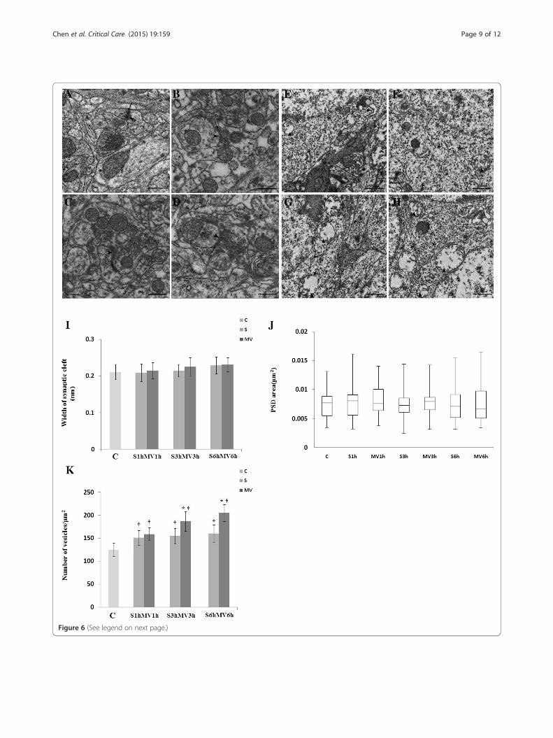
Figure 6 (See legend on next page.)
Chen et al. Critical Care (2015) 19:159 Page 9 of 12

(See figure on previous page.)Figure 6 Ultrastructural changes of synapses in the hippocampal CA1 region visualized by transmission electron microscopy. Theanimals were killed at 6 hours post-MV. Six-hour exposure to mechanical ventilation (MV) aggravated impairment in the synaptic ultrastructure at6 hours post-MV. (A) Control group (C). (B) Surgical group (S) at 6 hours (S6h). (C) MV group at 3 hours (MV3h). (D) MV group at 6 hours (MV6h).Arrows point to synaptic cleft; arrowheads point to postsynaptic density. Ultrastructural changes of the mitochondrion and endoplasmic reticulumwere visualized under transmission electron microscopy. The degree of rough endoplasmic reticulum degranulation in the hippocampus wasmore severe in the MV6h group (G) and (H) compared with the MV3h group (E) and (F). Scale bar =500 nm. Quantitation of the data ispresented for (I) width of synaptic cleft, (J) area of postsynaptic density (PSD) and (K) number of vesicles. Data for each group in (J) aresummarized by a box chart, in which the horizontal lines denote the 25th, 50th and 75th percentile values and the error bars denote the5th and 95th percentile values. *P <0.05, compared with group S; †P <0.05, compared with group C.
Chen et al. Critical Care (2015) 19:159 Page 10 of 12
Clinical relevanceThe etiology of cognitive impairment in critically illpatients is undoubtedly multifactorial and is the subjectof ongoing discussion [29]. Among different risk factorsidentified in observational studies, MV is one of themost relevant. The inflammatory response in the lungsdue to MV may lead to distal organ dysfunction, includingthe brain [30]. Nevertheless, inflammatory mechanisms ofMV-induced postoperative memory decline that is
Figure 7 Mechanical ventilation induced significantly greater apoptosinduced upregulation of cytochrome c (Cytc), cleaved caspase-3 and cleavesurgery (S). (A) Representative Western blot of Cytc, cleaved caspase-3 andof the ratio of Cytc to β-actin (B), the ratio of cleaved caspase-3 to β-actinassay data are expressed as mean ± standard error of the mean. C, Control
reflected in POD and POCD are poorly understood. Fewstudies have explored the influence of prolonged MV aftersurgery on neuroinflammation. Our findings regardingprolonged MV resulting in increasing peripheral andhippocampal cytokine expression, reactive microgliosis,apoptotic signals triggering caspase activation and be-havioral impairment might have implications for under-standing how POD or POCD happen in the intensivecare unit.
is in surgical mice. Six-hour exposure to mechanical ventilation (MV)d poly(ADP-ribose) polymerase 1 (PARP-1) in the hippocampus aftercleaved PARP-1. (B, C and D) The results of semiquantitative analysis(C) and the ratio of cleaved PARP-1 to β-actin (D). All Western blotgroup. n =6/group. *P <0.05, compared with group S.

Chen et al. Critical Care (2015) 19:159 Page 11 of 12
ConclusionsIn summary, neuroinflammation is associated with fullmaintenance of memory following postoperative pro-longed MV. The work reported here shows that MV-induced inflammation disrupted hippocampal memoryconsolidation, as evidenced by reduced contextual freez-ing time exhibited by mice that underwent MV aftersurgery. Our data suggest that prolonged MV further ag-gravates cognitive decline after surgery that may stemfrom upregulation of hippocampal IL-1β, IL-6 and TNFα,partially via activation of gliocytes in surgical mice.
Key messages
� Prolonged mechanical ventilation might affectpostoperative memory dysfunction in surgical miceand might be associated with neuroinflammation.
� This hypothesis is supported by evidence thatdetrimental effects of prolonged mechanicalventilation after surgery may affect the brain.
� Six-hour exposure to MV following surgery mightplay a synergistic role in hippocampal IL-1β,IL-6 and TNFα expression, as well as activationof gliocytes.
� We addressed an emerging area of research bystudying mechanical ventilation experimentally insurgical mice to determine how the cognitivedeficits expressed as POD happen.
AbbreviationsCNS: Central nervous system; Cytc: Cytochrome c; FC: Fear conditioning;FiO2: Fraction of inspiration oxygen; IL: Interleukin; MV: Mechanicalventilation; NF-κB: Nuclear factor κB; PARP: Poly(ADP-ribose) polymerase;POCD: Postoperative cognitive dysfunction; POD: Postoperative delirium;PSD: Postsynaptic density; TEM: Transmission electron microscopy;TNFα: Tumor necrosis factor α.
Competing interestsThe authors declare that they have no competing interests.
Authors’ contributionsCC finished most of the work of the experiments and wrote the manuscript.ZZ was responsible for the original design and provided critical revisionsimportant for the intellectual content. TC was responsible for datamanagement. MP carried out the statistical analysis and interpretation.XX completed fear conditioning tests. YW completed the analysis ofultrastructure of the hippocampus. All authors read, contributed to andapproved the final manuscript.
AcknowledgmentsThis work was supported by National Natural Science Foundation of China(81371195) and the excellent Youth Foundation of Hubei ScientificCommittee (2014CFA046).
Received: 12 January 2015 Accepted: 13 March 2015
References1. Quílez ME, López-Aguilar J, Blanch L. Organ crosstalk during acute lung
injury, acute respiratory distress syndrome, and mechanical ventilation.Curr Opin Crit Care. 2012;18:23–8.
2. Brummel NE, Jackson JC, Pandharipande PP, Thompson JL, Shintani AK,Dittus RS, et al. Delirium in the ICU and subsequent long-term disabilityamong survivors of mechanical ventilation. Crit Care Med.2014;42:369–77.
3. Van Rompaey B, Schuurmans MJ, Shortridge-Baggett LM, Truijen S, ElseviersM, Bossaert L. A comparison of the CAM-ICU and the NEECHAM ConfusionScale in intensive care delirium assessment: an observational study innon-intubated patients. Crit Care. 2008;12:R16.
4. López-Aguilar J, Fernández-Gonzalo MS, Turon M, Quílez ME, Gómez-SimónV, Jódar MM, et al. Lung–brain interaction in the mechanically ventilatedpatient. Med Intensiva. 2013;37:485–92.
5. Stevens RD, Puybasset L. The brain–lung–brain axis. Intensive Care Med.2011;37:1054–6.
6. López-Aguilar J, Villagrá A, Bernabé F, Murias G, Piacentini E, Real J, et al.Massive brain injury enhances lung damage in an isolated lung model ofventilator-induced lung injury. Crit Care Med. 2005;33:1077–83.
7. González-López A, Albaiceta GM, Talbot K. Newly identified precipitatingfactors in mechanical ventilation-induced brain damage: implications fortreating ICU delirium. Expert Rev Neurother. 2014;14:583.
8. González-López A, López-Alonso I, Aguirre A, Amado-Rodríguez L,Batalla-Solís E, Astudillo A, et al. Mechanical ventilation triggers hippocampalapoptosis by vagal and dopaminergic pathways. Am J Respir Crit Care Med.2013;188:693–702.
9. Fidalgo AR, Cibelli M, White JP, Nagy I, Maze M, Ma D. Systemicinflammation enhance surgery-induced cognitive dysfunction in mice.Neurosci Lett. 2011;498:63–6.
10. Degos V, Vacas S, Han Z, van Rooijen N, Gressens P, Su H, et al. Depletion ofbone marrow-derived macrophages perturbs the innate immune responseto surgery and reduces postoperative memory dysfunction. Anesthesiology.2013;118:527–36.
11. Das S, MacDonald K, Chang HY, Mitzner W. A simple method of mouselung intubation. J Vis Exp. 2013;73, e50318.
12. Culley DJ, Baxter MG, Yukhananov R, Crosby G. Long-term impairment ofacquisition of a spatial memory task following isoflurane-nitrous oxideanesthesia in rats. Anesthesiology. 2004;100:309–14.
13. Culley DJ, Baxter MG, Crosby CA, Yukhananov R, Crosby G. Impairedacquisition of spatial memory 2 weeks after isoflurane and isoflurane-nitrousoxide anesthesia in aged rats. Anesth Analg. 2004;99:1393–7.
14. Vizcaychipi MP, Xu L, Barreto GE, Ma D, Maze M, Giffard RG. Heat shockprotein 72 overexpression prevents early postoperative memory declineafter orthopedic surgery under general anesthesia in mice. Anesthesiology.2011;114:891–900.
15. Dos Santos CC, Shan Y, Akram A, Slutsky AS, Haitsma JJ. Neuroimmuneregulation of ventilator-induced lung injury. Am J Respir Crit Care Med.2010;183:471–82.
16. Bickenbach J, Biener I, Czaplik M, Nolte K, Dembinski R, Marx G, et al.Neurological outcome after experimental lung injury. Respir PhysiolNeurobiol. 2011;179:174–80.
17. Cibelli M, Fidalgo AR, Terrando N, Ma D, Monaco C, Feldmann M, et al.Role of interleukin-1β in postoperative cognitive dysfunction. Ann Neurol.2010;68:360–8.
18. Goshen I, Kreisel T, Ounallah-Saad H, Renbaum P, Zalzstein Y, Ben-Hur T,et al. A dual role for interleukin-1 in hippocampal-dependent memoryprocesses. Psychoneuroendocrinology. 2007;32:1106–15.
19. Toklu HZ, Kwon OS, Sakarya Y, Powers SK, Llinas K, Kirichenko N, et al.The effects of enalapril and losartan on mechanical ventilation-inducedsympathoadrenal activation and oxidative stress in rats. J Surg Res.2014;188:510–6.
20. Kamer AR, Galoyan SM, Haile M, Kline R, Boutajangout A, Li YS, et al.Meloxicam improves object recognition memory and modulates glialactivation after splenectomy in mice. Eur J Anaesthesiol. 2012;29:332–7.
21. Chirila AM, Brown TE, Bishop RA, Bellono NW, Pucci FG, Kauer JA. Long-termpotentiation of glycinergic synapses triggered by interleukin 1β. Proc NatlAcad Sci U S A. 2014;111:8263–8.
22. Barrientos RM, Higgins EA, Sprunger DB, Watkins LR, Rudy JW, Maier SF.Memory for context is impaired by post context exposure injection ofinterleukin-1 β into dorsal hippocampus. Behav Brain Res.2002;134:291–8.
23. Terrando N, Eriksson LI, Ryu JK, Yang T, Monaco C, Feldmann M, et al.Resolving postoperative neuroinflammation and cognitive decline. AnnNeurol. 2011;70:986–95.

Chen et al. Critical Care (2015) 19:159 Page 12 of 12
24. Webster CM, Kelly S, Koike MA, Chock VY, Giffard RG, Yenari MA.Inflammation and NFκB activation is decreased by hypothermia followingglobal cerebral ischemia. Neurobiol Dis. 2009;33:301–12.
25. Nicholson DW, Thornberry NA. Apoptosis. Life and death decisions.Science. 2003;299:214–5.
26. Annunziato L, Amoroso S, Pannaccione A, Cataldi M, Pignataro G, D’AlessioA, et al. Apoptosis induced in neuronal cells by oxidative stress: role playedby caspases and intracellular calcium ions. Toxicol Lett. 2003;139:125–33.
27. Harrison NA, Doeller CF, Voon V, Burgess N, Critchley HD. Peripheralinflammation acutely impairs human spatial memory via actions on medialtemporal lobe glucose metabolism. Biol Psychiatry. 2014;76:585–93.
28. Li RL, Zhang ZZ, Peng M, Wu Y, Zhang JJ, Wang CY, et al. Postoperativeimpairment of cognitive function in old mice: a possible role forneuroinflammation mediated by HMGB1, S100B, and RAGE. J Surg Res.2013;185:815–24.
29. Sobbi SC, van den Boogaard M. Inflammation biomarkers and delirium incritically ill patients: new insights? Crit Care. 2014;18:153.
30. Pelosi P, Rocco PR. The lung and the brain: a dangerous cross-talk.Crit Care. 2011;15:168.
Submit your next manuscript to BioMed Centraland take full advantage of:
• Convenient online submission
• Thorough peer review
• No space constraints or color figure charges
• Immediate publication on acceptance
• Inclusion in PubMed, CAS, Scopus and Google Scholar
• Research which is freely available for redistribution
Submit your manuscript at www.biomedcentral.com/submit