Embed Size (px)
Citation preview
ARTICLE
Received 1 Oct 2013 | Accepted 11 Apr 2014 | Published 15 May 2014
Programmable spin-state switching in amixed-valence spin-crossover iron gridTakuto Matsumoto1, Graham N. Newton1, Takuya Shiga1, Shinya Hayami2, Yuta Matsui3, Hiroshi Okamoto3,
Reiji Kumai4, Youichi Murakami4 & Hiroki Oshio1
Photo-switchable systems, such as discrete spin-crossover complexes and bulk iron–cobalt
Prussian blue analogues, exhibit, at a given temperature, a bistability between low- and
high-spin states, allowing the storage of binary data. Grouping different bistable
chromophores in a molecular framework was postulated to generate a complex that could be
site-selectively excited to access multiple electronic states under identical conditions. Here
we report the synthesis and the thermal and light-induced phase transitions of a tetranuclear
iron(II) grid-like complex and its two-electron oxidized equivalent. The heterovalent grid is
thermally inactive but the spin states of its constituent metal ions are selectively switched
using different laser stimuli, allowing the molecule to exist in three discrete phases.
Site-selective photo-excitation, herein enabling one molecule to process ternary data,
may have major ramifications in the development of future molecular memory storage
technologies.
DOI: 10.1038/ncomms4865 OPEN
1 Graduate School of Pure and Applied Sciences, Department of Chemistry, University of Tsukuba, 1-1-1 Tennodai, Tsukuba, Ibaraki 305-8571, Japan.2 Graduate School of Science and Technology, Department of Chemistry, Kumamoto University, 2-39-1 Kurokami, Kumamoto 860-8555, Japan. 3 Departmentof Advanced Materials Sciences, Graduate School of Frontier Sciences, The University of Tokyo, 5-1-5 Kashiwa, Chiba 277-8561, Japan. 4 Photon Factory andCondensed Matter Research Center, Institute of Materials Structure Science, High Energy Accelerator Research Organization (KEK), 1-1 Oho, Tsukuba, Ibaraki305-0801, Japan. Correspondence and requests for materials should be addressed to H.O. (email: [email protected]).
NATURE COMMUNICATIONS | 5:3865 | DOI: 10.1038/ncomms4865 | www.nature.com/naturecommunications 1
& 2014 Macmillan Publishers Limited. All rights reserved.
Bistable materials, which have two thermodynamically stablephases at a given temperature, have switchable electronicstates that can be manipulated through the application of
external stimuli and may have major applications in futuretechnologies as quantum logic operators or components inmemory storage devices1–3. The (SCO) phenomenon, by whichtransition metal ions can exhibit magnetic bistability, has beenreported in a wide range of systems, from mononuclearcomplexes to infinite coordination polymers4,5. Complexes oftransition metal ions with d4 to d7 electronic configurations mayshow reversible SCO between their high-spin (HS) and low-spin(LS) states when stimulated by temperature, light, pressure orguest absorption/desorption6,7. Their simple synthesis andfunctionality have made them excellent candidates as molecularswitches or sensors8. Iron(II) complexes are the most commonlyreported thermal SCO materials9, but there are various examplesbased on other metal ions10–14.
Iron(II) complexes also exhibit a light-induced LS-HS statetransition (LIESST: light-induced exited spin-state trapping)15,but usually only at low temperatures. The meta-stable HS state isgenerated by excitation of the d–d or metal-to-ligand chargetransfer (MLCT) absorption band, changing the spin state of theiron(II) ion from SLS¼ 0 to SHS¼ 2. The associated elongation(B0.2 Å) of the coordination bond lengths energeticallydisfavours thermal relaxation back to the LS state at lowtemperature, allowing the light-induced meta-stable state to betrapped16,17. LIESST has also been seen in iron(III) SCOcomplexes, usually through excitation of the ligand-to-metalcharge transfer (LMCT) band, but the shorter associated average(LS to HS) bond elongation (B0.1 Å) can make the meta-stableHS state more difficult to trap18.
Systems incorporating multiple bistable building units maydisplay more than two stable phases when stimulated by heator light, and have the potential to act as multi-responsive,multi-faceted19 or selective switches in nano-scale devices20.Approaches to the generation of multi-bistable systems includethe crystallization of mononuclear SCO complexes in asymmetricpacking environments21, the co-crystallization of differentbistable molecules22 and the linkage of two or more bistablebuilding blocks in a molecular system, in which the stabilizationof an intermediate state depends on the degree to whichneighbouring chromophores are electronically coupled23.Cooperative SCO behaviour, mediated by the steric andelectronic stress applied to neighbouring metal centres bymultidentate or bridging ligands, has been reported indinuclear complexes23, tetranuclear squares24,25 and grid-likecomplexes26–28. Indeed, the study of grid-type clusters is ofparticular interest, as their tunable electronic states29,30 have seenthem proposed as quantum cellular automata; molecular binarylogic devices that function through coulombic interactionsbetween metal centres on neighbouring cells31. Intramolecularelectronic cooperativity in {FeII
4} grids (consisting of fourequivalent chromophores) has been shown to lead to multi-stepthermal SCO, as geometric and electronic changes associated witheach spin transition have knock-on effects on the electronic statesof the neighbouring metal ions27. Step-wise thermal conversiondoes not, however, guarantee that multiple discrete phases can beaccessed photochemically.
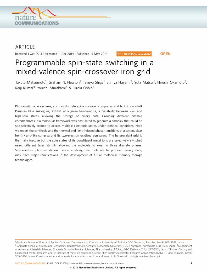
To address this, our strategy is to develop a molecule containingdifferent SCO-active chromophores, which will allow the cluster toexist in three discrete light-accessible states at low temperatures,that is, below B50 K (Fig. 1). Aromatic imidazolate ligands areused to bridge FeII and FeIII SCO chromophores in a grid-typearchitecture and, as anticipated, the heterovalent ions are selectivelyexcited by different laser stimuli, thus creating, to the best of ourknowledge, the first example of site-selective spin-state switching.
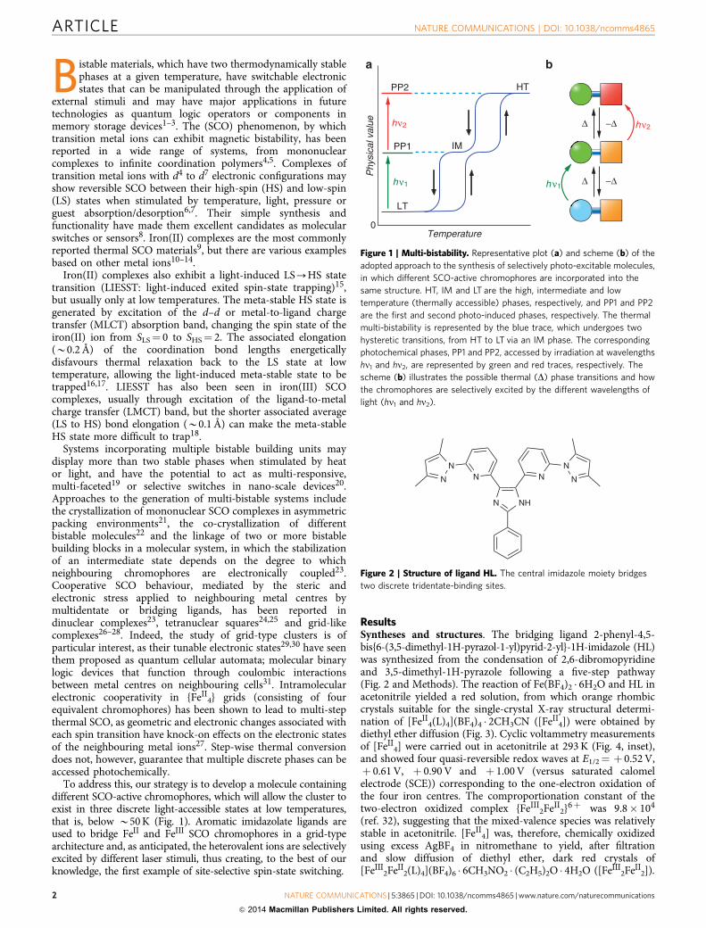
ResultsSyntheses and structures. The bridging ligand 2-phenyl-4,5-bis{6-(3,5-dimethyl-1H-pyrazol-1-yl)pyrid-2-yl}-1H-imidazole (HL)was synthesized from the condensation of 2,6-dibromopyridineand 3,5-dimethyl-1H-pyrazole following a five-step pathway(Fig. 2 and Methods). The reaction of Fe(BF4)2 � 6H2O and HL inacetonitrile yielded a red solution, from which orange rhombiccrystals suitable for the single-crystal X-ray structural determi-nation of [FeII
4(L)4](BF4)4 � 2CH3CN ([FeII4]) were obtained by
diethyl ether diffusion (Fig. 3). Cyclic voltammetry measurementsof [FeII
4] were carried out in acetonitrile at 293 K (Fig. 4, inset),and showed four quasi-reversible redox waves at E1/2¼ þ 0.52 V,þ 0.61 V, þ 0.90 V and þ 1.00 V (versus saturated calomelelectrode (SCE)) corresponding to the one-electron oxidation ofthe four iron centres. The comproportionation constant of thetwo-electron oxidized complex {FeIII
2FeII2}6þ was 9.8� 104
(ref. 32), suggesting that the mixed-valence species was relativelystable in acetonitrile. [FeII
4] was, therefore, chemically oxidizedusing excess AgBF4 in nitromethane to yield, after filtrationand slow diffusion of diethyl ether, dark red crystals of[FeIII
2FeII2(L)4](BF4)6 � 6CH3NO2 � (C2H5)2O � 4H2O ([FeIII
2FeII2]).
Temperature
Phy
sica
l val
ue
hν1
hν2
LT
HT
IMPP1
PP2
0
h ν1
hν2Δ
−Δ
−Δ
Δ
Figure 1 | Multi-bistability. Representative plot (a) and scheme (b) of the
adopted approach to the synthesis of selectively photo-excitable molecules,
in which different SCO-active chromophores are incorporated into the
same structure. HT, IM and LT are the high, intermediate and low
temperature (thermally accessible) phases, respectively, and PP1 and PP2
are the first and second photo-induced phases, respectively. The thermal
multi-bistability is represented by the blue trace, which undergoes two
hysteretic transitions, from HT to LT via an IM phase. The corresponding
photochemical phases, PP1 and PP2, accessed by irradiation at wavelengths
hn1 and hn2, are represented by green and red traces, respectively. The
scheme (b) illustrates the possible thermal (D) phase transitions and how
the chromophores are selectively excited by the different wavelengths of
light (hn1 and hn2).
NHN
NNN N
N N
Figure 2 | Structure of ligand HL. The central imidazole moiety bridges
two discrete tridentate-binding sites.
ARTICLE NATURE COMMUNICATIONS | DOI: 10.1038/ncomms4865
2 NATURE COMMUNICATIONS | 5:3865 | DOI: 10.1038/ncomms4865 | www.nature.com/naturecommunications
& 2014 Macmillan Publishers Limited. All rights reserved.
The crystallographically determined structures of [FeII4] and
[FeIII2FeII
2] are shown in Fig. 3. [FeII4] crystallized in the P �1
space group and temperature-dependent structural data werecollected at 18, 100 and 190 K (in heating mode) and at 293 K.[FeII
4] has a tetranuclear grid-like core, consisting of fourcrystallographically independent iron ions and ligands. Theligands adopt bis-tridentate coordination modes, bridging theiron ions through their central imidazolate moieties, ensuring thatall four iron ions are coordinated by two tridentate-binding sites,resulting in N6 coordination environments. Structural datacollected at both 18 K (Supplementary Fig. 1; SupplementaryTable 1) and 100 K (Supplementary Fig. 2; SupplementaryTable 2) allowed the characterization of the cluster in twopartially occupied, overlaid configurations (A and B), leading torelatively low precision on the observed bond distances. Despitethis, the bond lengths and angular distortion parameters (S)allowed us to suggest that Fe1A and Fe2A were HS and Fe3A andFe4A were LS ions, while Fe1B and Fe4B were HS and Fe2Band Fe3B were in the LS state; a ratio confirmed by lowtemperature magnetic susceptibility and Mossbauer data (Fig. 5,Supplementary Fig. 3; Supplementary Table 3). In both
configurations, the grid had a cis-2HS-2LS configuration,abbreviated as [(HS-FeII)2(LS-FeII)2], similar to that describedin the first crystallographically determined 2HS-2LS iron grid andthe subsequent theoretical studies thereupon33,34, although morerecently a trans-2HS-2LS grid has also been reported35.Increasing the measurement temperature from 100 K to 190 Kcaused the c axis to double in length, and the unit cell to containtwo crystallographically independent sites, both of whichwere occupied by two overlaid cluster configurations (seeSupplementary Fig. 4; Supplementary Table 4)36–38. Onceagain, the large number of atoms led to relatively low precisionon the bond distances. At site 1, in both configurations A and B,the bond lengths and angles suggested that ion Fe3A/B was in theLS state, while all other ions were in the HS state. At the second
Fe1
a b
Fe4
Fe2Fe3
Fe5
Fe6Fe7
Fe6*
Figure 3 | Crystal structures of [FeII4] and [FeIII
2FeII2]. (a) [FeII
4] (position A) and (b) [FeIII2FeII
2] at 100 K. HS-FeII centres are shown in green;
LS FeII, sky-blue; LS-FeIII, orange; C, grey and N, blue. For clarity, counteranions, hydrogen atoms and solvent molecules have been omitted.
E (V versus SCE)
1
LS-FeII
MLCT band
LS-FeIII
LMCT bandIVCT band
0.40 V
10 μA
0.80 V
1.4 1.2 1.0 0.8 0.6 0.4 0.2 0.0
1.20 V(versus SCE)
0
500 1,000 1,500
Wavelength (nm)
2,000 2,500
Abs
orba
nce
(a.u
.)
Figure 4 | Controlled potential absorption spectra of [FeII4]. The
absorption spectra show three key processes on increasing the applied
potential from 0.5 to 1.2 V: a decrease in LS-FeII MLCT band (501 nm)
intensity, an increase in LS-FeIII LMCT band (864 nm) intensity, and the
appearance and subsequent disappearance of an IVCT band (42,000 nm).
Inset: CV data of [FeII4].
15
12
9
6
3
0
χ mT
(e.
m.u
. mol
–1 K
)
3002001000
T (K)
[FeII4] [Fe
III2Fe
II2]
Cooling CoolingHeating Heating532 nm 532 nm808 nm 532 + 808 nm
LS
LS
LS
LS
HS
HS
HS
HS
HS
HS
LS
LS
HS
HS
LS
HS
Figure 5 | Magnetic susceptibility measurements. The thermal and
light-induced magnetic susceptibility data collected for [FeII4] (empty
markers; dark blue, heating mode; light blue, cooling mode; green, 532 nm
laser; red, 808 nm laser) and [FeIII2FeII
2] (filled markers; blue, heating
mode; light blue, cooling mode; green, 532 nm laser; red, 532 then 808 nm
lasers). [FeIII2FeII
2] showed very little thermal phase transition, but
irradiation with the different lasers had contrasting effects on the low
temperature wmT values.
NATURE COMMUNICATIONS | DOI: 10.1038/ncomms4865 ARTICLE
NATURE COMMUNICATIONS | 5:3865 | DOI: 10.1038/ncomms4865 | www.nature.com/naturecommunications 3
& 2014 Macmillan Publishers Limited. All rights reserved.

site, molecule C contained two LS ions at positions Fe2Cand Fe4C, while all ions in molecule D were in the HS state.Overall, the intermediate (IM) phase can be represented by theaverage formula [(HS-FeII)3(LS-FeII)], a ratio confirmed bymagnetic susceptibility and Mossbauer data (Fig. 5,Supplementary Fig. 3; Supplementary Table 3). Increasing themeasurement temperature to 293 K completed the transition tothe [(HS-FeII)4] HT phase (HS-FeII-Nav¼ 2.17(1) Å).Temperature-dependent crystallographic analysis showed that[FeII
4] displayed two-step intramolecular SCO behaviour whileretaining single crystallinity, allowing us to follow the spin-statechanges on each metal site as a function of temperature.
[FeIII2FeII
2] crystallized in the monoclinic space group C2/c at100 K (Fig. 3). The oxidation and spin states of the iron ionsin [FeIII
2FeII2] were confirmed by charge balance, magnetic
susceptibility measurements and Mossbauer spectra. The grid wasdiagonally bisected by a C2 axis, resulting in three crystal-lographically independent iron ions, suggesting that homovalentions were likely to be found on opposite corners. The Mossbauerspectrum of [FeIII
2FeII2] collected at 100 K confirmed that
LS-FeIII and LS-FeII ions existed in a 1/1 ratio in the grid, andthe observation of two LS-FeIII doublets and one LS-FeII doubletsuggested that the FeIII ions were located on the crystal-lographically inequivalent Fe5 and Fe7 positions (seeSupplementary Fig. 5; Supplementary Table 5). The averagecoordination bond lengths around the iron ions at 100 K wereFe5¼ 1.948(3) Å, Fe6¼ 1.957(3) Å and Fe7¼ 1.940(3) Å, respec-tively, suggesting that all iron ions were in the low-spin state at100 K. Increasing the data collection temperature to 250 K causedstructural disorder around the Fe7 centre, indicative of incom-plete single-site FeIII spin transition, an observation in line withthe Mossbauer spectra, in which a low-intensity HS-FeIII doubletprogressively appeared as temperature was increased (seeSupplementary Fig. 6 ; Supplementary Table 6).
Controlled potential absorption spectra. Full understanding ofthe relevant electronic transition energies within the moleculeswas key if selective LIESST was to be observed. The room tem-perature solution-state absorption spectra of [FeII
4] and [FeIII2-
FeII2] were comparable with those obtained from powdered
samples at 100 K, suggesting that the complexes were in their LTphases in solution (see Supplementary Figs 7 and 8). Controlledpotential Ultraviolet–visual measurements were conducted on asolution of [FeII
4] in acetonitrile to track the absorption bandsassociated with all accessible redox states (Fig. 4). Initial oxidationfrom {FeII
4}4þ to {FeIII2FeII
2}6þ caused the LS-FeII MLCT band(lmax¼ 501 nm) to weaken, while new absorption bandsappeared at 864 nm and 2,462 nm. Full oxidation to a {FeIII
4}8þ
species caused the band at 2,462 nm to disappear, while that at864 nm increased in intensity underwent a blue-shift, indicatingthe absorption at 2,462 nm to be an intervalence charge transfer(IVCT) band between heterovalent iron ions, and the band at864 nm to be the LS-FeIII LMCT band39. Absorption spectra werecollected from single crystals of [FeII
4] to observe the low-energyedge of the LS-FeII MLCT band at high resolution, and showed itto extend to around 850 nm (see Supplementary Fig. 9). Theobservation of the discrete MLCT (501 nm) and LMCT (864 nm)bands in the spectra corresponding to the {FeIII
2FeII2}6þ species
suggested that site-selective LIESST would be possible in[FeIII
2FeII2].
Temperature-dependent magnetic susceptibility. Magneticsusceptibility measurements were carried out on [FeII
4] and[FeIII
2FeII2] in heating (m) and cooling (k) modes (Fig. 5). [FeII
4]exhibited multi-step thermal SCO, in agreement with the
temperature-dependent structural data, from [(HS-FeII)4] atroom temperature, via a [(HS-FeII)3(LS-FeII)] intermediate pla-teau, to [(HS-FeII)2(LS-FeII)2] at around 100 K.
For [FeIII2FeII
2], the wmT value at 100 K was0.942 e.m.u. mol� 1 K, closely matching the predicted value of1.00 (g¼ 2.31) for two uncorrelated LS-FeIII ions (S¼ 1/2) andtwo LS-FeII ions (S¼ 0). With heating, the wmT value increasedgradually, reaching 2.36 e.m.u. mol� 1 K at 300 K. Further heatingled to some sample degradation, so all measurements were carriedout below 300 K. The susceptibility data suggest that thermal SCOoccurred gradually above 200 K, in agreement with the structuraland Mossbauer analyses40. The decrease in wmT values at lowtemperature indicates that antiferromagnetic interactions wereoperative between the LS-FeIII ions, and fitting the data collectedin heating mode up to 100 K gave a J value of � 2.47(9) cm� 1
and a g value of 2.31; the small J value, which may include bothintra- and inter-molecular interactions, supports the assignmentof trivalent ions on opposite corners of the grid (SupplementaryFig. 10). The LS-FeII ions in [FeIII
2FeII2] show greater thermal
stability than those in [FeII4], probably attributable to the more
pronounced distortion of the FeII coordination environments in[FeII
4] (see Supplementary Table 7)41,42.
Selective photoswitching. The combination of heterovalent ironchromophores within a SCO complex suggested that, for the firsttime, site-specific LIESST would be accessible. To investigate thewavelength-dependent photo-response of [FeII
4] and [FeIII2FeII
2],irradiation experiments were conducted inside the SQUIDmagnetometer. For the LIESST phenomenon to be observed,specific electronic absorption bands should be excited; usuallyd–d or MLCT for FeII and LMCT for FeIII. Ultraviolet–visible–near infrared measurements showed [FeII
4] to have a broadMLCT band centred at lmax¼ 501 nm and spreading to ca.850 nm, while [FeIII
2FeII2] had a separate LMCT band at 864 nm
originating from its LS-FeIII moieties (see SupplementaryFigs 7–9). These observations formed the basis for our site-selective LIESST excitation experiments.
Irradiation of a microcrystalline sample of [FeII4] at 532 nm
(10 mW cm� 2) at 5 K caused the initial wmT value to decrease(Fig. 5). As the sample was heated to 56 K, the wmT valueincreased rapidly to a maximum of 9.87 e.m.u. mol� 1 K, indicat-ing the occurrence of the LIESST effect due to excitation of theLS-FeII MLCT band. The photo-induced state then thermallyrelaxed to the initial phase on further temperature increase. When808 nm laser irradiation (10 mW cm� 2) was used, very similarbehaviour was observed, with a wmT maximum of9.54 e.m.u. mol� 1 K at 56 K. This behaviour is attributable tothe excitation of the low-energy tail of the broad LS-FeII MLCTband discussed above.
For [FeIII2FeII
2], a green light-induced excited state wasaccessed by irradiation with a 532 nm laser (10 mW cm� 2) at5 K, which led to an increase in the wmT values, reaching asaturation value of 1.24 e.m.u. mol� 1 K after 4 h (seeSupplementary Fig. 11). When the sample was then irradiatedwith red light (10 mW cm� 2) at 5 K, the wmT value increased to asecond saturation level of 1.46 e.m.u. mol� 1 K indicative of asecond greenþ red light-induced excited state (gþ rES).Subsequent temperature increases were mirrored by rises in thewmT values of both the green light-induced excited state andgþ rES, indicative of antiferromagnetic coupling, reachingmaxima at 22 K of 1.40 and 1.99 e.m.u. mol� 1 K, respectively,before thermally relaxing to the LS phase. Both species hadentirely reverted to the ground state by 100 K. When [FeIII
2FeII2]
was excited by only red light, the susceptibility reached saturationat 1.45 e.m.u. mol� 1 K after 6 h; very close to the value obtainedfor the gþ rES. It should be noted that the relatively low wmT
ARTICLE NATURE COMMUNICATIONS | DOI: 10.1038/ncomms4865
4 NATURE COMMUNICATIONS | 5:3865 | DOI: 10.1038/ncomms4865 | www.nature.com/naturecommunications
& 2014 Macmillan Publishers Limited. All rights reserved.
values reached on irradiation are likely to be due in part to theexistence of strong intramolecular antiferromagnetic coupling,originating from the charge transfer interactions, between theheterovalent metal centres. [FeIII
2FeII2] can be considered a
thermally inactive photo-switchable material, an unusualobservation in SCO materials research43.
Due to the low relaxation temperature and the existence of theintramolecular antiferromagnetic interactions at low temperature,the spin states of the excited species could not be elucidated bysusceptibility data alone. Light irradiation experiments werecarried out on single crystals of [FeII
4] and [FeIII2FeII
2].Irradiation at 532 and 808 nm to a single crystal of [FeII
4] causedthe bond distances to increase to lengths similar to those seen at293 K, characteristic of full [(HS-FeII)2(LS-FeII)2]-[(HS-FeII)4]transition (see Supplementary Table 8). No structural change wasobserved, however, on radiation of [FeIII
2FeII2] crystals, probably
as a result of their dark colour and impenetrability to light.To investigate the electronic characteristics of the light-induced
excited states with maximum sensitivity, 57Fe (95.5%)-enrichedsamples of [FeII
4] and [FeIII2FeII
2] were prepared. Mossbauermeasurements conducted at 5 K on [FeII
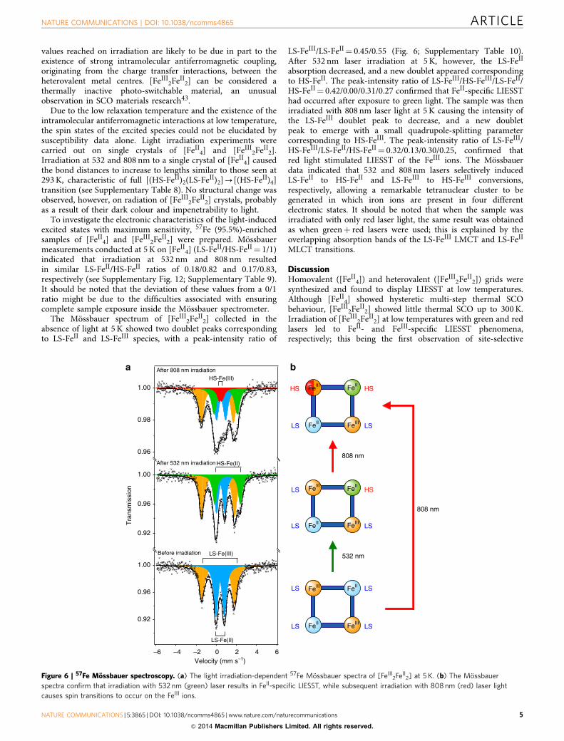
4] (LS-FeII/HS-FeII¼ 1/1)indicated that irradiation at 532 nm and 808 nm resultedin similar LS-FeII/HS-FeII ratios of 0.18/0.82 and 0.17/0.83,respectively (see Supplementary Fig. 12; Supplementary Table 9).It should be noted that the deviation of these values from a 0/1ratio might be due to the difficulties associated with ensuringcomplete sample exposure inside the Mossbauer spectrometer.
The Mossbauer spectrum of [FeIII2FeII
2] collected in theabsence of light at 5 K showed two doublet peaks correspondingto LS-FeII and LS-FeIII species, with a peak-intensity ratio of
LS-FeIII/LS-FeII¼ 0.45/0.55 (Fig. 6; Supplementary Table 10).After 532 nm laser irradiation at 5 K, however, the LS-FeII
absorption decreased, and a new doublet appeared correspondingto HS-FeII. The peak-intensity ratio of LS-FeIII/HS-FeIII/LS-FeII/HS-FeII¼ 0.42/0.00/0.31/0.27 confirmed that FeII-specific LIESSThad occurred after exposure to green light. The sample was thenirradiated with 808 nm laser light at 5 K causing the intensity ofthe LS-FeIII doublet peak to decrease, and a new doubletpeak to emerge with a small quadrupole-splitting parametercorresponding to HS-FeIII. The peak-intensity ratio of LS-FeIII/HS-FeIII/LS-FeII/HS-FeII¼ 0.32/0.13/0.30/0.25, confirmed thatred light stimulated LIESST of the FeIII ions. The Mossbauerdata indicated that 532 and 808 nm lasers selectively inducedLS-FeII to HS-FeII and LS-FeIII to HS-FeIII conversions,respectively, allowing a remarkable tetranuclear cluster to begenerated in which iron ions are present in four differentelectronic states. It should be noted that when the sample wasirradiated with only red laser light, the same result was obtainedas when greenþ red lasers were used; this is explained by theoverlapping absorption bands of the LS-FeIII LMCT and LS-FeII
MLCT transitions.
DiscussionHomovalent ([FeII
4]) and heterovalent ([FeIII2FeII
2]) grids weresynthesized and found to display LIESST at low temperatures.Although [FeII
4] showed hysteretic multi-step thermal SCObehaviour, [FeIII
2FeII2] showed little thermal SCO up to 300 K.
Irradiation of [FeIII2FeII
2] at low temperatures with green and redlasers led to FeII- and FeIII-specific LIESST phenomena,respectively; this being the first observation of site-selective
0.92
0.96
1.00
Tra
nsm
issi
on
HS-Fe(II)After 532 nm irradiation
0.96
0.98
1.00
a bHS-Fe(III)
After 808 nm irradiation
0.92
0.96
1.00
–6 –4 –2 0 2 4 6Velocity (mm s–1)
LS-Fe(II)
Before irradiation LS-Fe(III) 532 nm
808 nm
FeII
FeII FeIII
FeIIIHS HS
LS LS
FeIII FeII
FeII FeIIILS LS
LS LS
FeIII FeII
FeII FeIIILS LS
LS HS
808 nm
Figure 6 | 57Fe Mossbauer spectroscopy. (a) The light irradiation-dependent 57Fe Mossbauer spectra of [FeIII2FeII
2] at 5 K. (b) The Mossbauer
spectra confirm that irradiation with 532 nm (green) laser results in FeII-specific LIESST, while subsequent irradiation with 808 nm (red) laser light
causes spin transitions to occur on the FeIII ions.
NATURE COMMUNICATIONS | DOI: 10.1038/ncomms4865 ARTICLE
NATURE COMMUNICATIONS | 5:3865 | DOI: 10.1038/ncomms4865 | www.nature.com/naturecommunications 5
& 2014 Macmillan Publishers Limited. All rights reserved.
spin-state switching. The phenomenon of site-selective spin-statetransition allows the electronic state of a molecule to bephotochemically programmed between multiple levels. Moleculesdisplaying such extraordinary multistability have great potentialin future technologies as nano-scale logic devices, and theextension of such clusters into patterned arrays raises thepossibility of light-programmable molecular memory with vastdata storage density.
MethodsX-ray crystallography. A single crystal was removed from the mother liquor,mounted on a glass rod and intensity data were collected at 293 K, 190 K, 100 K,18 K (dark, after irradiation 532 nm, and 808 nm laser) for the divalent species, andat 250 K, and 100 K for the mixed-valence compound, using a Bruker SMARTAPEX II CCD system with Mo-Ka radiation (l¼ 0.71073 Å, 50 kV, 24 mA). SeeSupplementary Table 11 for all parameters. The structures were solved by directmethods and refined by full-matrix least-square techniques on F2 using SHELXTL.All data sets were treated with the SQUEEZE program from the PLATON suite toremove highly disordered solvent molecules from the calculations. In all [FeII
4]structures, electron density accounting for two acetonitrile molecules per formulaunit was removed, while electron densities equivalent to two nitromethane andfour water molecules, and four water molecules, were removed from the data setsof [FeIII
2FeII2] at 250 K and 100 K, respectively. See Supplementary Note 1;
Supplementary Data 1.
Magnetic measurements. Susceptibility data were collected under an appliedmagnetic field of 500 Oe using a Quantum Design MPMS-5S SQUID magnet-ometer. The temperature dependence was measured at 3.0 K increments in settlemode. The scan rate of the temperature was fixed to 10.0 K min� 1 above 10 K, andat 2.0 K min� 1 below 10 K, and each measurement was performed 30 s after thetemperature had stabilized. Magnetic data were corrected for the diamagnetism ofthe sample holder and of the sample using Pascal’s constants. In the lightirradiation experiments, a small amount of sample was used to maximize the lightconversion ratio. The sample was irradiated at 5 K by a DPSS laser (532 nm with10 mW cm� 2, Opto Tech 532.200.KE.01 and 808 nm with 10 mW cm� 2, InteliteI808-120G-CAP) through an optical fibre (Newport F-MBD; 3 m length, 1.0 mmcore size, 1.4 mm diameter). During irradiation, the magnetic moment wasrecorded at regular time intervals until saturation, after which point the lightirradiation was stopped. The temperature dependence of magnetic susceptibilityafter light irradiation was measured using an applied magnetic field of 500 Oe and ascan rate of 0.1 K min� 1 in sweep mode.
Mossbauer spectra. Mossbauer experiments were carried out using a 57Co/Rhsource in a constant-acceleration transmission spectrometer (Topologic Systems)equipped with an Iwatani HE05/CW404 cryostat. The spectrometer was calibratedusing standard a-Fe foil. All samples for Mossbauer experiments were obtainedusing 57Fe-enriched starting materials (95.5%). In the light irradiation experiments,the sample was irradiated at 5 K by a DPSS laser.
Single-crystal absorption spectroscopy. A specially designed spectrometer witha 25-cm-grating monochromator (JASCO M25-GT) and an optical microscope wasused. A 150-W tungsten–halogen lamp and a 250-W xenon lamp were employedas the light sources, and Si and Ge photodiodes were used with a photomultiplieras the detectors. The light sources and detectors were selected depending on themeasured energy range. Samples were cooled in a conduction-type cryostat.
Cyclic voltammetry. Cyclic voltammetry measurements were carried out in astandard one-compartment cell under a nitrogen atmosphere at 20 �C equipped witha platinum wire counter electrode, an SCE reference electrode, and a glassy carbonworking electrode using a BAS 620A electrochemical analyzer. The measurementswere performed in acetonitrile with 0.1 M tetra-n-butylammonium hexaflouropho-sphate (n-Bu4NPF6) as the supporting electrolyte at a scan rate of 50 mV s� 1.
Ultraviolet–visible–near infrared spectroscopy. Ultraviolet–visible–nearinfrared absorption spectra were recorded on Shimadzu UV-3150 spectrometer.The variable temperature dependence of ultraviolet–visible–near infrared spectrawas measured on KBr pellet samples using the Shimadzu UV-3150 spectrometerequipped with a Unisoku USP-203-A cryostat.
Controlled potential spectroscopy. Controlled potential spectroscopy experi-ments were done in a 0.5-mm path length quartz cell under a nitrogen atmosphere.A BAS 620A electrochemical analyzer was used as a potentiostat. Electrochemicalexperiments were performed in a three-electrode cell containing a platinum-meshworking electrode, a platinum wire counter electrode and the SCE referenceelectrode. Controlled potential spectra were recorded with a Shimadzu UV-3150
spectrometer. The measurements were performed in acetonitrile with 0.1 Mtetra-n-butylammonium hexaflourophosphate as the supporting electrolyte.
NMR measurements. 1H NMR and 13C NMR spectra were measured on a BrukerAVANCE400 spectrometer at room temperature. Chemical shifts in the NMR werereported in ppm (d), relative to the internal standard of tetramethylsilane. Thesignals observed were described as s (singlet), d (doublet), t (triplet), m (multiplets).The number of protons (n) for a given resonance is indicated as nH. Couplingconstants are reported as J in Hz.
Elemental analysis. Elemental analyses were performed using a Perkin Elmer2400 element analyzer.
General synthesis. All reagents were obtained from commercial suppliers andwere used without further purification except when noted, or additionally distilled(diglyme (bis(2-methoxyethyl)ether) over calcium hydride and diethyl ether oversodium/benzophenone) as required. For a scheme of the synthesis of HL seeSupplementary Fig. 13.
Preparation of 2-bromo-6-(3,5-dimethyl-1H-pyrazol-1-yl)pyridine (1).A solution of 3,5-dimethyl-1H-pyrazol (8.00 g, 83.2 mmol) in anhydrous diglyme(200 cm3) was stirred at 70 �C with sodium (1.80 g, 79.0 mmol) until the metaldissolved. To this solution was added 2,6-dibromopyridine (20.0 g, 84.4 mmol) inone portion. The resulting mixture was stirred at 60 �C for 42 h. The solvent wasremoved in vacuo, and water (200 cm3) was added. A crude white precipitate wascollected by filtration and dried. The solid was purified by column chromatographyon silica gel (eluting with dichloromethane) to give 1 (12.2 g, 48.3 mmol, 61% yield)as a crystalline white solid44: 1H NMR (CDCl3) d 7.84 (d, 1H, J¼ 8.0 Hz), 7.60 (dd,1H, J¼ 7.8 Hz), 7.29 (d, 1H, J¼ 7.2 Hz), 5.99 (s, 1H), 2.65 (s, 3H), 2.28 (s, 3H). Anal.(calc.) for C10H10N3Br (1): C, 47.81 (47.64); H, 4.07 (4.00); N, 16.69 (16.67)%.
Preparation of 6-(3,5-dimethyl-1H-pyrazol-1-yl)-2-pyridinecarboxylaldehyde(2). 1 (10.1 g, 40.0 mmol) was dissolved in anhydrous diethyl ether (150 cm3)under a nitrogen atmosphere. The solvent was cooled down to � 78 �C and n-buthyl lithium (2.6 M in hexane) (15.4 cm3, 40.0 mmol) was added slowly, keepingthe temperature under � 60 �C. After stirring for 1 h at � 78 �C, anhydrous N,N-dimethylformamide (6.74 cm3, 87.1 mmol) was added, ensuring that the reactiontemperature did not exceed � 70 �C. The mixture was stirred for one further hourat � 78 �C, before the reaction was quenched by the addition of 6 M hydrochloricacid. (15 cm3). The organic phase was collected and dried over anhydrous mag-nesium sulphate. After evaporating the solvent, the residue was purified by columnchromatography on a silica gel (eluting with dichloromethane/ethyl acetate¼ 20:1)to give 2 (4.94 g, 24.5 mmol, 61% yield) as a crystalline white solid45: 1H NMR(CDCl3) d 10.02 (s, 1H), 8.16 (d, 1H, J¼ 8.4 Hz), 7.95 (dd, 1H, J¼ 7.6 Hz), 7.80 (d,1H, J¼ 7.2 Hz), 6.05 (s, 1H), 2.75 (s, 3H), 2.31 (s, 3H). Anal. (calc.) for C11H11N3O(2): C, 65.43 (65.66); H, 5.61 (5.51); N, 20.56 (20.88)%.
Preparation of 1,2-bis[6-(3,5-dimethyl-1H-pyrazol-1-yl)pyrid-2-yl]-2-hydroxy-ethanone (3). 2 (4.94 g, 24.5 mmol) was dissolved in ethanol (100 cm3) and anaqueous solution (10 cm3) of potassium cyanide (325 mg, 5.00 mmol) was addedwhile the mixture was stirred. A yellow precipitate was collected by filtration andwashed with a small amount of water and ethanol to give, after drying, thecrystalline solid 3 (4.42 g, 11.3 mmol, 92% yield): 1H NMR (CDCl3) d 12.01 (s, 2H),7.95 (dd, 2H, J¼ 8.0 Hz), 7.81 (d, 2H, J¼ 7.8 Hz), 7.66 (d, 2H, J¼ 8.0 Hz), 6.07(s, 2H), 2.59 (s, 6H), 2.32 (s, 6H). 13C NMR (100 MHz, CDCl3) 154.09, 150.58,149.94, 140.53, 139.77, 135.26, 117.42, 115.73, 109.27, 13.59, 13.53. Anal. (calc.)for C22H22N6O2 (3): C, 65.53 (65.66); H, 5.60 (5.51); N, 20.73 (20.88)%. ESI–MSm/z (obs./calc.)¼ 403.3/403.2 for [3þH]þ (C22H23N6O2).
Preparation of bis-[6-(3,5-dimethyl-1H-pyrazol-1-yl)pyrid-2-yl]glyoxal (4).3 (4.42 g, 11.3 mmol) was dissolved in concentrated nitric acid (10 cm3). After theevolution of nitrogen dioxide gas had subsided, the solution was neutralized with asaturated aqueous solution of potassium hydroxide. A precipitate was collected byfiltration and purified by column chromatography on silica gel (eluting withdichloromethane/ethyl acetate¼ 20:1) to give 4 (2.68 g, 6.69 mmol, 59% yield) as apale yellow green solid: 1H NMR (CDCl3) d 8.20-8.18 (m, 2H), 8.00-7.99 (m, 4H),5.86 (s, 2H), 2.22 (s, 6H), 2.11 (s, 6H). 13C NMR (100 MHz, CDCl3) 196.46, 153.09,150.68, 149.35, 141.92, 139.42, 119.83, 118.32, 109.94, 14.37, 13.56. Anal. (calc.) forC22H20N6O2 (4): C, 66.04 (65.99); H, 5.12 (5.03); N, 20.98 (20.99)%. ESI–MS m/z(obs./calc.)¼ 401.3 / 401.2 for [4þH]þ (C22H21N6O2).
Preparation of 2-phenyl-4,5-bis[6-(3,5-dimethyl-1H-pyrazol-1-yl)pyrid-2-yl]-1H-imidazole (HL). 4 (2.68 g, 6.69 mmol), benzaldehyde (743 mg, 7.00 mmol) andammonium acetate (1.85 g, 24.0 mmol) were dissolved in acetic acid (80 cm3) andrefluxed for sixteen hours before cold water was added and the mixture wasneutralized by addition of saturated sodium hydroxide solution. The resulting
ARTICLE NATURE COMMUNICATIONS | DOI: 10.1038/ncomms4865
6 NATURE COMMUNICATIONS | 5:3865 | DOI: 10.1038/ncomms4865 | www.nature.com/naturecommunications
& 2014 Macmillan Publishers Limited. All rights reserved.
precipitate was extracted into dichloromethane, and the organic phase was driedover anhydrous magnesium sulphate. The solvent was removed in vacuo, and theresidue was purified by alumina column chromatography (activity stage I, elutingwith dichloromethane then methanol) to give HL (1.65 g, 3.38 mmol, 51% yield) asa white solid: 1H NMR (CDCl3) d 11.16 (s, 1H), 8.20 (d, 1H, J¼ 7.6 Hz), 8.03 (d,2H, J¼ 7.2 Hz), 7.00 (d, 1H, J¼ 7.6 Hz), 7.91 (dd, 1H, J¼ 8.0 Hz), 7.78 (d, 1H,J¼ 8.0 Hz), 7.61 (dd, 1H, J¼ 7.6 Hz), 7.50 (d, 1H, J¼ 7.6 Hz), 7.49 (dd, 2H,J¼ 7.4 Hz), 7.42 (dd, 1H, J¼ 7.4 Hz), 6.06 (s, 1H), 5.93 (s, 1H), 2.66 (s, 3H), 2.32,(s, 6H), 2.28 (s, 3H). 13C NMR (100 MHz, CDCl3) 152.29, 151.87, 149.87, 149.70,147.17, 146.37, 141.35, 140.69, 139.71, 139.01, 138.48, 129.73, 129.43, 129.15,128.92, 125.67, 120.30, 120.23, 114.52, 114.25, 109.03, 108.80, 14.32, 13.91, 13.63,13.62. Anal. (calc.) for C29H26N8 (HL): C, 71.31 (71.59); H, 5.60 (5.39); N, 22.59(23.03)%. ESI–MS m/z (obs./calc.)¼ 487.2/487.2 for [HLþH]þ (C29H27N8).
Preparation of [FeII4(L)4] complex. HL (97.3 mg, 0.20 mmol) in acetonitrile
(5.0 cm3) was added to Fe(BF4)2 � 6H2O (67.5 mg, 0.20 mmol) in acetonitrile(5.0 cm3). Diethyl ether was allowed to diffuse into the solution, resulting in theformation of orange blocks and yellow needle crystals. The crystals were thenwashed with methanol, and the yellow crystals removed. The orange crystals of[FeII
4](BF4)4 � 2CH3CN were collected by filtration (31.5 mg, 0.013 mmol, 25%yield). Anal. (calc.) for C116H100N32B4F16Fe4 ([FeII
4] (BF4)4): C, 55.32 (55.44);H, 4.31 (4.01); N, 17.70 (17.84)%. ESI–MS m/z (obs./calc.)¼ 541.5/541.4 for[M–4(BF4)]4þ (C116H100N32Fe4). The 57Fe-enriched samples for Mossbauerspectra measurements ware prepared using metallic 57Fe foils (95.5%).
Preparation of [FeIII2FeII
2(L)4] complex. Excess AgBF4 (12.0 mg, 0.062 mmol)was added to a nitromethane (5.0 cm3) solution of [FeII
4] (31.5 mg, 0.013 mmol).The mixture was stirred for 15 min at 50 �C, cooled to room temperature, and theprecipitate (silver) removed by filtration. Diethyl ether was allowed to diffuseinto the filtrate, resulting in the formation of dark red rhombic crystals of[FeIII
2FeII2](BF4)6 � 6CH3NO2 � (C2H5)2O � 4H2O, which were collected by filtration
(28.1 mg, 0.009 mmol, 74% yield). Anal. (calc.) for C123H127N35B6F24Fe4O11
([FeIII2FeII
2](BF4)6 � 3CH3NO2 � (C2H5)2O � 4H2O): C, 49.25 (48.99); H, 4.18 (4.24);N, 15.97 (16.26)%.
Details of magnetic measurements on [FeII4]. The wmT value of [FeII
4] at 100 K(m) was 6.89 e.m.u. mol� 1 K, close to the spin-only value of 6.63 e.m.u. mol� 1 K(g¼ 2.10), calculated from the sum of the uncorrelated spins of two HS-FeII (S¼ 2)and two LS-FeII (S¼ 0) ions, as predicted by the structural and Mossbauer data. Asthe sample was heated, the wmT value rapidly increased. At 190 K (m), the wmT plotshowed a small step with a value of 10.57 e.m.u. mol� 1 K, closely corresponding tothe spin-only value of 9.95 e.m.u. mol� 1 K expected for an average of three HS-FeII
ions and one diamagnetic LS-FeII centres per molecule. On further heating to300 K, the magnetic susceptibility reached a plateau with a wmT value of13.26 e.m.u. mol� 1 K, suggesting that all iron ions were in their HS states above250 K. Subsequent measurements in cooling mode echoed the two-step heatingprofile, but with a relatively wide thermal hysteresis. Mossbauer measurements(Supplementary Fig. 3) and structural analyses confirmed that the decrease in wmTvalues below 100 K was not due to SCO behaviour, but more likely a consequenceof the antiferromagnetic interactions between the HS iron ions. The thermalhysteresis and multi-step phase transition mean that [FeII
4] is a thermally multi-bistable molecule with the spin-state conversions of [(LS-FeII)2(HS-FeII)2]$[(LS-FeII)(HS-FeII)3]$[(HS-FeII)4].
References1. Aromı, G., Aguila, D., Gamez, P., Luisc, F. & Roubeau, O. Design of magnetic
coordination complexes for quantum computing. Chem. Soc. Rev. 41, 537–546(2012).
2. Bousseksou, A., Molnar, G., Demont, P. & Menegotto, J. Observation of athermal hysteresis loop in the dielectric constant of spin crossover complexes:towards molecular memory devices. J. Mater. Chem. 13, 2069–2071 (2003).
3. Bousseksou, A., Molnar, G., Salmon, L. & Nicolazzi, W. Molecular spincrossover phenomenon: recent achievements and prospects. Chem. Soc. Rev.40, 3313–3335 (2011).
4. Real, J. A. et al. Spin crossover in a catenane supramolecular system. Science268, 265–267 (1995).
5. Kahn, O. & Martinez, C. J. Spin-transition polymers: from molecular materialstoward memory devices. Science 279, 44–48 (1998).
6. Halder, G., Kepert, C. J., Moubaraki, B., Murray, K. S. & Cashion, J. D.Guest-dependent spin crossover in a nanoporous molecular frameworkmaterial. Science 298, 1762–1765 (2002).
7. Gutlich, P., Gaspar, A. B. & Garcia, Y. Spin state switching in iron coordinationcompounds. Beilstein J. Org. Chem. 9, 342–391 (2013).
8. Guetlich, P. & Goodwin, H. A. (eds.) Spin crossover in transition metalcompounds III. Top. Curr. Chem. 235, 268 (2004).
9. Halcrow, M. A. Trapping and manipulating excited spin states of transitionmetal compounds. Chem. Soc. Rev. 37, 278–289 (2008).
10. Halepto, D. M. et al. Spin crossover in chromium(II) complexes and the crystaland molecular structure of the high spin form of bis[1,2-bis(diethylphosphino)ethane]di-iodochromium(II). J. Chem. Soc., Chem.Commun. 1322–1323 (1989).
11. Sim, P. G. & Sinn, E. First manganese(III) spin crossover and first d4 crossover.comment on cytochrome oxidase. J. Am. Chem. Soc. 103, 241–243 (1981).
12. Hayami, S., Komatsu, Y., Shimizu, T., Kamihata, H. & Lee, Y. H. Spin-crossoverin cobalt(II) compounds containing terpyridine and its derivatives. Coord.Chem. Rev. 255, 1981–1990 (2011).
13. Nihei, M., Shiga, T., Maeda, Y. & Oshio, H. Spin crossover iron(III) complexes.Coord. Chem. Rev. 251, 2606–2621 (2007).
14. Venkataramani, S. et al. Magnetic bistability of molecules in homogeneoussolvent at room temperature. Science 331, 445–448 (2011).
15. Letard, J.-F. et al. Light induced excited pair spin state in an iron(II) binuclearspin-crossover compound. J. Am. Chem. Soc. 121, 10630–10631 (1999).
16. Hauser, A., Vef, A. & Adler, P. Intersystem crossing dynamics in Fe(II)coordination compounds. J. Chem. Phys. 95, 8710–8717 (1991).
17. Vef, A., Manthe, U., Guetlich, P. & A. Hauser, A. Intersystem crossingdynamics in the spin-crossover systems [M:Fe(pic)3]Cl2Sol (M¼Mn or Zn,Sol¼MeOH or EtOH). J. Chem. Phys. 101, 9326–9332 (1994).
18. Juhasz, G., Hayami, S., Sato, O. & Maeda, Y. Photo-induced spin transition foriron(III) compounds with p-p interactions. Chem. Phys. Lett. 364, 164–170(2002).
19. Hoshino, N. et al. Three-way switching in a cyanide-bridged [CoFe] chain.Nat. Chem. 4, 921–926 (2012).
20. Rotaru, A. et al. Nano-electromanipulation of spin crossover nanorods: towardsswitchable nanoelectronic devices. Adv. Mater. 25, 1745–1749 (2013).
21. Li, B. et al. Solvent-induced transformation of single crystals of a spin-crossover(SCO) compound to single crystals with two distinct SCO centers. J. Am. Chem.Soc. 132, 1558–1566 (2010).
22. Nihei, M. et al. Multiple bistability and tristability with dual spin-stateconversions in [Fe(dpp)2][Ni(mnt)2] � 2MeNO2. J. Am. Chem. Soc. 131,3553–3560 (2010).
23. Gaspar, A. B., Munoz, M. C. & Real, J. A. Dinuclear iron(II) spin crossovercompounds: singular molecular materials for electronics. J. Mater. Chem. 16,2522–2533 (2006).
24. Nihei, M. et al. Two-step spin conversion in a cyanide bridged ferrous square.Angew. Chem. Int. Ed. 44, 6484–6487 (2005).
25. Wei, R.-J., Hun, Q., Tao, J., Huang, R.-B. & Zheng, L.-S. Spin-crossover Fe4II
square: two-step complete spin transition and reversible single-crystal-to-single-crystal transformation. Angew. Chem. Int. Ed. 50, 8940–8943 (2011).
26. Breuning, E. et al. Spin crossover in a supermolecular FeII4 [2� 2] grid
triggered by temperature, pressure, and light. Angew. Chem. Int. Ed. 39,2504–2507 (2000).
27. Schneider, B., Demeshko, S., Dechert, S. & Meyer, F. A double-switchingmultistable fe4 grid complex with stepwise spin-crossover and redox transition.Angew. Chem. Int. Ed. 49, 9274–9277 (2010).
28. Wang, Y.-T. et al. Spin transitions in Fe(II) metallogrids modulated bysubstituents, counteranions, and solvents. J. Am. Chem. Soc. 135, 5942–5945(2013).
29. Sato, H. et al. Multiredox active [3� 3] copper grids. Inorg. Chem. 52,9714–9716 (2013).
30. Zhao, L., Matthews, C. J., Thompson, L. K. & Heath, S. L. A novel magneticallycoupled nanomanganese(II) 3� 3 portculluis-like gird involving just oxygenbridges, generated by strict self assembly of the metal cation and a singleheptadentate ligand. Chem. Commun. 265–266 (2000).
31. Lent, C. S. Molecular electronics: bypassing the transistor paradigm. Science288, 1597–1599 (2000).
32. Slater, J. W., D’Alessandro, D. M., Keene, F. R. & Steel, P. J. Metal-metalinteractions in dinuclear ruthenium complexes containing bridging 4,5-di(2-pyridyl)imidazolates and related ligands. Dalton Trans. 1954–1962 (2006).
33. Wu, D.-Y., Sato, O., Einaga, Y. & Duan, C.-Y. A spin-crossover cluster ofiron(II) exhibiting a mixed-spin structure and synergy between spin transitionand magnetic interaction. Angew. Chem. Int. Ed. 48, 1475–1478 (2009).
34. Zueva, E. M., Ryabikh, E. R. & Borshch, S. A. Theoretical analysis of spincrossover in iron(II) [2� 2] molecular grids. Inorg. Chem. 50, 11143–11151(2011).
35. Schneider, B., Demeshko, S., Neudeck, S., Dechert, S. & Meyer, F. Mixed-spin[2� 2] Fe4 grid complex optimized for quantum cellular automata. Inorg.Chem. 52, 13230–13237 (2013).
36. Brefuel, N. et al. Concerted Spin crossover and symmetry breaking yield threethermally and one light-induced crystallographic phases of a molecularmaterial. Angew. Chem., Int. Ed. 48, 9304–9307 (2009).
37. Griffin, M. et al. A symmetry-breaking spin-state transition in iron(III). Angew.Chem. Int. Ed. 50, 896–900 (2011).
38. Vieira, B. J. C. et al. [Fe(nsal2trien)]SCN, a new two-step iron(III) spincrossover compound, with symmetry breaking spin-state transition and anintermediate ordered state. Inorg. Chem. 52, 3845–3850 (2013).
NATURE COMMUNICATIONS | DOI: 10.1038/ncomms4865 ARTICLE
NATURE COMMUNICATIONS | 5:3865 | DOI: 10.1038/ncomms4865 | www.nature.com/naturecommunications 7
& 2014 Macmillan Publishers Limited. All rights reserved.
39. Hayami, S. et al. Photo-induced spin transition of iron(III) compounds withp-p intermolecular interactions. Chem. Eur. J. 15, 3497–3508 (2009).
40. Cook, C. et al. High-Temperature spin crossover behavior in a nitrogen-richFeIII-based system. Inorg. Chem. 52, 1825–1831 (2013).
41. Guionneau, P. et al. High pressure and very low temperature effects on thecrystal structures of some iron(II) complexes. C. R. Acad. Sci. Ser. IIc 4,161–171 (2001).
42. Guionneau, P., Marchivie, M., Bravic, G., Letard, J.-F. & Chasseau, D. Co(II)molecular complexes as a reference for the spin crossover in Fe(II) analogues.J. Mater. Chem. 12, 2546–2551 (2002).
43. Renz, F. et al. Strong field iron(II) Complex converted by light into a long-livedhigh-spin state. Angew. Chem. Int. Ed. 39, 3699–3700 (2000).
44. Watson, A. A., House, D. A. & Steel, P. J. Chiral heterocyclic ligands. 7.syntheses of some chiral 2,6-di-N-pyrazolylpyridines. J. Org. Chem. 56,4074–4076 (1991).
45. Zeng, F. & Yu, Z. Exceptionally efficient unsymmetrical ruthenium(II) NNNcomplex catalysts bearing a pyridyl-based pyrazolyl� imidazolyl ligand fortransfer hydrogenation of ketones. Organometallics 27, 2898–2901 (2008).
AcknowledgementsThis work was supported by a Grant-in-Aid for Scientific Research and for Priority Area(‘Coordination Programming’ area 2107, No. 21108006) from the Ministry of Education,Culture, Sports, Science and Technology, Japan, by a research grant (No. 2012G036)from KEK, and by a Grant-in-Aid for Scientific Research (No. 25248014) from the JapanSociety for the Promotion of Science (JSPS).
Author contributionsH.O. conceived and supervized the project. T.M. planned and implemented the syntheticand analytical experiments and co-wrote the manuscript with G.N.N., who helped to
analyse the data. T.S. directed the study and interpretation of the experiments. S.H.measured the light irradiation 57Fe Mossbauer spectra. Y.M. and H.O. contributed to thesingle-crystal absorption spectroscopy. R.K. and Y.M. performed the synchrotron X-raydata collections.
Additional informationAccession codes The X-ray crystallographic coordinates for structures reported in thisArticle have been deposited at the Cambridge Crystallographic Data Centre (CCDC),under deposition numbers CCDC 980494-980501. These data can be obtained free ofcharge from The Cambridge Crystallographic Data Centre via www.ccdc.cam.ac.uk/data_request/cif.
Supplementary Information accompanies this paper at http://www.nature.com/naturecommunications
Competing financial interests: The authors declare no competing financial interests.
Reprints and permission information is available online at http://npg.nature.com/reprintsandpermissions/
How to cite this article: Matsumoto, T. et al. Programmable spin-state switching in amixed-valence spin-crossover iron grid. Nat. Commun. 5:3865 doi: 10.1038/ncomms4865(2014).
This work is licensed under a Creative Commons Attribution-NonCommercial-NoDerivs 3.0 Unported License. The images or other
third party material in this article are included in the article’s Creative Commons license,unless indicated otherwise in the credit line; if the material is not included under theCreative Commons license, users will need to obtain permission from the license holderto reproduce the material. To view a copy of this license, visit http://creativecommons.org/licenses/by-nc-nd/3.0/
ARTICLE NATURE COMMUNICATIONS | DOI: 10.1038/ncomms4865
8 NATURE COMMUNICATIONS | 5:3865 | DOI: 10.1038/ncomms4865 | www.nature.com/naturecommunications
& 2014 Macmillan Publishers Limited. All rights reserved.
![Switching Magnetism and Superconductivity with Spin-Polarized Current ...PDF] Switching Magnet… · 1 Switching Magnetism and Superconductivity with Spin-Polarized Current in Iron-Based](https://img.dokumen.tips/doc/110x75/5f13e2be0b294765f40b22f3/switching-magnetism-and-superconductivity-with-spin-polarized-current-switching.jpg)