Embed Size (px)
Citation preview

Instructions for use
Title Study on the Energy Transfer Processes in Polynuclear Lanthanide Complexes
Author(s) 大曲, 駿
Citation 北海道大学. 博士(工学) 甲第13242号
Issue Date 2018-03-22
DOI 10.14943/doctoral.k13242
Doc URL http://hdl.handle.net/2115/70107
Type theses (doctoral)
File Information Shun_Omagari.pdf
Hokkaido University Collection of Scholarly and Academic Papers : HUSCAP

Study on the Energy Transfer
Processes in Polynuclear
Lanthanide Complexes
多核希土類錯体における
エネルギー移動過程に関する研究
Shun Omagari
Graduate School of Chemical Sciences and Engineering
Hokkaido University

i
I. List of Publications
For Thesis
1. Spin-orbit Coupling Dependent Energy Transfer in Luminescent
Nonanuclear Yb-Gd / Yb-Lu Clusters (Chapter 4)
S. Omagari, T. Nakanishi, Y. Kitagawa, T. Seki, K. Fushimi, H. Ito, A. Meijerink, Y.
Hasegawa, J. Lumin. 2017, submitted.
2. Origin of Concentration Quenching in Ytterbium Coordination Polymers:
Phonon-Assisted Energy Transfer (Chapter 3)
S. Omagari, T. Nakanishi, Y. Hirai, Y. Kitagawa, T. Seki, K. Fushimi, H. Ito, Y. Hasegawa,
Eur. J. Inorg. Chem. 2017, in publication.
3. Critical Role of Energy Transfer Between Terbium Ions for Suppression of
Back Energy Transfer in Nonanuclear Terbium Clusters (Chapter 6)
S. Omagari, T. Nakanishi, Y. Kitagawa, T. Seki, K. Fushimi, H. Ito, A. Meijerink, Y.
Hasegawa, Sci. Rep. 2016, 6, 37008.
4. Effective Photosensitized Energy Transfer of Nonanuclear Terbium Clusters
Using Methyl Salicylate Derivatives (Chapter 5)
S. Omagari, T. Nakanishi, T. Seki, Y. Kitagawa, Y. Takahata, K. Fushimi, H. Ito, Y.
Hasegawa, J. Phys. Chem. A 2015, 119, 1943-1947.
5. Synthesis and Photoluminescence Properties of Nonanuclear Tb(III)
Clusters with Long Alkyl Chain Group (Chapter 5)
S. Omagari, T. Nakanishi, Y. Kitagawa, K. Fushimi, Y. Hasegawa, e-J. Surf. Sci. Nanotech.
2015, 13, 27-30.

ii
Other Publications
1. Temperature dependence of 4fn−15d1→4fn luminescence of Ce3+ and Pr3+ ions
in Sr2GeO4 host
K. Fiaczyk, S. Omagari, A. Meijerink, E. Zych, J. Lumin. 2018, 198, 163-170.
2. A Luminescent Mechanochromic 9-Anthryl Gold(I) Isocyanide Complex with
an Emission Maximum at 900 nm after Mechanical Stimulation
T. Seki, N. Tokodai, S. Omagari, T. Nakanishi, Y. Hasegawa, T. Iwasa, T. Taketsugu, H.
Ito, J. Am. Chem. Soc. 2017, 139, 6514-6517.
3. Development of Ion-Conductive and Vapoluminescent Porous Coordination
Polymers Composed of Ruthenium(II) Metalloligand
A. Watanabe, A. Kobayashi, E. Satoh, Y. Nagao, S. Omagari, T. Nakanishi, Y. Hasegawa,
W. M. C. Sameera, M. Yoshida, M. Kato, Inorg. Chem. 2017, 56, 3005-3013.
4. Synthesis of Dipyridinogermole–Copper Complex as Soluble
Phosphorescent Material
K. Murakami, Y. Ooyama, S. Watase, K. Matsukawa, S. Omagari, T. Nakanishi, Y.
Hasegawa, K. Inumaru, J. Ohshita, Chem. Lett. 2016, 45, 502-504.
5. Drastically Improved Durability and Efficiency of Silicon Solar Cells Using
Hyper-Stable Lanthanide Coordination Polymer Beads
H. Kataoka, T. Nakanishi, S. Omagari, Y. Takabatake, Y. Kitagawa, Y. Hasegawa, Bull.
Chem. Soc. Jpn. 2016, 89, 103-109.
6. Photo-Degradation Analysis of Luminescent Polymers with Lanthanide
Complexes
H. Kataoka, S. Omagari, T. Nakanishi, Y. Hasegawa, J. Photopolym. Sci. Technol. 2015,
28, 247-254.
7. EVA Thin Film with Thermo- and Moisture-stable Luminescent Copolymer
Beads Composed of Eu(III) Complexes for Improvement of Energy
Conversion Efficiency on Silicon Solar Cell
H. Kataoka, S. Omagari, T. Nakanishi, Y. Hasegawa, Opt. Mater. 2015, 42, 411-416.

iii
II. List of Presentations
1. Concentration quenching mechanism of Ytterbium(III) coordination polymer
◯S. Omagari,T. Nakanishi,Y. Hirai,Y. Kitagawa,K. Fushimi,Y. Hasegawa,錯体化学会第
67 回討論会,2Fa-05,北海道大学札幌キャンパス,札幌,2017 年 9 月 18 日. (Oral
Presentation)
2. 九核希土類クラスターにおけるエネルギー移動と発光特性に関する研究
◯大曲駿,中西貴之,北川裕一,伏見公志,長谷川靖哉,第 78 回応用物理学会秋季学術講
演会,7p-A414-1,福岡国際会議場,福岡,2017年 9月 7日.(Invited Oral Presentation)
3. テルビウムクラスターにおけるエネルギー移動と発光特性
◯大曲駿,中西貴之,北川裕一,伏見公志,長谷川靖哉,錯体化学若手の会夏の学校 2017,
西浦温泉ホテルたつき,蒲郡,2017年 8月 2日.(Invited Oral Presentation)
4. Mechanism Elucidation and Suppression of Back Energy Transfer in
Nonanuclear Tb3+ Clusters
◯S. Omagari,T. Nakanishi,Y. Kitagawa,K. Fushimi,Y. Hasegawa,Nagoya Univ.-Tsinghua
Univ.-Toyota Motor Corp.-Hokkaido Univ. Joint Symposium,P02,Takayama Municipal Cultural
Hall,Takayama,September 13, 2017.(Poster)
5. Energy Transfer and Luminescence Properties of Nonanuclear Rare-Earth
Clusters including Terbium and Gadolinium
◯S. Omagari,T. Nakanishi,Y. Kitagawa,K. Fushimi,Y. Hasegawa,第 64回応用物理学会春
季学術講演会,14p-411-10,パシフィコ横浜,横浜,2017年 3月 13日.(Oral Presentation)
6. Molecular Design for Improving Photoluminescence Efficiency in
Lanthanide Complexes
◯S. Omagari,Y. Hasegawa,9th HOPE meeting,JP1601,Tokyo International Forum,Tokyo,
February 26, 2017.(Poster)
7. Dependency of Spin of Lanthanide Electrons on Ligand Emission in
Lanthanide Clusters
◯S. Omagari,T. Nakanishi,Y. Kitagawa,K. Fushimi,Y. Hasegawa,2016年光化学討論会,
1C03,東京大学駒場第一キャンパス,東京,2016年 9月 6日.(Oral Presentation)
8. Excited State Dynamics of Nonanuclear Tb(III) Clusters
◯S. Omagari,T. Nakanishi,Y. Kitagawa,K. Fushimi,Y. Hasegawa,Rare Earths 2016,C09-
23,北海道大学札幌キャンパス,札幌,2016年 6月 10日.(Oral Presentation)

iv
9. Effective photosensitized energy transfer of nonanuclear terbium clusters
using methyl salicylate derivatives
◯S. Omagari,T. Nakanishi,T. Seki,Y. Kitagawa,Y. Takahata,K. Fushimi,H. Ito,Y.
Hasegawa,The International Chemical Congress of Pacific Basin Societies 2015 (Pacifichem 2015),
55 (1074),Honolulu (HI, USA),December 15, 2015.(Poster)
10. テルビウムクラスターにおける配位子の電子構造と光増感エネルギー移動の関
係
◯大曲駿,中西貴之,北川裕一,伏見公志,長谷川靖哉,日本化学会北海道支部 2015 年夏
季研究発表会,E15,北海道教育大学函館キャンパス,函館,2015 年 7 月 18 日.(Oral
Presentation)
11. エネルギー移動のループを利用した希土類クラスターの発光増強
◯大曲駿,中西貴之,北川裕一,関朋宏,伏見公志,伊藤肇,長谷川靖哉,化学系学協会北
海道支部 2015年冬季研究発表会,1B15,北海道大学札幌キャンパス,札幌,2015年 1月 26
日.(Oral Presentation)
12. Ln-O格子を有する九核ランタニドクラスターの合成とその光物性
◯大曲駿,中西貴之,北川裕一,伏見公志,長谷川靖哉,第 4 回 CSJ 化学フェスタ 2014,
4P061,タワーホール船堀,東京,2014年 10月 15日.(Poster)
13. 九核ランタニドクラスターにおける発光メカニズムの解明
◯大曲駿,中西貴之,伏見公志,長谷川靖哉,2014 年光化学討論会,1P087,北海道大学札
幌キャンパス,札幌,2014年 10月 11日.(Poster)
14. Synthesis and Photoluminescence Properties of Nonanuclear Tb(III)
Clusters with Long Alkyl Chain
◯S. Omagari,T. Nakanishi,Y. Kitagawa,K. Fushimi,Y. Hasegawa,15th Chitose International
Forum on Photonics & Technology,P9,Chitose Institute of Science and Technology,Chitose,
October 2, 2014.(Poster)
15. テルビウムとガドリニウムで構成された九核ランタニドクラスターの発光メカ
ニズム
◯大曲駿,中西貴之,伏見公志,長谷川靖哉,第 26回配位化合物の光化学討論会,P30,首
都大東京南大沢キャンパス,東京,2014年 8月 7日.(Poster)

v
16. Photophysical Properties of Nonanuclear Tb(III) Clusters with Intramolecular
Interactions
◯S. Omagari,T. Nakanishi,Y. Kitagawa,K. Fushimi,Y. Hasegawa, 10th International
Workshop on Supramolecular Nanoscience of Chemically Programmed Pigments,P19,Ritsumeikan
University Biwako-Kusatsu Campus,Kusatsu,May 31, 2014.(Poster)
17. 長鎖アルキル基を導入した九核 Tb(III)クラスターの合成と光機能評価
◯大曲駿,中西貴之,伏見公志,長谷川靖哉,日本化学会第 94 期春季年会,2F3-08,名古
屋大学東山キャンパス,名古屋,2014年 3月 28日.(Oral Presentation)
III. Achievements
1. 「学生講演賞」,2017年錯体化学会第 67回討論会.
2. 「講演奨励賞」,2017年第 64回応用物理学会春季学術講演会.
3. 「優秀学生発表賞(口頭)」,2016年光化学討論会.
4. “Student Poster Award”,The International Chemical Congress of Pacific Basin
Societies 2015 (Pacifichem 2015).
5. Featured in the Chem-station’s “Spotlight Research” in response to the Student
Poster Award in Pacifichem 2015.
6. 「優秀講演賞(口頭部門)」,日本化学会北海道支部 2015年夏季研究発表会.
7. 「優秀講演賞(口頭部門)」,化学系学協会北海道支部 2015年冬季研究発表
会.
8. 「優秀ポスター発表賞」,2014年度錯体化学若手の会北海道支部第 3回勉強
会.
9. 「優秀学生発表賞(ポスター)」,2014年光化学討論会.
10. 「優秀ポスター賞」,第 26回配位化合物の光化学討論会.

vi

vii
Table of Contents
I. List of Publications …………………………………………………………...... i
II. List of Presentations ……………………………………………………….…. iii
III. Achievements …………………………………………………………………. v
Chapter 1: General Introduction
1.1. Introduction ……………………………………………………………………. 2
1.1.1. Descriptions of Light
1.1.2. Photoluminescence
1.2. Lanthanides …………………………………………………………………… 8
1.2.1. Electron Configuration and Chemistry
1.2.2. Luminescence
1.3. Lanthanide Complexes …………………………………………………..… 14
1.3.1. Luminescence Mechanism
1.3.2. Polynuclear Lanthanide Complexes
1.4. Outline ……………………………………………………………………..… 20
1.5. References …………………………………………………………………... 23
1.6. Abbreviations ………………………………………………………………... 28
Chapter 2: Theory
2.1. Introduction ………………………………………………………………..… 30
2.2. Lanthanides …………………………………………………………………. 30
2.1.1. Energy Levels and Wavefunctions
2.1.2. Radiative Rate Constants
2.1.3. Energy Transfer Rate Constants
2.3. Kinetic Analysis of Excited State ………………………………………….. 39
2.2.1. Rate Equation for Impulse Excitation
2.2.2. Rate Equation for Constant Excitation
2.2.3. Simple Mononuclear Case
2.4. References …………………………………………………………………... 46

viii
Chapter 3: Concentration Quenching in Ytterbium Coordination
Polymers
3.1. Introduction ………………………………………………………………….. 48
3.2. Experimental Section ………………………………………………………. 50
3.2.1. Material
3.2.2. Apparatus
3.2.3. Synthesis
3.2.4. Spectroscopy
3.2.5. Crystallography
3.3. Theoretical Methods ………………………………………………………... 54
3.3.1. Rate Constants
3.3.2. Rate Equations
3.4. Results and Discussion …………………………………………………….. 60
3.4.1. Structure
3.4.2. Photophysical Properties
3.4.3. Theoretical Lifetimes
3.5. Conclusion …………………………………………………………………… 68
3.6. Appendix …………………………………………………………………….. 70
3.6.1. Absorption Spectra of Yb(III) Ions
3.6.2. MATLAB Script for Calculation of Theoretical Lifetimes
3.6.3. Quantitative Considerations of Theoretical Lifetimes
3.7. References …………………………………………………………………... 76
Chapter 4: Spin-Orbit Coupling and Energy Transfer in Nonanuclear
Lanthanide Clusters
4.1. Introduction ………………………………………………………………….. 80
4.2. Experimental Section ………………………………………………………. 81
4.2.1. Material
4.2.2. Apparatus
4.2.3. Synthesis
4.2.4. Spectroscopy
4.2.5. Crystallography

ix
4.3. Results and Discussion …………………………………………………….. 84
4.3.1. Structure and Identification
4.3.2. Photophysical Properties
4.4. Conclusion …………………………………………………………………… 94
4.5. Appendix …………………………………………………………………….. 94
4.5.1. Notes on Spin-Orbit Coupling
4.5.2. Continuous Shape Measure and Shape Measure
4.6. References …………………………………………………………………... 96
Chapter 5: Back Energy Transfer in Terbium Clusters
5.1. Introduction ……………………………………………………………….. 100
5.2. Experimental Section ……………………………………………………. 101
5.2.1. Material
5.2.2. Apparatus
5.2.3. Synthesis
5.2.4. Spectroscopy
5.2.5. Computational Details
5.3. Results and Discussion …………………………………………………… 104
5.3.1. Structure
5.3.2. Intramolecular Interactions and Photophysical Properties
5.3.3. Mechanism of Back Energy Transfer
5.4. Conclusion …………………………………………………………………. 116
5.5. Appendix …………………………………………………………………… 117
5.5.1. Intrinsic Quantum Yield Measurements
5.5.2. BET Rate Constant
5.5.3. BET and T1 Decay
5.6. References …………………………………………………………………. 120
Chapter 6: Suppression of Back Energy Transfer by Energy
Transfer between Terbium Ions
6.1. Introduction ………………………………………………………………… 124
6.2. Theoretical Motivation …………………………………………………….. 125
6.2.1. Theoretical Method

x
6.2.2. Results
6.3. Experimental Section …………………………………………………...… 132
6.3.1. Material
6.3.2. Apparatus
6.3.3. Synthesis
6.3.4. Spectroscopy
6.4. Results and Discussion …………………………………………………… 134
6.4.1. Structure and Identification
6.4.2. Photophysical Properties
6.5. Conclusion …………………………………………………………………. 141
6.6. Appendix …………………………………………………………………… 142
6.6.1. Dependence of TbTbET in Absence of BET
6.6.2. MATLAB Script for Theoretical Calculation of Tb9-Bu
6.7. References …………………………………………………………………. 146
Chapter 7: Summary and Outlook
7.1. Summary and Conclusion ………………………………………………... 150
7.2. Outlook ……………………………………………………………………... 151
Acknowledgements

Chapter 1
GENERAL INTRODUCTION

General Introduction
2
1.1. Introduction
“Light brings us the news of the Universe,” said W. H. Bragg in The Universe of Light in
1933.[1] He was indeed right that it does. Production, manipulation, and utilization of light have
been one of the most extensively studied areas in science, and also one of the most practical.
Humanity has become increasingly reliant on technology utilizing light throughout the history,
and it is now difficult to find a society that does not rely on light. Plants (and even some
bacteria) also absorb sunlight for photosynthesis, which is vital to the ecosystem including our
existence. Utilization of light has also lead to other scientific discoveries, with one of the most
recent (2017) Nobel Prize in Physics going to R. Weiss, K. Throne, and B. Barish for the
observation of gravitational waves using laser interferometer gravitational-wave observatory
(LIGO).
The history of science and technology based on light is quite long and in a sense could date
all the way back to the Stone Age when human had used fire for heat and light. However,
modern (commercialized) lighting was only available since the early 20th century with
incandescent light bulbs. Today, fluorescent lamps and light-emitting diode (LED) lamps are
Figure 1.1 Examples of application of light. Images taken from ref [2] (top left), ref [3] (top right),
ref [4] (bottom left), and ref [5] (bottom right).

Chapter 1
3
usually used for general lighting, but high-pressure sodium lamps are commonly used for street
lights. Cathode ray tube (CRT) based displays are rarely produced now and are replaced by
liquid crystal (LCD) and (briefly) plasma displays. More recently, organic light-emitting diode
(OLED) displays have emerged as another type of displays. Lasers (light amplification by
stimulated emission of radiation) are also one of the most important technologies that use light,
being invented by T. H. Maiman in 1960. Applications include, but not limited to,
telecommunication, quantum computing, optical memory, medicine, military weapons, and
cutting/welding in industry. As a tool for the further development in science, lasers are used in
time-resolved spectroscopy, Fourier-transform infrared spectroscopy (FTIR), Raman
spectroscopy, fluorescence microscopy, dynamic light scattering (DLS), laser fusion, laser
cooling, and ion trapping (Figure 1.1).[2–5]
1.1.1. Descriptions of Light
There are several quantitative descriptions of light (or more precisely, electromagnetic
radiation), but the two main descriptions are intensity and energy of the light. Some other
descriptions are coherence and polarity, but it is not the focus of this thesis. The difference
between the intensity and energy of light can be visually explained by particle-like
interpretation of light called “photon” (Figure 1.2). The energy of light refers to photon energy,
meaning the energy carried by a single photon. The unit used can be wavenumber [cm-1],
electronvolt [eV], or wavelength [nm], all of which can be easily converted. As shown in the
photon energy spectrum in Figure 1.2a, light is classified into radio waves, (microwaves),
infrared light, visible light, ultraviolet light, X-rays, and gamma rays depending on the energy
carried by a photon. The visible region and the regions below and above the energy of this
region (near-infrared and ultraviolet) is of particular importance in this thesis. As the name
implies, the visible region of light is the region that the human eyes can detect. A color of light
could be composed of light of single energy (color) or combination of light of different energies.
The primary colors (Figure 1.3) are the (minimum number of) pure colors to be able to express
any color by a combination of them. To express a certain color, one can use additive or
subtractive mixing of primary colors. In additive mixing, one combines light of different
energies. For example, a total mixture of the conventional red, green, and blue (RGB) primary
colors yields white (Figure 1.3a). In subtractive mixing, one combines pigments that absorb
light of different energies. For example, a total mixture of yellow, magenta, and cyan pigments
yields black by absorbing all colors of light, rendering it unable for the human eyes to receive

General Introduction
4
Figure 1.2 a) Energy and b) intensity of the light in the particle (rough) description.
Figure 1.3 a) Additive mixing (primary colors: blue, green, and red) and b) subtractive mixing
(primary colors: cyan, yellow, and magenta).
any light (Figure 1.3b). Near-infrared (NIR) and ultraviolet (UV) lights are not visible to human
eyes, but there are many applications. NIR light is used for bioimaging for their high
transmission in biological tissues, and opto-telecommunication for their low loss in optical
fibers. Most compounds absorb light of UV region. Therefore, they can be used for forensic
analysis and drug detection. High energy UV is potentially hazardous to organisms as it can
disrupt DNA molecules, and can be used as disinfectants.
a) b)
a) b)

Chapter 1
5
The intensity of light roughly refers to the number of photons that are detected by the
observer (Figure 1.2b). However, most units used to describe the intensity of light do not count
the number of photons due to the instrumental difficulty in precisely detecting the number of
photons (of single energy). Most detectors measure the total energy resulting from the photons
that enter the detector instead. The most common unit used to describe the intensity of light is
irradiance (also known as flux density) expressed in W m-2 (watts per meter squared), which is
radiant flux received by a surface of unit area. Radiant flux, expressed in W (or J s-1), refers to
the total energy of light (photons) emitted per unit time. As can be inferred from the definition
of radiant flux, irradiance includes information on the energy of light and not just the intensity
of light. In general, a light may be composed of photons with various energy. In spectroscopy,
where one measures the intensity of each photon energy, it is often a poor practice to judge the
intensity of light based on the irradiance without the information of the photon energy that the
light is composed of.
1.1.2. Photoluminescence
A material that can emit photons is called luminescent material. Emission of photons
requires that the material is electronically excited by some excitation source. Depending on the
excitation source, a prefix is added to the word “luminescence”. For example, if a material is
excited by light, the resulting luminescence is termed “photoluminescence”. Let it be an applied
voltage, and it is termed “electroluminescence”. Likewise, if the excitation source is chemical
reactions, mechanical stimulation, or applied heat, the corresponding luminescence is called
chemiluminescence, mechanoluminescence, or thermoluminescence, respectively.
The quantum mechanical description of photoluminescence is provided by quantum field
theory. In this theory, the electromagnetic field is quantized and the interaction of this field
with the compound in question is calculated. Nonetheless, they can be roughly approximated
to quantum mechanics in the non-relativistic Schrödinger equation for one photon process, as
will be treated in this thesis. In any compounds there are multiple electronic states, vibrational
states, and spin states. The lowest state is called the “ground state”, and any of the higher
energy states are called “excited state”. Any processes that goes between such states are called
“transition”. Photoluminescence in this case is the transition between two electronic states
involving an absorption or emission photon corresponding to the energy gap between the states.
Under adiabatic approximation (such as Born-Huang, Born-Oppenheimer, or crude adiabatic),
the movement of electrons is assumed to move much faster than the nuclei so that vibrational

General Introduction
6
Figure 1.4 a) Potential energy curve for a diatomic compound and photoluminescence. Up arrow
represents absorption and down arrow represents emission. The waves on each of the
potential depicts the probability density of the vibronic modes. ν represents the
quantum number of the vibronic mode. b) Classical depiction of the vibration of a two-
atom molecule and their corresponding energies on the potential curve. c) Absorption
and emission spectra resulting from a). Wavelength is the inverse of energy. Longer
wavelength means lower energy photon and shorter wavelength means larger energy
photon.
states are determined once an electronic state for a given configuration of the nuclei of the
compound is solved. This produces a potential energy surface, and for diatomic molecules a
potential energy curve (Figure 1.4a). Both the potential energy of ground and excited states are
depicted as quadratic curves. This is because for a diatomic compound, two atoms can be
considered to be bonded together like a spring, classically speaking. By the Hooke’s law, this
produces a quadratic potential curve (Figure 1.4b). The x-axis (nuclear coordinate) position of
the minimum of the potential energy curves is called the “equilibrium position”. The
equilibrium position of the two potential curves is depicted as being different because generally
the different electronic states usually involve changes to the bonding length and strength, and
thus the energetically stable point as well. Quantum mechanically, vibrations of the molecules
a) b)
c)

Chapter 1
7
are quantized (quantum harmonic oscillator) and thus there are equal gaps in energy between
the vibrational states. In reality, a compound is comprised of much more than two atoms, which
increases the degree of freedom in the movement of the nuclei, resulting in multiple vibrational
modes. Moreover, the potential energy surface is not quadratic, especially in higher energy
limit because bonds can significantly change (anharmonic).
Electronic transition (or luminescence) typically proceeds from the lowest vibrational states
of an electronic state (higher vibrational states can still be populated by thermal excitation).
Under Franck-Condon approximation (which assumes Born-Oppenheimer approximation) an
electronic transition is fully vertical as shown in the arrows of Figure 1.4a because the slow
moving nuclei cannot change its configuration faster than the electrons. In practice, absorption
and emission spectra, which show the dependence of photon energy on the intensity of the
transition, are in the form of that shown in Figure 1.4c. The vibrational bands are observed
because the electromagnetic field couples vibrational states of an electronic state with another
electronic state, and the emission intensity depends on the amplitude of the overlap of
vibrational states. The degree of coupling depends on the displacement of the equilibrium
position of the ground and excited state.
All photophysical processes are kinetic. This means that luminescence can be expressed in
terms of how “fast” they emit a photon from an excited state. The rate constant of this process
is called “radiative rate constant” and is in the unit of emission of photon per second [s-1]. Many
of the light sources seem like they are continuously emitting. Microscopically, if only one
single emitting center is considered, blinking of an emitting center at a rate mentioned above
is observed.[6] Macroscopically, numerous emitting centers blink that make the material looks
as if it is continuously emitting. The efficiency of photoluminescence of a material is expressed
by “quantum yield” Φ defined by the number of emitted photons per number of absorbed
photons:
Φ =(Number of emitted photons)
(Number of absorbed photons) [1.1]
In practice, the quantum yield of a photoluminescent material is rarely unity (100%). This is
because excited material can deactivate to a ground state without photon emission in the form
of heat through vibration of the compound and/or the surrounding matrix. This phenomenon is
called internal conversion (but often confusing called “vibrational relaxation”).[7]
Internal conversion is a nonadiabatic process. This means that the adiabatic approximation
used in the case of luminescence cannot be directly applied. Quantum mechanically, the off-
diagonal matrix elements resulting from the nuclear kinetic energy operator cause coupling of

General Introduction
8
vibrational and electronic states even without electromagnetic field. The nonradiative rate
constant of this process is exponentially dependent on the energy gap between two electronic
states. This is the “energy gap law”, and it is also the basis of Kasha’s rule.[8] Kasha’s rule
states that an emission will occur from the lowest vibrational state of the lowest excited
electronic state. In many molecules, the energy gap between two electronically excited states
is too small that internal conversion between these two states proceed much faster than
luminescence from the second excited state. In contrast, energy gap between the first excited
and ground states are usually larger allowing luminescence. The implication of this rule is that
NIR luminescence (emission of a photon of small energy) is typically more inefficient than
UV-visible luminescence (emission of a photon of large energy).
Spin multiplicity is another factor that greatly affects the emission process. A transition
between two electronic states of the same spin multiplicity is an allowed process and is called
“fluorescence.” On the other hand, a transition between those of different multiplicity is a
forbidden process and is called “phosphorescence.” Typically, fluorescence have a large
radiative rate constant ranging from 107 – 1011 s-1 while phosphorescence has a small radiative
rate constant ranging from 101 – 104 s-1. Consequently, phosphorescence is usually not
observed in room temperature as internal conversion dominates.
There are numerous types of photoluminescent materials available today, but the common
(and commercially available) ones used today include Ce:YPO4 (blue), Tb:YPO4 (green),
Eu:Y2O3 (red) (these three are used for fluorescent lamps), and Ce:Y3Al5O12 (green-yellow,
used for white LED). These materials can be considered as spectral converters, which convert
light of certain wavelength (or energy) into the light of different wavelength. Other potential
applications of spectral converters are films to produce an optimal wavelength of light for solar
panels and crops. Some photoluminescent materials are used as solid-state laser medium such
as Ti-sapphire lasers, ruby lasers, and Nd:YAG lasers.
1.2. Lanthanides
Lanthanides (Ln) are 15 metallic elements in the periodic table with atom number 57 to 71
(Figure 1.5, blue elements). They are also called “rare-earth” along with scandium (Sc) and
yttrium (Y) for their similar chemical properties (Figure 1.5, yellow and blue elements). The
name rare-earth originated from their low availability at the time they were discovered (1787)
due to the difficulty in purification. Lanthanides could also be classified as “f-elements” along

Chapter 1
9
Figure 1.5 Rare-earths (shaded in yellow and blue), lanthanides (blue), and actinides (red) in the
periodic table. The numbers are the atomic numbers.
Figure 1.6 a) Depiction of the real wavefunctions of 4f-orbitals reprinted from ref [10]. b) Radial
part of the wavefunction of the orbitals reprinted from ref [9].
with actinides because electron gradually fills seven 4f-orbitals (5f-orbitals for actinides) with
increasing atomic number (Figure 1.5, blue and red elements). Contrary to its classification
name “rare earth”, they are relatively abundant in Earth’s crust. Cerium (Ce) is the 35th most
abundant element in Earth’s crust. The least abundant (or nonexistent) is promethium (Pm) in
which there is no stable isotope available and is produced artificially by nuclear reaction. Rare
a) b)

General Introduction
10
earth as a whole is estimated to be up to 242 ppm in the Earth’s crust, more than the amount of
carbon (200 ppm).[9]
The filling of seven 4f-orbitals[10] (Figure 1.6a) in the lanthanides gives rise to many unique
physical properties. The chemical properties of lanthanides have also been considered
significant in synthetic chemistry as a catalyst. General chemical properties of lanthanides
include similar ionic radii, reduction potentials, oxidization states, and electronegativity.
Physical properties of lanthanides include luminescence and magnetism, which both are
intrinsic and usually remain rather unaffected by outer environment. Lanthanides have been
playing an important role in the current technologies such as lasers, radars, telecommunication,
lighting equipment, and MRIs.[11] This section will mainly describe the basic properties of
lanthanide ions (Ln(III) ions) with greater emphasis on the photophysical properties.
1.2.1. Electron Configuration and Chemistry
The electron configuration of lanthanide is the main reason for their chemical and physical
properties. The electron configuration of lanthanides (as well as Sc and Y) is shown in Table
1.1. In its metallic state (0 valence state), the 6s-orbital is fully occupied, and sometimes a
single electron occupies the 5d-orbital as well. However, lanthanides in its metallic state are
quite reactive (their reactivity is similar to alkali earth metal) and quickly oxidize to a trivalent
state. In the trivalent state, lanthanide (Ln(III)) loses three electrons: two from the 6s-orbital
and one from either the 4f-orbital or 5d-orbital if it was occupied. This results in a Ln(III) ion
with 4f-orbitals that is not completely filled (except lanthanum (La), which do not have any 4f
electrons, and lutetium (Lu), which is filled). As shown in Figure 1.6b, 4f-orbitals are narrowly
distributed closer to the nucleus than the 5s, 5p, 5d, and 6s-orbitals.[9] Therefore, 4f-orbitals are
“protected” by the outer orbitals and do not contribute to bonding. As a result, the chemical
and physical properties are similar throughout the lanthanide series.
Lanthanides are commonly in their trivalent state, but under some conditions, divalent and
quadrivalent states are also possible. La and Lu have none and fully occupied 4f-orbitals,
respectively, and thus are stable electron configurations. Gadolinium (Gd) has seven 4f-
electrons making all of the 4f- orbitals half filled, and is in a relatively stable electron
configuration as well. Ce and terbium (Tb) take the relatively stable quadrivalent state by taking
the same electron configuration as La and Gd, respectively. Similarly, europium (Eu) and
ytterbium (Yb) also reduce into a divalent state by possessing the electron configuration of Gd

Chapter 1
11
Table 1.1 Electron configuration, ionic radii, and electronegativity of lanthanides.
Element Atomic
Number
Configuration
of atom
Configuration
of trivalent ion
Ionic radii /
Å a
Electro-
negativity b
Scandium Sc 21 [Ar]3d14s2 [Ar] 0.87 1.20
Yttrium Y 39 [Kr]4d15s2 [Kr] 1.019 1.11
Lanthanum La 57 [Xe]5d16s2 [Xe] 1.16 1.08
Cerium Ce 58 [Xe]4f15d16s2 [Xe]4f1 1.143 1.08
Praseodymium Pr 59 [Xe]4f36s2 [Xe]4f2 1.126 1.07
Neodymium Nd 60 [Xe]4f46s2 [Xe]4f3 1.109 1.07
Promethium Pm 61 [Xe]4f56s2 [Xe]4f4 1.093 1.07
Samarium Sm 62 [Xe]4f66s2 [Xe]4f5 1.079 1.07
Europium Eu 63 [Xe]4f76s2 [Xe]4f6 1.066 1.01
Gadolinium Gd 64 [Xe]4f75d16s2 [Xe]4f7 1.053 1.11
Terbium Tb 65 [Xe]4f96s2 [Xe]4f8 1.04 1.10
Dysprosium Dy 66 [Xe]4f106s2 [Xe]4f9 1.027 1.10
Holmium Ho 67 [Xe]4f116s2 [Xe]4f10 1.015 1.10
Erbium Er 68 [Xe]4f126s2 [Xe]4f11 1.004 1.11
Thulium Tm 69 [Xe]4f136s2 [Xe]4f12 0.994 1.11
Ytterbium Yb 70 [Xe]4f146s2 [Xe]4f13 0.985 1.06
Lutetium Lu 71 [Xe]4f145d16s2 [Xe]4f14 0.977 1.14
a) Shannon Radii of 8-coordinate Ln(III) ion. b) Value of Ln(III) ion with Pauling scale.
and Lu, respectively. These divalent and quadrivalent states are not stable and require synthesis
in a controlled condition.
Relatively large ionic radii of Ln(III) ions compared to other metal ions give rise to variable
coordination number of ligating atoms ranging from 3 to 12. Ionic radii of Ln(III) ions

General Introduction
12
gradually contract from 1.16 Å of La to 0.977 Å of Lu, known as the “lanthanide contraction.”
This phenomenon occurs due to the increase in the number of electrons in the inner orbitals
that cannot adequately shield the increasing positive charge of nuclei, causing the outer orbitals
to be drawn closer to the nuclei. The slight difference in ionic radii is important in purification
and extraction of each Ln(III) ions from oxides. However, the effect of lanthanide contraction
is small enough to maintain their similarity in chemical properties and thus considered to have
similar ionic radii.[10,12]
1.2.2. Luminescence
Ln(III) ions show distinctive luminescence properties mainly deriving from the 4f-orbitals.
This includes spectrally sharp absorption and emission bands (similar to atomic spectra), long
emission lifetimes, and small absorption coefficients. This subsection covers basic
luminescence properties of Ln(III) ions, namely the energy levels, the spectral width of the
emission bands, and emission lifetimes. Further details of what is explained below are provided
in Chapter 2.
Energy of 4f-states in Ln(III) ions in its free ion state is fixed for each Ln(III) ions, and thus
the absorption and emission wavelength of Ln(III) ions are fixed.[13–16] The electronic states
are determined by electrostatic coupling and spin-orbit coupling of 4f-electrons to the first-
order. As stated before, 4f-orbitals are shielded by 5s-, 5p-, 5d-, and 6s-orbitals, and do not
contribute to bonding. Therefore, unlike the d-orbitals in transition metals, the crystal-field is
a minor perturbation in the determination of the 4f-states. The energy level of 4f-states is shown
in the “Dieke diagram” in Figure 1.7a.[17] Each energy level can be labeled by a Russell-
Saunders term symbol: 2S+1LJ, where S, L, and J are the total spin quantum number (2S+1
represents spin multiplicity), total orbital quantum number, and total angular quantum number,
respectively. Combination of S, L, and J gives a level, and each level contains 2J+1 possible
microstates. These intrinsic energy levels are advantageous for designing a luminescent
material as one can easily expect what wavelength the material is excited and emits.
The absorption and emission of Ln(III) ions are spectrally sharp, similar to an atomic
spectrum.[18,19] Figure 1.7b shows the potential energy of two 4f-states in comparison with a
typical luminescent center. Since the 4f-orbitals do not contribute to bonding, the shape of the
potential curve, which is determined by bonding and position of the atoms, is unaffected for
all 4f-states. Furthermore, the lack of involvement in the bonding keeps the vibrational
coupling (a.k.a. electron-phonon coupling) of 4f-states very weak.[9,20] As a result, absorption

Chapter 1
13
Figure 1.7 a) Dieke diagram representing 4f-state energy levels. Reprinted from ref [17]. b)
Schematic image of the potential energy curve of the ground and excited 4f-states
(top) and typical luminescent center (bottom).
and emission between two 4f-states are sharp. The splitting usually observed in a single band
of Ln(III) ions is called Stark splitting, caused by a crystal-field that lifts some of the
degeneracy of 2J+1 microstates. Thus, a different spectral shape is observed for different
coordination environment. Sometimes, the vibronic structure is observed in Ln(III) absorption
and emission, called phonon-sidebands.[21–23] Nonetheless, these bands are very low in intensity.
a) b)

General Introduction
14
The emission lifetimes of Ln(III) ions are typically long; they are in the order of
microseconds to milliseconds. Emission lifetime 𝜏obs in its simplest form is given by the
following equation:
𝜏obs =1
𝑘𝑟+ 𝑘𝑛𝑟 [1.2]
where 𝑘𝑟 and 𝑘𝑛𝑟 are radiative and nonradiative rate constants, respectively. In lanthanides,
both of these rate constants are small in the order of 102 – 103 s-1. The kr of Ln(III) ions is small,
meaning the electronic transitions are weak (small oscillator strength), due to the 4f-4f
transitions being Laporte forbidden.[10,24] Absorption and emission can still be observed in
practice because crystal-field causes a mixture of 4f5d-states into 4f-states, leading to partially
allowed 4f-4f transitions. Absorption coefficient is no more than 10 M-1 cm-1 and in most cases
smaller than 1 M-1 cm-1. Qualitatively, the oscillator strength can be made stronger by distorting
the symmetry of ligand field (coordination geometry).[25,26] The oscillator strength of dipole
transitions in a Ln(III) ion can be quantitatively calculated using semi-empirical Judd-Ofelt
theory.[27,28] The 𝑘𝑛𝑟 (relaxation via vibrational states) is also small in 4f-4f transitions. As
mentioned in the paragraph above, the shape and the nuclear configurational position of the
potential curves between 4f-states are nearly identical and the vibrational coupling is thus very
weak. This means that relaxation from the crossing point between potentials is low in
probability and relaxation through overlap with the vibrational modes is very small as well.
Additionally, the wavefunction of the vibrational mode of the ground and excited states tends
to be smaller for higher vibrational states of the ground state, further reducing the overlap.
The small absorption coefficient of Ln(III) ions makes it impractical to use the 4f-4f
transition for exciting the ions directly. Only few Ln(III) ions such as Tb(III) and Eu(III) ions
have, respectively, 4f-4f5d and charge transfer transition in the UV-violet region with relatively
large absorption coefficient that can be used to excite 4f-states.[10,29] Ln(III) ions have many
intrinsic properties that distinguish themselves from other elements, making them a necessity
for current and future technology. If the absorption band can be fully tuned with very large
absorption coefficient, Ln(III) ions can be used efficiently and would be necessary for long-
term technological advances.
1.3. Lanthanide Complexes
Lanthanides, as with other metal elements, form chemical bonds with nonmetal elements.
Since the 4f-orbitals do not contribute to chemical bonding and the ionic radii is large, the

Chapter 1
15
chemical bonds with lanthanides are mainly ionic. Such property leads to Ln(III) complexes
(Figure 1.8a) forming a variety of coordination number and structure and is heavily dependent
on the steric effect. Coordination number of Ln(III) complexes is primarily eight (and less
commonly nine), but complexes with other coordination numbers (3 to 12) are reported
although rare. Due to lanthanide contraction, coordination number is sometimes smaller in the
heavier lanthanides even when using the same ligand. Some of the common ligands used are
shown in Figure 1.8b, namely, β-diketonate, bipyridine, DOTA (1,4,7,10-tetraaza-
cyclododecane-1,4,7,10-tetraacetic acid), and PyBOX (pyridine-2,6-bisoxazoline).[9,10,30–36]
From the perspective of photoluminescence, one of the most important features of Ln(III)
complexes is the photosensitization by organic ligands. The absorption coefficient of Ln(III)
complexes is usually in the order of 103 to 104 M-1 cm-1 due to multiple organic ligands.[31]
Energy transfer (ET) from the organic ligands to Ln(III) ions efficiently produces Ln(III)
excited states, leading to strong luminescence of Ln(III) ions. Additionally, functionalization
Figure 1.8 a) Schematic depiction of Ln(III) complex. b) Common ligands used for Ln(III)
complexes. c) Luminescence mechanism of Ln(III) complexes. Ln* means excited
Ln(III) ion.
a)
b)
c)

General Introduction
16
is possible by the design of the ligands. For example, target-specific organic ligands can be
used as Ln(III) complex for bioimaging agents.[32,37–40] Other potential applications include
lasers, lighting, OLEDs, sensors, and spectral converters.[39,41–48]
Despite the advantages of Ln(III) complexes, the quantum yield of Ln(III) complexes tends
to be lower than the ceramic counterpart. This is due to the additional processes before the
Ln(III) ions are excited as well as the use of organic ligands that induce vibrational
relaxation.[49] In this section, the mechanism of photoluminescence of Ln(III) complexes is
described first, and then the strategies to improve the quantum yield.
1.3.1. Luminescence Mechanism
Mechanism of luminescence in a typical Ln(III) complex is shown in Figure 1.8c. First, the
π-conjugated organic ligands absorb a photon to be electronically excited from the singlet
ground state (S0) to the singlet excited state (S1). Strong spin-orbit coupling of Ln(III) ion
promotes intersystem crossing (ISC) to the triplet excited state (T1). This process is often said
to be caused primarily by the “heavy atom effect”. ET usually occurs from the T1 state to an
excited level of Ln(III) ion. For the convenience of the discussion throughout this thesis,
T1→Ln ET will be denoted as “FET” (“forward energy transfer”), although this is not the usual
convention in this area of study. With this mechanism, quantum yield of Ln(III) complexes by
ligand excitation Φligand can be calculated from the following equation:
Φligand = 𝜂sens × ΦLn , [1.3]
where 𝜂sens is the overall sensitization efficiency of the Ln(III) ion by the organic ligands, and
ΦLn is the quantum yield of Ln(III) complexes by direct excitation of Ln(III) ion. ΦLn is given
by the following equation:
ΦLn =𝑘𝑟
𝑘𝑟+ 𝑘𝑛𝑟 . [1.4]
Finally, using the equation of emission lifetime 𝜏obs (Equation [1.2]) and “intrinsic” emission
lifetime 𝜏rad (= 1 𝑘𝑟⁄ , emission lifetime of Ln(III) in the ideal state where there are no
quenching), Equation [1.3] can be rewritten as follows:
Φligand = 𝜂sens ×𝜏obs
𝜏rad . [1.5]
As can be interpreted from Equations [1.3] and [1.4], improving the sensitization efficiency
and raising the radiative rate constant, while reducing the nonradiative rate constant would
increase the quantum yield. The strategy to enhance radiative rate constant was already

Chapter 1
17
explained in paragraph four of 1.2.2. Below explains the reported method to suppress
nonradiative process and improve sensitization efficiency.
Nonradiative relaxation to the ground state of Ln(III) ions occurs through vibrational states.
Suppressing nonradiative relaxation requires the elimination of the contributing vibrational
mode(s). In the case of organic ligands, high energy vibrational modes like C-H (stretching
3000 – 3300 cm-1) and O-H (stretching 3200 – 3700 cm-1) can lead to a high nonradiative rate
constant.[25,50] This is especially prominent for Ln(III) ions with a small energy gap between
4f-states. To reduce nonradiative rate constant, high energy vibrational modes like C-H and O-
H needs to be eliminated as much as possible. An example of the reduction by elimination of
C-H vibronic mode were provided by Y. Hasegawa et al. for Nd(III) complex[51,52] and by C.
Doffek et al. for Nd(III), Er(III), and Yb(III) complexes.[53] They showed that replacing C-H
bonds with C-D (deuterated) bonds leads to smaller nonradiative rate constants. The
suppression of nonradiative relaxation has been extensively studied.
The improvement of sensitization efficiency is complicated compared to the reduction of
the nonradiative rate constant because of the multiple factors that affect the efficiency.
Breaking down the sensitization efficiency, and also considering the radiative and nonradiative
rate constant for S1 and T1 states, leads to:
𝜂sens = 𝜂ISC × 𝜂FET
=𝑘ISC
𝑘𝑟S1+𝑘𝑛𝑟S1+𝑘ISC×
𝑘FET
𝑘𝑟T1+𝑘𝑛𝑟T1+𝑘FET [1.6]
where 𝑘ISC, 𝑘FET, are S1→T1 ISC, FET rate constants, respectively. Once again, 𝑘𝑟 and 𝑘𝑛𝑟
are radiative and nonradiative rate constants and the subscript denotes the states in which they
apply to. All of these constants influence the sensitization efficiency. Further complication here
is that these rate constants are not fully independent variables, although some of them can be
approximated as so. For example, spin-orbit coupling mixes states of different multiplicity, and
causes 𝑘ISC, 𝑘𝑟T1, and 𝑘𝑛𝑟T1
to increase while 𝑘𝑟S1 and 𝑘𝑛𝑟S1
to decrease.
In order to promote fast FET, an optimum level of the T1 state energy in relative to the
excited state of the Ln(III) ion is required. From a survey of the available reports on T1 energy,
the T1 energy of 2000 cm-1 above the excited state of a Ln(III) ion is optimal.[54–58] Below this,
the reverse process of FET called “back energy transfer” (BET) occurs and reduces the
sensitization efficiency. BET is known to be particularly prominent in Tb(III) complexes,
where T1 state energy lower than 1850 cm-1 significantly reduces the quantum yield (Figure
1.9).[59] The factors that affect the BET rate constant other than the T1 state energy are not yet

General Introduction
18
Figure 1.9 Relationship between the T1 state energy and the quantum yield of Tb(III) complexes.
Reproduced from ref [59].
reported. Studying the details of the BET mechanism may provide new insight into suppressing
BET and achieving high sensitization efficiency.
Raising the S1→T1 ISC efficiency has been largely neglected since they are considered to
proceed efficiently due to the heavy atom effect of lanthanides.[60,61] In a more general sense,
S1→T1 ISC is induced by spin-orbit coupling, which includes the heavy atom effect and the
“paramagnetic effect” (caused by the intrinsic magnetic moment produced by the 4f-
electrons).[62,63] Reports are limited in numbers, but available reports showed that within the
lanthanide series, the effect of heavy atom effect is not as pronounced as the paramagnetic
effect. D. M. Guldi et al. demonstrated the effect of spin-orbit coupling on various processes
within the ligands. In particular, Gd(III) complex showed the larger S1→T1 ISC rate constant
of 3.8×10⁹ s-1 compared to that of Lu(III) complex of 1.9×10⁹ s-1, despite the latter being the
heavier atom. Furthermore, the T1 nonradiative process is also enhanced for the Gd(III)
complex.[63] This is expected because spin-orbit coupling mixes states of different multiplicity,
i.e., S1 and T1 states as well as T1 and S0 states.[64]

Chapter 1
19
1.3.2. Polynuclear Lanthanide Complexes
Polynuclear Ln(III) complex is a general term for Ln(III) complexes with multiple Ln(III)
ions within a single molecule. The advantage of polynuclear Ln(III) complexes is that different
Ln(III) ions can be used in a single molecule, allowing multiple emission bands.[65,66] Moreover,
when there are two or more Ln(III) ions, ET between Ln(III) ions (LnLnET) occurs depending
on the distance between them.[65,67] Figure 1.10 shows several of the reported polynuclear
Ln(III) complexes.[68–70]
In general, ET is a distance-dependent process. Förster mechanism and Dexter mechanism
are two widely known mechanisms of the ET process, each corresponding to the direct
Coulomb interaction and exchange interaction, respectively, of the approximated electrostatic
coupling matrix element.[71,72] Both are distance dependent, but Förster mechanism[72] has a
distance dependence of R-6 while Dexter mechanism[71] has an exp(2R/L) dependence (L is the
Figure 1.10 Examples of a) polynuclear Ln(III) complexes, b) Ln(III) coordination polymers, and
c) Ln(III) clusters. 1: ref [68], 2: ref [69], 3: ref [70], 4: ref [73], 5: ref [74], 6: ref [75],
7: ref [84], 8: ref [85], and 9: ref [86].
b) a) c)
1
2
3
4
5
6
7
8
9

General Introduction
20
effective Bohr radius). Ln(III) coordination polymers are polynuclear Ln(III) complexes with
polymer structure (Figure 1.10b).[73–75] They are characterized by chain of Ln(III) ions and
bridging ligands between them. Porous coordination polymers (PCP), also known as metal-
organic frameworks (MOF, Figure 1.10b bottom), are coordination polymers with large spatial
vacancy between the chains.[75] Depending on the ligand design, some of these coordination
polymers have high thermostability.[74,76,77] MOF, in particular, can be used as an adsorbing
agent for their porous structure.[78,79] Regarding LnLnET, numerous reports are available that
utilize temperature dependent LnLnET for thermosensing functions.[80–83]
Ln(III) clusters are polynuclear complexes with Ln(III) rich core and multiple ligands
coordinating around the core (Figure 1.10c).[84–86] The Ln(III) ions in the core are often bridged
by OH- ions and therefore have very short Ln(III)-Ln(III) distance. It is not easy to “design” a
synthetic procedure to produce Ln(III) clusters, and their formation remains serendipitous.[87–
89] The numbers of reports of Ln(III) clusters are limited compared to those of other class of
polynuclear Ln(III) complexes likely because of the unpredictable nature of the synthesis of
the clusters. Reports regarding luminescent properties of Ln(III) clusters are further
limited.[86,90–92] Structurally, Ln(III) clusters share the traits of both coordination polymers and
mononuclear complexes; they involve LnLnET but with a discrete structure (such as size, shape,
and molecular weight). Considering the close Ln(III)-Ln(III) distance, LnLnET is expected to
be very fast compared to the other polynuclear complexes and could lead to new properties that
cannot be achieved in mononuclear complexes or coordination polymers. The structural and
luminescent properties of clusters potentially provide a new aspect in Ln(III) complexes.
1.4. Outline
The objective of the study is to elucidate various unexplored photophysical processes that
could affect the photoluminescence efficiency with emphasis on energy transfer processes
between the ligand and Ln(III) ion or between Ln(III) ions. Ln(III) coordination polymers and
Ln(III) clusters are the central part of the study. This thesis provides new insight into strategies
of molecular design for highly efficient Ln(III) complexes. The outline of this thesis is
summarized in Scheme 1.1.
In order to analyze the photophysics of a complicated system of coordination polymers and
clusters, quantum mechanical analysis and kinetic analysis are employed. In Chapter 2, the
theories that are relevant to the discussion in the following chapters are discussed. The chapter

Chapter 1
21
Scheme 1.1 The outline of this thesis.
covers the (quantum mechanical) theory of the transition and energy transfer of Ln(III) ions,
as well as derivation of rate equations that model the excited state dynamics. The former
describes the photophysical property of individual Ln(III) ion, whereas the latter describes how
the interaction of these Ln(III) ions through energy transfer results in the actual observed
photophysical properties. These two theories are useful in describing complicated
photophysical processes where microscopic and macroscopic effects are needed to be
accounted for.
Concentration quenching is considered to be an inevitable effect when Ln-to-Ln energy
transfer (LnLnET) occurs in Ln(III) doped ceramics.[93,94] In Ln(III) complexes, this is
experimentally considered as a small effect, although their mechanism is not known.[80,95,96] In
Chapter 3, the concentration quenching mechanism in Yb(III) coordination polymer (in which
this host is already reported to show LnLnET) is described both experimentally and
theoretically. The theoretical results qualitatively match well with the experimental results
when considered with the phonon-assisted energy transfer relaxation model,[97] a type of
concentration quenching observed only for high-purity solids.

General Introduction
22
Strong spin-orbit coupling promotes intersystem crossing from the singlet excited state (S1)
to the triplet excited state (T1) but at the same time promotes relaxation from T1 state to the
ground state (S0). The latter is an unfavorable effect (Equation [1.6]) but is experimentally
difficult to confirm because spin-orbit coupling is intrinsic for each Ln(III) ion, and changing
Ln(III) ions changes all the other processes unrelated to spin-orbit coupling as well. Ln(III)
clusters make this investigation possible because Ln(III) ions can be mixed. Chapter 4
describes the effect of spin-orbit coupling induced by the Gd(III), Yb(III), or Lu(III) ions on
the photophysical processes within the organic ligands and their effect on the Yb sensitization
efficiency (𝜂sens) using nonanuclear Yb/Gd or Yb/Lu mixed clusters.
Regarding ligand-Ln energy transfer, back energy transfer (BET) is the reverse process of
forward energy transfer (FET) that reduces the overall sensitization efficiency 𝜂sens . In
Chapter 5, the effect of the excited state energy of the ligands on the FET and BET rate
constants in nonanuclear Tb(III) clusters is described. The factors that affect BET rate constant
is derived. This chapter also describes the rigidity of the core structure.
Tb-to-Tb energy transfer (TbTbET) is a competitive process with BET since they both occur
from the excited state of a Tb(III) ion. In the case of Tb(III) clusters, the distance between
Tb(III) ions is short, and fast TbTbET can be expected. Chapter 6 describes the effect of Tb-
to-Tb energy transfer (TbTbET) on the photophysical properties of nonanuclear Tb(III) clusters
in the presence of BET.
Chapter 7 summarizes the study described from Chapters 1 to 6 along with a new insight
into the strategy for molecular design to achieve Ln(III) complexes with high luminescence
efficiency. The chapter is concluded with an outlook.

Chapter 1
23
1.5. References
[1] W. Bragg, The Universe of Light, The Hegeler Institute, 1935.
[2] “New York City Picture,” can be found under http://www.grayline.com/tours/new-york-
city/night-tour-5870_25/, 2017.
[3] “LED Display,” can be found under https://www.currys.co.uk/gbuk/tv-and-home-
entertainment/televisions/televisions/jvc-lt-42c550-42-led-tv-10142379-pdt.html, 2017.
[4] “Optical Fiber,” can be found under http://www.m2optics.com/blog/what-is-optical-
fiber, 2017.
[5] “Laser Picture,” can be found under
http://www.directindustry.com/prod/continuum/product-27505-1290451.html, 2017.
[6] H. Imada, K. Miwa, M. Imai-Imada, S. Kawahara, K. Kimura, Y. Kim, Nature 2016, 538,
364–367.
[7] B. Di Bartolo, V. Goldberg, Radiationless Processes, Plenum Press, 1980.
[8] M. Kasha, Faraday Discuss. 1950, 9, 14–19.
[9] C. Huang, Rare Earth Coordination Chemistry: Fundamentals And Applications, John
Wiley & Sons (Asia) Pte. Ltd., 2010.
[10] J. G. Bünzli and S. V Eliseeva, Basics of Lanthanide Photophysics, in Springer Series on
Fluorescence, Springer, 2011.
[11] O. A. Blackburn, N. F. Chilton, K. Keller, C. E. Tait, W. K. Myers, E. J. L. McInnes, A.
M. Kenwright, P. D. Beer, C. R. Timmel, S. Faulkner, Angew. Chem. Int. Ed. 2015, 54,
10783–10786.
[12] G. Liu, B. Jacquier, Spectroscopic Properties of Rare Earths in Optical Materials,
Springer, 2005.
[13] W. T. Carnall, P. R. Fields, K. Rajnak, J. Chem. Phys. 1968, 49, 4424–4442.
[14] W. T. Carnall, P. R. Fields, K. Rajnak, J. Chem. Phys. 1968, 49, 4443–4446.
[15] W. T. Carnall, P. R. Fields, K. Rajnak, J. Chem. Phys. 1968, 49, 4447–4449.
[16] W. T. Carnall, P. R. Fields, K. Rajnak, J. Chem. Phys. 1968, 49, 4450–4455.
[17] G. H. Dieke and H. M. Crosswhite, Appl. Opt. 1963, 2, 675–686.
[18] W. T. Carnall, P. R. Fields, B. G. Wybourne, J. Chem. Phys. 1965, 42, 3797–3806.
[19] W. T. Carnall, P. R. Fields, K. Rajnak, J. Chem. Phys. 1968, 49, 4412–4423.
[20] M. D. Marcantonatos, J. Chem. Soc. Faraday Trans. 2 1986, 82, 381–393.
[21] K. K. Pukhov, T. T. Basiev, F. Auzel, F. Pellé, J. Heber, J. Lumin. 2001, 94–95, 737–
741.

General Introduction
24
[22] T. Miyakawa, D. L. Dexter, Phys. Rev. B 1970, 1, 2961–2969.
[23] F. Auzel, Chem. Rev. 2004, 104, 139–173.
[24] K. Binnemans, Coord. Chem. Rev. 2015, 295, 1–45.
[25] N. B. D. Lima, S. M. C. Gonçalves, S. A. Júnior, A. M. Simas, Sci. Rep. 2013, 3, 2395.
[26] K. Yanagisawa, Y. Kitagawa, T. Nakanishi, T. Akama, M. Kobayashi, T. Seki, K.
Fushimi, H. Ito, T. Taketsugu, Y. Hasegawa, Eur. J. Inorg. Chem. 2017, 3843–3848.
[27] B. R. Judd, Phys. Rev. 1962, 127, 750–761.
[28] G. S. Ofelt, J. Chem. Phys. 1962, 37, 511–520.
[29] M. P. Hehlen, M. G. Brik, K. W. Krämer, J. Lumin. 2013, 136, 221–239.
[30] J.-C. G. Bünzli, S. V. Eliseeva, Chem. Sci. 2013, 4, 1939–1949.
[31] Y. Hasegawa, Y. Wada, S. Yanagida, J. Photochem. Photobiol. C 2004, 5, 183–202.
[32] J.-C. G. Bünzli, Chem. Rev. 2010, 110, 2729–2755.
[33] J.-C. G. Bünzli, C. Piguet, Chem. Soc. Rev. 2005, 34, 1048–1077.
[34] S. V. Eliseeva, J.-C. G. Bünzli, New J. Chem. 2011, 35, 1165.
[35] A. Rodríguez-Rodríguez, D. Esteban-Gómez, R. Tripier, G. Tircsó, Z. Garda, I. Tóth, A.
de Blas, T. Rodríguez-Blas, C. Platas-Iglesias, J. Am. Chem. Soc. 2014, 136, 17954–
17957.
[36] N. Souri, P. Tian, A. Lecointre, Z. Lemaire, S. Chafaa, J.-M. Strub, S. Cianférani, M.
Elhabiri, C. Platas-Iglesias, L. J. Charbonnière, Inorg. Chem. 2016, 55, 12962–12974.
[37] A.-S. Chauvin, S. Comby, B. Song, C. D. B. Vandevyver, J.-C. G. Bünzli, Chem. Eur. J.
2008, 14, 1726–1739.
[38] S. V. Eliseeva, J.-C. G. Bünzli, Chem. Soc. Rev. 2010, 39, 189–227.
[39] R. Y. Tsien, L. Ernst, A. Waggoner, Fluorophores for Confocal Microscopy:
Photophysics and Photochemistry, in Handbook of Biological Confocal Microscopy,
Springer, 2006.
[40] V. Placide, A. T. Bui, A. Grichine, A. Duperray, D. Pitrat, C. Andraud, O. Maury, Dalton
Trans. 2015, 44, 4918–4924.
[41] K. Nakamura, Y. Hasegawa, H. Kawai, N. Yasuda, N. Kanehisa, Y. Kai, T. Nagamura,
S. Yanagida, Y. Wada, J. Phys. Chem. A 2007, 111, 3029–3037.
[42] S. Marchionna, F. Meinardi, M. Acciarri, S. Binetti, A. Papagni, S. Pizzini, V. Malatesta,
R. Tubino, J. Lumin. 2006, 118, 325–329.
[43] T. Gunnlaugsson, J. P. Leonard, Chem. Commun. 2005, 3114–3131.
[44] S. J. Bradberry, A. J. Savyasachi, M. Martinez-Calvo, T. Gunnlaugsson, Coord. Chem.
Rev. 2014, 273–274, 226–241.

Chapter 1
25
[45] I. V. Taydakov, A. A. Akkuzina, R. I. Avetisov, A. V. Khomyakov, R. R. Saifutyarov, I.
C. Avetissov, J. Lumin. 2016, 177, 31–39.
[46] J. Kido, Y. Okamoto, Chem. Rev. 2002, 102, 2357–2368.
[47] A. de Bettencourt-Dias, Dalton Trans. 2007, 2229–2241.
[48] P. K. Shahi, A. K. Singh, S. B. Rai, B. Ullrich, Sens. Actuators, A 2015, 222, 255–261.
[49] E. S. Teixeira, B. B. Neto, P. M. C. de Oliveira, R. L. Longo, J. Lumin. 2016, 170, 602–
613.
[50] A. Beeby, I. M. Clarkson, R. S. Dickins, S. Faulkner, D. Parker, L. Royle, A. S. de Sousa,
J. A. G. Williams, M. Woods, J. Chem. Soc. Perkin Trans. 2 1999, 493–504.
[51] Y. Hasegawa, Y. Kimura, K. Murakoshi, Y. Wada, J.-H. Kim, N. Nakashima, T.
Yamanaka, S. Yanagida, J. Phys. Chem. 1996, 100, 10201–10205.
[52] Y. Hasegawa, K. Murakoshi, Y. Wada, S. Yanagida, J.-H. Kim, N. Nakashima, T.
Yamanaka, Chem. Phys. Lett. 1996, 248, 8–12.
[53] C. Doffek, N. Alzakhem, C. Bischof, J. Wahsner, T. Güden-Silber, J. Lügger, C. Platas-
Iglesias, M. Seitz, J. Am. Chem. Soc. 2012, 134, 16413–16423.
[54] J. Andres, A.-S. Chauvin, Phys. Chem. Chem. Phys. 2013, 15, 15981–94.
[55] R. Q. Albuquerque, N. B. Da Costa, R. O. Freire, J. Lumin. 2011, 131, 2487–2491.
[56] F. Gutierrez, C. Tedeschi, L. Maron, J.-P. Daudey, R. Poteau, J. Azema, P. Tisnès, C.
Picard, Dalton Trans. 2004, 1334–1347.
[57] F. Gutierrez, C. Tedeschi, L. Maron, J.-P. Daudey, J. Azema, P. Tisnès, C. Picard, R.
Poteau, J. Mol. Struct. (Theochem.) 2005, 756, 151–162.
[58] M. B. S. Botelho, M. D. Gálvez-Lõpez, L. De Cola, R. Q. Albuquerque, A. S. S. de
Camargo, Eur. J. Inorg. Chem. 2013, 5064–5070.
[59] M. Latva, H. Takalo, V.-M. Mukkala, C. Matachescu, J. C. Rodríguez-Ubis, J. Kankare,
J. Lumin. 1997, 75, 149–169.
[60] M.-H. Ha-Thi, J. A. Delaire, V. Michelet, I. Leray, J. Phys. Chem. A 2010, 114, 3264–
3269.
[61] L.-M. Fu, X.-C. Ai, M. Y. Li, X. F. Wen, R. Hao, Y.-S. Wu, Y. Wang, J.-P. Zhang, J.
Phys. Chem. A 2010, 114, 4494–4500.
[62] S. Tobita, M. Arakawa, I. Tanaka, J. Phys. Chem. 1985, 89, 5649–5654.
[63] D. M. Guldi, T. D. Mody, N. N. Gerasimchuk, D. Magda, J. L. Sessler, J. Am. Chem.
Soc. 2000, 122, 8289–8298.
[64] C. M. Marian, WIREs Comput. Mol. Sci. 2012, 2, 187–203.

General Introduction
26
[65] D. J. Lewis, P. B. Glover, M. C. Solomons, Z. Pikramenou, J. Am. Chem. Soc. 2011, 133,
1033–1043.
[66] A. D’Aléo, F. Pointillart, L. Ouahab, C. Andraud, O. Maury, Coord. Chem. Rev. 2012,
256, 1604–1620.
[67] A. Zaïm, S. V Eliseeva, L. Guénée, H. Nozary, S. Petoud, C. Piguet, Chem. Eur. J. 2014,
20, 12172–12182.
[68] R. Ilmi, K. Iftikhar, Polyhedron 2015, 102, 16–26.
[69] F. Habib, J. Long, P.-H. Lin, I. Korobkov, L. Ungur, W. Wernsdorfer, L. F. Chibotaru,
M. Murugesu, Chem. Sci. 2012, 3, 2158.
[70] Y. Hirai, T. Nakanishi, Y. Kitagawa, K. Fushimi, T. Seki, H. Ito, H. Fueno, K. Tanaka,
T. Satoh, Y. Hasegawa, Inorg. Chem. 2015, 54, 4364–4370.
[71] D. L. Dexter, J. Chem. Phys. 1953, 21, 836–850.
[72] T. Förster, Disc. Faraday Soc. 1959, 7–17.
[73] C. Seidel, C. Lorbeer, J. Cybińska, A.-V. Mudring, U. Ruschewitz, Inorg. Chem. 2012,
51, 4679–4688.
[74] K. Miyata, T. Ohba, A. Kobayashi, M. Kato, T. Nakanishi, K. Fushimi, Y. Hasegawa,
Chempluschem 2012, 77, 277–280.
[75] Y. Cui, H. Xu, Y. Yue, Z. Guo, J. Yu, Z. Chen, J. Gao, Y. Yang, G. Qian, B. Chen, J.
Am. Chem. Soc. 2012, 134, 3979–3982.
[76] A. Nakajima, T. Nakanishi, Y. Kitagawa, T. Seki, H. Ito, K. Fushimi, Y. Hasegawa, Sci.
Rep. 2016, 6, 24458.
[77] H. Zhang, L. Zhou, J. Wei, Z. Li, P. Lin, S. Du, J. Mater. Chem. 2012, 22, 21210–21217.
[78] Y. Zhu, M. Zhu, L. Xia, Y. Wu, H. Hua, J. Xie, Sci. Rep. 2016, 6, 29728.
[79] W.-G. Lu, D.-C. Zhong, L. Jiang, T.-B. Lu, Cryst. Growth Des. 2012, 12, 3675–3683.
[80] Y. Cui, B. Chen, G. Qian, Coord. Chem. Rev. 2014, 273–274, 76–86.
[81] Y. Cui, R. Song, J. Yu, M. Liu, Z. Wang, C. Wu, Y. Yang, Z. Wang, B. Chen, G. Qian,
Adv. Mater. 2015, 27, 1420–1425.
[82] Y. Hirai, T. Nakanishi, K. Miyata, K. Fushimi, Y. Hasegawa, Mater. Lett. 2014, 130, 91–
93.
[83] J. Rocha, C. D. S. Brites, L. D. Carlos, Chem. Eur. J. 2016, 22, 14782–14795.
[84] S. Liao, X. Yang, R. A. Jones, Cryst. Growth Des. 2012, 12, 970–974.
[85] T. Nakanishi, Y. Suzuki, Y. Doi, T. Seki, H. Koizumi, K. Fushimi, K. Fujita, Y. Hinatsu,
H. Ito, K. Tanaka, Y. Hasegawa, Inorg. Chem. 2014, 53, 7635–7641.

Chapter 1
27
[86] D. T. Thielemann, A. T. Wagner, E. Rösch, D. K. Kölmel, J. G. Heck, B. Rudat, M.
Neumaier, C. Feldmann, U. Schepers, S. Bräse, P. W. Roesky, J. Am. Chem. Soc. 2013,
135, 7454–7457.
[87] S. Petit, F. Baril-Robert, G. Pilet, C. Reber, D. Luneau, Dalton Trans. 2009, 6809–6815.
[88] R. Wang, D. Song, S. Wang, Chem. Commun. 2002, 368–369.
[89] Z. Zheng, Chem. Commun. 2001, 2521–2529.
[90] A. Kornienko, T. J. Emge, G. A. Kumar, R. E. Riman, J. G. Brennan, J. Am. Chem. Soc.
2005, 127, 3501–3505.
[91] D. I. Alexandropoulos, S. Mukherjee, C. Papatriantafyllopoulou, C. P. Raptopoulou, V.
Psycharis, V. Bekiari, G. Christou, T. C. Stamatatos, Inorg. Chem. 2011, 50, 11276–
11278.
[92] E. C. Mazarakioti, K. M. Poole, L. Cunha-Silva, G. Christou, T. C. Stamatatos, Dalton
Trans. 2014, 43, 11456–11460.
[93] F. Auzel, G. Baldacchini, L. Laversenne, G. Boulon, Opt. Mater. 2003, 24, 103–109.
[94] F. Auzel and F. Pellé, J. Lumin. 1996, 69, 249–255.
[95] B. Yan, Y. Bai, Z. Chen, J. Mol. Struct. 2005, 741, 141–147.
[96] J. Heine and K. Müller-Buschbaum, Chem. Soc. Rev. 2013, 43, 9232–9242.
[97] F. Auzel, J. Lumin. 2002, 100, 125–130.

General Introduction
28
1.6. Abbreviations
This thesis contains many abbreviations to prevent ambiguities in combining technical
terms and words. Below is the comprehensive list of abbreviations used in this thesis.
BET: Back Energy Transfer (Ln→T1 ET)
ET: Energy Transfer
FET: Forward Energy Transfer (T1→Ln ET)
ISC: Intersystem Crossing
KS: Killer-Site
Ln: Lanthanide
LnLnET: Ln-to-Ln Energy Transfer
NIR: Near-Infrared
PAETR: Phonon-Assisted Energy Transfer Relaxation
S0: Singlet Ground State
S1: Singlet Excited State
SOC Spin-Orbit Coupling
T1: Triplet Excited State
UV: Ultraviolet

Chapter 2
THEORY

Theory
30
2.1. Introduction
Photophysical properties of a material can be characterized by emission and absorption
spectroscopy, emission lifetime measurements, and emission quantum yield measurements.
These spectroscopic methods measure photophysical properties of an ensemble of multiple
emission centers within a material. The photophysical properties of a single emission center
can be measured, provided that the measurement apparatus is observing an ideal system, i.e.,
the emission centers are homogeneous and non-interacting. In a system with inhomogeneous
and interacting emission centers, the obtained data requires statistically rigorous treatment.
Many of the photophysical properties of lanthanide complexes described in this thesis result
from the emission from an ensemble of species that are interacting with each other. For instance,
Chapter 3 describes Yb(III) ions that are distributed inhomogeneously throughout the
coordination polymer host. Chapter 6 discusses the energy transfer between Tb(III) ions as well
as the energy transfer of Tb(III) ions to and from the organic ligands. Although the details will
be discussed in their respective chapters, these inhomogeneous and interacting system
complicates the interpretation of the data.
The theory that models the dynamics of a system, in general, comprises of microscopic and
macroscopic theories that are independent of each other. To a microscopic scale, each species
is emitting a photon, nonradiatively relaxing to ground state, and/or transferring its energy to
other species. Such properties can be calculated quantum mechanically. In section 2.2., the
theories of radiative transitions and energy transfer in Ln(III) ions are reviewed. The theory of
radiative transitions for Ln(III) ions is widely known as the Judd-Ofelt theory.[1,2] The theory
of energy transfer was provided by T. Kushida in 1973 under the framework of the Judd-Ofelt
theory.[3] In section 2.3., the theory for modeling and deriving the rate equation of
photophysical properties of a system of interacting species is reviewed.[4] A simple model of
mononuclear Ln(III) complex is provided as an example.
2.2. Lanthanides
As mentioned in Chapter 1, the radiative transition and nonradiative energy transfer of
Ln(III) ions have been extensively investigated in the early 1900s. The breakthrough in
understanding the photophysical properties of Ln(III) ions came after B. R. Judd and G. S.
Ofelt who separately reported a semi-empirical, quantum mechanical theory in 1962.[1,2] The
theory is commonly referred to as the “Judd-Ofelt theory,”[5] and it is the framework for the
Chapter 2
31
theory of energy transfer between Ln(III) ions by T. Kushida[3] and the theory that was later
revisited by O. L. Malta.[6] The following subsections describe the essential part of the theory
with emphasis on the applicative aspect as they will become important in the following chapters.
The rigorous derivation of the equations is beyond the scope of this work and will be kept to a
minimum.
The calculation of radiative transition rate constant in Judd-Ofelt theory is a multistep
calculation. The basic procedure is as follows:[5]
1) Calculation of reduced matrix elements for various tensor operators arising from transition
dipole moment operator.
2) Fit the Slater radial integrals and spin-orbit coupling parameters to the experimental 4f-state
energy levels to obtain the “mixed” 4f-state wavefunctions.
3) Calculation of theoretical oscillator strengths for electric dipole and magnetic dipole
transitions between 4f-states using the “mixed” 4f-state wavefunctions.
4) Fit the “Judd-Ofelt” parameters (which includes 4f5d-state mixed by crystal field
perturbation) to the experimental 4f-state transition oscillator strengths to obtain the
transition rate constants.
The calculation of energy transfer rate constant in the study reported by T. Kushida and O.
L. Malta is based on the Judd-Ofelt theory. As such, most of the parameters needed for the
calculation can be directly taken from the parameters obtained in the above procedure. The
focus here is the resonance mechanism, and the exchange mechanism is neglected. The details
of the entire procedure are explained below.
2.1.1. Energy Levels and Wavefunctions
The 4f-electronic states in Judd-Ofelt theory are treated as a hydrogen-like atom under a
central-field approximation. The intermediate coupling (electrostatic and spin-orbit
interactions in a fairly equal contribution) can then be considered to provide a first-order
description to the wavefunction and the energy levels of 4f-states.[7] Finally, the effect of
crystal-field is treated once again as a first-order perturbation in which experimental parameters
are used.[5]
First, the quantum numbers relevant to the initial wavefunctions (the “pure” 4f-states) are
reviewed. Since the electrons only in the 4f-orbitals are being considered, the principal
quantum number is n = 4 and the orbital angular momentum (a.k.a. azimuthal) quantum number
is l = 3. Electrons also have intrinsic angular momentum called spin with electron spin

Theory
32
(projection) quantum number 𝑚𝑠. For a given 4fN electron configuration (N being the number
of electrons), there are N electron spins each with angular momentum 𝑠𝑖 (quantum number
𝑚𝑠 = ±1
2) and N 4f-orbitals each with angular momentum 𝑙𝑖 (quantum number 𝑚𝑙 =
−3,… , 3), where i = 1, 2, … , N. These two angular momenta are combined to give a total spin
angular momentum 𝑆 = ∑ 𝑠𝑖𝑖 and total orbital angular momentum 𝐿 = ∑ 𝑙𝑖𝑖 . Each of these
momentum has quantum numbers of 𝑀𝑠 = −𝑆,… , 𝑆 and 𝑀𝑙 = −𝐿,… , 𝐿 , respectively. The
combination must be within the Pauli’s exclusion principle, so for N 4f-electrons, there are
14!
𝑁!(14−𝑁)! possible combinations. At this point, the initial quantum numbers 𝑚𝑠 and 𝑚𝑙 can no
longer represent each state and instead S and L become good quantum numbers. It should be
noted that some lanthanide 4fN-states have two or more states of the same S and L. These two
states are distinguished by seniority quantum number 𝛼 . The splitting of the zeroth-order
degenerate 4fN-states into these new states is caused by electrostatic coupling between electrons.
Furthermore, spin-orbit coupling causes magnetic moment arising from the electron spin to be
coupled with the magnetic moment arising from the electron’s orbit. The resulting total angular
momentum quantum number J becomes a good quantum number in this coupling. In the LS-
coupling scheme (in contrast to jj-coupling), the states produced by the electrostatic coupling
are further split into 𝐽 = |𝐿 − 𝑆|, |𝐿 − 𝑆 + 1|, … , |𝐿 + 𝑆|. These states are described using the
Russel-Saunders term symbol 2S+1LJ. Finding all of the combination of angular momenta for
all lanthanides shows that a lanthanide with N electrons will have same 2S+1LJ term symbols as
that with 14-N electrons. The term “intermediate coupling” refers to a case where the
electrostatic coupling and spin-orbit coupling are approximately in the same order of energy.[5]
Using the wavefunctions of “pure” Russel-Saunders 4f-states as a basis, the “mixed” 4f-
states under the intermediate coupling scheme are calculated. The Hamiltonian for the
intermediate coupling scheme is
𝐻 = 𝐻ee +𝐻SO , [2.1]
where 𝐻ee is the electrostatic coupling Hamiltonian and 𝐻SO is the spin-orbit coupling
Hamiltonian. Since a hydrogen-like atom is being considered, the wavefunction 𝛹(𝑟, 𝜃, ∅) is a
product of radial wavefunction 𝑅𝑛,𝑙(𝑟) and spherical harmonics 𝑌𝑙,𝑚(𝜃, ∅), i.e., 𝛹(𝑟, 𝜃, ∅) =
, 𝑅𝑛,𝑙(𝑟)𝑌𝑙,𝑚(𝜃, ∅) . For spherical harmonics, tensor operator technique can be utilized.
Electrostatic coupling Hamiltonian for 4f-electrons is given by:
𝐻ee = ∑𝑒2
4𝜋𝜀0
1
𝑟𝑖𝑗𝑖≠𝑗 =
1
4𝜋𝜀0∑ ∑ 𝐶𝑖
(𝑘)𝐶𝑗(𝑘)
𝑘=0.2.4.6𝑖≠𝑗 , [2.2]

Chapter 2
33
where 𝑒, 휀0 , and 𝑟𝑖𝑗 are elementary charge, vacuum permittivity, and the distance between
electron 𝑖 and 𝑗. 𝐶𝑞(𝑘)= (
4𝜋
2𝑘+1)
1
2𝑌𝑘,𝑞 is the tensor operator with spherical harmonics 𝑌𝑘,𝑞. The
matrix element of 𝐻ee is expressed for 4f-electrons as:
⟨𝑙𝑁𝑆𝐿|𝐶𝑖(𝑘)𝐶𝑗
(𝑘)|𝑙𝑁𝑆′𝐿′⟩ = 𝛿𝑆𝑆′𝛿𝐿𝐿′(−1)4×3(−1)𝐿(2𝐿 + 1)2
× ∑ (3 𝑘 30 0 0
)2
{3 3 𝑘3 3 𝐿
}𝑘=0,2,4,6 𝐹(𝑘) , [2.3]
where (𝑎 𝑏 𝑐𝑑 𝑒 𝑓
) and {𝑎 𝑏 𝑐𝑑 𝑒 𝑓
} are the Wigner 3-j symbol and Wigner 6-j symbol,
respectively. These coefficients arise from the coupling of angular momentum, and allow great
simplification of the calculation of the matrix elements. 𝐹(𝑘) is the Slater integrals. The Dirac’s
delta arises from the fact that 𝐻ee does not act on a spin. In order to prevent off-diagonal
elements due to some lanthanides having multiple 4fN-states of the same S and L, C. W. Nielson
and G. F. Koster have provided matrix element in the form:
⟨𝑙𝑁𝑆𝐿|𝐻ee|𝑙𝑁𝑆′𝐿′⟩ = ∑ 𝑒𝑝𝐸
(𝑝)𝑝=0.1.2.3 [2.4]
where 𝑒𝑝 are coefficients listed in the book by the two authors, and 𝐸(𝑝) are expressed with
Slater integrals as:
𝐸(0) = 𝐹(0) − 10𝐹(2) − 33𝐹(4) − 286𝐹(6) , [2.5a]
𝐸(1) =1
9(70𝐹(2) + 231𝐹(4) + 2002𝐹(6)) , [2.5b]
𝐸(2) =1
9(𝐹(2) − 3𝐹(4) + 7𝐹(6)) , [2.5c]
𝐸(3) =1
3(5𝐹(2) + 6𝐹(4) − 91𝐹(6)) . [2.5d]
𝐹(0) is usually considered to be zero since it will only shift all of the energies in uniform and
does not provide any meaningful information since the ground state energy is considered to be
zero.[5]
As for the spin-orbit coupling 𝐻SO:
𝐻SO = ∑ 𝜉(𝑟𝑖)𝑖 𝑠𝑖 ∙ 𝑙𝑖 = ∑ħ2
𝑚e2𝑐2𝑟𝑖
𝑑𝑈(𝑟𝑖)
𝑑𝑟𝑖𝑖 𝑠𝑖 ∙ 𝑙𝑖 , [2.6]
where ħ, 𝑚e , and 𝑐 are Planck constant, electron mass, and the speed of light in vacuum,
respectively. 𝜉(𝑟𝑖) is the spin-orbit coupling parameter that includes 𝑈(𝑟𝑖), the central-field
potential arising from the central-field approximation.[7] The spin-orbit coupling parameter is
independent of spin and angular momenta so it is the same for all 4fN-states for a given N. The
matrix element of 𝐻SO is expressed in a simplified form under Wigner-Eckart theorem[8] as:

Theory
34
⟨𝑙𝑁𝑆𝐿𝐽|𝐻SO|𝑙𝑁𝑆′𝐿′𝐽′⟩ = 𝜉(−1)𝐽+𝐿+𝑆′√3(3 + 1)(23 + 1)
× {𝑆 𝑆′ 𝑘𝐿′ 𝐿 𝐽
} ⟨𝑙𝑁𝑆𝐿||𝑉(11)||𝑙𝑁𝑆′𝐿′⟩ , [2.7]
where ⟨𝑙𝑁𝑆𝐿||𝑉(11)||𝑙𝑁𝑆′𝐿′⟩ is the reduced matrix element with tensor operator 𝑉(11) .
Equation [2.5] shows that the matrix element of 𝐻SO can be split into two factors: Clebsch-
Gordan coefficient (which is directly related to the Wigner 6-j symbol hence their existence in
Equation [2.5]) and the reduced matrix element.[5,7,8] The former is a geometrical factor that
does not depend on the nature of the tensor operator while the latter depends on the nature of
the tensor operator but does not depend on the geometrical factor. This greatly simplifies the
calculation. The 𝑉(11) is given by:
⟨𝑙𝑁𝑆𝐿||𝑉(11)||𝑙𝑁𝑆′𝐿′⟩ = 𝑁√1
2(1
2+ 1) (2𝑆 + 1)(2𝐿 + 1)(2𝑆′ + 1)(2𝐿′ + 1)
× ∑ (𝑙𝑁−1𝑆̅�̅�|𝑙𝑁𝑆𝐿)𝜓(𝑙𝑁−1) {𝑆 𝑆′ 11
2
1
2𝑆̅} {𝐿 𝐿′ 13 3 �̅�
} (−1)�̅�+�̅�+𝑆+𝐿+1
2+3+1+1
, [2.8]
where (𝑙𝑁−1𝑆̅�̅�|𝑙𝑁𝑆𝐿) is coefficient of fractional parentage and they are summed over all the
4fN-1-states (4f states of the previous atomic number). The values are available from the works
of C. W. Nielson and G. F. Koster.[9] It should be noted that the relationship between 4fN and
4f14-N lanthanides for the reduced matrix element of tensor operator of 𝑉(11) is:
⟨𝑙14−𝑁𝑆𝐿||𝑉(11)||𝑙14−𝑁𝑆′𝐿′⟩ = −⟨𝑙𝑁𝑆𝐿||𝑉(11)||𝑙𝑁𝑆′𝐿′⟩ . [2.9]
𝐻SO contains off-diagonal matrix elements and mixes all states of the same J.
Using Equations [2.4] and [2.8], the matrix element of Equation [2.1] can be calculated.
Diagonalization of Equation [2.1] along with experimental energies of 4fN-states derived from
absorption spectrum provides an overdetermined system of equations. Nonlinear optimization
of the system of equations provides 𝐹(2), 𝐹(4), 𝐹(6), and 𝜉. Using these parameters, the “mixed”
wavefucntions of the 4fN-states |𝑙𝑁𝑆𝐿𝐽mixed⟩ can be derived from linear combinations of “pure”
wavefunctions |𝑙𝑁𝑆′𝐿′𝐽pure⟩:
|𝑙𝑁𝑆𝐿𝐽mixed⟩ = ∑ 𝑐𝑖|𝑙𝑁𝑆′𝐿′𝐽pure⟩𝑖 . [2.10]
The importance of considering electrostatic and spin-orbit coupling is that these coupling
sometimes leads to a significant mixture of pure wavefunctions, leading to mixed
wavefunctions that do not retain almost 50% of its pure state.[5] It is also worth mentioning that
the Slater integrals 𝐹(2) , 𝐹(4) , 𝐹(6) , and spin-orbit coupling parameter 𝜉 are host-dependent
values although they are similar among hosts for a same lanthanide. This means that the

Chapter 2
35
optimization of the wavefunctions must be performed for every host material that is being
considered.
Several things can be said about the energy levels of lanthanides presented here from the
details of the theory. First, looking at the Dieke diagram[10] (Figure 1.7a in Chapter 1) in the
large energy scale, the energy levels seem symmetric in respect to Gd(III) ion. The number of
configurations of 4f-electrons is equal (and so is the term symbols) for lanthanide with N and
14-N electrons, and the number of configurations increases from N = 1 to 7 and then decreases
from N = 7 to 13 (hence Equation [2.9]).
Second, looking at the small energy scale, the energy gap between 4fN-states of the same S
and L is larger with an increase in the number of 4f-electrons. For example, Ce(III) and Yb(III)
ions both have the same 2F7/2 and 2F5/2 terms but Yb(III) ion shows more than twice as large
energy gap between these two terms. This is due to the stronger spin-orbit coupling parameter
𝜉 of lanthanide with larger N (heavier atom).
2.1.2. Radiative Rate Constants
The 4f-4f transition of lanthanide is a parity forbidden transition (also known as Laporte
forbidden transition). A transition moment �⃗�𝑖𝑓 between an initial 𝛹𝑖 and a final state 𝛹𝑓 is
expressed with an electric dipole moment operator �⃗� as �⃗�𝑖𝑓 = ∫𝛹𝑖�⃗�𝛹𝑓 𝑑𝜏. The electric dipole
moment operator and 4f-orbitals have odd parity. Therefore, the product in the integral also
have odd parity if the 4fN-wavefunctions of a lanthanide are odd (odd number of 4f-electrons)
or even (even number of 4f-electrons). The integration over the product leads to a value of zero.
This is the origin of the Laporte forbidden transition of 4f-4f transition. In reality, however, the
4f-4f transitions of lanthanides are observed both in the absorption and emission spectra. The
premise of the Judd-Ofelt theory is that the energetically close state, i.e., the 4fN-15d-state, is
mixed into the 4fN-state wavefunctions by crystal field perturbation:[1,2,5]
|𝑙𝑁𝑆𝐿𝐽actual⟩ = |𝑙𝑁𝑆𝐿𝐽mixed⟩ + ∑ {
⟨𝑙𝑁𝑆𝐿𝐽mixed|𝑉|𝜑𝑛,𝑙⟩
𝐸(𝑙𝑁𝑆𝐿𝐽mixed)−𝐸(𝜑𝑛,𝑙)|𝜑𝑛,𝑙⟩}𝜑𝑛,𝑙 , [2.11]
where |𝑙𝑁𝑆𝐿𝐽actual⟩ and |𝜑𝑛,𝑙⟩ are wavefunction of 4fN-state after mixture of even parity states
and wavefunction of the even parity state, respectively. V represents the crystal field interaction
operator. With an electric dipole moment operator in tensor operator form, the matrix element
of the electric dipole moment is:

Theory
36
⟨𝑙𝑁𝑆𝐿𝐽actual|𝐷𝑞(1)|𝑙𝑁𝑆′𝐿′𝐽′actual⟩ = ∑ {
⟨𝑙𝑁𝑆𝐿𝐽mixed|𝑉|𝜑𝑛,𝑙⟩⟨𝑙𝑁𝑆′𝐿′𝐽′mixed|𝐷𝑞
(1)|𝜑𝑛,𝑙⟩
𝐸(𝑙𝑁𝑆𝐿𝐽)−𝐸(𝜑𝑛,𝑙)𝜑𝑛,𝑙
+⟨𝑙𝑁𝑆′𝐿′𝐽′mixed|𝑉|𝜑𝑛,𝑙⟩⟨𝑙
𝑁𝑆𝐿𝐽mixed|𝐷𝑞(1)|𝜑𝑛,𝑙⟩
𝐸(𝑙𝑁𝑆′𝐿′𝐽′)−𝐸(𝜑𝑛,𝑙)} . [2.12]
In order to simplify Equation [2.12], two rough assumptions are made: 1) even parity states are
all degenerate, and 2) 𝐸(𝑙𝑁𝑆𝐿𝐽) − 𝐸(𝜑𝑛,𝑙) = 𝐸(𝑙𝑁𝑆′𝐿′𝐽′) − 𝐸(𝜑𝑛,𝑙). With these assumptions,
the oscillator strength of electric dipole transition 𝑓ED:
𝑓ED = 8𝜋2𝑚𝑒
3ℎ
𝑣abs
2𝐽+1𝜒abs ∑ 𝛺𝜆𝜆=2,4,6 |⟨𝑙𝑁𝑆𝐿𝐽mixed‖𝑈
(𝜆)‖𝑙𝑁𝑆′𝐿′𝐽′mixed⟩|2 , [2.13]
where 𝑣abs , ℎ , and 𝜒abs are the absorption frequency, Planck constant, and local-field
correction (𝜒abs = {(n2+2)/3}2 in the Lorentz model, where n is refractive index). 𝛺𝜆 are the
Judd-Ofelt parameters. The reduced matrix element ⟨𝑙𝑁𝑆𝐿𝐽mixed‖𝑈(𝜆)‖𝑙𝑁𝑆′𝐿′𝐽′mixed⟩ can be
expanded using Equation [2.10] as:
⟨𝑙𝑁𝑆𝐿𝐽mixed‖𝑈(𝜆)‖𝑙𝑁𝑆′𝐿′𝐽′mixed⟩ = ∑ 𝑐𝑖𝑐𝑗⟨𝑙
𝑁𝑆𝐿𝐽pure‖𝑈(𝜆)‖𝑙𝑁𝑆′𝐿′𝐽′pure⟩𝑖,𝑗 . [2.14]
⟨𝑙𝑁𝑆𝐿𝐽pure‖𝑈(𝜆)‖𝑙𝑁𝑆′𝐿′𝐽′pure⟩ is expressed by tensor operator technique as:
⟨𝑙𝑁𝑆𝐿𝐽‖𝑈(𝜆)‖𝑙𝑁𝑆′𝐿′𝐽′⟩
= (−1)𝑆+𝐿′+𝐽+𝜆√(2𝐽 + 1)(2𝐽′ + 1) {
𝐽 𝐽′ 𝜆
𝐿′ 𝐿 𝑆} ⟨𝑙𝑁𝑆𝐿‖𝑈(𝜆)‖𝑙𝑁𝑆𝐿′⟩ , [2.15]
in which ⟨𝑙𝑁𝑆𝐿‖𝑈(𝜆)‖𝑙𝑁𝑆𝐿′⟩ can be further reduced to:
⟨𝑙𝑁𝑆𝐿‖𝑈(𝜆)‖𝑙𝑁𝑆𝐿′⟩
= 𝑁√(2𝐿 + 1)(2𝐿′ + 1)∑ (𝑙𝑁−1𝑆𝐿̅̅ ̅|𝑙𝑁𝑆𝐿)𝜓(𝑙𝑁−1) {𝐿 𝑙 �̅�𝑙 𝐿′ 𝜆
} (−1)�̅�+𝐿+𝑙+𝜆 . [2.16]
Here, like Equation [2.9]:
⟨𝑙14−𝑁𝑆𝐿‖𝑈(𝜆)‖𝑙14−𝑁𝑆𝐿′⟩ = −(−1)𝜆⟨𝑙𝑁𝑆𝐿‖𝑈(𝜆)‖𝑙𝑁𝑆𝐿′⟩ . [2.17]
Meanwhile, the oscillator strength of magnetic dipole transition 𝑓MD is:
𝑓MD = ℎ𝑣abs
6𝑚𝑒𝑐2𝑛
2𝐽+1|⟨𝑙𝑁𝑆𝐿𝐽mixed‖𝐿 + 𝑔𝑆‖𝑙
𝑁𝑆′𝐿′𝐽′mixed⟩|2 . [2.18]
Similarly to Equation [2.13], the reduced matrix element can be expanded with pure
wavefunctions. Magnetic-dipole transitions are allowed when ΔJ = 0, ±1. The reduced matrix
element can be calculated by:
⟨𝑙𝑁𝑆𝐿𝐽‖𝐿 + 𝑔𝑆‖𝑙𝑁𝑆𝐿𝐽′⟩ = (𝑔 − 1)√(𝑆+𝐿+𝐽+1)(𝐽+𝐿−𝑆)(𝐽+𝑆−𝐿)(𝑆+𝐿−𝐽+1)
4𝐽 , [2.19]
for 𝐽′ = 𝐽 – 1 and
⟨𝑙𝑁𝑆𝐿𝐽‖𝐿 + 𝑔𝑆‖𝑙𝑁𝑆𝐿𝐽′⟩ = 𝑔√𝐽(𝐽 + 1)(2𝐽 + 1) , [2.20]

Chapter 2
37
for 𝐽′ = 𝐽, where 𝑔 = 2.002319304362… is the g-factor of the electron. Unlike the oscillator
strength of electric dipole transition 𝑓ED, there is no Judd-Ofelt parameter 𝛺𝜆. Therefore, the
value of 𝑓MD can be calculated unlike 𝑓ED , which requires optimization of Judd-Ofelt
parameters 𝛺𝜆 from experimental oscillator strengths.
From the oscillator strength obtained from the absorption spectra of a lanthanide in a
material under investigation, the value of total oscillator strength 𝑓tot is derived:
𝑓tot = 𝑓ED + 𝑓MD . [2.21]
After obtaining oscillator strength for multiple transitions, they are fitted to Equation [2.13] to
yield an overdetermined system of equations. Through optimization, the values of the Judd-
Ofelt parameters 𝛺𝜆 can be obtained.
The radiative rate constant 𝐴rad is composed of electric dipole transition 𝐴rad(ED) and
magnetic dipole transition 𝐴rad(MD) and they are given by the following equations:
𝐴rad = 𝐴rad(ED) + 𝐴rad(MD) , [2.22]
𝐴rad(ED) = 2𝜋𝑣em
2𝑒2
𝜀0𝑐3𝑚𝑛2𝑓ED , [2.23]
𝐴rad(MD) = 2𝜋𝑣em
2𝑒2
𝜀0𝑐3𝑚𝑛2𝑓MD . [2.24]
2.1.3. Energy Transfer Rate Constants
The derivation of energy transfer matrix element between two Ln(III) ions can be expressed
by using the operator for Coulomb interaction between two Ln(III) ions. As stated in the
beginning of this section, exchange interaction will be neglected. With tensor operator
technique for diatomic molecules, the operator for resonance interaction is:
𝐻AB = 𝑒2∑ (−1)𝑘1 {
(2𝑘1+2𝑘2+1)
(2𝑘1)!(2𝑘2)!}
1
2(𝑘1 𝑘2 𝑘1 + 𝑘2𝑞1 𝑞2 −(𝑞1 + 𝑞2)
)𝐶𝑞1+𝑞2𝑘1+𝑘2
∗
𝑅𝑘1+𝑘2+1𝑘1,𝑘2𝑞1,𝑞2
𝐷𝐴𝑞1(𝑘1)𝐷𝐵𝑞2
(𝑘2) ,
[2.25]
where R is the distance between atom A and B, and 𝐷A𝑞1(𝑘1) and 𝐷B𝑞2
(𝑘2) are the electric multipole
operator (𝐷𝑞(𝑘) = ∑ 𝑟𝑖
𝑘𝐶𝑞(𝑘)
𝑖 ) for A and B centers, respectively. Calculating the squared matrix
elements using this operator leads to infinite series of interaction types, e.g., dipole-dipole,
dipole-quadrupole (and vise-versa), quadrupole-quadrupole, quadrupole-hexadecapole, and so
on.[11] However, the theoretical energy transfer rate constant reported by T. Kushida[12] under
the formulation of Judd-Ofelt theory concluded that higher-order interactions above
quadrupole-quadrupole could be neglected in most cases. Since the interaction between Ln(III)

Theory
38
ions are very small, 𝐻AB can be considered as a perturbation to the system. Inserting the matrix
element of the energy transfer into the Fermi’s Golden rule, the rate constant of the energy
transfer is given by:
𝑊ET =2𝜋
ħ|⟨𝐴𝐵|𝐻AB|𝐴′𝐵′⟩|
2𝐹 , [2.26]
where 𝐹 (=∫𝑆𝐴→𝐴′(𝐸)𝑆𝐵→𝐵′(𝐸)𝑑𝐸) is the spectral overlap between the transitions of 𝐴 and 𝐵
to 𝐴′ and 𝐵′ , respectively. For dipole-dipole, dipole-quadrupole (and vice-versa), and
quadrupole-quadrupole interaction of energy transfer between two Ln(III) ions A and B, the
rate constants are:
𝑊ET(dd) =1
(2𝐽a+1)(2𝐽b+1)
2
3
4𝜋2
ℎ
𝑒4
𝑅6[∑ 𝛺𝜆
𝐴𝜆=2,4,6 |⟨𝑙𝑁𝑆𝐿𝐽‖𝑈(𝜆)‖𝑙𝑁𝑆′𝐿′𝐽′⟩|
2]
× [∑ 𝛺𝜆𝐵
𝜆=2,4,6 |⟨𝑙𝑁𝑆𝐿𝐽‖𝑈(𝜆)‖𝑙𝑁𝑆′𝐿′𝐽′⟩|2] 𝐹 , [2.27]
𝑊ET(dq) =1
(2𝐽a+1)(2𝐽b+1)
4𝜋2
ℎ
𝑒4
𝑅8[∑ 𝛺𝜆
𝐴𝜆=2,4,6 |⟨𝑙𝑁𝑆𝐿𝐽‖𝑈(𝜆)‖𝑙𝑁𝑆′𝐿′𝐽′⟩|
2]
× [|⟨4f‖𝑟𝐵2‖4f⟩|2|⟨𝑙‖𝐶(2)‖𝑙⟩|
2|⟨𝑙𝑁𝑆𝐿𝐽‖𝑈(2)‖𝑙𝑁𝑆′𝐿′𝐽′⟩|
2] 𝐹 , [2.28]
𝑊ET(qq) =1
(2𝐽a+1)(2𝐽b+1)
14
5
4𝜋2
ℎ
𝑒4
𝑅10|⟨𝑙‖𝐶(2)‖𝑙⟩|
4[|⟨4f‖𝑟𝐴
2‖4f⟩|2|⟨𝑙𝑁𝑆𝐿𝐽‖𝑈(2)‖𝑙𝑁𝑆′𝐿′𝐽′⟩|2]
× [|⟨4f‖𝑟𝐵2‖4f⟩|2|⟨𝑙𝑁𝑆𝐿𝐽‖𝑈(2)‖𝑙𝑁𝑆′𝐿′𝐽′⟩|
2] 𝐹 , [2.29]
where R, ⟨𝑙‖𝐶(2)‖𝑙⟩, ⟨4f‖𝑟𝐴2‖4f⟩, and ⟨𝑙𝑁𝑆𝐿𝐽‖𝑈(2)‖𝑙𝑁𝑆′𝐿′𝐽′⟩ are the distance between Ln(III)
ions, one-electron reduced matrix element, and reduced matrix elements of the tensor operator
𝑈(2) that is calculated from Equation [2.15] and [2.16]. The mixed wavefunctions due to
intermediate coupling must also be accounted for in the reduced matrix elements. ⟨𝑙‖𝐶(2)‖𝑙⟩ is
calculated by the following equation:
⟨𝑙‖𝐶(2)‖𝑙⟩ = (−1)𝑙(2𝑙 + 1) (𝑙 2 𝑙0 0 0
) . [2.30]
The values of ⟨4f‖𝑟𝐴2‖4f⟩ for Ln(III) ions are provided in the literature.[3] F is conveniently
expressed as the overlap integral of emission and absorption given by the following equation:[6]
𝐹 = ln 2
√𝜋
1
ħ𝛾Dħ𝛾A√{(
1
ħ𝛾D)2
+ (1
ħ𝛾A)2
} ln 2 × exp [1
4
(2𝛥
ħ𝛾D2 ln2)
2
{(1
ħ𝛾D)2
+(1
ħ𝛾A)2
} ln2
− (𝛥
ħ𝛾D)2
ln 2] ,
[2.31]
where ħ𝛾D, ħ𝛾A, and Δ are FWHM of emission, FWHM of absorption, and the centroid-to-
centroid gap between emission and absorption, respectively.
𝑊ET(dd) and 𝑊ET(dq) (Equations [2.27] and [2.28]) contain the Judd-Ofelt parameter 𝛺𝜆 .
Under the assumption that the 4f-state wavefunctions are already optimized (|𝑙𝑁𝑆𝐿𝐽mixed⟩), the
Chapter 2
39
energy transfer rate constants can still be calculated without 𝛺𝜆 because the equation only
requires the value for the entire sum of the reduced matrix element. Looking at the total
oscillator strength 𝑓tot in Equation [2.21], which can be derived experimentally through
absorption spectroscopy, the oscillator strength of electric dipole transition 𝑓ED can be
determined once 𝑓MD is calculated from Equation [2.18]. From Equation [2.13], the sum of the
matrix elements can be calculated, and thus the values for 𝑊ET(dd) and 𝑊ET(dq) as well.
2.3. Kinetic Analysis of Excited State
Kinetics dominate any photophysical processes in a material. In the simplest case (such as
two-level system), it is sometimes easy to visualize the excited state dynamics. However, a
process such as energy transfer that could cause interaction between emitting species to span
over a large spatial area could lead to a complicated excited state dynamics. The photophysical
properties of a system with interactions are sometimes hard to intuitively understand and
analyze. This is especially common for an inhomogeneous system where local dynamics are
significantly different from the other parts of the system. Kinetic analysis is a powerful tool to
model and analyze a system mathematically.[4]
Consider N species where each species 𝑖 (i = 1, 2, …, N) is transitioning from ground to
excited state and vise-versa, with or without interaction with another species. Let all of the
processes to be a linear processes, meaning that the order of process of all processes are one,
in contrast to a nonlinear process where the order of process is not one. Quantum cutting is one
example of a nonlinear process because it produces two excited states from one excited state
in a single process. The population density of the entire system is constant and normalized in a
closed system, that is:
∑ 𝑥𝑖(𝑡)𝑖 +∑ 𝑥𝑖′(𝑡)𝑖 = 1 [2.32]
where 𝑥𝑖(𝑡) and 𝑥𝑖′(𝑡) are the ith species at excited and ground state, respectively. Let the
population density of ground states be significantly larger than excited states (excitation source
with reasonable power is being used), i.e. ∑ 𝑥𝑖(𝑡)𝑖 ≪ ∑ 𝑥𝑖′(𝑡)𝑖 = 1 (steady state
approximation). With these premises, a system of N linear differential equations are expressed
as:
𝑑
𝑑𝑡𝑥1(𝑡) = 𝐴1,1𝑥1(𝑡) + 𝐴2,1𝑥2(𝑡) + ∙ ∙ ∙ +𝐴𝑁,1𝑥𝑁(𝑡) ,
𝑑
𝑑𝑡𝑥2(𝑡) = 𝐴1,2𝑥1(𝑡) + 𝐴2,2𝑥2(𝑡) + ∙ ∙ ∙ +𝐴𝑁,2𝑥𝑁(𝑡) ,

Theory
40
∙
∙
∙
𝑑
𝑑𝑡𝑥𝑁(𝑡) = 𝐴1,𝑁𝑥1(𝑡) + 𝐴2,𝑁𝑥2(𝑡) + ∙ ∙ ∙ +𝐴𝑁,𝑁𝑥𝑁(𝑡) , [2.33]
where 𝐴𝑖,𝑗 ( 𝑖 ≠ 𝑗) is the interaction rate constant involving 𝑥𝑖(𝑡) and 𝑥𝑗(𝑡) . 𝐴𝑖,𝑖 is the
relaxation rate constant of 𝑥𝑖(𝑡). At this point, the system is closed and there is no excitation
source to produce any excited state. So we add input functions 𝐽𝑖(𝑡) to Equations [2.33]:
𝑑
𝑑𝑡𝑥1(𝑡) = 𝐴1,1𝑥1(𝑡) + 𝐴2,1𝑥2(𝑡) + ∙ ∙ ∙ +𝐴𝑁,1𝑥𝑁(𝑡) + 𝐽1(𝑡) ,
𝑑
𝑑𝑡𝑥2(𝑡) = 𝐴1,2𝑥1(𝑡) + 𝐴2,2𝑥2(𝑡) + ∙ ∙ ∙ +𝐴𝑁,2𝑥𝑁(𝑡) + 𝐽2(𝑡) ,
∙
∙
∙
𝑑
𝑑𝑡𝑥𝑁(𝑡) = 𝐴1,𝑁𝑥1(𝑡) + 𝐴2,𝑁𝑥2(𝑡) + ∙ ∙ ∙ +𝐴𝑁,𝑁𝑥𝑁(𝑡) + 𝐽𝑁(𝑡) . [2.34]
This is now a system of inhomogeneous N linear differential equations. It is possible to write
Equation [2.32] in the matrix form:
𝑑
𝑑𝑡𝑿(𝑡) = 𝑨𝑿(𝑡) + 𝑱(𝑡) , [2.35]
where 𝑿(𝑡) = (
𝑥1(𝑡)
𝑥2(𝑡)⋮
𝑥𝑁(𝑡)
) , [2.36]
𝑨 =
(
𝐴1,1 𝐴2,1 ⋯ 𝐴𝑁,1𝐴1,2 𝐴2,2 ⋮
⋮ ⋱ ⋮𝐴1,𝑁 ⋯ ⋯ 𝐴𝑁,𝑁)
, [2.37]
𝑱(𝑡) = (
𝐽1(𝑡)
𝐽2(𝑡)⋮
𝐽𝑁(𝑡)
) . [2.38]
There is no mathematical difficulty in deriving Equation [2.35]. The matrix multiplication in
Equation [2.35] will easily yield Equation [2.34]. Let the excitation begin at t = 0, and the
analytical solution of [2.35] is:
𝑿(𝑡) = ∫ exp{(𝑡 − 𝑡′)𝑨}𝑱(𝑡′)d𝑡′𝑡
0 . [2.39]
Using this equation, it is possible to solve for 𝑿(𝑡) with 𝑱(𝑡) of impulse function,
corresponding to a pulse excitation, or a constant, corresponding to a steady state excitation.

Chapter 2
41
2.2.1. Rate Equation for Impulse Excitation
Solving the rate equation 𝑿(𝑡) with 𝑱(𝑡) of impulse function leads to population density
directly related to emission lifetimes. Emission lifetimes are linearly proportional to the
population density. Let only the species i = 1 is excited, and then the input function 𝑱(𝑡) can
be expressed as:
𝑱(𝑡) = (
𝐽1(𝑡)
𝐽2(𝑡)⋮
𝐽𝑁(𝑡)
) = 𝑗1(
𝛿(𝑡)0⋮0
) , [2.40]
where 𝑗1 is the amplitude of the input, which for convenience will be set to 1. The impulse
function, or the Dirac delta function 𝛿(𝑥) is defined (actually one of the definitions) as:
∫ 𝑓(𝑥)𝛿(𝑥 − 𝑎)𝑑𝑥𝑎+𝜖
𝑎−𝜖= 𝑓(𝑎) (ϵ > 0) . [2.41]
Using Equation [2.41], Equation [2.39] with input function defined in Equation [2.40] is further
solved to give:
𝑿(𝑡) = exp(𝑨𝑡)𝑩 , [2.42]
where 𝑩 = (
𝑗10⋮0
) = (
10⋮0
) . [2.43]
exp(𝑨𝑡) is called matrix exponential. Equation [2.42] is now an eigenvalue problem and
diagonalization of the matrix exponential leads to the rate equations for each 𝑥𝑖(𝑡). Equation
[2.42] may not be solved analytically for N > 4 with some exception. However, unlike the
numerical methods to solve for differential equation in the form of Equation [2.34] (such as 4th
order Runge-Kutta method), many numerical (iterative) methods for the diagonalization of
matrices are comparatively accurate and faster in the practical level.
2.2.2. Rate Equation for Constant Excitation
Solving the rate equation 𝑿(𝑡) with 𝑱(𝑡) of constant leads to population density related to
quantum yields. Let only the species i = 1 is excited, and then the input function 𝑱(𝑡) can be
expressed as:
𝑱(𝑡) = (
𝐽1(𝑡)
𝐽2(𝑡)⋮
𝐽𝑁(𝑡)
) = 𝑗1(
10⋮0
) , [2.44]

Theory
42
where 𝑗1 is the amplitude of the input, which for convenience will be set to 1 once again.
Equation [2.39] with input function defined in Equation [2.44] is further solved to give:
𝑿(𝑡) = 𝑨−1{exp(𝑨𝑡) − 𝑰}𝑩 , [2.45]
where 𝑩 = (
𝑗10⋮0
) = (
10⋮0
) . [2.46]
2.2.3. Simple Mononuclear Case
The simplest form of mononuclear Ln(III) complex is shown in Figure 1.8a and the simplest
mechanism is shown in Figure 1.8b of Chapter 1. The rate equations are derived for this simple
case with BET as an example of kinetic analysis. Absorption of a photon leads to a transition
from singlet ground state S0 of the ligand to singlet excited state S1 and is immediately followed
by intersystem crossing to the triplet excited state T1. Energy transfer from the T1 state to two-
level Ln(III) ion leads to excited state of Ln(III) ion (Ln*). The rate equation for this system
is:
𝑑
𝑑𝑡𝑿(𝑡) = 𝑨𝑿(𝑡) + 𝑱(𝑡) , [2.35]
𝑿(𝑡) = (
S1(𝑡)
T1(𝑡)
Ln∗(𝑡)) , [2.47]
𝑨 = (−𝑎11 0 0𝑎21 −𝑎22 𝑎230 𝑎32 −𝑎33
) , [2.48]
where
𝑎11 = 𝑘𝑟S1 + 𝑘𝑛𝑟S1 + 𝑘ISC , [2.49]
𝑎21 = 𝑘ISC , [2.50]
𝑎22 = 𝑘𝑟T1 + 𝑘𝑛𝑟T1 + 𝑘FET , [2.51]
𝑎23 = 𝑘BET , [2.52]
𝑎33 = 𝑘𝑟Ln∗ + 𝑘𝑛𝑟Ln∗ + 𝑘BET , [2.53]
𝑎32 = 𝑘FET , [2.54]
and 𝑘ISC, kr, knr, 𝑘FET, and 𝑘BET are defined as intersystem-crossing, radiative, nonradiative,
FET, and BET, respectively. The subscript indicates a species.
For both the impulse and constant excitations, analytical solution is available because
solving for 𝑿(𝑡) is an eigenvalue problem of 3×3 square matrix. Eigenvalue problem of 3×3

Chapter 2
43
square matrix is essentially about solving the cubic equation in which an algebraic solution is
available. For impulse excitation, the rate equations are:
S1(𝑡) = exp(−𝑎11𝑡) , [2.55]
T1(𝑡)=
𝑎21
4(𝑎11𝑎22+𝑎11𝑎33−𝑎22𝑎33+𝑎23𝑎32−𝑎112){4(𝑎11 − 𝑎33)exp(−𝑎11𝑡) +
(−𝑎22+𝑎33−√𝑎222−2𝑎22𝑎33+𝑎33
2+4𝑎23𝑎32)(2𝑎11−𝑎22−𝑎33+√𝑎222−2𝑎22𝑎33+𝑎33
2+4𝑎23𝑎32)
√𝑎222−2𝑎22𝑎33+𝑎33
2+4𝑎23𝑎32exp (−
𝑎22+𝑎33+√𝑎222−2𝑎22𝑎33+𝑎33
2+4𝑎23𝑎32
2𝑡) −
(𝑎22+𝑎33+√𝑎222−2𝑎22𝑎33+𝑎33
2+4𝑎23𝑎32)(−2𝑎11+𝑎22+𝑎33+√𝑎222−2𝑎22𝑎33+𝑎33
2+4𝑎23𝑎32)
√𝑎222−2𝑎22𝑎33+𝑎33
2+4𝑎23𝑎32exp (−
𝑎22+𝑎33−√𝑎222−2𝑎22𝑎33+𝑎33
2+4𝑎23𝑎32
2𝑡)} ,
[2.56]
Ln∗(𝑡) = 𝑎21𝑎32
2(𝑎11𝑎22+𝑎11𝑎33−𝑎22𝑎33+𝑎23𝑎32−𝑎112){−2exp(−𝑎11𝑡) +
2𝑎11−𝑎22−𝑎33+√𝑎222−2𝑎22𝑎33+𝑎332+4𝑎23𝑎32
√𝑎222−2𝑎22𝑎33+𝑎332+4𝑎23𝑎32exp (−
𝑎22+𝑎33+√𝑎222−2𝑎22𝑎33+𝑎332+4𝑎23𝑎32
2𝑡) +
−2𝑎11+𝑎22+𝑎33+√𝑎222−2𝑎22𝑎33+𝑎332+4𝑎23𝑎32
√𝑎222−2𝑎22𝑎33+𝑎332+4𝑎23𝑎32exp (−
𝑎22+𝑎33−√𝑎222−2𝑎22𝑎33+𝑎332+4𝑎23𝑎32
2𝑡)} .
[2.57]
These population density equations are exponential equations. The emission decay profile and
population density are equivalent regarding lifetimes since emission intensity is proportional
to the population density. Thus the emission lifetime is defined as the amount of time it takes
for the intensity to drop to 1 𝑒⁄ of the initial intensity. The equations are quite complicated but
by breaking down the equations into terms and interpreting them each, the equations provide
meaningful information. Equation [2.55] is a straight forward equation that represents the time
evolution of the S1 state. Equation [2.56] has three terms. The first term represents the
contribution of S1 state decay and corresponds to the rise of the time evolution of the T1 state.
The second term represents the decay of the T1 state. The last term represents the contribution
of the BET from the Ln* state. For Equation [2.57], it can be seen that the BET contributes
directly into the Ln* state, similar to the second term of Equation [2.56]. BET mixes pure T1
and Ln* state dynamics into mixed dynamics. In the absence of BET, the equations become
much simpler:
S1(𝑡) = exp(−𝑎11𝑡) , [2.55’]
T1(𝑡) =𝑎21
2(𝑎22−𝑎33)(𝑎11𝑎22+𝑎11𝑎33−𝑎22𝑎33−𝑎112){2(𝑎22 − 𝑎33)(𝑎11 − 𝑎33)exp(−𝑎11𝑡)
−2(𝑎11 − 𝑎33)exp(−𝑎22𝑡) + 𝑎22(𝑎11 − 𝑎22)exp(−𝑎33𝑡)} , [2.56’]
Ln∗(𝑡) =𝑎21𝑎32
(𝑎22−𝑎33)(𝑎11𝑎22+𝑎11𝑎33−𝑎22𝑎33−𝑎112){−(𝑎22 − 𝑎33)exp(−𝑎11𝑡)
+(𝑎11 − 𝑎33)exp(−𝑎22𝑡) − (𝑎11 − 𝑎22)exp(−𝑎33𝑡)} . [2.57’]

Theory
44
Now the exponential terms of these three equations are pure. In most cases, S1 and T1 states
have very short lifetimes (𝑎11 ≫ 𝑎22 ≫ 𝑎33), and Equation [2.57’] can be further simplified
to:
Ln∗(𝑡) =𝑎21𝑎32
(𝑎22−𝑎33)(𝑎11−𝑎33)exp(−𝑎33𝑡) = Ln
∗(0)exp(−𝑎33𝑡) . [2.57’’]
This equation is now the commonly seen emission decay with lifetime of 1
𝑎33=
1
𝑘𝑟Ln∗+𝑘𝑛𝑟Ln∗=
𝜏obs in literature. Equation [2.57’’] is merely an equation derived with the assumption that the
S1 and T1 states have short lifetimes compared to the excited state of Ln(III) ion. Sometimes,
however, T1 states may have a long lifetime (still shorter than Ln(III)) with those of Ln(III) ion.
In that case, the emission decay profile of Ln(III) will have an initial rise in the intensity
corresponding to the donor lifetime (= T1 state).
For constant excitation, the rate equations are:
S1(𝑡) = −1
𝑎11{exp(−𝑎11𝑡) − 1} , [2.58]
T1(𝑡) =
−𝑎21
𝑎22𝑎33−𝑎23𝑎32[
𝑎33
4(𝑎11𝑎22−𝑎112+𝑎11𝑎33−𝑎22𝑎33+𝑎23𝑎32)
{4(𝑎11 − 𝑎33)exp(−𝑎11𝑡)
−(𝑎22−𝑎33+√𝑎22
2−2𝑎22𝑎33+𝑎332+4𝑎23𝑎32)(2𝑎11−𝑎22−𝑎33+√𝑎22
2−2𝑎22𝑎33+𝑎332+4𝑎23𝑎32)
√𝑎222−2𝑎22𝑎33+𝑎33
2+4𝑎23𝑎32 exp (−
𝑎22+𝑎33−√𝑎222−2𝑎22𝑎33+𝑎33
2+4𝑎23𝑎32
2𝑡)
−(𝑎22−𝑎33−√𝑎22
2−2𝑎22𝑎33+𝑎332+4𝑎23𝑎32)(𝑎22−2𝑎11+𝑎33+√𝑎22
2−2𝑎22𝑎33+𝑎332+4𝑎23𝑎32)
√𝑎222−2𝑎22𝑎33+𝑎33
2+4𝑎23𝑎32 exp (−
𝑎22+𝑎33−√𝑎222−2𝑎22𝑎33+𝑎33
2+4𝑎23𝑎32
2𝑡)}
+𝑎23𝑎32
2(𝑎11𝑎22−𝑎112+𝑎11𝑎33−𝑎22𝑎33+𝑎23𝑎32)
{−2exp(−𝑎11𝑡)
+2𝑎11−𝑎22−𝑎33+√𝑎22
2−2𝑎22𝑎33+𝑎332+4𝑎23𝑎32
√𝑎222−2𝑎22𝑎33+𝑎33
2+4𝑎23𝑎32exp (−
𝑎22+𝑎33+√𝑎222−2𝑎22𝑎33+𝑎33
2+4𝑎23𝑎32
2𝑡)
+𝑎22−2𝑎11+𝑎33+√𝑎22
2−2𝑎22𝑎33+𝑎332+4𝑎23𝑎32
√𝑎222−2𝑎22𝑎33+𝑎33
2+4𝑎23𝑎32 exp (−
𝑎22+𝑎33−√𝑎222−2𝑎22𝑎33+𝑎33
2+4𝑎23𝑎32
2𝑡)}
+𝑎33
𝑎11{exp(−𝑎11𝑡) − 1}] , [2.59]
Ln∗(𝑡) =
−𝑎21𝑎32
𝑎22𝑎33−𝑎23𝑎32[
1
4(𝑎11𝑎22+𝑎11𝑎33−𝑎22𝑎33+𝑎23𝑎32−𝑎112){4(𝑎11 − 𝑎33)exp(−𝑎11𝑡)
−(𝑎22−𝑎33+√𝑎22
2−2𝑎22𝑎33+𝑎332+4𝑎23𝑎32)(2𝑎11−𝑎22−𝑎33+√𝑎22
2−2𝑎22𝑎33+𝑎332+4𝑎23𝑎32)
√𝑎222−2𝑎22𝑎33+𝑎33
2+4𝑎23𝑎32exp (−
𝑎22+𝑎33+√𝑎222−2𝑎22𝑎33+𝑎33
2+4𝑎23𝑎32
2𝑡)
− (𝑎22−𝑎33−√𝑎222−2𝑎22𝑎33+𝑎33
2+4𝑎23𝑎32)(𝑎22−2𝑎11+𝑎33+√𝑎222−2𝑎22𝑎33+𝑎33
2+4𝑎23𝑎32)
√𝑎222−2𝑎22𝑎33+𝑎33
2+4𝑎23𝑎32exp (−
𝑎22+𝑎33−√𝑎222−2𝑎22𝑎33+𝑎33
2+4𝑎23𝑎32
2𝑡)}
+𝑎22
2(𝑎11𝑎22−𝑎112+𝑎11𝑎33−𝑎22𝑎33+𝑎23𝑎32)
{−2exp(−𝑎11𝑡)
+1
(2𝑎11−𝑎22−𝑎33+√𝑎222−2𝑎22𝑎33+𝑎33
2+4𝑎23𝑎32)√𝑎222−2𝑎22𝑎33+𝑎33
2+4𝑎23𝑎32exp (−
𝑎22+𝑎33+√𝑎222−2𝑎22𝑎33+𝑎33
2+4𝑎23𝑎32
2𝑡)
+1
(𝑎22−2𝑎11+𝑎33+√𝑎222−2𝑎22𝑎33+𝑎33
2+4𝑎23𝑎32)√𝑎222−2𝑎22𝑎33+𝑎33
2+4𝑎23𝑎32exp (−
𝑎22+𝑎33−√𝑎222−2𝑎22𝑎33+𝑎33
2+4𝑎23𝑎32
2𝑡)}
+1
𝑎11(exp(−𝑎11t) − 1)] . [2.60]
The rate equation seems complicated at first, but considering that the emission lifetime of
Ln(III) ions is in the order of milliseconds, at sufficient elapsed time (order of seconds), above
equations reduce to:
S1 =1
𝑎11=
1
𝑘𝑟S1+𝑘𝑛𝑟S1+𝑘ISC , [2.58’]

Chapter 2
45
T1 =𝑎21𝑎33
𝑎11(𝑎22𝑎33−𝑎23𝑎32)=
𝑘ISC
𝑘𝑟S1+𝑘𝑛𝑟S1+𝑘ISC×
𝑘𝑟Ln∗+𝑘𝑛𝑟Ln∗+𝑘BET
(𝑘𝑟T1+𝑘𝑛𝑟T1+𝑘FET)(𝑘𝑟Ln∗+𝑘𝑛𝑟Ln∗+𝑘BET)−𝑘BET𝑘FET
= 𝜂ISC ×𝑘𝑟Ln∗+𝑘𝑛𝑟Ln∗+𝑘BET
(𝑘𝑟T1+𝑘𝑛𝑟T1+𝑘FET)(𝑘𝑟Ln∗+𝑘𝑛𝑟Ln∗+𝑘BET)−𝑘BET𝑘FET , [2.59’]
Ln∗ =𝑎21𝑎32
𝑎11(𝑎22𝑎33−𝑎23𝑎32)=
𝑘ISC
𝑘𝑟S1+𝑘𝑛𝑟S1+𝑘ISC×
𝑘FET
(𝑘𝑟T1+𝑘𝑛𝑟T1+𝑘FET)(𝑘𝑟Ln∗+𝑘𝑛𝑟Ln∗+𝑘BET)−𝑘BET𝑘FET
= 𝜂ISC ×𝑘FET
(𝑘𝑟T1+𝑘𝑛𝑟T1+𝑘FET)(𝑘𝑟Ln∗+𝑘𝑛𝑟Ln∗+𝑘BET)−𝑘BET𝑘FET . [2.60’]
The obtained rate equations (now constant at all time) each represents the (average) amount of
time the species requires until any photophysical process proceeds. Therefore, by multiplying
the rate constant of the particular process in question to the rate equation, the efficiency of the
process can be derived. In the absence of BET, the equations above are simplified to:
S1 =1
𝑘𝑟S1+𝑘𝑛𝑟S1+𝑘ISC , [2.58’’]
T1 = 𝜂ISC ×1
(𝑘𝑟T1+𝑘𝑛𝑟T1+𝑘FET) , [2.59’’]
Ln∗ = 𝜂sens ×𝑘FET
(𝑘𝑟T1+𝑘𝑛𝑟T1+𝑘FET)(𝑘𝑟Ln∗+𝑘𝑛𝑟Ln∗)
= 𝜂ISC × 𝜂FET ×1
𝑘𝑟Ln∗+𝑘𝑛𝑟Ln∗= 𝜂sens ×
1
𝑘𝑟Ln∗+𝑘𝑛𝑟Ln∗ . [2.60’’]
By multiplying the radiative rate constant of Ln(III) ion 𝑘𝑟Ln∗, Equation [2.60’’] becomes:
Ln∗𝑘𝑟Ln∗ = 𝜂sens ×𝑘𝑟Ln∗
𝑘𝑟Ln∗+𝑘𝑛𝑟Ln∗= 𝜂sens × ΦLn = Φligand . [2.61]
Now the equation is equivalent to Equation [1.3] in Chapter 1 and defines the quantum yield
of the Ln(III) complexes.
For more complicated models such as those described in this thesis, these kinetic analyses
become necessary since the rate equations become sophisticated and the oversimplified model
shown in this section may or may not apply.

Theory
46
2.4. References
[1] B. R. Judd, Phys. Rev. 1962, 127, 750–761.
[2] G. S. Ofelt, J. Chem. Phys. 1962, 37, 511–520.
[3] T. Kushida, J. Phys. Soc. Japan 1973, 34, 1334–1337.
[4] M. E. Starzak, Mathematical Methods in Chemistry and Physics, Springer, 1989.
[5] M. P. Hehlen, M. G. Brik, K. W. Krämer, J. Lumin. 2013, 136, 221–239.
[6] O. L. Malta, J. Non. Cryst. Solids 2008, 354, 4770–4776.
[7] B. R. Judd, Operator Techniques in Atomic Spectroscopy, Princeton University Press,
2014.
[8] J. J. Sakurai, J. Napolitano, Modern Quantum Mechanics, Addison-Wesley, 2011.
[9] C. W. Nielson and G. F. Koster, Spectroscopic Coefficients for the pn, dn, and fn
Configurations, M.I.T. Press, 1963.
[10] G. H. Dieke, H. M. Crosswhite, B. Dunn, J. Opt. Soc. Am. 1961, 51, 820.
[11] B. Dibartolo, Energy Transfer Processes in Condensed Matter, Springer, 2012.
[12] T. Kushida, J. Phys. Soc. Japan 1973, 34, 1327–1333.

Chapter 3
CONCENTRATION QUENCHING IN
YTTERBIUM COORDINATION POLYMERS

Concentration Quenching in Ytterbium Coordination Polymers
48
3.1. Introduction
In 1.1., the energy transfer (ET) between Ln(III) ions and the unique properties arising from
it was briefly mentioned. However, one of the major consequences of ET between Ln(III) ions
is the concentration quenching.[1,2] Concentration quenching has been extensively studied in
Ln(III)-doped ceramics, and are known to reduce the emission quantum yield significantly.[3–
7] For example, L. G. Jacobsohn showed that bulk Tb:Y2O3 with 1% Tb(III) concentration
exhibit only half of the emission intensity of that of 0.5% Tb(III) concentration.[8] At 5% Tb(III)
concentration, the intensity falls further down to 5%. Most commercially available Ln(III)
doped materials thus contain only a few percent of Ln(III) ions to prevent the ET between them.
Meanwhile, Ln(III) complexes, specifically Ln(III) coordination polymers, are known to show
the less pronounced effect of concentration quenching.[9–11] Elucidation of the concentration
quenching mechanism in Ln(III) coordination polymers could become important not only in
achieving insights into the fundamental differences between Ln(III) coordination polymers and
Ln(III)-doped ceramics, but also in obtaining strategies to suppress concentration quenching.
There are mainly four models for concentration quenching: self-trapping model, killer-site
model, cross-relaxation model, and phonon-assisted ET relaxation (PAETR) as depicted in
Figure 3.1.[3,12] In the self-trapping model (Figure 3.1a), the emitted light of a Ln(III) ion is
Figure 3.1 Concentration quenching models reported for Ln(III)-doped ceramics. a) Self-trapping,
b) killer-site (KS), c) cross-relaxation, and d) phonon-assisted ET relaxation (PAETR).

Chapter 3
49
reabsorbed by the same species.[3] The killer-site model is characterized by nonradiative ET
between multiple Ln(III) ions (energy migration) to “killer-sites” (impurities and defect sites,
Figure 3.1b).[12,13] The self-quenching model can be further divided into limited-diffusion and
fast-diffusion cases. The former refers to the case in which energy migration rate is comparable
to the decay rate of the Ln(III) ions and is locally quenched. The latter refers to the case in
which energy migration rate is much faster than the decay rate of the Ln(III) ions and the
excited state spans over a large spatial area, probabilistically. Cross-relaxation occurs when ET
from a Ln(III) ion with multiple energy levels populates different levels of identical Ln(III) ion
and non-radiatively decays to the ground state (Figure 3.1c).[14] Finally, PAETR is a process
where the energy from a donor is directly transferred to the phonon modes of the acceptor
(Figure 3.1d).[15] This was proposed by F. Auzel in 2002 under the framework of phonon-
assisted ET by Miyakawa and Dexter.[16] Concentration quenching in a material is usually
comprised of more than one of these mechanisms and must be separately considered.
Experimental determination of the mechanism of concentration quenching in a material
usually relies on Ln(III) concentration dependence of emission lifetimes instead of emission
quantum yield.[3–7] Quantum yield only shows the number of emitted photons per absorbed
photons, and thus it will simply decrease in value with increasing concentration of Ln(III) ions
regardless of the mechanism. There is also an intrinsic problem in this method when used for
Ln(III) coordination polymers because the ligands are excited instead of Ln(III) ions. This
means that a change in Ln(III) concentration inevitably changes ligand-to-Ln(III) sensitization
efficiency and thus the quantum yield. On the other hand, emission lifetimes (or more precisely,
emission decay profiles) provide information on the excited state dynamics as long as the
dynamics involve the Ln(III) ions that is being monitored. For example, self-trapping is known
to increase the emission lifetimes because there is a time lag between emitting and absorbing
Ln(III) ions without any lifetime changes in the individual Ln(III) ions. In the limited-diffusion
scheme of the killer-site model, the lifetimes of Ln(III) ions surrounding the killer-site is
decreased while other Ln(III) far from the killer-site remains the same, leading to a multi-
exponential decay profile.[17] In this study, relying mainly on emission lifetimes, the
mechanism of concentration quenching in Ln(III) coordination polymers is elucidated.
Some reports consider concentration quenching in Ln(III) coordination polymers to arise
from cross-quenching.[10] This is indeed a plausible explanation when considering Ln(III) ions
with multiple excited states such as Er(III) ions. However, a report by Utochnikova and her
coworkers indicate that concentration quenching occurs for coordination polymers with Yb(III)
ions, which only have two 4f-states, namely the ground state 2F7/2 and the excited state 2F5/2.[18]

Concentration Quenching in Ytterbium Coordination Polymers
50
This two-level system simplifies ET from the ligands, ET between Yb(III) ions (YbYbET), and
emission from the Yb(III) ions, and thus cross-quenching can be omitted from consideration.
Thus, cross-quenching is not necessarily the only reason for concentration quenching.
Furthermore, the nonradiative rate constant is generally large in Ln(III) coordination polymers,
and this results in the lowered contribution of concentration quenching as well. Thus a small
decrease in the emission lifetimes with increasing concentration of Ln(III) ions could be an
indirect effect caused by the large nonradiative rate constant and not the fundamental property
of Ln(III) coordination polymers. Judging concentration quenching as a small effect in Ln(III)
coordination polymers based on a small decrease in the emission lifetimes could be a poor
practice. This chapter focuses on [Ln(hfa)3dpbp]n (hfa: hexafluoroacetylacetonate, dpbp: 4,4’-
bis(diphenylphosphoryl)-biphenyl) to investigate Yb(III) concentration quenching.[19,20] First,
the energy transfer between Ln(III) ions (LnLnET) has been confirmed to occur for this system,
which means that the concentration quenching is also likely to be observed. Second, the
minimum distance between Ln(III) ions is 10.4 Å, allowing complete omission of any
contribution from exchange interactions.
We synthesized [YbxGd1-x(hfa)3dpbp]n (x = 0.001, 0.005, 0.01, 0.05, 0.1, 0.5, 1, product of
Scheme 3.1) and investigated the effect of concentration quenching of Yb(III) emission. The
periodic structure and the local coordination geometry around Yb(III) ions of the prepared
coordination polymers were characterized by powder XRD and diffuse reflectance spectra of
the 4f-4f transition of Yb(III) ions, respectively. Emission spectra and lifetimes were measured
to elucidate the concentration quenching mechanisms. It is suggested that the concentration
mechanism in [YbxGd1-x(hfa)3dpbp]n is the self-generated quenching center by the PAETR,
which was suggested in the report of Auzel in 2002. This is further qualitatively supported
from theoretical calculations of the emission lifetimes. This investigation shows the
fundamental difference between concentration quenching in Ln(III)-doped inorganic hosts and
Ln(III) coordination polymers, and also provides a strategy to reduce the concentration
quenching in Ln(III) coordination polymers.
3.2. Experimental Section
3.2.1. Material
Yb(OOCCH3)3·4H2O (99.9%) were purchased from Sigma-Aldrich Co.
Gd(OOCCH3)3·4H2O (>99.9) were purchased from Wako Pure Chemical Industries, Ltd. 4,4-

Chapter 3
51
Scheme 3.1 General synthetic method of [YbxGd1-x(hfa)3dpbp]n coordination polymers (three step
process). Ln = Yb or Gd.
dibromobiphenyl (>98.0%), chlorodiphenylphosphine (>97.0%), and hexafluoroacetylacetone
(>95.0%) were purchased from Tokyo Chemicals Industries Co. Ltd.
3.2.2. Apparatus
ESI-MS was measured on a JEOL JMS-T100LP. Elemental analyses were performed by an
Exter Analytical CE440. Infrared spectra were recorded on a JASCO FT/IR-4600 spectrometer.
XRD patterns were characterized by a RIGAKU X-ray diffractometer RINT 2200. ICP-AES
were obtained by using a Shimadzu ICPE-9000.
3.2.3. Synthesis
The general synthetic method of the precursors and the coordination polymers are shown in
Scheme 3.1.[21] The synthetic procedures of the linker ligand, the precursor complexes, and the
coordination polymers are based on the previous reports. Dpbp, Yb(hfa)3(H2O)2,
Gd(hfa)3(H2O)2, and the coordination polymers were synthesized also following the procedures
previously reported. The procedure for the synthesis of [YbxGd1-x(hfa)3dpbp]n (x = 1, 0.5, 0.1,
0.05, 0.01, 0.005, 0.001) coordination polymers is as follows: required amount of
Yb(hfa)3(H2O)2 and Gd(hfa)3(H2O)2 (total 1 mmol) were dissolved in 10 mL of methanol and

Concentration Quenching in Ytterbium Coordination Polymers
52
stirred for 5 minutes. Dpbp ligand (1 mmol, 0.56 g) dispersed in 20 mL of methanol was added
subsequently and stirred for 5 hours. The resulting white precipitate was vacuum filtered,
washed with methanol, and dried in vacuum.
[YbxGd1-x(hfa)3dpbp]n coordination polymers were identified by infrared spectroscopy (IR),
electron spray ionization mass spectroscopy (ESI-MS), and elemental analysis. Yb(III) ion
concentration in each of the coordination polymers was determined by inductively coupled
plasma atomic emission spectroscopy (ICP-AES). Diluted 1000 ppm standards of Yb and Gd
nitrate aqueous solution were used as calibration standards. Calibration curves were made from
three emission wavelengths of Yb (289.138, 328.937, 369.419 nm) and Gd (335.047, 336.223,
342.47 nm). The concentration of each coordination polymers were determined from the
emission intensity of the three Yb and the three Gd wavelengths.
[Yb0.001Gd0.999(hfa)3dpbp]n: Yield: 1.21 g (91%, monomer). Selected IR (KBr, cm-1): 3141
(w, arC-H), 3067 (w, arC-H), 1654 (st, C=O), 1599 (m, arC=C), 1551 (m, arC=C), 1527 (st,
arC=C), 1498 (st, arC=C), 1439 (m, arP-C), 1254 (st, P=O), 1100 – 1200 (st, C-F). ESI-MS:
m/z = 1126.06 [Gd(hfa)2dpbp]+, 1142.07 [Yb(hfa)2dpbp]+, 2459.10
[Gd(hfa)3dpbp][Gd(hfa)2dpbp]+, 3792.13 [Gd(hfa)3dpbp]2[Gd(hfa)2dpbp]+. Elemental analysis
calculated for [C51H31F18O8P2Yb0.001Gd0.999]n: C, 45.95%, H, 2.34%. Found: C, 46.18%, H,
2.23%. Yb and Gd ratio from ICP-AES: Yb:Gd = 0.001517:0.9915 (calcd. 0.002:0.998).
[Yb0.005Gd0.995(hfa)3dpbp]n: Yield: 1.23 g (92%, monomer). Selected IR (KBr, cm-1): 3141
(w, arC-H), 3067 (w, arC-H), 1654 (st, C=O), 1599 (m, arC=C), 1551 (m, arC=C), 1527 (st,
arC=C), 1498 (st, arC=C), 1439 (m, arP-C), 1254 (st, P=O), 1100 – 1200 (st, C-F). ESI-MS:
m/z = 1126.07 [Gd(hfa)2dpbp]+, 1142.08 [Yb(hfa)2dpbp]+, 2459.09
[Gd(hfa)3dpbp][Gd(hfa)2dpbp]+, 3791.10 [Gd(hfa)3dpbp]2[Gd(hfa)2dpbp]+. Elemental analysis
calculated for [C51H31F18O8P2Yb0.005Gd0.995]n: C, 45.95%, H, 2.34%. Found: C, 46.15%, H,
2.23%. Yb and Gd ratio from ICP-AES: Yb:Gd = 0.00278:0.9944 (calcd. 0.003:0.997).
[Yb0.01Gd0.99(hfa)3dpbp]n: Yield: 1.25 g (93%, monomer). Selected IR (KBr, cm-1): 3141 (w,
arC-H), 3067 (w, arC-H), 1654 (st, C=O), 1599 (m, arC=C), 1551 (m, arC=C), 1527 (st, arC=C),
1498 (st, arC=C), 1439 (m, arP-C), 1254 (st, P=O), 1100 – 1200 (st, C-F). ESI-MS: m/z =
1126.04 [Gd(hfa)2dpbp]+, 1142.05 [Yb(hfa)2dpbp]+, 2459.06 [Gd(hfa)3dpbp][Gd(hfa)2dpbp]+,
3791.7 [Gd(hfa)3dpbp]2[Gd(hfa)2dpbp]+. Elemental analysis calculated for
[C51H31F18O8P2Yb0.01Gd0.99]n: C, 45.95%, H, 2.34%. Found: C, 46.07%, H, 2.22%. Yb and Gd
ratio from ICP-AES: Yb:Gd = 0.005969:1.0175 (calcd. 0.006:0.994).
[Yb0.05Gd0.95(hfa)3dpbp]n: Yield: 1.22 g (92%, monomer). Selected IR (KBr, cm-1): 3141 (w,
arC-H), 3067 (w, arC-H), 1654 (st, C=O), 1599 (m, arC=C), 1551 (m, arC=C), 1527 (st, arC=C),

Chapter 3
53
1498 (st, arC=C), 1439 (m, arP-C), 1254 (st, P=O), 1100 – 1200 (st, C-F).. ESI-MS: m/z =
1126.04 [Gd(hfa)2dpbp]+, 1142.05 [Yb(hfa)2dpbp]+, 2459.06 [Gd(hfa)3dpbp][Gd(hfa)2dpbp]+,
3791.07 [Gd(hfa)3dpbp]2[Gd(hfa)2dpbp]+. Elemental analysis calculated for
[C51H31F18O8P2Yb0.05Gd0.95]n: C, 45.93%, H, 2.34%. Found: C, 46.19%, H, 2.24%. Yb and Gd
ratio from ICP-AES: Yb:Gd = 0.03498:0.9605 (calcd. 0.04:0.96).
[Yb0.1Gd0.9(hfa)3dpbp]n: Yield: 1.18 g (89%, monomer). Selected IR (KBr, cm-1): 3141 (w,
arC-H), 3067 (w, arC-H), 1654 (st, C=O), 1599 (m, arC=C), 1551 (m, arC=C), 1527 (st, arC=C),
1498 (st, arC=C), 1439 (m, arP-C), 1254 (st, P=O), 1100 – 1200 (st, C-F). ESI-MS: m/z =
1126.04 [Gd(hfa)2dpbp]+, 1142.05 [Yb(hfa)2dpbp]+, 2459.06 [Gd(hfa)3dpbp][Gd(hfa)2dpbp]+,
2475.09 [Gd(hfa)3dpbp][Yb(hfa)2dpbp]+ and [Yb(hfa)3dpbp][Gd(hfa)2dpbp]+, 3791.08
[Gd(hfa)3dpbp]2[Gd(hfa)2dpbp]+. Elemental analysis calculated for
[C51H31F18O8P2Yb0.1Gd0.9]n: C, 45.90%, H, 2.34%. Found: C, 46.11%, H, 2.22%. Yb and Gd
ratio from ICP-AES: Yb:Gd = 0.07413:0.9383 (calcd. 0.07:0.93).
[Yb0.5Gd0.5(hfa)3dpbp]n: Yield: 1.21 g (91%, monomer). Selected IR (KBr, cm-1): 3141 (w,
arC-H), 3067 (w, arC-H), 1654 (st, C=O), 1599 (m, arC=C), 1551 (m, arC=C), 1527 (st, arC=C),
1498 (st, arC=C), 1439 (m, arP-C), 1254 (st, P=O), 1100 – 1200 (st, C-F). ESI-MS: m/z =
1126.05 [Gd(hfa)2dpbp]+, 1142.06 [Yb(hfa)2dpbp]+, 2459.08 [Gd(hfa)3dpbp][Gd(hfa)2dpbp]+,
2475.09 [Gd(hfa)3dpbp][Yb(hfa)2dpbp]+ and [Yb(hfa)3dpbp][Gd(hfa)2dpbp]+, 3791.09
[Gd(hfa)3dpbp]2[Gd(hfa)2dpbp]+, 3808.12 [Yb(hfa)3dpbp][Gd(hfa)3dpbp][Gd(hfa)2dpbp]+,
3822.13 [Gd(hfa)3dpbp][Yb(hfa)3dpbp][Yb(hfa)2dpbp]+, 3839.15
[Yb(hfa)3dpbp]2[Yb(hfa)2dpbp]+. Elemental analysis calculated for
[C51H31F18O8P2Yb0.5Gd0.5]n: C, 45.68%, H, 2.33%. Found: C, 45.87%, H, 2.22%. Yb and Gd
ratio from ICP-AES: Yb:Gd = 0.4001:0.5965 (calcd. 0.4:0.6).
[Yb1(hfa)3dpbp]n: Yield: 1.25 g (93%, monomer). Selected IR (KBr, cm-1): 3141 (w, arC-H),
3067 (w, arC-H), 1654 (st, C=O), 1599 (m, arC=C), 1551 (m, arC=C), 1527 (st, arC=C), 1498
(st, arC=C), 1439 (m, arP-C), 1254 (st, P=O), 1100 – 1200 (st, C-F). ESI-MS: m/z = 1142.06
[Yb(hfa)2dpbp]+, 2491.10 [Yb(hfa)3dpbp][Yb(hfa)2dpbp]+, 3839.13
[Yb(hfa)3dpbp]2[Yb(hfa)2dpbp]+. Elemental analysis calculated for [C51H31F18O8P2Yb]n: C,
45.42%, H, 2.32%. Found: C, 45.48%, H, 2.20%. Yb and Gd ratio from ICP-AES: Yb:Gd =
0.9917:0 (calcd. 1:0).

Concentration Quenching in Ytterbium Coordination Polymers
54
3.2.4. Spectroscopy
Absorption spectra were obtained by using a JASCO V-670 spectrometer. Emission spectra
and lifetimes were measured using a Horiba/Jobin-Yvon FluoroLog-3ps spectrofluorometer.
The spectrofluorometer was equipped with a Hamamatsu R5509-43 photomultiplier for
detecting NIR emission, and SpectraLED 356 nm light source was used as the excitation light
source for the lifetime measurements.
3.2.5. Crystallography
Single crystals of [Yb1(hfa)3dpbp]n coordination polymer were obtained by liquid-liquid
diffusion of dpbp dichloromethane solution and Yb(hfa)3(H2O)2 methanol solution. One of the
obtained colorless block crystals was mounted on a glass fiber by epoxy resin glue and cooled
to -150°C. All measurements were made using a Rigaku RAXIS RAPID imaging plate area
detector with graphite-monochromated MoKα radiation. Corrections for decay and Lorentz-
polarization effects were made using a spherical absorption correction, solved by direct
methods, and expanded using Fourier techniques. Non-hydrogen atoms were refined
anisotropically. Hydrogen atoms were refined using the riding model. The final cycle of full-
matrix least-squares refinement was based on observed reflections and variable parameters. All
calculations were performed using a CrystalStructure crystallographic software package. The
CIF was checked using the checkCIF/PLATON service. The CIF was deposited to The
Cambridge Crystallographic Data Center (CCDC) with the deposition number CCDC-1046310
([Yb1(hfa)3dpbp]n). The data can be obtained free of charge from CCDC via
www.ccdc.cam.ac.uk/data_request/cif.
3.3. Theoretical Methods
3.3.1. Rate Constants
The rate constants for the radiative process (electric dipole and magnetic dipole transitions),
as well as energy transfer of various interactions, are provided in 2.2. Here, the relevant
equations are listed. The radiative rate constants are:[22–24]
𝐴rad(ED) = 2𝜋𝑣em
2𝑒2
𝜀0𝑐3𝑚𝑛2𝑓ED , [2.23]
𝐴rad(MD) = 2𝜋𝑣em
2𝑒2
𝜀0𝑐3𝑚𝑛2𝑓MD , [2.24]
where

Chapter 3
55
𝑓ED = 8𝜋2𝑚𝑒
3ℎ
𝑣abs
2𝐽+1𝜒abs ∑ 𝛺𝑘𝑘=2,4,6 |⟨𝑙𝑁𝑆𝐿𝐽mixed‖𝑈(𝑘)‖𝑙𝑁𝑆′𝐿′𝐽′mixed⟩|
2 , [2.13]
𝑓MD =ℎ𝑣abs
6𝑚𝑒𝑐2
𝑛
2𝐽+1|⟨𝑙𝑁𝑆𝐿𝐽mixed‖𝐿 + 𝑔𝑆‖𝑙𝑁𝑆′𝐿′𝐽′mixed⟩|
2 . [2.18]
The energy transfer rate constants are:[25,26]
𝑊ET(dd) = 1
(2𝐽a+1)(2𝐽b+1)
2
3
4𝜋2
ℎ
𝑒4
𝑅6 [∑ 𝛺𝑘𝐴
𝑘=2,4,6 |⟨𝑙𝑁𝑆𝐿𝐽‖𝑈(𝑘)‖𝑙𝑁𝑆′𝐿′𝐽′⟩|2]
× [∑ 𝛺𝑘𝐵
𝑘=2,4,6 |⟨𝑙𝑁𝑆𝐿𝐽‖𝑈(𝑘)‖𝑙𝑁𝑆′𝐿′𝐽′⟩|2] 𝐹 , [2.27]
𝑊ET(dq) = 1
(2𝐽a+1)(2𝐽b+1)
4𝜋2
ℎ
𝑒4
𝑅8 [∑ 𝛺𝑘𝐴
𝑘=2,4,6 |⟨𝑙𝑁𝑆𝐿𝐽‖𝑈(𝑘)‖𝑙𝑁𝑆′𝐿′𝐽′⟩|2]
× [|⟨4f‖𝑟𝐵2‖4f⟩|2|⟨𝑙‖𝐶(2)‖𝑙⟩|
2|⟨𝑙𝑁𝑆𝐿𝐽‖𝑈(2)‖𝑙𝑁𝑆′𝐿′𝐽′⟩|
2] 𝐹 , [2.28]
𝑊ET(qq) = 1
(2𝐽a+1)(2𝐽b+1)
14
5
4𝜋2
ℎ
𝑒4
𝑅10 |⟨𝑙‖𝐶(2)‖𝑙⟩|4[|⟨4f‖𝑟𝐴
2‖4f⟩|2|⟨𝑙𝑁𝑆𝐿𝐽‖𝑈(2)‖𝑙𝑁𝑆′𝐿′𝐽′⟩|2]
× [|⟨4f‖𝑟𝐵2‖4f⟩|2|⟨𝑙𝑁𝑆𝐿𝐽‖𝑈(2)‖𝑙𝑁𝑆′𝐿′𝐽′⟩|
2] 𝐹 . [2.29]
In general, these rate constants require several fitting procedures with experimental data to
calculate the values. However, below is an explanation how this is not necessary for this
investigation.
Ce(III) ion can be considered as a one-electron system (only 2FJ term) as far as the 4f-
electrons are concerned. Since the terms for lanthanides with N and 14-N electrons are the same
(see Chapter 2), Yb(III) ions can also be considered as a one-electron system. In such case,
electrostatic coupling does not exist, and only spin-orbit coupling contribute to the splitting of
4fN-states. Furthermore, spin-orbit coupling mixes states of the same J, which does not exist
for Ce(III) and Yb(III) ions. Therefore, the wavefunctions of these ions are not mixed in the
intermediate coupling scheme. The reduced matrix element in Equations [2.13], [2.18], [2.27],
and [2.28] can now be directly calculated without accounting for mixed wavefunctions, and
therefore Equation [2.23] and [2.24] as well.
Yb(III) ions only have one excited state (i.e., 2F5/2). Since 𝑓tot = 𝑓ED + 𝑓MD (Equation
[2.21]), the value of the only 𝑓ED can be calculated simply from oscillator strength measured
from absorption spectra. The entire sum ∑ 𝛺𝑘𝑘=2,4,6 |⟨𝑙𝑁𝑆𝐿𝐽mixed‖𝑈(𝑘)‖𝑙𝑁𝑆′𝐿′𝐽′mixed⟩|2 can
be calculated from Equation [2.13]. The low solubility of [Yb(hfa)3dpbp]n coordination
polymers rendered the oscillator strength of Yb(III) ions undetermined, so instead a typical
oscillator strength 𝑓tot = 3 × 10⁻⁶ was used.[25,26] Therefore, the discussion is restricted to the
qualitative side of the theoretical results and not the quantitative side.
Phonon-assisted ET relaxation (PAETR) is an additional relaxation process that occurs
when two luminescent centers are close in the distance. The principle of this type of ET is the

Concentration Quenching in Ytterbium Coordination Polymers
56
same as the phonon-assisted ET, where one luminescent center in its excited state transfers its
energy to a ground state luminescent center with different excited state energy, assisted with a
phonon(s) to compensate for the difference in energy. Auzel proposed in 2002 that phonon-
assisted ET can also occur as a relaxation process when one luminescent center transfers
directly to the phonon state of the neighboring luminescent center (hence can be considered as
being “fully phonon-assisted”).[15] The rate constant of phonon-assisted ET 𝑊PAET is given by
the following equation provided by Miyakawa and Dexter:[16]
𝑊PAET = 𝑊tot 𝑒−
𝛥𝐸
ħ𝜔ln
𝑁
𝑠𝑒(𝑛+1)(1+𝑠1
𝑠2⁄ ) [3.1]
where 𝑊tot is phonon-unassisted ET rate constant (= 𝑊ET(dd) + 𝑊ET(qd) + 𝑊ET(dq) +
𝑊ET(qq)), ΔE is energy gap between the excited state of two Ln(III) ions, ħω is the phonon
energy relevant to the assist, s is the Huang-Rhys parameter (𝑠1 and 𝑠2 refers to donor and
acceptor), n is number of phonon at a given temperature, N is the multi-phonon order given by
the integer part of N = 𝛥𝐸
ħ𝜔. In the case of PAETR, Auzel described that ΔE is the energy gap
between ground and excited state of the donor (= 𝑣em). When the donor and the acceptor are
the same species, 𝑠1 and 𝑠2 are identical and Equation [3.1] can be modified for PAETR and
becomes:
𝑊PAETR = 𝑊tot 𝑒−
𝛥𝐸
ħ𝜔ln
𝑁
2𝑠𝑒(𝑛+1) [3.2]
s is provided by the Stokes shift between absorption and emission spectra 𝛥 , which for
lanthanides are typically in the order of 1×10⁻²:
𝑠 =𝛥
2×ħ𝜔 [3.3]
n can be derived from Boltzmann distribution:
𝑛 =1
𝑒
ħ𝜔𝑘B𝑇−1
[3.4]
The C-H stretching mode of the alpha position of hfa ligands (3141 cm-1, see 3.2.3.) was
considered as the relevant phonon for this process since it has been shown in previous reports
that this vibration is important in suppressing vibrational relaxation of lanthanide complexes.
The various constants (experimental and calculated) are summarized in Table 3.1.

Chapter 3
57
Table 3.1 Values for various constants needed for rate equation calculation.
Constant Value
Constant Value
𝑣abs 1.039×104 cm-1
𝑓ED 2.729×10⁻⁶
𝑣em 1.007×104 cm-1 ∑ 𝛺𝑘
𝑘=2,4,6
|⟨𝑙𝑁𝑆𝐿𝐽‖𝑈(𝑘)‖𝑙𝑁𝑆′𝐿′𝐽′⟩|2 9.652×10⁻²¹ cm2
ħ𝛾D 5.845×102 cm-1
𝐴rad(MD) 4.395×101 s-1
ħ𝛾A 6.794×102 cm-1
𝐴rad(ED) 4.420×10² s-1
𝐴tot 4.611×10⁴ s-1
𝐴nrad 4.562×10⁴ s-1
𝜒abs 2.007
𝑊ET(dd) 9.688×10⁸×R-6 [s-1]
Δ 3.178×102 cm-1
𝑊ET(dq) 1.289×10¹¹×R-8 [s-1]
F 4.804×10⁻⁴ cm
𝑊ET(qq) 3.201×10¹³×R-10 [s-1]
𝑓𝑡𝑜𝑡𝑎𝑙 3×10⁻⁶
ħ𝜔 3141 cm-1
⟨𝑙𝑁𝑆𝐿𝐽‖𝐿 + 𝑔𝑆‖𝑙𝑁𝑆𝐿(𝐽 − 1)⟩ 1.856
s 0.096
⟨𝑙𝑁𝑆𝐿‖𝑈(𝑘)‖𝑙𝑁𝑆𝐿′⟩ –1.000 (k = 2, 4, 6)
n 2.869×10⁻⁷
⟨𝑙𝑁𝑆𝐿𝐽‖𝑈(𝑘)‖𝑙𝑁𝑆𝐿′𝐽′⟩ 0.3499 (k = 2)
0.6389 (k = 4)
0.9258 (k = 6)
N 3
⟨𝑙‖𝐶(2)‖𝑙⟩ –1.366
𝑊PAETR 𝑊tot×3.670×10⁻³ [s-1]
𝑓MD 2.713×10⁻⁷
3.3.2. Rate Equations
The concept of the theory and the program to automatically generate rate equations are
briefly explained in this section. The flow chart of the calculation method is summarized in
Scheme 3.2. As mentioned in the main text, “active sphere” considered for calculation consists
of 135 Ln(III) ions, which is approximately 40 Å in diameter (Figure 3.2). In this active sphere,
the minimum concentration is 0.01 where only the center Ln(III) ion is Yb(III) ion (red dot),
and the rest is considered to be Gd(III) ions, and the maximum concentration is unity where all
of the sites are Yb(III) ions. With reasonable excitation power (under 0.02 W mm-2), multiple
excitations in the same sphere are neglected,[27] thus for this calculation, initial excitation can
be considered to begin at the center Yb(III) ion. With these considerations, this active sphere
can be modeled as a system of inhomogeneous linear differential equations, and its matrix form
(See 2.3.) is given by the following equation:[28]
d𝑿(𝑡)
d𝑡 = AX(t) + J(t) , [2.33]

Concentration Quenching in Ytterbium Coordination Polymers
58
where X(t) is the vector of population density, and each of the elements represents each excited
Yb(III) ion, and J(t) is the input function of the system. A is the “interaction matrix,” a 135×135
square matrix consisting of rate constants that models the interaction between Yb(III) ions as
well as the fundamental decay rate constant of each Yb(III) ion:
A =
[ −(𝐴tot + 𝑊tot−67) 𝑊ET−66,−67 ⋯ 𝑊ET0,−67 ⋯ ⋯ 𝑊ET67,−67
𝑊ET−67,−66 ⋱ ⋱ ⋮ ⋮
⋮ ⋱ −(𝐴tot + 𝑊tot−1) 𝑊ET0,−1 ⋱ ⋮
𝑊ET−67,0 ⋯ 𝑊ET−1,0 −(𝐴tot + 𝑊tot0) 𝑊ET1,0 ⋯ 𝑊ET67,0
⋮ 𝑊ET0,1 −(𝐴tot + 𝑊tot1) ⋱ ⋮
⋮ ⋮ ⋱ ⋱ 𝑊ET67,66
𝑊ET−67,67 ⋯ ⋯ 𝑊ET0,67 ⋯ 𝑊ET66,67 −(𝐴tot + 𝑊tot67)]
, [3.5]
where 𝐴tot = 𝐴rad+𝐴nrad, 𝑊ET𝑖,𝑗 is the ET rate constant between ith and jth Yb(III) ions, and
𝑊tot𝑖 is the total rate constant (= 𝑊ET𝑖,1 + 𝑊ET𝑖,2 + ⋯ + 𝑊ET𝑖,135) of the ET from the ith
Yb(III) ion. The diagonal elements represent decay rate of each Yb(III) ion, and the off-
diagonal elements represent the ET rate constant between Yb(III) ions. Delta function is used
Scheme 3.2 The flow chart of the rate equation calculation.

Chapter 3
59
Figure 3.2 “Active sphere” of the calculation consisting of 135 Ln(III) ions. The grey spheres
represent Yb(III) ions. The red sphere represents the center Yb(III) ion. The
coordinates were taken from single crystal x-ray analysis. The diameter is
approximately 40 Å.
for the center Yb(III) ion as the input function J(t) for the calculation of emission decay profile,
and thus Equation [2.33] can be solved to give:
𝑿(𝑡) = exp(𝑨𝑡)𝑩 , [2.40]
where B is the vector of the initial population density for all Yb(III) ions, which is zero for all
elements except for the element corresponding to the center Yb(III) ion (68th element). exp(𝑨)
is the matrix exponential of the interaction matrix. Diagonalization of exp(𝑨) in Equation
[2.40] solves for X(t). The calculated X(t) provides the population density decay profiles of the
active sphere. This procedure applies for all of the calculations in this section.
The following part of this section explains the modification of the interaction matrix A
(Equation [3.5]) corresponding to the specified concentration and the concentration quenching
model. In order to account for the inhomogeneity of the distribution of Yb(III) ions, the row
and column were changed to zero for those corresponding to the Yb(III) ions randomly
specified by uniformly distributed random integer generator. Then all the elements of each

Concentration Quenching in Ytterbium Coordination Polymers
60
column except the diagonal element were added, and the diagonal element was replaced with
the sum along with 𝐴rad and 𝐴nrad. This efficiently erases all the rate constants existent in the
system that involves the specified Yb(III) ion.
In the killer-site (KS) model, an abstract form of “killer-site” was introduced. Identifying
the specific type of killer-site is difficult and is beyond the scope of this work. For the sake of
discussion, non-radiative impurity-like killer-site was considered, which is acting as strong
quenching center (rate constant of the ET to this site is 1000 times larger than the YbYbET).
The concentration of the killer-site is decided randomly from 0 to 1 (corresponds to approx.
0.007 of all Ln(III) ion sites), and the distribution is also decided randomly, both using
uniformly distributed random integer generator. The column corresponding to the killer-site
chosen by the uniformly random integer generator is set to zero, and the diagonal element is
changed to 𝐴nrad only, which removes all of the ET back to the Yb(III) ions and decays without
emission. The row corresponding to the killer-site is multiplied by 1000 to model the killer-
site as strong quenching center.
In the PAETR model, an additional relaxation rate is described by Equation [3.2]. 𝑊tot is
the total ET rate constant for individual Yb(III) ion, and this is the origin of concentration
dependence. The interaction matrix A is modified for PAETR model by adding the exponential
part of Equation S15 multiplied with 𝑊tot of individual Yb(III) ion to the diagonal elements.
Above method describes single calculation for a single active sphere. Experimentally, the
emission lifetime of the combination of multiple active spaces is observed, each with a different
distribution of Yb(III) ions. To account for this inhomogeneity, 500 cycles of the calculation
were performed and combined by adding each of the derived equations. The calculation was
done in MATLAB R2015b, and the scripts are provided in 3.6.2.
3.4. Results and Discussion
3.4.1. Structure
The crystal structure of [Yb1(hfa)3dpbp]n coordination polymer was determined by single-
crystal X-ray analysis (Figure 3.3a). [Yb1(hfa)3dpbp]n forms a polymer structure with each of
the Yb(III) ions coordinated by two dpbp and three hfa ligands. As shown in Figure 3.3b, the
shortest pair of Yb(III) is between the neighboring polymer chains, separated by 10.4 Å. The
structure is similar to that of the previously reported [Eu(hfa)3dpbp]n.[21] Powder XRD analyses
of [YbxGd1-x(hfa)3dpbp]n (x = 1, 0.5, 0.1, 0.05, 0.01, 0.005, 0.001) coordination polymers

Chapter 3
61
Figure 3.3 a) Molecular structure of the [Yb1(hfa)3dpbp]n coordination polymer, showing 50%
probability displacement ellipsoids. Hydrogen atoms are omitted for clarity. Blue: Yb,
red: O, gray: C, yellow: F, and orange: P. b) Packing structure of [Yb1(hfa)3dpbp]n
polymer chains and the distance between Yb(III) ions in angstroms (Å). c) Powder
XRD patterns of [YbxGd1-x(hfa)3dpbp]n.
a)
b)
c)

Concentration Quenching in Ytterbium Coordination Polymers
62
allows comparison of the periodic structures. As shown in Figure 3.3c, the main peaks at around
10.2, 20.1, and 21.5 degrees of the polymers are within negligibly small shifts of 0.1 degrees,
with similar FWHMs. The diffuse reflectance spectra, which is directly related to the
absorption spectra, in the NIR region of [Yb1(hfa)3dpbp]n and [Yb0.01Gd0.99(hfa)3dpbp]n were
recorded to determine the dependence of concentration on the local coordination geometry
around Yb(III) ions (Figure A3.1, see 3.6.1.). The emission and absorption spectral shapes of
Ln(III) ions are known to be sensitive to the local coordination geometry around Ln(III) ion.[29]
The resulting spectrum was not of high quality for [Yb0.01Gd0.99(hfa)3dpbp]n, and thus it was
not possible to compare the spectral shape. The diffuse reflectance spectrum of
[Yb0.01Gd0.99(hfa)3dpbp]n took about 11 days to complete due to the small absorption
coefficient of Yb(III) ions while that of [Yb1(hfa)3dpbp]n took only about 10 hours. The
resolution, sensitivity, correction, and the baseline might have changed during such extreme
time duration differences. Nonetheless, the lack of notable change in the powder XRD patterns
shows that the structures of [YbxGd1-x(hfa)3dpbp]n coordination polymers are identical.
3.4.2. Photophysical Properties
In order to consider the effect of self-trapping, emission spectra of [YbxGd1-x(hfa)3dpbp]n
coordination polymers in the NIR region were measured in powder state (excitation at 356
Figure 3.4 a) Emission spectra of [YbxGd1-x(hfa)3dpbp]n coordination polymers (λEX = 356 nm).
The spectra were normalized at the 1025 nm peak, and offsets were made for clarity.
b) Diffuse reflectance spectrum (solid line) and emission spectrum (dotted line) of
[Yb0.01Gd0.99(hfa)3dpbp]n.
a) b)

Chapter 3
63
nm). The emission spectra of the polymers are shown in Figure 3.4a. Multiple emission peaks
from 900 to 1100 nm attributed to the 2F5/2 → 2F7/2 transition of Yb(III) ions with Stark splitting
were observed. The height of the peaks at a shorter wavelength decreased with increasing
concentration of Yb(III) ions. Figure 3.4b shows the comparison of diffuse reflectance and
emission spectra of [Yb0.01Gd0.99(hfa)3dpbp]n. The longer wavelength region of the diffuse
reflectance spectrum overlaps with the shorter wavelength region of the emission spectrum.
The overlap leads to the reabsorption of emission in the shorter wavelength region when the
Figure 3.5 a) Emission lifetime decay profile of [YbxGd1-x(hfa)3dpbp]n coordination polymers (λEX
= 356 nm, λEM = 1007 nm). IRF: instrument response function. b) Plot of emission
lifetimes for all Yb(III) concentration in [YbxGd1-x(hfa)3dpbp]n coordination polymers.
a)
b)

Concentration Quenching in Ytterbium Coordination Polymers
64
Yb(III) ion concentration is high. This result confirms the existence of self-trapping in [YbxGd1-
x(hfa)3dpbp]n coordination polymers.
Emission lifetimes provide information on the mechanism of concentration quenching.
Emission decay profiles of [YbxGd1-x(hfa)3dpbp]n coordination polymers excited at 356 nm and
monitored at 1007 nm (Yb(III) ion emission) are shown in Figure 3.5a. The emission decay
profiles were single-exponential at all concentrations of Yb(III) ions in the polymers. The
emission lifetimes remained constant at approximately 21.8 µs for x = 0.001 to x = 0.01, and
gradually decreased for higher concentrations (Figure 3.5b). The emission lifetimes ultimately
decreased to 19.2 µs for x = 1. According to the previous reports, self-trapping leads to an
increase in emission lifetimes, while self-quenching leads to a decrease in emission
lifetimes.[12,30] An increase in the emission lifetimes was not observed, indicating the dominant
effect of self-quenching over the effect of self-trapping. The weak effect of self-trapping is
because the optical measurements were performed on the fine powder sample. The long
minimum Yb(III) – Yb(III) distance of 10.4 Å typically means a limited-diffusion type of
concentration quenching due to the slow energy migration. Since this type of concentration
quenching leads to multi-exponential decay at high concentration in the presence of killer-sites,
there is an inconsistency with the experimental result that showed single exponential decay.
Therefore, interpreting the concentration quenching mechanism from the shape of the decay
profiles is not simple. In the following section, the concentration quenching mechanisms is
elucidated by qualitatively comparing the experimental lifetimes to those of theoretically
calculated lifetimes.
3.4.3. Theoretical Lifetimes
The theoretical lifetimes were calculated for “no concentration quenching (NCQ),” “killer-
site (KS),” and “phonon-assisted ET relaxation” (PAETR) models by the method provided in
3.3. Theoretical Methods. The theoretical emission decay profiles of [YbxGd1-x(hfa)3dpbp]n
coordination polymers in the NCQ model for x = 0.01, 0.05, 0.1, 0.5, and 1 are given in Figure
3.6a. All emission lifetimes were identical both in the shape of the decay profile (being single-
component) and the slope. The result indicates that the emission lifetimes do not depend on the
concentration of Yb(III) ions (and thus the energy migration rate), in the absence of
concentration quenching even when the distribution of Yb(III) ions is inhomogeneous.
In the presence of concentration quenching, the coordination polymers exhibit different
photophysical behavior between the KS model and the PAETR model. In the presence of a
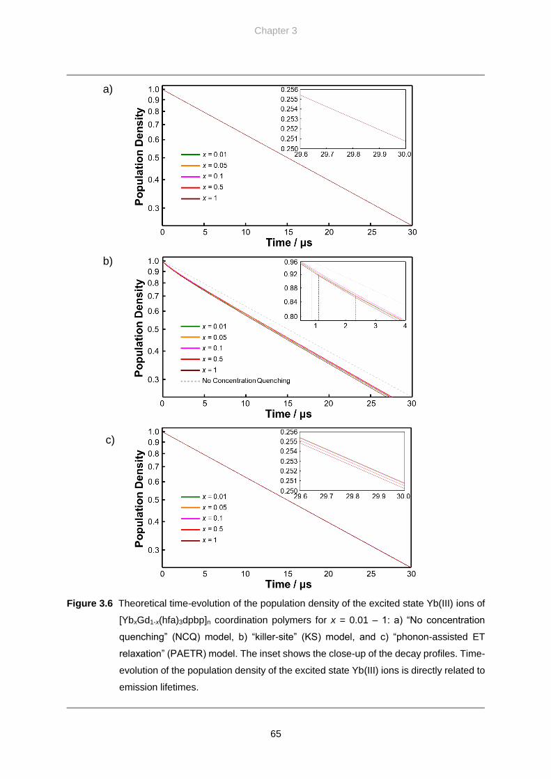
Chapter 3
65
Figure 3.6 Theoretical time-evolution of the population density of the excited state Yb(III) ions of
[YbxGd1-x(hfa)3dpbp]n coordination polymers for x = 0.01 – 1: a) “No concentration
quenching” (NCQ) model, b) “killer-site” (KS) model, and c) “phonon-assisted ET
relaxation” (PAETR) model. The inset shows the close-up of the decay profiles. Time-
evolution of the population density of the excited state Yb(III) ions is directly related to
emission lifetimes.
a)
b)
c)

Concentration Quenching in Ytterbium Coordination Polymers
66
killer-site, the shape of the decay profile was multi-component for all concentration, even at x
= 0.01 (Figure 3.6b). The average lifetimes did not change significantly with concentration,
contrary to what would generally be expected for this model. There are several points of the
decay profiles of each concentration that crosses (Figure 3.6b inset), indicating that the
concentrations considered in this calculation are within the range of transition phase from a
limited-diffusion to a fast-diffusion scheme. These features are not observed in the case of
PAETR model as the decay profile remains single-component throughout all concentrations
and gradually decreases with increasing concentration (Figure 3.6c). The difference in the
photophysical behavior between the two concentration quenching mechanisms arises from the
degree of localization of the quenching. Concentration quenching in the KS model is a localized
mechanism where the excited state energy is delivered to the killer-site via energy migration.
In the case of [YbxGd1-x(hfa)3dpbp]n coordination polymers, the maximum ET rate for the
central Yb(III) ion (red dot in Figure 3.2) is 20378 s-1, which is approximately only the half of
the fundamental decay rate constant (𝐴rad+𝐴nrad) of 46109 s-1. This implies that energy
migration does not diffuse to large spatial area and average out the lifetimes of individual
Yb(III) ion. Depending on the position of the killer-site relative to the excited Yb(III) ion, the
effect of the quenching can change drastically. In total, the emission from multiple spherical
ensembles with the various position of killer-sites lead to the multi-component decay. On the
other hand, concentration quenching in the PAETR model is a well-distributed effect since
Figure 3.7 Plot of theoretical lifetimes for the PAETR model.

Chapter 3
67
phonon-assisted ET relaxation occurs between all Yb(III) ions that are close. This results in the
decay profile that is apparently single-component for all concentrations.
Of the three different models, the experimental emission decay profiles (Figure 3.5a) are
more consistent with the theoretical emission decay profiles for the PAETR model (Figure
3.6c). Both show similar shape for the decrease in the emission lifetimes with concentration
beyond x = 0.01 (Figure 3.5b and 3.7), and the profiles are single-component at all
concentrations. Meanwhile, the rate of decrease in the lifetimes is significantly smaller for the
theoretical lifetime. This is likely because of two reasons: the increased electron-phonon
coupling between Yb(III) ion pairs and the larger actual ET rate than the calculated values (see
3.6.3.). Auzel[31,32] and Vink[33] both reported that electron-phonon coupling strength itself
increases with increased concentration of luminescent center. This could slightly increase in
both 𝐴nrad and 𝑊PAETR. Also, the theoretical lifetimes are calculated based on typical Yb(III)
oscillator strength of 3×10⁻⁶ but can range between 1×10⁻⁶ – 8×10⁻⁶ according to the previous
studies.[34–38] Since the ET rate constant was calculated from the oscillator strength (by deriving
values for ∑ 𝛺𝑘𝑘=2,4,6 |⟨𝑙𝑁𝑆𝐿𝐽‖𝑈(𝑘)‖𝑙𝑁𝑆′𝐿′𝐽′⟩|2 in Equations [2.23] and [2.24]), deviation in
the oscillator strength (and refractive index) can lead to deviations in the ET rate constant
roughly from 0.04 to 36 times the calculated value (which also increases 𝑊PAETR around this
order). In total, the deviations in the quantitative values in this case are within expected order.
The single component lifetime and the similar shape of the plot of the emission lifetimes
qualitatively suggest that the concentration quenching mechanism in [YbxGd1-x(hfa)3dpbp]n
coordination polymers is the phonon-assisted ET relaxation.
The nonradiative rate constant, ET rate constant, and the model of concentration quenching
are important factors that affect lifetimes, and they must be considered. As stated above, the
ET rate constant is comparatively smaller than the fundamental decay rate constant in [YbxGd1-
x(hfa)3dpbp]n coordination polymers. The maximum ET efficiency is 30.6%, and thus the
excited Yb(III) ions are rather localized. The large nonradiative rate constant 𝐴nrad of 45623
s-1 is due to the use of organic ligands with high-frequency vibration modes. In this sense, the
small effect of concentration quenching in Ln(III) coordination polymers is not only because
of the PAETR mechanism. The effect of concentration quenching is also lowered by the
reduction in the energy migration due to the large fundamental decay rate constant of Ln(III)
ions. This especially applies for NIR emitting Ln(III) ions because they are significantly more
vulnerable to multi-phonon relaxation, as in the case of [Yb(hfa)3dpbp]n coordination polymer.
Therefore, an extra care needs to be taken before judging that concentration quenching is a

Concentration Quenching in Ytterbium Coordination Polymers
68
small effect in lanthanide coordination polymers, because it may simply be due to the large
nonradiative rate constant and not the fundamental property of lanthanide coordination
polymers.
The fundamental difference between Ln(III) doped inorganic host and Ln(III) coordination
polymers is likely the concentration quenching mechanism. Inorganic hosts usually show
multi-component decay, suggesting the existence of killer-sites. On the other hand, various
reported Ln(III) coordination polymers, including [Yb(hfa)3dpbp]n, at maximum concentration
still show single-component emission lifetimes,[39–43] implying that coordination polymers in
general lack or have an extremely low concentration of killer-sites. Therefore, the
concentration quenching for coordination polymers is suggested to be dominated by the
PAETR, a small effect compared to strongly quenching killer-sites. Other than to lower the
concentration of Ln(III) ions, the strategy to reduce the effect of concentration quenching in
both types of the host is different. For inorganic hosts, this is reducing the concentration of
killer-sites (high purity host). For Ln(III) coordination polymers, the general strategy to achieve
high emission quantum yield in lanthanide complexes of eliminating phonon modes
responsible for multi-phonon relaxation is also effective in reducing concentration quenching.
Equation [3.2] indicates that smaller phonon energy ħω leads to smaller PAETR rate constant
𝑊PAETR . The results provides a new meaning in using Ln(III) coordination polymers as
functional luminescent materials based on energy transfer.
3.5. Conclusion
The concentration quenching mechanism and the extent of its effect in [YbxGd1-
x(hfa)3dpbp]n (x = 0.001, 0.005, 0.01, 0.05, 0.1, 0.5, 1) coordination polymers were investigated.
Emission spectra revealed that the shorter wavelength region of the Yb(III) emission is
deformed with increasing concentration of Yb(III) ions, indicating self-trapping. Emission
lifetimes were single-component at all concentrations and decreased with increasing
concentration of Yb(III) ions. The minimum Yb–Yb distance of the polymer of 10.4 Å
suppresses long-range energy migration. For this reason, the concentration mechanism is in the
limited-diffusion regime. Theoretical calculation of emission lifetimes was performed for the
killer-site model and phonon-assisted energy transfer relaxation model. The calculated and
experimental results were qualitatively more consistent in the latter model, suggesting that
[YbxGd1-x(hfa)3dpbp]n acts as a high-purity host in terms of luminescence. This report is

Chapter 3
69
another step forward in understanding the fundamentals of lanthanide chemistry, especially
lanthanide coordination polymers and lanthanide-doped inorganic hosts.
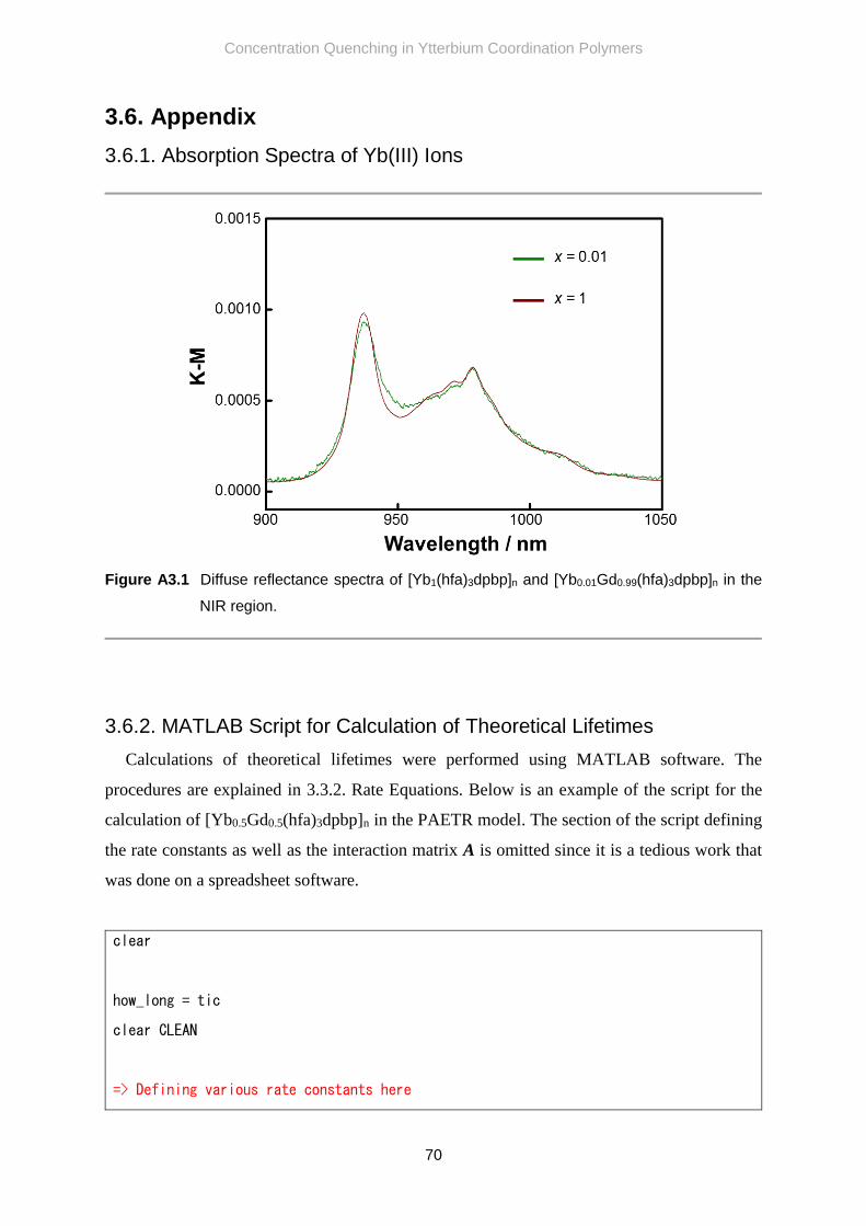
Concentration Quenching in Ytterbium Coordination Polymers
70
3.6. Appendix
3.6.1. Absorption Spectra of Yb(III) Ions
Figure A3.1 Diffuse reflectance spectra of [Yb1(hfa)3dpbp]n and [Yb0.01Gd0.99(hfa)3dpbp]n in the
NIR region.
3.6.2. MATLAB Script for Calculation of Theoretical Lifetimes
Calculations of theoretical lifetimes were performed using MATLAB software. The
procedures are explained in 3.3.2. Rate Equations. Below is an example of the script for the
calculation of [Yb0.5Gd0.5(hfa)3dpbp]n in the PAETR model. The section of the script defining
the rate constants as well as the interaction matrix A is omitted since it is a tedious work that
was done on a spreadsheet software.
clear
how_long = tic
clear CLEAN
=> Defining various rate constants here

Chapter 3
71
=> Defining Interaction Matrix A here
m = 500 % Cycle of calculation
%Yb0.5
S1 = sym(zeros(1, m))
clear S2 t
syms t S2(t)
S2(t) = 0
wb = waitbar(0, 'Calculating for x = 0.5')
CLEAN = onCleanup(@() close(wb))
for n = 1:m
waitbar(n/m, wb, 'Calculating for x = 0.5');
A2 = A1
% Random generation of a quenching center(s)
h = 0 % max# of QCs
r = h + 1
B1 = setdiff(1:135,68) .'
q = randi(r,1) - 1 % Random number of QCs
if q == 0
B2 = [] % Do nothing to B1
else
B2 = zeros(q,1)
for i = 1:q
k = randi(length(B1),1)
B2(i) = B1(k)
B1(k) = []
A2(:, B2(i)) = 0
A2(B2(i), :) = A2(B2(i), :)*1000 % Strong Quenching Center
A2(B2(i),B2(i)) = -Anrad
end
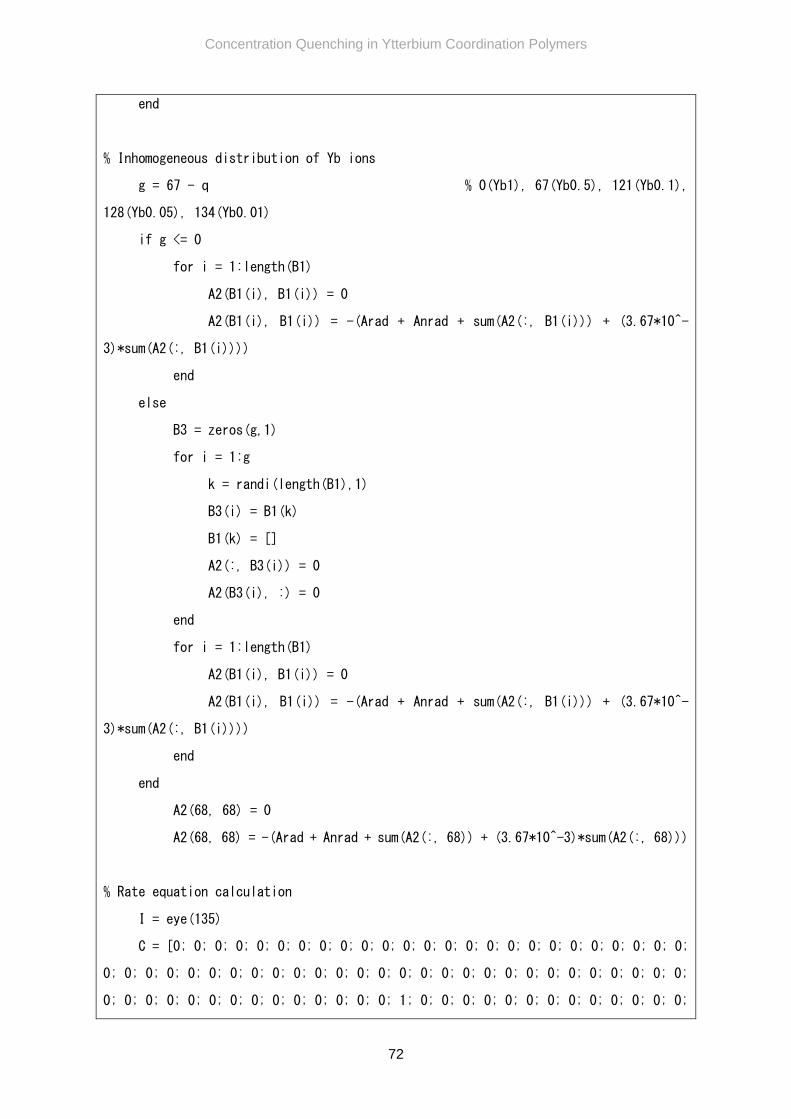
Concentration Quenching in Ytterbium Coordination Polymers
72
end
% Inhomogeneous distribution of Yb ions
g = 67 - q % 0(Yb1), 67(Yb0.5), 121(Yb0.1),
128(Yb0.05), 134(Yb0.01)
if g <= 0
for i = 1:length(B1)
A2(B1(i), B1(i)) = 0
A2(B1(i), B1(i)) = -(Arad + Anrad + sum(A2(:, B1(i))) + (3.67*10^-
3)*sum(A2(:, B1(i))))
end
else
B3 = zeros(g,1)
for i = 1:g
k = randi(length(B1),1)
B3(i) = B1(k)
B1(k) = []
A2(:, B3(i)) = 0
A2(B3(i), :) = 0
end
for i = 1:length(B1)
A2(B1(i), B1(i)) = 0
A2(B1(i), B1(i)) = -(Arad + Anrad + sum(A2(:, B1(i))) + (3.67*10^-
3)*sum(A2(:, B1(i))))
end
end
A2(68, 68) = 0
A2(68, 68) = -(Arad + Anrad + sum(A2(:, 68)) + (3.67*10^-3)*sum(A2(:, 68)))
% Rate equation calculation
I = eye(135)
C = [0; 0; 0; 0; 0; 0; 0; 0; 0; 0; 0; 0; 0; 0; 0; 0; 0; 0; 0; 0; 0; 0; 0; 0; 0;
0; 0; 0; 0; 0; 0; 0; 0; 0; 0; 0; 0; 0; 0; 0; 0; 0; 0; 0; 0; 0; 0; 0; 0; 0; 0; 0; 0;
0; 0; 0; 0; 0; 0; 0; 0; 0; 0; 0; 0; 0; 0; 1; 0; 0; 0; 0; 0; 0; 0; 0; 0; 0; 0; 0; 0;

Chapter 3
73
0; 0; 0; 0; 0; 0; 0; 0; 0; 0; 0; 0; 0; 0; 0; 0; 0; 0; 0; 0; 0; 0; 0; 0; 0; 0; 0; 0;
0; 0; 0; 0; 0; 0; 0; 0; 0; 0; 0; 0; 0; 0; 0; 0; 0; 0; 0; 0; 0; 0; 0; 0; 0; 0]
[P,D] = eig(A2)
V = (P*expm(D*t)*P^-1)*C
S1(n) = sum(V(B1,1)) + V(68,1)
end % End of entire cycle
% Storing to final rate equation
S2(t) = sum(S1)
clear CLEAN
diary EEPCYb05.txt
S2(t)
% Graph
t = 0:0.0000001:0.00003
y = vpa(S2(t))
plot(t,y,'color','y','Linewidth',3)
Title = title('Yb Concentration Quenching','FontSize',15)
ylabel('Population Density','FontSize',10)
xlabel('Time / s','FontSize',10)
axis([0 0.00003 0 m])
S1
toc(how_long)
diary off
The calculation for each model took anywhere from 3 – 6 days to complete (on a 64-bit
Windows 7, Intel Xeon E3-1241 v3 (8M cache, 3.50 GHz, 4 cores, 8 threads), 32 GB RAM
computer) depending on the concentration of Yb(III) ions, the number of erasing operations

Concentration Quenching in Ytterbium Coordination Polymers
74
done to the interaction matrix A to account for the inhomogeneity, and the complexity of the
equation for each cycle. For example, the calculation for the KS model took approximately 4.6
days to complete while the calculation for the PAETR model took approximately 6.1 days to
complete.
3.6.3. Quantitative Considerations of Theoretical Lifetimes
The plot of the experimental (Figure 3.5b) and theoretical lifetimes (Figure 3.7) differ in the
order of decrease; the former decreased from 21.7 µs to 19.2 µs while the latter decreased from
21.69 µs to only 21.65 µs. As explained in 3.4.3., this is likely due to assumed oscillator
strength and Huang-Rhys parameter. In order to account for this, the absorption spectrum of
Yb(hfa)3(TPPO)2[44] was measured to estimate the oscillator strength. This Yb(III) complex
have similar ligands (hfa and phosphine oxide ligands) that are coordinated in a similar
geometrical form as that of [Yb(hfa)3dpbp]n. Thus, the oscillator strength between these two
complexes are likely similar as well.
The absorption spectra of Yb(hfa)3(TPPO)2 in 1.0×10⁻² M chloroform solution (n = 1.43 at
1000 nm[45–47]) is shown in Figure A3.2. The oscillator strength calculated from the area of the
Figure A3.2 Absorption spectrum of Yb(hfa)3(TPPO)2 in 1.0×10⁻² M chloroform solution.

Chapter 3
75
absorption spectra was 7.24×10⁻⁶. This is more than twice as stronger than the assumed
oscillator strength (3×10⁻⁶), which means the energy transfer rate constant is significantly
increased and thus the PAETR rate constant as well.
The Huang-Rhys parameter was determined from absorption and emission spectra, but it is
known from previous reports that this will not provide a very accurate value, and instead should
be calculated from the ratio of intensity of phonon sideband and zero-phonon band.[48–50]
Because of the complex absorption spectra (Figure A3.1), it is difficult to consider the phonon
sideband. As such, we instead assume a value. Huang-Rhys parameter s can range from 10-2 to
10-1 order in lanthanides.[48–50] Here, the s = 2×10⁻¹ were used based on ref 49. Also, in order
to keep the discussion general, the refractive index was set to 1. The reconsidered parameters
are given in Table A3.1.
The recalculated theoretical lifetimes for PAETR model is shown in Figure A3.3. The decay
profile is still shown to be single exponential for all concentration and gradually decreases with
increasing concentration. The lifetimes decreased from 21.69 µs to 18.31 µs. The decrease is
now well within the order of decrease seen in the experiment.
Table A3.1 Values reconsidered for the theoretical calculation.
Constant Value Constant Value
𝜒abs 1 ħωa 3141 cm-1 (ArC-H)
𝑓𝑡𝑜𝑡𝑎𝑙 7.24×10⁻⁶ (n = 1.43) 3068 cm-1 (ArC-H)
𝑓MD 3.808 ×10⁻⁷ 1654 cm-1 (C=O)
𝑓ED 6.981×10⁻⁶ 1599 cm-1 (ArC=C)
𝐴tot 4.611×10⁴ s-1 1551 cm-1 (ArC=C)
∑ 𝛺𝑘
𝑘=2,4,6
|⟨𝑙𝑁𝑆𝐿𝐽‖𝑈(𝑘)‖𝑙𝑁𝑆′𝐿′𝐽′⟩|2 2.732×10⁻²⁰ cm2
1527 cm-1 (ArC=C)
𝐴rad(MD) 3.808×101 s-1 1439 cm-1 (P-C)
𝐴rad(ED) 1.028×10² s-1 1254 cm-1 (P=O)
𝐴nrad 4.504×10⁴ s-1 1200 cm-1 (C-F)
𝑊ET(dd) 7.729×10⁹×R-6 [s-1] N a 3 (ArC-H)
𝑊ET(dq) 3.640×10¹¹×R-8 [s-1] 3 (ArC-H)
𝑊ET(qq) 3.201×10¹³×R-10 [s-1] 6 (C=O)
s 0.2 6 (ArC=C)
𝑊PAETR 𝑊tot×7.443×10⁻² [s-1] 6 (ArC=C)
6 (ArC=C)
6 (P-C)
8 (P=O)
8 (C-F)
[a] Vibration below ArC-H have minor influence on the value of 𝑊PAETR

Concentration Quenching in Ytterbium Coordination Polymers
76
Figure A3.3 a) Recalculated theoretical time-evolution of the population density of the excited
state Yb(III) ions of [YbxGd1-x(hfa)3dpbp]n coordination polymers for x = 0.01 – 1 for
PAETR model. b) Plot of the theoretical lifetimes for the PAETR model.
3.7. References
[1] D. L. Dexter and J. H. Schulman, J. Chem. Phys. 1954, 22, 1063–1070.
[2] B. Di Bartolo and V. Goldberg, Radiationless Processes, Plenum Press, 1980.
[3] F. Auzel, G. Baldacchini, L. Laversenne, G. Boulon, Opt. Mater. 2003, 24, 103–109.
[4] F. Benz and H. P. Strunk, AIP Adv. 2012, 2, 2–7.
[5] W. Wei, G. Chen, A. Baev, G. S. He, W. Shao, J. Damasco, P. N. Prasad, J. Am. Chem.
Soc. 2016, 138, 15130–15133.

Chapter 3
77
[6] P. Yang, P. Deng, Z. Yin, J. Lumin. 2002, 97, 51–54.
[7] M. Ito, G. Boulon, A. Bensalah, Y. Guyot, C. Goutaudier, H. Sato, J. Opt. Soc. Am. B
2007, 24, 3023–3032.
[8] L. G. Jacobsohn, B. L. Bennett, R. E. Muenchausen, J. F. Smith, D. Wayne Cooke,
Radiat. Meas. 2007, 42, 675–678.
[9] Y. Cui, B. Chen, G. Qian, Coord. Chem. Rev. 2014, 273–274, 76–86.
[10] J. Heine and K. Müller-Buschbaum, Chem. Soc. Rev. 2013, 43, 9232–9242.
[11] B. Yan, Y. Bai, Z. Chen, J. Mol. Struct. 2005, 741, 141–147.
[12] F. Auzel, F. Bonfigli, S. Gagliari, G. Baldacchini, J. Lumin. 2001, 94–95, 293–297.
[13] J. Knoester, J. E. Van Himbergen, J. Chem. Phys. 1987, 86, 3571.
[14] F. Auzel, Chem. Rev. 2004, 104, 139–173.
[15] F. Auzel, J. Lumin. 2002, 100, 125–130.
[16] T. Miyakawa and D. L. Dexter, Phys. Rev. B 1970, 1, 2961–2969.
[17] R. Balda, J. Fernández, I. Saéz de Ocáriz, J. L. Adam, A. Mendioroz, E. Montoya, Opt.
Mater. 1999, 13, 159–165.
[18] V. V. Utochnikova, A. S. Kalyakina, I. S. Bushmarinov, A. A. Vashchenko, L.
Marciniak, A. M. Kaczmarek, R. Van Deun, S. Bräse, N. P. Kuzmina, J. Mater. Chem.
C 2016, 4, 9848–9855.
[19] K. Miyata, Y. Konno, T. Nakanishi, A. Kobayashi, M. Kato, K. Fushimi, Y. Hasegawa,
Angew. Chem. Int. Ed. 2013, 52, 6413–6416.
[20] Y. Hirai, T. Nakanishi, K. Miyata, K. Fushimi, Y. Hasegawa, Mater. Lett. 2014, 130,
91–93.
[21] K. Miyata, T. Ohba, A. Kobayashi, M. Kato, T. Nakanishi, K. Fushimi, Y. Hasegawa,
ChemPlusChem 2012, 77, 277–280.
[22] B. R. Judd, Phys. Rev. 1962, 127, 750–761.
[23] G. S. Ofelt, J. Chem. Phys. 1962, 37, 511–520.
[24] M. P. Hehlen, M. G. Brik, K. W. Krämer, J. Lumin. 2013, 136, 221–239.
[25] T. Kushida, J. Phys. Soc. Jpn. 1973, 34, 1318–1326.
[26] O. L. Malta, J. Non. Cryst. Solids 2008, 354, 4770–4776.
[27] L. Aboshyan-Sorgho, H. Nozary, A. Aebischer, J. C. G. Bünzli, P. Y. Morgantini, K. R.
Kittilstved, A. Hauser, S. V. Eliseeva, S. Petoud, C. Piguet, J. Am. Chem. Soc. 2012,
134, 12675–12684.
[28] M. E. Starzak, Mathematical Methods in Chemistry and Physics, Springer, 1989.
[29] J.-C. G. Bünzli and S. V Eliseeva, Basics of Lanthanide Photophysics, in Springer Series

Concentration Quenching in Ytterbium Coordination Polymers
78
on Fluorescence, Springer, 2011.
[30] M. Ito, C. Goutaudier, Y. Guyot, K. Lebbou, T. Fukuda, G. Boulon, J. Phys. Condens.
Matter 2004, 16, 1501–1521.
[31] F. Auzel, G. F. de Sá, W. M. de Azevedo, J. Lumin. 1980, 21, 187–192.
[32] F. Auzel and F. Pellé, J. Lumin. 1996, 69, 249–255.
[33] A. P. Vink, A. Meijerink, Spectrochim. Acta Part A 1998, 54, 1755–1761.
[34] W. T. Carnall, P. R. Fields, K. Rajnak, J. Chem. Phys. 1968, 49, 4412.
[35] F. Auzel, A. M. Jean-Louis, Y. Toudic, J. Appl. Phys. 1989, 66, 3952–3955.
[36] K. Venkata Krishnaiah, R. Rajeswari, K. Upendra Kumar, S. Surendra Babu, I. R.
Martin, C. K. Jayasankar, J. Quant. Spectrosc. Radiat. Transf. 2014, 140, 37–47.
[37] E. A. Seregina, A. A. Seregin, G. V Tikhonov, Opt. Spectrosc. 2014, 116, 438–453.
[38] S. Balaji, A. K. Mandal, K. Annapurna, Opt. Mater. 2012, 34, 1930–1934.
[39] W.-G. Lu, D.-C. Zhong, L. Jiang, T.-B. Lu, Cryst. Growth Des. 2012, 12, 3675–3683.
[40] R. Decadt, K. van Hecke, D. Depla, K. Leus, D. Weinberger, I. van Driessche, P. Van
Der Voort, R. van Deun, Inorg. Chem. 2012, 51, 11623–11634.
[41] S. Chen, R. Fan, C. Sun, P. Wang, Y. Yang, Q. Su, Y. Mu, Cryst. Growth Des. 2012,
12, 1337–1346.
[42] Z. Y. Li, J. W. Dai, N. Wang, H. H. Qiu, S. T. Yue, Y. L. Liu, Cryst. Growth Des. 2010,
10, 2746–2751.
[43] A. R. Ramya, D. Sharma, S. Natarajan, M. L. P. Reddy, Inorg. Chem. 2012, 51, 8818–
26.
[44] S. Kishimoto, T. Nakagawa, T. Kawai, Y. Hasegawa, Bull. Chem. Soc. Jpn. 2011, 84,
1–5.
[45] A. Samoc, J. Appl. Phys. 2003, 94, 6167–6174.
[46] H. El-Kashef, Opt. Mater. 2002, 20, 81–86.
[47] S. Kedenburg, M. Vieweg, T. Gissibl, H. Giessen, Opt. Mater. Express 2012, 2, 1588.
[48] C. de Mello Donegá, A. Meijerink, G. Blasse, J. Phys. Condens. Matter 1992, 4, 8889–
8902.
[49] C. de Mello Donegá, S. Schenker, H. F. Folkerts, A. Meijerink, G. Blasse, J. Phys.
Condens. Matter 1994, 6, 6043–6056.
[50] M. P. Hehlen, A. Kuditcher, S. C. Rand, M. A. Tischler, J. Chem. Phys. 1997, 107,
4886–4892.

Chapter 4
SPIN-ORBIT COUPLING AND ENERGY TRANSFER
IN NONANUCLEAR LANTHANIDE CLUSTERS

Spin-Orbit Coupling and Energy Transfer in Nonanuclear Lanthanide Clusters
80
4.1. Introduction
Spin-orbit coupling (SOC) plays an important role in Ln(III) complexes to efficiently
produce the T1 state of organic ligands, which is the state that FET proceeds from.[1–3] SOC
mixes states of different multiplicity, and thus S1 and T1, as well as T1 and S0 states are mixed
(see 4.5.1.).[4,5] This makes ISC and phosphorescence partially allowed processes. There are
two ISC processes S1→T1 and T1→S0. So the relaxation of the T1 state includes partially
allowed phosphorescence and ISC to the ground state.
The SOC in Ln(III) complexes is strong due to 1) the large effective nuclear charge of
Ln(III) ions (the “heavy atom effect”), and 2) the spin and orbital angular momentum of 4f-
electrons (the “paramagnetic effect”). As a result, S1→T1 ISC rate in Ln(III) complexes are
assumed to be fast and ranges anywhere between 107 to 1010 s-1.[6–8] Reports by S. Tobita and
D. M. Guldi both showed that the difference in SOC arises predominantly from the
paramagnetic effect rather than the heavy atom effect in the lanthanide series. For example,
Gd(III) complex with methyl salicylate ligands showed an order of magnitude larger S1→T1
ISC and T1→S0 relaxation rates compared to Lu(III) complex despite Lu(III) being the heavier
atom. D. M. Guldi and his coworkers further demonstrated that both S1→T1 ISC and T1→S0
relaxation rates are roughly proportional to the magnetic moment of the lanthanides.[8]
Here, the effect of SOC on the sensitization efficiency of Ln(III) complexes is considered.
As mentioned in the 1.3.1., the sensitization efficiency 𝜂sens is given by the following
equation:
𝜂sens = 𝜂ISC × 𝜂FET
=𝑘ISC
𝑘𝑟S1+𝑘𝑛𝑟S1+𝑘ISC×
𝑘FET
𝑘𝑟T1+𝑘𝑛𝑟T1+𝑘FET , [4.1]
where 𝑘ISC, 𝑘FET, are S1→T1 ISC, FET rate constants, respectively. 𝑘𝑟 and 𝑘𝑛𝑟 are radiative
and nonradiative rate constants, respectively, and the subscript denotes the states in which they
apply to. Since SOC increases 𝑘ISC, 𝑘𝑟T1, and 𝑘𝑛𝑟T1
, it can be inferred from Equation [4.1] that
strong SOC enhances 𝜂ISC while reducing 𝜂FET. The opposing effect that SOC has on 𝜂ISC and
𝜂FET indicate that the sensitization efficiency 𝜂sens, which is the product of 𝜂ISC and 𝜂FET ,
should have an optimal point in terms of SOC strength.
Experimentally investigating the existence of the optimal point, however, is difficult. SOC
is determined by the specific Ln(III) ion that is being used. Changing the Ln(III) ion also
changes the FET rate constant 𝑘FET as well (all Ln(III) ions have different 4f-state energy

Chapter 4
81
levels) and thus, direct comparison of the effect of SOC induced by Ln(III) cannot be
determined spectroscopically. Herein, nonanuclear Ln(III) clusters was selected as the suitable
material.[9] The clusters allow control of SOC by mixing different Ln(III) ions without
significantly distorting the structure. Yb(III), Gd(III), and Lu(III) ions were chosen for this
study. The Yb(III) ion with its two-level 4f-states (2F7/2 and 2F5/2 states) is suitable as the
emissive center for the simplicity in discussing T1→Yb(2F5/2) ET process (the FET). Gd(III)
and Lu(III) ions were chosen as the non-emissive paramagnetic and diamagnetic centers,
respectively.
In this chapter, the effect of SOC strength induced by paramagnetic and diamagnetic Ln(III)
ions on the sensitization efficiency is described. Nonanuclear YbnGd9-n and YbnLu9-n clusters
with butyl salicylate ligands ([Ln9(μ-OH)10(butyl salicylate)16]NO3, Ln = YbnGd9-n / YbnLu9-n,
n = 0, 1, 3, 7, 9) were synthesized. The clusters in the chapter are abbreviated as YbnGd9-n-Bu
and YbnLu9-n-Bu (Bu = butyl salicylate ligands). The clusters were identified by IR, mass
spectroscopy, and elemental analysis. The structural analysis was performed by the
combination of powder and single-crystal XRD measurements. The effect of SOC was
elucidated by emission spectra and lifetimes. This study explore whether the sensitization
efficiency is improved or reduced in the presence of a strong SOC. Either result provides
important insight into fundamental photophysics of Ln(III) complexes as well as factors to
consider when designing them to optimize the energy transfer efficiency.
4.2. Experimental Section
4.2.1. Material
Gd(NO3)3·6H2O (>99.95%) and Lu(NO3)3·4H2O (>99.95) were purchased from Kanto
Chemical Co., and Yb(NO3)3·5H2O (99.9%) was purchased from Sigma-Aldrich Japan.
Butylsalicylate was purchased from Tokyo Chemical Industries. Methanol for spectroscopy
was purchased from Wako Pure Chemical Industries, Ltd. All reagents were used without
further purification.
4.2.2. Apparatus
FAB-MS and ESI-MS spectra were measured on a JEOL JMS-700TZ and a JEOL JMS-
T100LP, respectively. Elemental analyses were performed by an Exter Analytical CE440.
Infrared spectra were recorded on a JASCO FT/IR-4600 spectrometer. XRD spectra were

Spin-Orbit Coupling and Energy Transfer in Nonanuclear Lanthanide Clusters
82
characterized by a RIGAKU SmartLab X-ray diffractometer. Single crystal X-ray diffractions
were made on a RIGAKU RAXIS RAPID imaging plate area detector.
4.2.3. Synthesis
General synthetic procedures of YbnGd9-n-Bu and YbnLu9-n-Bu: The general synthetic
procedures are shown in Scheme 4.1. 2.7 mmol of butyl salicylate ligand (Bu) was dissolved
in methanol, and triethylamine (0.44 mL, 4.40 mmol) was added to this solution while stirred
at 40 ˚C. 1.52 mmol of the appropriate ratio of Gd(NO3)3·6H2O and Yb(NO3)3·5H2O or
Lu(NO3)3·4H2O dissolved in methanol was added dropwise to this solution and further stirred
for 60 minutes. The resulting white precipitate (the target cluster) was vacuum filtered and
dried in vacuum. The clusters were recrystallized by evaporation method from a concentrated
methanol solution.
Gd9-Bu: Yield: 68%. Selected IR (ATR, cm-1): 3573 (w, O–H), 3235 (w, O–H), 2956 (m, C–
H), 1319 (s, C–O). FAB-MS: m/z = 4676.8 [Gd9(µ-OH)10(Bu)16]+. Elemental analysis
calculated for C176H218NO61Gd9: C, 44.61%, H, 4.64%, N, 0.30%. Found: C, 44.30%, H, 4.53%,
N, < 0.30%.
Yb1Gd8-Bu: Yield: 67%. Selected IR (ATR, cm-1): 3572 (w, O–H), 3235 (w, O–H), 2957 (m,
C–H), 1319 (s, C–O). FAB-MS: m/z = 4692.2 [Yb1Gd8(µ-OH)10(Bu)16]+. Elemental analysis
calculated for C176H218NO61Yb1Gd8: C, 44.46%, H, 4.62%, N, 0.29%. Found: C, 43.98%, H,
Scheme 4.1 Synthetic procedure of Ln9-Bu. Ln9 = YbnGd9-n or YbnLu9-n.

Chapter 4
83
4.45%, N, < 0.30%.
Yb3Gd6-Bu: Yield: 78%. Selected IR (ATR, cm-1): 3572 (w, O–H), 3223 (w, O–H), 2957 (m,
C–H), 1319 (s, C–O). FAB-MS: m/z = 4724.3 [Yb3Gd6(µ-OH)10(Bu)16]+. Elemental analysis
calculated for C176H218NO61Yb3Gd6: C, 44.17%, H, 4.59%, N, 0.29%. Found: C, 43.77%, H,
4.41%, N, < 0.30%.
Yb7Gd2-Bu: Yield: 85%. Selected IR (ATR, cm-1): 3582 (w, O–H), 3210 (w, O–H), 2957 (m,
C–H), 1321 (s, C–O). FAB-MS: m/z = 4785.8 [Yb7Gd2(µ-OH)10(Bu)16]+. Elemental analysis
calculated for C176H218NO61Yb7Gd2: C, 43.59%, H, 4.53%, N, 0.29%. Found: C, 43.27%, H,
4.36%, N, < 0.30%.
Yb9-Bu: Yield: 78%. Selected IR (ATR, cm-1): 3585 (w, O–H), 3206 (w, O–H), 2956 (m, C–
H), 1320 (s, C–O). FAB-MS: m/z = 4819.0 [Yb9(µ-OH)10(Bu)16]+. Elemental analysis
calculated for C176H218NO61Yb9: C, 43.31%, H, 4.50%, N, 0.29%. Found: C, 42.99%, H, 4.37%,
N, < 0.30%.
Yb7Lu2-Bu: Yield: 80%. Selected IR (ATR, cm-1): 3585 (w, O–H), 3226 (w, O–H), 2956 (m,
C–H), 1321 (s, C–O). ESI-MS: m/z = 4822.74 [Yb7Lu2(µ-OH)10(Bu)16]+. Elemental analysis
calculated for C176H218NO61Yb7Lu2: C, 43.27%, H, 4.50%, N, 0.29%. Found: C, 42.94%, H,
4.37%, N, < 0.30%.
Yb3Lu6-Bu: Yield: 28%. Selected IR (ATR, cm-1): 3584 (w, O–H), 3206 (w, O–H), 2957 (m,
C–H), 1321 (s, C–O). ESI-MS: m/z = 4830.73 [Yb3Lu6(µ-OH)10(Bu)16]+. Elemental analysis
calculated for C176H218NO61Yb3Lu6: C, 43.21%, H, 4.49%, N, 0.29%. Found: C, 42.73%, H,
4.31%, N, 0.25%.
Yb1Lu8-Bu: Yield: 81%. Selected IR (ATR, cm-1): 3586 (w, O–H), 3208 (w, O–H), 2956 (m,
C–H), 1321 (s, C–O). ESI-MS: m/z = 4834.75 [Yb1Lu8(µ-OH)10(Bu)16]+. Elemental analysis
calculated for C176H218NO61Yb1Lu8: C, 43.17%, H, 4.49%, N, 0.29%. Found: C, 43.04%, H,
4.36%, N, < 0.30%.
Lu9-Bu: Yield: 80%. Selected IR (ATR, cm-1): 3586 (w, O–H), 3207 (w, O–H), 2957 (m, C–
H), 1321 (s, C–O). FAB-MS: m/z = 4836.1 [Lu9(µ-OH)10(Bu)16]+. Elemental analysis
calculated for C176H218NO61Lu9: C, 43.16%, H, 4.49%, N, 0.29%. Found: C, 42.82%, H, 4.33%,
N, < 0.30%.
4.2.4. Spectroscopy
Absorption spectra of the clusters were obtained by using a JASCO V-670 spectrometer.
Emission spectra and lifetimes were measured using a Horiba/Jobin-Yvon FluoroLog-3ps

Spin-Orbit Coupling and Energy Transfer in Nonanuclear Lanthanide Clusters
84
spectrofluorometer. The emission spectra of UV-vis and NIR region were connected using
Eu(hfa)3(TPPO)2 complex as a reference to match the detection level of both detectors. A
combination of JASCO FP-6600 spectrometer and Oxford Instruments OptistatDN2 cryostat
was used to measure emission spectra at low temperature. The absolute quantum yield was
measured by a combination of JASCO FP-6600 and an integration sphere.
4.2.5. Crystallography
Colorless single crystal of Gd9-Bu, Yb9-Bu, and Lu9-Bu obtained from solutions in methanol
were mounted on a glass fiber by using epoxy resin glue. All measurements were made using
a Rigaku RAXIS RAPID imaging plate area detector with graphite-monochromated MoKα
radiation. Corrections for decay and Lorentz-polarization effects were made using a spherical
absorption correction, solved by direct methods, and expanded using Fourier techniques. Non-
hydrogen atoms were refined anisotropically except for disordered atoms. Hydrogen atoms
were refined using the riding model. The final cycle of full-matrix least-squares refinement
was based on observed reflections and variable parameters. All calculations were performed
using a CrystalStructure crystallographic software package. The CIF data were confirmed by
using the checkCIF/PLATON service. CCDC-1479981 (“Gd9 cluster”), CCDC-1578015
(“Yb9 cluster”), and CCDC-1578016 (“Lu9 cluster”) contain the crystallographic data for this
paper. These data can be obtained free of charge from The Cambridge Crystallographic Data
Center via www.ccdc.cam.ac.uk/data_request/cif.
4.3. Results and Discussion
4.3.1. Structure and Identification
The crystal structures of Gd9-Bu, Yb9-Bu, and Lu9-Bu are shown in Figure 4.2 and the
crystallographic data in Table 4.1. The crystallographic analysis showed that the Ln(III) ions
in the cluster form an “hour-glass” structure in which the upper four and lower four Ln(III)
ions are connected to the center Ln(III) ion via oxygen. All Ln (III) ions have an 8-coordination
structure. The center Ln(III) ion is coordinated only by oxygen atoms and not by Bu ligands
while each of the Ln(III) ions in the outer unit is coordinated by both Bu ligands and oxygen
atoms. In the outer unit, the upper and the lower parts are twisted approximately 45 degrees
with respect to the vertical axis that goes through the two μ4-O and the center Ln(III) ion.

Chapter 4
85
Figure 4.2 ORTEP drawing (50% probability displacement ellipsoids) of a) Gd9-Bu, b) Yb9-Bu,
and c) Lu9-Bu. d) center Ln(III) ion and e) other Ln(III) ions (taken from Gd9-Bu as an
example).
Continuous shape measure analysis (CShM) was employed for the Ln(III) ions in the
clusters to investigate the coordination geometry.[10,11] Continuous Shape Measures (CShM)
was calculated using SHAPE program.[12] The CShM criterion S is given by the following
equation:
𝑆CShM = min ∑ |𝑸𝑘−𝑷𝑘|2𝑁
𝑘
∑ |𝑸𝑘−𝑸0|2𝑁𝑘
× 100 , [4.2]
a) b)
c) d)
e)

Spin-Orbit Coupling and Energy Transfer in Nonanuclear Lanthanide Clusters
86
Table 4.1 Crystallographic information of Gd9-Bu, Yb9-Bu, and Lu9-Bu
Gd9-Bu Yb9-Bu Lu9-Bu
Chemical formula C176H218Gd9NO61 C176H208O58Yb9 C176H208O58Lu9
Formula weight 4738.88 4808.90 4826.24
Crystal system Orthorhombic Tetragonal Tetragonal
Space group Pbcn P42/n (#86) P42/n (#86)
a / Å 29.1572(7) 23.5449(3) 23.857(3)
b / Å 21.2906(5) 23.5449(3) 23.857(3)
c / Å 31.7940(7) 36.1157(7) 36.2570(10)
α / deg 90 90 90
β / deg 90 90 90
γ / deg 90 90 90
V / Å3 19736.9(8) 20021.2(5) 20636(4)
Z 4 4 4
Dcalcd / g cm-3 1.595 1.595 1.553
T / K 123.15 93.15 173.15
μ (MoKα) / cm-1 30.655 42.345 43.357
Max 2θ / deg 55.0 56.6 50.7
No. of measured reflections 183154 74859 120501
No. of unique reflections 22608 22456 18839
R a 0.1215 0.0696 0.1265
wR2 b 0.2134 0.1726 0.2115
a R = Σ||F0| – |Fc|| / Σ|F0|
b wR2 = [Σw(F02 – Fc
2)2 / Σw(F02)2]1/2

Chapter 4
87
where 𝑸𝑘 is the vertices of an actual structure, 𝑸0 is the center of mass of an actual
structure, 𝑷𝑘 is the vertices of an ideal structure, and N is the number of vertices. The S value
represents the degree of the deviation from a specified ideal coordination geometry. The
smaller the value of S, the closer the structure is to a specified ideal geometry. For example, S
= 0 means that the structure is fully identical with the ideal geometry. CShM were chosen over
another method called “Shape Measure” (ShM)[13] because it accounts for the distortion of the
center metal ion from the center of mass (see 4.5.2.). The S values calculated for the clusters
are summarized in Table 4.2. For the center Ln(III) ion (Figure 4.2d), the S values ranged from
0.077 to 0.105 when calculated for 8-coordinated square antiprism (8-SAP) geometry and
2.371 to 2.481 for 8-coordinated trigonal dodecahedron (8-TDH) geometry. Therefore, the
coordination geometry of the center Ln(III) ions is attributed to the 8-SAP structure. There
was no specific dependence on the ionic radii of Ln(III) ions on the S values calculated for the
clusters as 8-SAP. On the other hand, the other Ln(III) ions (Figure 4.2e) showed smaller S
values when calculated for 8-TDH than 8-SAP. Therefore, the coordination geometry of the
Table 4.2 Continuous Shape Measure calculation results for Gd9-Bu, Yb9-Bu, and Lu9-Bu.
Gd9 cluster Yb9 cluster Lu9 cluster
8-TDH 8-SAP 8-TDH 8-SAP 8-TDH 8-SAP
Ln1 2.976 4.725 2.573 4.578 2.565 4.455
Ln2 2.836 4.813 2.545 4.546 2.481 4.633
Ln3 2.704 4.588 2.413 4.776 2.447 4.622
Ln4 2.513 4.822 2.404 4.623 2.400 4.507
Ln5 2.976 4.725 2.573 4.578 2.565 4.455
Ln6 2.836 4.813 2.545 4.546 2.481 4.633
Ln7 2.704 4.588 2.413 4.776 2.447 4.622
Ln8 2.513 4.822 2.404 4.623 2.400 4.507
Ln9a 2.481 0.082 2.396 0.077 2.371 0.105
a) Center Ln(III) ion.

Spin-Orbit Coupling and Energy Transfer in Nonanuclear Lanthanide Clusters
88
Figure 4.3 Powder XRD patterns of YbnGd9-n-Bu and YbnLu9-n-Bu.
Ln(III) ions other than the center Ln(III) is attributed to the 8-TDH structure. Unlike the center
Ln(III) ion calculated for 8-SAP, the other Ln(III) ions showed larger S values for Ln(III) ions
with larger ionic radii when calculated for 8-TDH. However, the differences are still quite small,
and these clusters can be considered to have the same structure. These structural results are
similar to that of the previously reported Tb9-Bu.[9]
As a crystal, these three clusters have different crystal system with Yb9-Bu and Lu9-Bu
being triclinic while Gd9-Bu being orthorhombic (Table 4.1). This may be due to the larger
ionic radii of Gd(III) ions with minor influence on the microscopic level but is large on the
larger scale. To compare the structure of the other clusters (YbnGd9-n-Bu and YbnLu9-n-Bu, n =
1, 3, and 7), XRD measurements were performed. The XRD patterns for the clusters shown in
Figure 4.3 are similar, but the peaks shift to the lower angle with increasing number of Yb(III)
and Lu(III) ions compared to Gd9-Bu, and the peak heights are quite different among the
clusters. The spectroscopic measurements were performed in methanol solution, and
intermolecular interaction is minimized. Therefore, any spectroscopic differences of the
clusters should arise from the structure of a single molecule. This will become important for
the later discussion because a distortion of ligand alignment may affect both the ligand and
Yb(III) emissions.

Chapter 4
89
4.3.2. Photophysical Properties
The emission intensity of the fluorescence and phosphorescence from the ligands in YbnGd9-
n-Bu and YbnLu9-n-Bu provides information on photophysical properties of the ligands (i.e.,
SOC) under the influence of the paramagnetic effect of Ln(III) ions. On the other hand, the
emission intensity of Yb(III) emission implies the sensitization efficiency 𝜂sens. The emission
spectra of YbnGd9-n-Bu and YbnLu9-n-Bu in 1.0×10⁻⁴ M degassed methanol solution in the
visible and NIR region excited at 371 nm are shown in Figure 4.4a. The peak at 410 nm
corresponds to the fluorescence from the butyl salicylate ligands. The fluorescence intensity is
the lowest for Yb9-Bu and increases with increasing number of Gd(III) or Lu(III) ions.
Increasing number of Lu(III) ions showed greater enhancement in emission intensity compared
to those with Gd(III) ions (Figure 4.4b). From these spectra, the strength of SOC in this cluster
is in the order of Yb(III) > Gd(III) > Lu(III). A similar trend was reported by D. M. Guldi[8].
The absolute quantum yield of the fluorescence of Lu9-Bu was below 1% (calculated as 0.2%).
Phosphorescence was observed at low temperature (180 K) for Gd9-Bu and Yb9-Bu at around
465 nm (Figure 4.5). The intensity of the phosphorescence in relative to the fluorescence is
significantly reduced for Yb9-Bu due to the presence of T1 → Ln energy transfer.
Phosphorescence was not observed for Lu9-Bu because of the high fluorescence intensity, and
low S1→T1 ISC and T1→S0 relaxation rate compared to those of Gd9-Bu.
Emission peak at 976 nm in Figure 4.4b originates from the 2F5/2→2F7/2 transition of Yb(III)
ions. The spectral shape of the Yb(III) near-infrared (NIR) emission was identical for all
compositions. The spectral shape of Ln(III) emission is known to be sensitive to the
coordination geometry, thus the NIR emission spectra show that the coordination geometries
around Yb(III) ions are identical for all clusters.[14,15] The continuous shape measure
calculations (see 4.5.2.) support this analysis. Therefore, the radiative rate constants of Yb(III)
ions in these clusters are equal.
The NIR emission lifetimes monitored at 976 nm (λEX = 371 nm) were single-exponential
for all compositions (Figure 4.6a). Emission lifetime values derived from single-exponential
fitting showed that the lifetimes of the clusters gradually increased with decreasing number of
Gd(III) ions and increasing number of Lu(III) ions (Figure 4.6b). In particular, the emission
lifetimes of Yb1Gd8-Bu, Yb9-Bu, and Yb1Lu8-Bu were 568, 598, and 740 ns, respectively. The
difference in the ionic radii of Gd(III), Yb(III), and Lu(III) ions may have contributed to the
difference in the values of nonradiative rate constants of Yb(III) ions in the clusters.

Spin-Orbit Coupling and Energy Transfer in Nonanuclear Lanthanide Clusters
90
Figure 4.4 a) Emission spectra of YbnGd9-n-Bu / YbnLu9-n-Bu in 1.0×10⁻⁴ M methanol solution (𝜆EX
= 371 nm). The spectra for the UV-Vis and the NIR regions were measured separately,
and connected by correcting the detectors with the previously reported
Eu(hfa)3(TPPO)2 complex. b) Plot of the emission intensity (𝜆EM = 410 nm) of the
ligand fluorescence.
a)
b)

Chapter 4
91
Figure 4.5 Emission spectra of a) Gd9-Bu, b) Yb9-Bu, and c) Lu9-Bu in 1.0×10⁻⁴ M methanol
solution at 210 K (𝜆EX = 371 nm). A Raman peak is observed at around 430 nm for
Yb9-Bu.
Figure 4.7a shows the plot of the NIR emission intensity of the clusters, normalized by the
intensity of Yb9-Bu. The emission intensities in the plot indirectly mean the sensitization
efficiency. The emission intensity of Yb1Gd8-Bu was 1.6 times higher than that of Yb1Lu8-Bu.
For these two clusters, the main contributor to the sensitization efficiency is the S1→T1 ISC
efficiency due to the SOC induced by Gd(III) or Lu(III) ions (region A). Stronger SOC by
Gd(III) ions leads to more efficient S1→T1 ISC (Figure 4.7b, green arrow) in Yb1Gd8-Bu
compared to that of Yb1Lu8-Bu. On the other hand, Yb7Gd2-Bu, Yb9-Bu, and Yb7Lu2-Bu
showed the same emission intensity. Normally, reduced number of emissive Ln(III) ions leads
to a decrease in T1→Ln energy transfer efficiencies, but this phenomenon was not observed.
Thus, for these three clusters, the T1→S0 relaxation is the main contributor to the sensitization
efficiency (region B). Gd(III) and Lu(III) ions mitigated the fast T1→S0 relaxation induced by
a)
b)
c)

Spin-Orbit Coupling and Energy Transfer in Nonanuclear Lanthanide Clusters
92
the Yb(III) ions (Figure 4.7c, blue arrow) and thus the sensitization efficiency did not drop.
These results suggest that there is an optimal strength of SOC for a high sensitization efficiency
in Ln(III) complexes.
Figure 4.6 a) Emission decay profiles of YbnGd9-n-Bu / YbnLu9-n-Bu in 1.0×10⁻⁴ M methanol
solution (𝜆EX = 371 nm, 𝜆EM = 976 nm). b) Lifetime plot of the clusters.
a)
b)

Chapter 4
93
Figure 4.7 a) Plot of the NIR emission intensities normalized by the peak intensity of Yb9-Bu. The
shaded area represents the two different regions in which the dominating photophysical process
differs. b) Comparison of the photophysical processes in Yb1Gd8-Bu and Yb1Lu8-Bu (region A) and
c) Yb9-Bu, Yb7Lu2-Bu, Yb7Gd2-Bu (region B). The thickness of the arrows represents the yield
resulting from the process (not the rate constant of the process).
a)
b)
c)

Spin-Orbit Coupling and Energy Transfer in Nonanuclear Lanthanide Clusters
94
4.4. Conclusion
In this chapter, the effect of SOC strength induced by paramagnetic and diamagnetic Ln(III)
ions on the sensitization efficiency in nonanuclear Ln(III) clusters was explored. Nonanuclear
YbnGd9-n-Bu and YbnLu9-n-Bu ([Ln9(μ-OH)10(Bu)16]+NO3
-, Ln = YbnGd9-n / YbnLu9-n, n = 0, 1,
3, 7, 9) were synthesized and investigated their photophysical properties by emission spectra
and lifetimes. The result implies that SOC affects sensitization efficiency both positively and
negatively depending on the strength of SOC, and thus there is an optimal region in the SOC
strength. It is also suggested that mixing Ln(III) ions with different SOC in Ln(III) clusters is
an effective method to optimize sensitization efficiency. The findings are another step forward
in understanding the fundamental photophysical processes in Ln(III) complexes, as well as
providing a possible novel strategy to enhance the luminescence efficiency.
4.5. Appendix
4.5.1. Notes on Spin-Orbit Coupling
To be precise, spin-orbit coupling Hamiltonian 𝐻SO does not always mix S1 and T1 state
since this depends on the symmetry of the two states. In fact, spin-orbit coupling may mix
higher singlet excited states Sn with another higher triplet excited states Tn. For example,
mixing between singlet and triplet nπ* state as well as singlet and triplet ππ* states is not large.
On the other hand, mixing of nπ* and ππ* state of different multiplicity is large (known as the
El-Sayed rule).[16] Nonetheless, the convention is that the intersystem crossing is written as a
process that occurs between S1 to T1 for simplicity.[17] Since this study focuses on nonanuclear
YbnGd9-n and YbnLu9-n with the same butylsalicylate organic ligands (Bu), the mixing problem
of S1 and T1 states can be neglected as long as the comparison is between these clusters.

Chapter 4
95
4.5.2. Continuous Shape Measure and Shape Measure
Continuous shape measure (CShM) calculates the deviation of the vertices of an actual
structure from the vertices of an ideal structure.[10] The CShM criterion 𝑆CShM is given by the
following equation:
𝑆CShM = min ∑ |𝑸𝑘−𝑷𝑘|2𝑁
𝑘
∑ |𝑸𝑘−𝑸0|2𝑁𝑘
× 100 [4.2]
where 𝑸𝑘 is the vertices of an actual structure, 𝑸0 is the center of mass of an actual structure,
𝑷𝑘 is the vertices of an ideal structure, and N is the number of vertices. In the case of Gd9-Bu,
for each Gd(III) ion, N equals 9 (Gd(III) ion and eight oxygen atoms). CShM calculation takes
accounts of the deviation of the metal atom from the center of mass.
On the other hand, shape measure (ShM)[13] calculates the deviation of the dihedral angles
of an actual structure from the dihedral angles of an ideal structure. The ShM criterion 𝑆ShM is
given by the following equation:
𝑆ShM = min √1
𝑀∑ (𝛿𝑖 − 𝜃𝑖)2𝑀
𝑖 × 100 [4.3]
where 𝛿𝑖 is the angle of two planes of the ideal structure, 𝜃𝑖 is the angle of two planes of the
actual structure, and M is the number of dihedral angles. In the case of Gd9-Bu, for each Gd(III)
ion, M equals 18. Since dihedral angles are independent of the effect of the distortion of the
metal ion, ShM calculations do not take into consideration of the distortion of the metal ion.
Table A4.1 summarizes the difference in 𝑆CShM values when the metal ion is taken into
consideration to those of when the metal ion is not considered. The importance of considering
the deviation of the metal atom from the center of mass of a structure lies in the fact that without
such consideration, the S values were smaller. This indicates that when the metal ion is not
considered, the actual structure is calculated to be closer to ideal. Therefore, the inclusion of
the metal ion is indeed important to derive the more accurate result. It is essential to point out
that the 𝑆CShM value of Gd9 (center Gd(III) ion) calculated with and without the metal ion was
almost identical. This implies that the distortion of the structure of Gd9 was purely due to the
position of the oxygen atoms.

Spin-Orbit Coupling and Energy Transfer in Nonanuclear Lanthanide Clusters
96
Table A4.1 Calculated 𝑆CShM values for all Gd(III) ions with and without including metal ion.
Gd(III) ion 𝑆SAP (without) 𝑆SAP (with) 𝑆TDH (without) 𝑆TDH (with)
Gd1 4.670 4.725 2.920 2.976
Gd2 4.752 4.813 2.774 2.836
Gd3 4.532 4.588 2.646 2.704
Gd4 4.746 4.822 2.435 2.513
Gd5 4.670 4.725 2.920 2.976
Gd6 4.752 4.813 2.774 2.836
Gd7 4.532 4.588 2.646 2.704
Gd8 4.746 4.822 2.435 2.513
Gd9 0.082 0.082 2.481 2.481
𝑆SAP and 𝑆TDH are S values calculated for an 8-coordinated square antiprism and an 8-
coordinated trigonal dodecahedron, respectively. Label in parentheses indicates whether
metal ion has been taken into consideration or not.
4.6. References
[1] E. G. Moore, A. P. S. Samuel, K. N. Raymond, Acc. Chem. Res. 2009, 42, 542–552.
[2] C. Huang, Rare Earth Coordination Chemistry: Fundamentals and Applications, John
Wiley & Sons (Asia) Pte. Ltd., 2010.
[3] N. B. D. Lima, S. M. C. Gonçalves, S. A. Júnior, A. M. Simas, Sci. Rep. 2013, 3, 2395.
[4] C. M. Marian, WIREs Comput. Mol. Sci. 2012, 2, 187–203.
[5] L. S. Forster, Coord. Chem. Rev. 2006, 250, 2023–2033.
[6] S. Tobita, M. Arakawa, I. Tanaka, J. Phys. Chem. 1985, 89, 5649–5654.
[7] S. Tobita, M. Arakawa, I. Tanaka, J. Phys. Chem. 1984, 88, 2697–2702.
[8] D. M. Guldi, T. D. Mody, N. N. Gerasimchuk, D. Magda, J. L. Sessler, J. Am. Chem.
Soc. 2000, 122, 8289–8298.
[9] T. Nakanishi, Y. Suzuki, Y. Doi, T. Seki, H. Koizumi, K. Fushimi, K. Fujita, Y. Hinatsu,
H. Ito, K. Tanaka, Y. Hasegawa, Inorg. Chem. 2014, 53, 7635–7641.

Chapter 4
97
[10] D. Casanova, M. Llunell, P. Alemany, S. Alvarez, Chem. Eur. J. 2005, 11, 1479–1494.
[11] M. Pinsky, D. Avnir, Inorg. Chem. 1998, 37, 5575–5582.
[12] SHAPE, version 2.1. Continuous shape measures calculations. Electronic Structure
Group, Universitat de Barcelona: Spain, 2010.
[13] J. Xu, E. Radkov, M. Ziegler, K. N. Raymond, Inorg. Chem. 2000, 39, 4156–4164.
[14] Y. Hasegawa, T. Ohkubo, T. Nakanishi, A. Kobayashi, M. Kato, T. Seki, H. Ito, K.
Fushimi, Eur. J. Inorg. Chem. 2013, 5911–5918.
[15] K. Miyata, T. Nakagawa, R. Kawakami, Y. Kita, K. Sugimoto, T. Nakashima, T. Harada,
T. Kawai, Y. Hasegawa, Chem. A Eur. J. 2011, 17, 521–528.
[16] M. A. El-Sayed, J. Chem. Phys. 1963, 38, 2834–2838.
[17] J.-C. G. Bünzli, C. Piguet, Chem. Soc. Rev. 2005, 34, 1048–1077.

Spin-Orbit Coupling and Energy Transfer in Nonanuclear Lanthanide Clusters
98

Chapter 5
BACK ENERGY TRANSFER IN
NONANUCLEAR TERBIUM CLUSTERS

Back Energy Transfer in Nonanuclear Terbium Clusters
100
5.1. Introduction
Sensitization efficiency in Ln(III) complexes is one of the most critical factors that affect
the quantum yield of Ln(III) complexes.[1,2] Chapter 4 described the effect of SOC on the
photophysical processes within the ligands of Ln(III) complexes that affects the sensitization
efficiency. In this chapter, the FET process is the primary focus. FET efficiency is known to
be dependent on the T1 state energy in relative to the excited 4f-state of a Ln(III) ion.[1,3] In
particular, T1 energy of Tb(III) complexes has a strong dependence on the quantum yield (and
emission lifetimes) due to BET as shown in Figure 1.9.[4] Within the region of the T1 state
energy below 1850 cm-1, some Tb(III) complexes deviate from such trend (emission lifetimes
as well). This is an important fact because it implies that there is a factor(s) other than the T1-
Tb(5D4) energy gap that affects the BET.
Herein, nonanuclear Tb(III) clusters were used for the investigation of the mechanism of
BET.[5] The main objective is to derive the parameters that affect the BET rate constant. By
employing methyl salicylate with methyl substituent on the aromatic part as a ligand, the
excited state energies of the ligand can be altered. Methyl is a weak electron-donating
substituent with only of inductive effect (no resonance effect), allowing only a slight
modification to the electronic structure and the excited state energies of the ligand. The reason
for using nonanuclear Tb(III) clusters is similar to that explained in section 4.1. the structure
of the cluster core is likely maintained. Since the luminescence property of Ln(III) ions is
sensitive to the crystal field (see 1.2.2.),[6–9] maintaining the core structure is necessary for
direct comparison of the effects caused by the different ligands. However, there is a difference
in the modifications mentioned in Chapter 4 and those in this chapter. In Chapter 4, different
Ln(III) ions were mixed within the cluster whereas, in this chapter, different ligands are used.
Therefore, the effect of the ligands on the luminescent properties of the cluster core must first
be elucidated.
This chapter is composed of two parts: 1) elucidation of the effect of intramolecular
interactions of the ligands on the photophysical properties of the clusters, and 2) investigation
of the mechanism of BET and factors that determine the BET rate constants. The organic
ligands used for this study are provided in Scheme 5.1. Alkylsalicylate ligands of different
alkyl chain length, namely ethyl (Et), propyl (Pr), butyl (Bu), and hexyl (He), which changes
the intramolecular interaction between ligands, were employed for 1). Methylsalicylate ligands
(Me) with methyl substituent on the 4-position (4Me) and 5-position (5Me) of the aromatic
part were used for 2). The synthesized nonanuclear Tb(III) clusters with these ligands above

Chapter 5
101
([Tb9(μ-OH)10(L)16]NO3, where L = Me, 4Me, 5Me, Et, Pr, Bu, or He) were identified by IR,
mass spectroscopy, and elemental analysis. The photophysical properties were investigated by
emission and absorption spectroscopy, absolute quantum yield measurements, and emission
lifetimes.
Scheme 5.1 Organic ligands used for the study described in this chapter. a) Me, b) 4Me, c) 5Me,
d) Et, e) Pr, f) Bu, g) He.
5.2. Experimental Section
5.2.1. Material
Salicylic acid (C6H4(OH)COOH), methanol (CH3OH), ethanol (C2H5OH), propanol
(C3H7OH), n-butanol (C4H9OH), Tb(III) nitrate hexahydrate (Tb(NO3)3·6H2O) and
triethylamine (C6H15N) were purchased from Kanto Chemical Co. Inc. Gd(III) nitrate
hexahydrate (Gd(NO3)3·6H2O) were purchased from Sigma-Aldrich Co. Inc. n-hexanol
(C6H13OH), 4-methyl-methylsalicylate (C11H14O3) and 5-methyl-methylsalicylate (C11H14O3)
were purchased from Tokyo Chemical Industry Co., LTD. All other chemicals and solvents
were reagent grade and were used without further purification.
a) b) c)
d) e)
f) g)

Back Energy Transfer in Nonanuclear Terbium Clusters
102
5.2.2. Apparatus
Infrared spectra were recorded on a ThermoNicolet Avatar 320 FTIR spectrometer. XRD
spectra were characterized by a RIGAKU X-ray diffractometer SmartLab. FAB-MS spectra
were measured on a JEOL JMS-700TZ. Elemental analyses were performed by a MICRO
CORDER JM10.
5.2.3. Synthesis
General synthetic procedures of Tb9 Clusters with long alkyl chain: The general synthetic
scheme is provided in Scheme 4.1 (4.2.3. Synthesis) and will be omitted in this section.
Appropriate alkyl salicylate ligand (2.7 mmol) was dissolved in methanol, and triethylamine
(0.44 mL, 4.40 mmol) was added to this solution while stirred at 40 °C. Tb(NO3)3·6H2O (0.686
g, 1.52 mmol) dissolved in methanol was added dropwise to this solution and further stirred
for 40 minutes. The resulting white precipitate (the target cluster) was vacuum filtered and
dried in vacuum. The clusters were recrystallized by evaporation method from a concentrated
methanol solution.
General synthetic procedures of Ln9 clusters with methyl-methylsalicylate ligands: The
synthetic procedures are the same as the procedures above but with a different scale.
Appropriate methylsalicylate ligand (5.40 mmol) was dissolved in methanol, and triethylamine
(1.22 ml, 8.80 mmol) was added to this solution while stirred at 40 °C. Ln(NO3)3·6H2O (3.04
mmol) dissolved in methanol was added dropwise to this solution and further stirred for 40
minutes. The resulting white precipitate (the target cluster) was vacuum filtered and dried in
vacuum.
Tb9-Et ([Tb9(µ-OH)10(Et)16]+NO3-): Elemental analysis calculated for C144H154NO61Tb9: C,
40.17%, H, 3.61%, N, 0.33%. Found: C, 39.91%, H, 3.59%, N, 0.34%. FAB-MS: m/z = 4242.2
([Tb9(μ-OH)10(Et)16]+).
Tb9-Pr ([Tb9(µ-OH)10(Pr)16]+NO3-): Elemental analysis calculated for C160H186NO61Tb9: C,
42.43%, H, 4.14%, N, 0.31%. Found: C, 42.12%, H, 4.10%, N, 0.34%. FAB-MS: m/z = 4466.6
([Tb9(μ-OH)10(Pr)16]+).
Tb9-Bu ([Tb9(µ-OH)10(Bu)16]+NO3-): Elemental analysis calculated for C176H218NO61Tb9: C,
44.47%, H, 4.62%, N, 0.29%. Found: C, 44.16%, H, 4.57%, N, 0.51%. FAB-MS: m/z = 4691.3
([Tb9(μ-OH)10(Bu)16]+).

Chapter 5
103
Tb9-He ([Tb9(µ-OH)10(He)16]+NO3-): Elemental analysis calculated for C208H282NO61Tb9: C,
48.02%, H, 5.46%, N, 0.27%. Found: C, 47.8%, H, 5.28%, N, 0.31%. FAB-MS: m/z = 5140.0
([Tb9(μ-OH)10(He)16]+).
Tb9-Me ([Tb9(µ-OH)10(Me)16]+NO3-): Selected IR (ATR, cm-1): 3573 (w, O–H), 3235 (w, O–
H), 2956 (m, C–H), 1319 (s, C–O). FAB-MS: m/z = 4018.8 ([Tb9(μ-OH)10(Me)16]+).
Tb9-4Me ([Tb9(µ-OH)10(4Me)16]+NO3-): Selected IR (ATR, cm-1): 3573 (w, O–H), 3235 (w,
O–H), 2956 (m, C–H), 1319 (s, C–O). FAB-MS: m/z = 4242.1 ([Tb9(μ-OH)10(4Me)16]+).
Elemental analysis calculated for C148H160NO61Tb9: C, 40.17%, H, 3.61%, N, 0.33%. Found:
C, 39.45%, H, 3.66%, N, 0.31%.
Tb9-5Me ([Tb9(µ-OH)10(5Me)16]+NO3-): Selected IR (ATR, cm-1): 3573 (w, O–H), 3235 (w,
O–H), 2956 (m, C–H), 1319 (s, C–O). FAB-MS: m/z = 4242.6 ([Tb9(μ-OH)10(5Me)16]+).
Elemental analysis calculated for C148H160NO61Tb9: C, 40.17%, H, 3.61%, N, 0.33%. Found:
C, 39.40%, H, 3.69%, N, 0.35%.
Gd9-Me ([Gd9(µ-OH)10(Me)16]+NO3-): Selected IR (ATR, cm-1): 3573 (w, O–H), 3235 (w,
O–H), 2956 (m, C–H), 1319 (s, C–O). FAB-MS: m/z = 4002.0 ([Gd9(μ-OH)10(Me)16]+).
Gd9-4Me ([Gd9(µ-OH)10(4Me)16]+NO3-): Selected IR (ATR, cm-1): 3573 (w, O–H), 3235 (w,
O–H), 2956 (m, C–H), 1319 (s, C–O). FAB-MS: m/z = 4228.2 ([Gd9(μ-OH)10(4Me)16]+).
Gd9-5Me ([Gd9(µ-OH)10(5Me)16]+NO3-): Selected IR (ATR, cm-1): 3573 (w, O–H), 3235 (w,
O–H), 2956 (m, C–H), 1319 (s, C–O). FAB-MS: m/z = 4229.6 ([Gd9(μ-OH)10(5Me)16]+).
5.2.4. Spectroscopy
UV/Vis absorption spectra (1.0×10⁻⁴ M chloroform solution) and diffuse reflectance
(powdered samples) were recorded using a JASCO V-670 spectrometer. Emission spectra were
recorded using a Horiba FluoroLog-3ps. Emission lifetimes were measured using the third
harmonics (355 nm) of a Q-switched Nd:YAG laser (Spectra Physics, INDI-50, FWHM = 5
ns, λ = 1064 nm) and a photomultiplier (Hamamatsu photonics, R928). The Nd:YAG laser
response was monitored with a digital oscilloscope (Sony Tektronix, TDS3052, 500 MHz)
synchronized to the single-pulse excitation. Emission lifetimes were determined from the slope
of logarithmic plots of the decay profiles.
The absolute emission quantum yields were estimated using a JASCO FP-6600
spectrometer attached with a JASCO ILF-533 integrating sphere unit (φ = 100 mm). The
wavelength dependences of the detector response and the beam intensity of Xe light source for
each spectrum were calibrated using a standard light source. Quantum yield can be calculated

Back Energy Transfer in Nonanuclear Terbium Clusters
104
by [Φ = (Ec – Ea)/(La – Lc)] with Ec being the integrated emission spectrum of the sample, Ea
the integrated “blank” emission spectrum, La the “blank” absorption, and Lc the sample
absorption at the excitation wavelength.
5.2.5. Computational Details
DFT geometry optimizations and MO calculations of simplified clusters were performed
with Gaussian 09 (Revision B.01) by employing the three-parameter hybrid functional of
Becke based on the correlation functional of Lee, Yang, and Parr (B3LYP). The 6-31G(d) basis
set was used for all atoms.
5.3. Results and Discussion
5.3.1. Structure
Identification of the Tb9 clusters was performed by fast-atomic bombardment mass
spectroscopy (FAB-MS). The m/z for Tb9-Me, Tb9-4Me, Tb9-5Me, Tb9-Et, Tb9-Pr, Tb9-Bu,
and Tb9-He were determined to be 4018.8, 4242.1, 4242.6 4242.2, 4466.6, 4691.3, and 5140.0,
respectively. The obtained m/z can be attributed to Tb9 clusters without the NO3- counter-anion.
The basic molecular structures of Ln9 clusters are provided in Chapter 4 (see 4.3.1.). The single
crystal X-ray analyses of the Tb9-Me, Tb9-Bu, and Tb9-He, in particular, are already
reported.[5,10] Given the mass spectrometry results, the other clusters without single crystal X-
ray analysis also have the same basic structure. In Chapter 4, it was mentioned that the Ln9
clusters possess nine oxygen-bridged Ln(III) ions with an “hour-glass” structure surrounded
by sixteen alkyl salicylate ligands (Figure 4.2). Focusing on the packing structure of the Tb9
clusters (Tb9-He as an example taken from ref [7]), a chain of intramolecular CH-π and π-π
interactions between ligands can be seen(Figure 5.1). The π-π interactions are observed
between two aromatic rings (center-to-center distance of ~4.25 Å) around the equator of the
cluster. The CH-π interactions are also found between alkyl chain and aromatic ring (>2.75 Å)
near the top and bottom of the cluster. From the structure, the methyl substituent on the
aromatic part of methylsalicylate ligands (4Me and 5Me) would face outside of the cluster with
very small steric hindrance. This is likely the reason behind the retained structure of Tb9-4Me
and Tb9-5Me as nonanuclear Ln(III) clusters.

Chapter 5
105
Figure 5.1 Intramolecular CH-π and π-π interactions between ligands in Tb9-He. The crystal
structure was redrawn from the CIF provided in ref [7].
5.3.2. Intramolecular Interactions and Photophysical Properties
In order to investigate the influence of intramolecular interaction on the photophysical
properties of the organic ligands, absorption spectra of Et, Pr, Bu, He, and the Tb9 clusters with
these ligands in 1.0×10⁻⁵ M chloroform solution were measured. The spectra of the ligands and
the clusters are shown in Figures 5.3a and 5.3b, respectively. All ligands showed a negligibly
small difference in absorption spectra. Meanwhile, a difference in absorption coefficient ε is
observed for the Tb9 clusters with the different alkyl chain, although there is no proportional
relationship between the length of alkyl chain and the absorption coefficient. Tb9-Bu showed
the largest absorption coefficient of 55508 M-1 cm-1 while Tb9-Et showed the smallest
absorption coefficient of 46432 M-1 cm-1. The change in the oscillator strength is suggested to
be caused by changes in the intramolecular interactions. However, given the lack of
proportional relationship between the length of alkyl chain and the absorption coefficient, the
effect is complicated and is not easily predictable.

Back Energy Transfer in Nonanuclear Terbium Clusters
106
Figure 5.3 Absorption spectra of a) the ligands dissolved in 1.0×10⁻⁴ M chloroform solution, and
b) the clusters in 1.0×10⁻⁵ M chloroform solution.
The influence of intramolecular interactions on the photophysical properties of the Tb(III)
ions in the clusters was investigated by the emission spectra of the Tb9 clusters. The emission
of Ln(III) ions is known to be sensitive to the surrounding environment in terms of spectral
shape (Stark-splitting) and radiative rate constant.[6–8] The emission spectra of Tb9-Et, Tb9-Pr,
Tb9-Bu, and Tb9-He in 1.0×10⁻⁴ M chloroform solution are shown in Figure 5.4a. The clusters
were excited at 375 nm. The emission spectra were normalized at the 5D4→7F5 emission. The
emission bands of the Tb9 clusters were observed at approximately 487, 548, 582, 623, 651,
and 681 nm, which are attributed to the typical 4f-4f transition of Tb(III) ions (5D4→7FJ, where
J = 6 – 1). The spectral shape was identical for all the clusters, indicating that the clusters have
identical coordination environment despite the difference in the intramolecular interactions.
The identical spectral shapes also imply that the radiative rate constants are the same for these
clusters.
The emission decay profiles and the emission lifetimes 𝜏obs of Tb9-Et, Tb9-Pr, Tb9-Bu, and
Tb9-He in 1.0×10⁻⁴ M chloroform solution are shown in Figure 5.4b and Table 5.1, respectively.
Tb9-Et had the longest lifetime of 1.26 ms. Tb9-Pr and Tb9-Bu showed similar lifetime of 1.13
and 1.17 ms, respectively. Tb9-He exhibited the shortest lifetime of 1.00 ms. Since the radiative
rate constants of these clusters are likely the same, the difference in the lifetimes likely arose
from the difference in the nonradiative rate constants. The quantum yields of the clusters by
ligand excitation at 375 nm (Φππ∗) are around 40% (Table 5.1). The emission lifetime and the
a) b)

Chapter 5
107
Figure 5.4 a) Emission spectra of Tb9-Et, Tb9-Pr, Tb9-Bu, and Tb9-He in 1.0×10⁻⁴ M chloroform
solution (𝜆EX = 375 nm). The emission spectra were normalized and given an offset.
b) Emission decay profiles of the clusters in 1.0×10⁻⁴ M chloroform solution.
Table 5.1 Photophysical properties of Tb9-Et, Tb9-Pr, Tb9-Bu, and Tb9-He.
Cluster ε / M-1 cm-1 Φππ* Φff a ηsens a τobs / ms τrad / ms a kr / s-1 a knr / s-1 a
Tb9-Et 46432 0.42 0.69 0.61 1.26 1.82 548 246
Tb9-Pr 50665 0.40 0.56 0.72 1.13 2.03 493 392
Tb9-Bu 55508 0.39 0.42 0.93 1.17 2.79 358 497
Tb9-He 52897 0.42 0.86 0.49 1.00 1.16 859 137
a) These values are unreliable due to the uncertainties in the measurement of Φff.
a)
b)

Back Energy Transfer in Nonanuclear Terbium Clusters
108
quantum yield results altogether imply that the increase in the length of the alkyl chain leads
to the increase in the nonradiative rate constant of Tb(III) ions, but also in the sensitization
efficiency (𝜂sens) as well.
It should be note here that the quantum yields of the clusters by Tb(III) ion excitation at 487
nm (Φff) were also measured, which in principle allow the calculation of the radiative and
nonradiative rate constants (kr and knr, respectively). The values are summarized in Table 5.1.
However, the small absorption coefficient of the Tb(III) ions (approximately 10 M-1 cm-1 for a
cluster containing nine Tb(III) ions) and thus the very low emission intensity observed in the
absolute quantum yield measurement makes these values unreliable (see Figure A5.9 in 5.5.1.).
Moreover, there exist two different coordination sites in the clusters as mentioned in 4.3.1. in
these clusters. When ligands are excited first, the Tb(III) ions coordinated directly to the ligands
will be excited first and then the center Tb(III) ion that is not coordinated to alkyl salicylate
ligands. On the other hand, when Tb(III) ions are directly excited, all of the Tb(III) ions are
excited at first. This fundamental difference in the initial excitation of Tb(III) ions renders the
Φππ∗ and Φff to be a (directly) incompatible values. This will be explained in Chapter 6 in
details.
Provided with the results above, it is suggested that the intramolecular interactions mainly
affect the photophysical properties on the ligand side than the cluster core side. The effect of
ionic radii of Ln(III) ions on the photophysical property of the clusters was also briefly
mentioned in Chapter 4 where the Yb(III) emission was monitored when the cluster was mixed
with Gd(III) and Lu(III) ions. It was found that ionic radii do not affect the radiative rate
constant of Yb(III) ions, but only the nonradiative rate constant. Overall, the nonanuclear
Ln(III) clusters contain a rigid core and maintain the fundamental photophysical properties of
Ln(III) ions. Such property of the Tb9 clusters sets the stage for the next step where the
influence of using methyl salicylate derivatives on the energy transfer rate constant is
investigated. It is preferred for such investigation that the fundamental properties of Tb(III)
ions core is maintained as much as possible so that the effect arising from the ligands can be
directly compared.
5.3.3. Mechanism of Back Energy Transfer
The effect of the methyl substituent on the photophysical properties of methyl salicylate
ligands was investigated by quantum chemical calculations (DFT with B3LYP functional).
The calculations were performed for Me, 4Me, and 5Me ligands bonded with Li(I) ion to

Chapter 5
109
Figure 5.5 a) Molecular orbitals of Li-Me, Li-4Me, and Li-5Me calculated by DFT (B3LYP functional
with 6-31G(d) basis set). b) Energy diagram of the orbital energy of HOMO and LUMO.
Dotted horizontal lines are the HOMO and LUMO energy level of Li-Me.
a)
b)

Back Energy Transfer in Nonanuclear Terbium Clusters
110
simplify the discussion. These complexes will be referred to as Li-Me, Li-4Me, and Li-5Me.
The highest-occupied and lowest-unoccupied molecular orbitals (HOMO and LUMO,
respectively) of Li-Me, Li-4Me, and Li-5Me are depicted in Figure 5.5a, and their energies are
summarized in Figure 5.5b. Focusing on Li-Me, both the HOMO and LUMO are distributed
throughout the aromatic part of Me ligand including the carboxyl part of the ester group. The
HOMO was poorly distributed on the 4-position carbon of the aromatic ring (opposite side of
the ester group) while LUMO was poorly distributed on the 5-position. Li-4Me and Li-5Me
did not show much difference in the distribution of the HOMO and LUMO compared to those
of Li-Me, but the energies of these two orbitals were different. In Li-4Me, the LUMO energy
was higher than that of Li-Me (0.095 eV) while the HOMO did not show much shift (0.044
eV). The opposite was observed for Li-5Me where the HOMO rose in energy (0.136 eV) while
the LUMO only slightly shifted (0.035 eV). Li-4Me and Li-5Me showed the opposite effect
regarding which orbital energy rises because of the distribution differences of HOMO and
LUMO on 4- and 5-positions. The resulting HOMO-LUMO energy gaps of Li-Me, Li-4Me,
and Li-5Me were 4.14, 4.19, and 4.04 eV, respectively.
The rise in the energy is caused by the methyl substituent, which donates the electron into
HOMO or LUMO leading the rise in the electron density and thus the energy as well. Methyl
substituent is a weak electron donor with only the inductive effect. This shows that using
methyl substituent is an effective method for slightly modifying the HOMO-LUMO gap
without significantly altering the electronic structure. The clusters are composed of 16
methylsalicylate ligands, and the result above is not necessarily accurate in predicting the effect
of the methyl substituent because of the intramolecular interaction between the ligands as
explained in 5.3.2. Nonetheless, the goal here is to slightly modify the energy gap between the
T1 and S0 states so that the FET process is changed. The calculation results justify the use of
these ligands to investigate the effect of electronic energy on the energy transfer processes.
The diffuse reflectance spectra of Tb9-Me, Tb9-4Me, and Tb9-5Me were carried out to
compare the absorption edge of the ligands (Figure 5.6a). The clusters were measured as a
powdered state because of the poor solubility. The absorption edge slightly shifted toward the
longer wavelength in the order of Tb9-Me, Tb9-4Me, and Tb9-5Me. TD-DFT calculation of Li-
Me, Li-4Me, and Li-5Me showed that the S0 – S1 transition of these molecules are of HOMO-
to-LUMO origin (π-π* transition). The absorption of the clusters can be assigned to the π-π*
transition of the methylsalicylate ligands.
Figure 5.6b shows the emission spectra of Gd9-Me, Gd9-4Me, and Gd9-5Me excited at 380
nm at 100 K. Gd(III) ions have excited states above 32000 cm-1, which is well above of the

Chapter 5
111
Figure 5.6 a) Diffuse reflectance spectra of Tb9-Me, Tb9-4Me, and Tb9-5Me. b) Emission spectra
of Gd9-Me, Gd9-4Me, and Gd9-5Me at 100 K. c) Emission spectra of Tb9-Me, Tb9-4Me,
and Tb9-5Me at room temperature. The inset shows the comparison of spectral shape
between Tb9-Bu and Tb9-Me. The measurement was performed as powder, and the
excitation wavelength was 380 nm for the emission spectra.
a)
b)
c)

Back Energy Transfer in Nonanuclear Terbium Clusters
112
ligands and energy transfer do not proceed from the ligands to Gd(III) ions. As a result,
emission from the ligands is observed, and at lower temperature phosphorescence from the
triplet excited state. This is a common method to identify the T1 state energy of Ln(III)
complexes.[3,4] These result clearly indicate a gradual shift to longer wavelength in the order of
Gd9-Me, Gd9-4Me, and Gd9-5Me, similar to the diffusion reflectance spectra results. The
energy of the T1 state (T1–S0 gap) of Gd9-Me, Gd9-4Me, and Gd9-5Me were estimated to be
21277, 21053, and 20408 cm-1, respectively. The combination of diffuse reflectance spectra of
the Tb9 clusters and emission spectra of the Gd9 clusters confirms that the slight modification
of the (singlet and triplet) excited states of the ligands using methyl substituents was successful.
The steady-state emission spectra of Tb9-Me, Tb9-4Me, and Tb9-5Me in solid-state excited at
380 nm are shown in Figure 5.6c. The clusters were poorly soluble in solvents and had to be
performed in the solid state. The spectral shape of Tb(III) emission of these clusters was
identical with those of Tb9-Et, Tb9-Pr, Tb9-Bu, and Tb9-He (Figure 5.6c inset). Therefore,
coordination geometry of Tb(III) ions is once again similar among all of the clusters mentioned
in this chapter. The excitation spectra of the clusters monitored at 548 nm also showed a shift
to the longer wavelength region in the order of Tb9-Me, Tb9-4Me, and Tb9-5Me, which are
consistent with the diffuse reflection spectra (Figure 5.6a) and triplet excited state
phosphorescence spectra (Figure 5.6b). The result further supports that Ln(III) ions core of the
cluster is rigid and retains its core structure even with changes in the intramolecular interactions.
From the emission spectra of Tb9-Me, Tb9-4Me, and Tb9-5Me as well as their Gd(III) ion
analogs, the energy gaps between the T1 state and the 5D4 state of the Tb(III) ions in the clusters
ΔE (= 𝐸T1− 𝐸 D4
5 ) are summarized in Table 5.2. Tb9-Me showed the largest ΔE of 740 cm-1
while Tb9-5Me showed the smallest ΔE of -130 cm-1. Generally, the sensitization efficiency is
known to be proportional to ΔE especially when the value is less than 1850 cm-1 since BET is
known to occur under this energy gap.[4,11] The clusters therefore likely show the effect of BET.
Table 5.2 Photophysical properties of Tb9-Me, Tb9-4Me, and Tb9-5Me.
Cluster ΔE / cm-1 𝜏obs / μs 𝐸𝑎 / cm-1 D / s-1 Φππ∗
Tb9-Me +740 284 1822 5.76×10⁵ 0.07
Tb9-4Me +520 815 2082 1.17×10⁶ 0.31
Tb9-5Me -130 190 1496 1.17×10⁶ 0.02

Chapter 5
113
Figure 5.7 a) Emission decay profiles of Tb9-Me, Tb9-4Me, and Tb9-5Me at room temperature. b)
Temperature dependence of emission lifetimes of the clusters. All measurements were performed
as powder and excited at 355 nm.
The emission decay profiles (excited at 355 nm) and the absolute quantum yields (excited
at 380 nm) of the clusters were measured. The values are summarized in Table 5.2. The profiles
and its value 𝜏obs of the clusters revealed single-exponential decays as shown in Figure 5.7a.
The largest 𝜏obs was observed for Tb9-4Me (815 µs), while the smallest was observed for Tb9-
5Me (190 µs). Tb9-Me showed a value in the middle (264 µs). The emission quantum yields
a)
b)

Back Energy Transfer in Nonanuclear Terbium Clusters
114
showed a similar trend where the values were in the order from large to small Tb9-4Me (Φππ∗
= 31%), Tb9-Me (Φππ∗ = 6.7%), and Tb9-5Me (Φππ∗ = 2.4%). These results are inconsistent
with the trend observed for the energy gap ΔE.
In order to explain this inconsistency, the focus is now primarily on the BET rate constant
𝑘BET, which would directly provide information on the energy transfer processes. BET can be
considered as a temperature dependent process because the process needs to be assisted by
thermal energy to compensate for the energy difference between 5D4 state and T1 state. Ignoring
temperature dependence of nonradiative rate constant of Tb(III) ions, at sufficiently low
temperature, BET does not proceed. The BET rate constant 𝑘BET can be expressed using
emission lifetimes by:
𝑘BET = (𝑘𝑟 + 𝑘𝑛𝑟 + 𝑘BET) − (𝑘𝑟 + 𝑘𝑛𝑟) =1
𝜏(𝑇)−
1
𝜏(87K) , [5.1]
where 𝑘𝑟 and 𝑘𝑛𝑟 are radiative and nonradiative rate constants, respectively. 𝜏(𝑇) is the
emission lifetime at a given temperature T, and 𝜏(87K) is the emission lifetime at 87 K. Since
this process is temperature dependent, Arrhenius plot can be applied[12,13] (see 5.5.2. and 5.5.3.
for details):
𝑘BET = 𝐷 exp (−𝐸𝑎
𝑘B𝑇) , [5.2]
where D is the frequency factor. By combining Equations [5.1] and [5.4], the following
equation is derived:
𝑘BET =1
𝜏(𝑇)−
1
𝜏(87K)= 𝐷 exp (−
𝐸𝑎
𝑘B𝑇) . [5.3]
The equation implies that by plotting 1
𝜏(𝑇)−
1
𝜏(87K) against temperature and fitting the right side
of the equation leads to the derivation of the frequency factor D and the activation energy of
BET 𝐸𝑎.
The temperature dependence (87 – 380 K) of emission lifetimes for Tb9-Me, Tb9-4Me, and
Tb9-5Me are provided in Figure 5.7b. The derived frequency factor D and the activation energy
𝐸𝑎 are summarized in Table 5.2. The activation energy 𝐸𝑎 for Tb9-Me, Tb9-4Me, and Tb9-
5Me were 1822 cm-1, 2082 cm-1, and 1496 cm-1, respectively. The values were clearly larger
than the energy gap ΔE by more than 1000 cm-1. Furthermore, the frequency factor D for Tb9-
Me, Tb9-4Me, and Tb9-5Me were 5.76×10⁵, 1.17×10⁶, and 1.17×10⁶ s-1, respectively. These
value of the frequency factors is proportional to the fundamental electronic correlation between
the organic ligands and the Ln(III) ion in a Ln(III) complex (|⟨S0, D45 |𝑯ET|T1, F6
7 ⟩|), and has
been the central discussion of the ET between ligand and Ln(III) ions. The values indicate that
fundamentally, the T1 state of 4Me and 5Me ligand interacts strong electronically with the 5D4

Chapter 5
115
level of Tb(III) ions compared to that of Me ligands. The results also imply that the FET and
BET processes can be expressed by potential energy surface as shown in Figure 5.8. This
argument on the existence of the activation energy in the ET process between ligands and
Tb(III) ions is consistent with the previous report.[14] On the other hand, the nature of the other
constants has been overlooked. In the particular case of these clusters, the energy gap is
inconsistent with the lifetime results, but now can be explained by the activation energy and
frequency factor. All of these three parameters, i.e., the energy gap ΔE, the activation energy
𝐸𝑎 , and the frequency factor were shown to be important in discussing BET in Ln(III)
complexes.
Figure 5.8 Schematic diagram of the potential energy curve of T1 and 5D4 states of the clusters.

Back Energy Transfer in Nonanuclear Terbium Clusters
116
5.4. Conclusion
A series of nonanuclear Tb(III) clusters with ester salicylate derivatives, Tb9-Me, Tb9-4Me,
Tb9-5Me, Tb9-Et, Tb9-Pr, Tb9-Bu, and Tb9-He has been synthesized and characterized by their
photophysical properties. The difference in the photophysical properties arising from the
ligands depended on the length of the alkyl chain of the ligands. Meanwhile, the emission
spectra of Tb(III) emission showed that the photophysical properties arising from the core
structure of the cluster remained unaffected. The length of the alkyl chain of the ligands affects
the CH-π and π-π interactions, which systematically changes the packing structure of the
clusters, but does not affect the core structure of the cluster. It is suggested that the core
structure of the cluster is rigid.
The photophysical properties of Tb9-Me, Tb9-4Me, and Tb9-5Me, where the ligands have
the same alkyl chain length but the different position of the methyl substituent on the aromatic
right, were drastically different. Tb9-4Me showed more than four times the increase in the
lifetime compared to Tb9-Me, and three times the increase compared to Tb9-5Me. DFT
calculations revealed that the position of the methyl group is directly linked to the HOMO and
LUMO energy of the ligands. The introduction of the methyl group and its position on the
aromatic ring also slightly altered the S0-S1 and S0-T1 energy gap. This affected the
photophysical properties of the clusters significantly due to BET that is sensitive to the T1-5D4
energy gap ΔE. Meanwhile, the energy gaps and the emission lifetimes were not consistent: the
energy gap was the largest for Tb9-Me, but Tb9-4Me showed the longest lifetime. The plot of
emission lifetime temperature dependence was fitted with Arrhenius equation and lead to the
conclusion that the activation energy 𝐸𝑎 and the frequency factor D of the energy transfer was
the main contributor to the lifetimes, not the energy gap ΔE, for these clusters. The results
provide new insight into the BET and FET mechanism of Ln(III) complexes.

Chapter 5
117
5.5. Appendix
5.5.1. Intrinsic Quantum Yield Measurements
Absolute quantum yield measurement uses integration sphere. The measurement monitors
the emission intensity of blank sample and the cluster sample. This requires that the cluster
sample is emitting enough for adequate quantitative detection. As shown in Figure A5.9, due
to the low intensity of the emission observed for direct excitation of Tb(III) ions, the value
obtained here, is still unreliable.
The intrinsic emission quantum yield ΦLn can also be calculated by Equation [1.4]:
ΦLn =𝑘𝑟
𝑘𝑟+ 𝑘𝑛𝑟=
𝜏obs
𝜏rad , [1.4]
where 𝜏rad (= 1 𝑘𝑟⁄ ) is the emission lifetime of Ln(III) in the ideal state where there are no
quenching. Radiative rate constant 𝑘𝑟 is estimated as emission lifetime at 100 K under the
assumption that lifetime at low temperature is purely radiative (non-radiative rate constant knr
is negligible).[15,16] This assumption is not accurate and also does not yield a very reliable values.
The results are summarized in Table A5.1.
Figure A5.9 Emission spectra measured in integration sphere with corrections.

Back Energy Transfer in Nonanuclear Terbium Clusters
118
Table A5.1 Photophysical properties of Tb9-Me, Tb9-4Me, and Tb9-5Me.
Cluster Φππ* ΦLn ηsens τobs / ms τrad / ms kr / s-1 knr / s-1
Tb9-Me 31% 57% 54% 0.815 1.44 696 531
Tb9-4Me 2.4% 16% 15% 0.190 1.22 823 4430
Tb9-5Me 6.7% 17% 39% 0.264 1.53 654 3127
5.5.2. BET Rate Constant
BET can be considered as a transition from 5D4 state to T1 state coupled by Coulomb
interaction between electrons. Taking the Coulomb potential as a perturbation in a system (the
cluster), the transition rate constant can be expressed by Fermi’s Golden Rule. Applying the
Marcus-type equation to ET rate constant under the assumption that the BET is a nonadiabatic
process, 𝑘BET can be expressed in the following equation:[17–19]
𝑘BET = 2𝜋
ħ|⟨S0, D4
5 |𝑯ET|T1, F67 ⟩|
2 1
√4𝜋𝜆𝑘B𝑇exp (−
𝐸𝑎
𝑘B𝑇) , [5.4]
where ħ, ⟨S0, D45 |𝑯ET|T1, F6
7 ⟩, 𝜆, 𝑘B, T, 𝐸𝑎, are Planck constant, matrix element of the ET
from S0,5D4 to T1,
7F6 states (BET), reorganization energy, Boltzmann constant, temperature,
and activation energy of BET. Reorganization energy 𝜆 is given by:
𝜆 = 2𝐸𝑎 (1 −∆𝐸
2𝐸𝑎+ √1 −
∆𝐸
𝐸𝑎) . [5.5]
By combining Equations [5.1] and [5.2], the following is derived:
𝑘BET =1
𝜏(𝑇)−
1
𝜏(87K)=
2𝜋
ħ|⟨S0, D4
5 |𝑯ET|T1, F67 ⟩|
2 1
√4𝜋𝜆𝑘B𝑇exp (−
𝐸𝑎
𝑘B𝑇) . [5.6]
Equation [5.4] is often simplified to Equation [5.2]:
𝑘BET = 𝐷 exp (−𝐸𝑎
𝑘B𝑇) , [5.2]
where D becomes the frequency factor that includes matrix element of the BET. 1
√4𝜋𝜆𝑘B𝑇 is also
temperature dependent in Equation [5.6] but will be neglected here. For example, 1
√4𝜋𝜆𝑘B𝑇
changes by a factor of 0.48 when the temperature changes from 87 K to 380 K. Such
temperature changes can be ignored as long as the clusters are considered at a same temperature.
The Arrhenius plot using Equation [5.2] is given in Figure A5.10.

Chapter 5
119
Figure A5.10 Arrhenius plots of the emission lifetimes temperature dependence of the clusters.
5.5.3. BET and T1 Decay
As explained in 2.2.3., in the presence of BET, the emission lifetime is not precisely
expressed simply by Equation [5.1]. Looking at Equation [2.57] and under the circumstance
that in most cases, S1 and T1 states have very short lifetimes (𝑎11 ≫ 𝑎22 ≫ 𝑎33), the total
relaxation rate constant for a Tb(III) ion 𝑘Tb,actual is expressed as:
𝑘Tb,actual =𝑘T1+𝑘Tb+𝑘FET+𝑘BET−√(𝑘T1−𝑘Tb)2+(𝑘FET+𝑘BET)2+2(𝑘T1−𝑘Tb)(𝑘FET+𝑘BET)
2 , [5.7]
where 𝑘T1 and 𝑘Tb are intrinsic relaxation rate of T1 state and Ln excited state, respectively.
Under the assumption that the T1 state and the 5D4 state of the clusters can be expressed with
the potential energy diagrams shown in Figure 5.8, 𝑘FET = 𝑘BET1
exp(−∆𝐸
𝑘𝑇) and the frequency
factor can be assumed same for FET and BET near the intersection, and thus Equation [5.7]
converts to:
𝑘Tb,actual =
𝑘T1+𝑘Tb+𝑘BET(1
exp(−∆𝐸𝑘𝑇
)+1)−√(𝑘T1−𝑘Tb)2+𝑘BET
2(1
exp(−∆𝐸𝑘𝑇
)+1)
2
+2𝑘BET(𝑘T1−𝑘Tb)(1
exp(−∆𝐸𝑘𝑇
)−1)
2 . [5.8]

Back Energy Transfer in Nonanuclear Terbium Clusters
120
Equation [5.8] implies the decay rate of T1 state also affect the emission lifetimes of 5D4 state
(not just BET rate constant).
Meanwhile, this study concerns comparison between three Tb9 clusters with different
ligands. As long as the 𝑘T1 is identical between the ligands, the comparison can still be
restricted to the BET rate and the energy gap between T1 and 5D4 states. TD-DFT calculation
of the S1 state of Li-Me, Li-4Me, and Li-5Me resulted in an S1→S0 oscillator strengths of f =
0.1377, 0.1368, and 0.1363, respectively (corresponding to a pure HOMO-LUMO transition).
Along with the close structure of the ligands, we can assume that the 𝑘T1 are nearly identical
between these three clusters. Therefore, the comparison can still be restricted to the BET rate
constants 𝑘BET of the clusters.
5.6. References
[1] L. Smentek and A. Kędziorski, J. Lumin. 2010, 130, 1154–1159.
[2] Y. Hasegawa, Y. Wada, S. Yanagida, J. Photochem. Photobiol. C 2004, 5, 183–202.
[3] F. Gutierrez, C. Tedeschi, L. Maron, J.-P. Daudey, R. Poteau, J. Azema, P. Tisnès, C.
Picard, Dalton Trans. 2004, 1334–1347.
[4] M. Latva, H. Takalo, V.-M. Mukkala, C. Matachescu, J. C. Rodríguez-Ubis, J. Kankare,
J. Lumin. 1997, 75, 149–169.
[5] T. Nakanishi, Y. Suzuki, Y. Doi, T. Seki, H. Koizumi, K. Fushimi, K. Fujita, Y. Hinatsu,
H. Ito, K. Tanaka, Y. Hasegawa, Inorg. Chem. 2014, 53, 7635–7641.
[6] N. B. D. Lima, S. M. C. Gonçalves, S. a Júnior, A. M. Simas, Sci. Rep. 2013, 3, 2395.
[7] K. Miyata, T. Nakagawa, R. Kawakami, Y. Kita, K. Sugimoto, T. Nakashima, T. Harada,
T. Kawai, Y. Hasegawa, Chem. Eur. J. 2011, 17, 521–528.
[8] K. Yanagisawa, T. Nakanishi, Y. Kitagawa, T. Seki, T. Akama, M. Kobayashi, T.
Taketsugu, H. Ito, K. Fushimi, Y. Hasegawa, Eur. J. Inorg. Chem. 2015, 4769–4774.
[9] Y. Hasegawa, T. Ohkubo, T. Nakanishi, A. Kobayashi, M. Kato, T. Seki, H. Ito, K.
Fushimi, Eur. J. Inorg. Chem. 2013, 5911–5918.
[10] K. Manseki, Y. Hasegawa, Y. Wada, H. Ichida, Y. Kanematsu, T. Kushida, J. Lumin.
2007, 122–123, 262–264.
[11] S. Katagiri, Y. Tsukahara, Y. Hasegawa, Y. Wada, Bull. Chem. Soc. Jpn. 2007, 80,
1492–1503.
[12] K. Yanagisawa, Y. Kitagawa, T. Nakanishi, T. Akama, M. Kobayashi, T. Seki, K.

Chapter 5
121
Fushimi, H. Ito, T. Taketsugu, Y. Hasegawa, Eur. J. Inorg. Chem. 2017, 3843–3848.
[13] K. Miyata, Y. Konno, T. Nakanishi, A. Kobayashi, M. Kato, K. Fushimi, Y. Hasegawa,
Angew. Chem. Int. Ed. 2013, 52, 6413–6416.
[14] M. Hatanaka, K. Morokuma, J. Chem. Theory Comput. 2014, 10, 4184–4188.
[15] I. Nasso, N. Geum, G. Bechara, B. Mestre-Voegtlé, C. Galaup, C. Picard, J. Photochem.
Photobiol. A 2014, 274, 124–132.
[16] A. P. Bassett, S. W. Magennis, P. B. Glover, D. J. Lewis, N. Spencer, S. Parsons, R. M.
Williams, L. De Cola, Z. Pikramenou, J. Am. Chem. Soc. 2004, 126, 9413–9424.
[17] R. A. Marcus and N. Sutin, Biochim. Biophys. Acta 1985, 811, 265–322.
[18] R. A. Marcus, J. Chem. Phys. 1984, 81, 4494–4500.
[19] L. Sudha Devi, M. K. Al-Suti, C. Dosche, M. S. Khan, R. H. Friend, A. Köhler, Phys.
Rev. B 2008, 78, 1–8.

Back Energy Transfer in Nonanuclear Terbium Clusters
122
Chapter 6
SUPPRESSION OF BACK ENERGY TRANSFER BY
ENERGY TRANSFER BETWEEN TERBIUM IONS

Suppression of Back Energy Transfer by Energy Transfer between Terbium Ions
124
6.1. Introduction
So far, various factors that affect the ET processes in Ln(III) complexes have been discussed.
In Chapter 3, the effect of YbYbET on the photophysical properties of Yb(III) coordination
polymers was elucidated. Chapter 4 described the effect of SOC on the sensitization efficiency
by using nonanuclear Yb(III)/Gd(III) and Yb(III)/Lu(III) clusters. The effect of BET and its
mechanism in Chapter 5 were explored using nonanuclear Tb(III) clusters. These chapters
provided new insights into LnLnET of polynuclear Ln(III) complexes as well as some
fundamental properties of nonanuclear Ln(III) clusters and factors that affect sensitization
efficiency. Now it is set to combine these insights and focus on LnLnET in nonanuclear Ln(III)
clusters and its effect on the photophysical properties.
The hypothesis here is that the ET between Tb(III) ions (TbTbET) can suppress BET in
Tb(III) complexes. Considering that both BET and TbTbET occur from the same 4f-excited
state of Tb(III) ion (5D4), these two processes are competitive. When TbTbET is adequately
larger than BET, the overall contribution of BET could be suppressed. In this regard,
nonanuclear Tb(III) clusters are a good candidate since the Ln-Ln distance is quite short at
around 3.65 Å. The structural feature of nonanuclear Tb(III) cluster is also important. The
center Tb(III) ion is coordinated only with OH- ions (see section 4.3.1.) and thus the center
Tb(III) ion is expected not to involve (or involve negligibly little) in FET and BET compared
Figure 6.1 Schematic 3D energy diagram depicting the ET processes in nonanuclear Tb(III)
clusters.

Chapter 6
125
to those of the other Tb(III) ions (Figure 6.1). Therefore, TbTbET to this center Tb(III) ion
overall raises the probability of the cluster emitting from the Tb(III) ions than BET to the
ligands. The details of this concept are discussed in section 6.2 of this chapter. This new
strategy implies that BET can be suppressed without changing T1 state energy, which may
become important for applications such as spectral converters where both the absorption and
emission wavelength are specified.[1–3] For example, two-color white LED consists of blue
GaN-based LED chip with blue absorbing, yellow emitting Ce:YAG spectral converter.
Fluorescent lamps use spectral converters that absorb in the UV region and emit in blue, green,
and red.[4]
In this study, Gd(III) ions were introduced to nonanuclear Tb(III) clusters to investigate the
relationship between TbTbET and BET. Gd(III) ions were used as spacers between Tb(III) ions
to modulate the TbTbET processes. Novel nonanuclear Ln(III) clusters containing Tb(III) and
Gd(III) ions [TbnGd9-n(µ-OH)10(Bu)16NO3 (n = 0, 1, 2, 5, 8, 9) (referred to as TbnGd9-n-Bu)
were synthesized by complexation reaction of Tb(III)/Gd(III) nitrate salt and Bu ligands. Their
characteristic structures were evaluated using mass spectroscopy and powder XRD. Emission
spectroscopy, quantum yield, and emission lifetime measurements were performed for the
clusters in a chloroform solution. Temperature dependence of emission lifetimes was also
measured for the estimation of the activation energy of BET. The experimental results are
compared with that of theoretical motivation (section 6.2.) to qualitatively see the suppression
effect. The promising properties of Ln(III) clusters and their potential as a new class of highly
efficient luminescent materials are presented in this chapter.
6.2. Theoretical Motivation
6.2.1. Theoretical Method
The importance of TbTbET and a Tb(III) ion site unaffected by BET and FET in suppressing
BET has been briefly mentioned in 6.1. This section elaborates on this hypothesis by taking
nonanuclear Tb(III) cluster as a model and qualitatively calculate the theoretical emission
lifetimes and BET efficiencies by rate equation analysis.[5]
First, the Tb(III) ions are labeled as “Tb1”, “Tb2”, … , and “Tb9” (where 9 is the center
Tb(III) ion) as shown in Figure 6.2 to distinguish between Tb(III) ions of a different site. The
dynamics model of the energy transfer processes in Tb9-Bu is shown in Figure 6.3. Three
assumptions are made here that does not affect the conclusion of the section in any way but

Suppression of Back Energy Transfer by Energy Transfer between Terbium Ions
126
Figure 6.2 Labeling of the Tb(III) ions in Tb9-Bu.
Figure 6.3 Excited state dynamics model of Tb9-Bu. a) Between a Tb(III) ion and ligands, b)
closest Tb(III) ion pair of the same outer unit, c) Tb(III) ion pair on the opposite of the
same outer unit, d) closest Tb(III) ion pair of different outer unit, e) Tb(III) ion pair on
the opposite of the different outer unit, and f) Tb9 and all the other Tb(III) ions.
a) b) c)
d) e) f)

Chapter 6
127
are important in the calculations for the sake of discussion and simplicity. 1) Both FET and
BET are ignored for Tb9 (the center Tb(III) ion). Tb9 is not directly coordinated with the Bu
ligands. Due to the strong distance sensitivity of ET, the ET between Bu ligands and Tb9 is
significantly more inefficient compared to the other Tb(III) ions that are coordinated directly
with the Bu ligands (m = 1, 2, …, 8). This means that Tb9 can only be populated by TbTbET.
2) TbTbET is calculated based on the R-6 distance dependence (dipole-dipole interaction), and
all the other interactions are neglected (i.e., exchange, dipole-quadrupole, and quadrupole-
quadrupole interactions).[6,7] While the Tb-Tb distance of 3.65 Å is close enough for all types
of interactions, considering these interactions would only enhance TbTbET, thereby enhancing
the BET suppression effect. Thus, for this qualitative investigation, calculating the dipole-
dipole interaction is adequate. 3) The rate constants used for the calculations are based on the
previous reports on different systems. The rate constants are arbitrary (as long as they are not
zero), but to keep the rate constant within a reasonable scale, values previously reported were
used.
The excited state dynamics can be expressed as a system of linear differential equations in
the matrix form as introduced in 2.3.:
𝑑𝑿(𝑡)
𝑑𝑡= 𝑨𝑿(𝑡) + 𝑱(𝑡) , [2.33]
where 𝑿(𝑡) is the vector of population density of a specific state. In this case, there are 11
excited states, namely singlet excited state S1(𝑡), triplet excited state T1(𝑡), excited Tb(III)
ions in the outer unit Tb𝑚(𝑡) (m = 1, 2, … , 8), and the center Tb(III) ion (Tb9(𝑡)):
𝑿(𝑡) =
(
S1(𝑡)
T1(𝑡)
Tb1(𝑡)⋮
Tb8(𝑡)
Tb9(𝑡))
. [6.1]
A is the “interaction matrix” that models the system. With the assumptions above, the
interaction matrix A for Tb9-Bu becomes:

Suppression of Back Energy Transfer by Energy Transfer between Terbium Ions
128
A =
[ −
1
𝜏S1
0 0 0 0 0 0 0 0 0 0
𝑘isc −1
𝜏T1
𝑘BET 𝑘BET 𝑘BET 𝑘BET 𝑘BET 𝑘BET 𝑘BET 𝑘BET 0
0 𝑘FET −1
𝜏Tb𝑚
𝑘TbTbET 𝑘TbTbET 0.13𝑘TbTbET 0.07𝑘TbTbET 0.02𝑘TbTbET 0.07𝑘TbTbET 0.02𝑘TbTbET 𝑘TbTbET
0 𝑘FET 𝑘TbTbET −1
𝜏Tb𝑚
0.13𝑘TbTbET 𝑘TbTbET 0.07𝑘TbTbET 0.07𝑘TbTbET 0.02𝑘TbTbET 0.02𝑘TbTbET 𝑘TbTbET
0 𝑘FET 𝑘TbTbET 0.13𝑘TbTbET −1
𝜏Tb𝑚
𝑘TbTbET 0.02𝑘TbTbET 0.07𝑘TbTbET 0.02𝑘TbTbET 0.07𝑘TbTbET 𝑘TbTbET
0 𝑘FET 0.13𝑘TbTbET 𝑘TbTbET 𝑘TbTbET −1
𝜏Tb𝑚
0.02𝑘TbTbET 0.02𝑘TbTbET 0.07𝑘TbTbET 0.07𝑘TbTbET 𝑘TbTbET
0 𝑘FET 0.07𝑘TbTbET 0.07𝑘TbTbET 0.02𝑘TbTbET 0.02𝑘TbTbET −1
𝜏Tb𝑚
𝑘TbTbET 𝑘TbTbET 0.13𝑘TbTbET 𝑘TbTbET
0 𝑘FET 0.02𝑘TbTbET 0.07𝑘TbTbET 0.07𝑘TbTbET 0.02𝑘TbTbET 𝑘TbTbET −1
𝜏Tb𝑚
0.13𝑘TbTbET 𝑘TbTbET 𝑘TbTbET
0 𝑘FET 0.07𝑘TbTbET 0.02𝑘TbTbET 0.02𝑘TbTbET 0.07𝑘TbTbET 𝑘TbTbET 0.13𝑘TbTbET −1
𝜏Tb𝑚
𝑘TbTbET 𝑘TbTbET
0 𝑘FET 0.02𝑘TbTbET 0.02𝑘TbTbET 0.07𝑘TbTbET 0.07𝑘TbTbET 0.13𝑘TbTbET 𝑘TbTbET 𝑘TbTbET −1
𝜏Tb𝑚
𝑘TbTbET
0 0 𝑘TbTbET 𝑘TbTbET 𝑘TbTbET 𝑘TbTbET 𝑘TbTbET 𝑘TbTbET 𝑘TbTbET 𝑘TbTbET −1
𝜏Tb9 ]
, [6.2]
where
1
𝜏S1 = 𝑘rS1 + 𝑘nrS1 + 𝑘isc , [6.3]
1
𝜏T1 = 𝑘rT1 + 𝑘nrT1 + 8𝑘FET , [6.4]
1
𝜏Tb𝑚 = 𝑘rTb + 𝑘nrTb + 3.31𝑘TbTbET + 𝑘BET , [6.5]
1
𝜏Tb9 = 𝑘rTb + 𝑘nrTb + 8𝑘TbTbET , [6.6]
𝑘isc , 𝑘r , 𝑘nr , 𝑘FET , 𝑘BET , and 𝑘TbTbET are defined as intersystem-crossing, radiative,
nonradiative, FET, BET, and TbTbET rate constants, respectively. The subscript indicates a
species in Tb9-Bu. Finally, vector 𝑱(𝑡) represents the input function of each species in Tb9-Bu:
𝑱(𝑡) =
(
𝐽S1(𝑡)𝐽T1(𝑡)𝐽Tb1(𝑡)
⋮𝐽Tb8(𝑡)𝐽Tb9(𝑡))
. [6.7]
In Tb9-Bu, the ligands are excited to the singlet excited state S1, and all the other states are not
directly excited by outside sources. Here, doubly excited states were ignored since excitation
with reasonably low intensity (under 20 W mm-2) is considered.[8] Thus, 𝐽T1(𝑡) = 𝐽Tb1(𝑡) = ⋯
= 𝐽Tb8(𝑡) = 𝐽Tb9(𝑡) = 0. 𝐽S1(𝑡) depends on the purpose of the calculation. If the emission
lifetimes are to be calculated, then laser pulse excitation is assumed and 𝐽S1(𝑡) is expressed by
an impulse function 𝛿(𝑡) (Dirac’s delta function):

Chapter 6
129
𝑱(𝑡) =
(
𝛿(𝑡)𝐾00⋮00 )
= 𝑗1
(
𝛿(𝑡)00⋮00 )
, [6.8]
where the constant 𝑗1 is the amplitude of the excitation (the intensity of the excitation source).
Since the intensity in terms of lifetimes are arbitrary, constant 𝑗1 can be any value other than
zero. For simplicity, 𝑗1 = 1, which means that the excitation intensity is normalized. Calculation
under this input function will lead to time-dependent population density, which is linearly
proportional to emission lifetimes. If the quantum yields are to be calculated, then we are
assuming steady excitation and 𝐽S1(𝑡) is expressed by a constant K:
𝑱(𝑡) =
(
𝐾00⋮00)
= 𝑗1
(
100⋮00)
, [6.9]
where the constant K, once again, is the amplitude of the excitation. If the excitation intensity
is normalized (K = 1), then the efficiency of a process can be calculated. Here, the focus is the
emission quantum yield 𝛷𝜋𝜋∗,calc and the BET efficiency 𝜂BET,calc . The BET efficiency
𝜂BET,calc is defined as the percentage of loss of population density of Tb(III) ions in the
presence of BET to when BET is absent:
𝜂BET,calc =[Tb1(𝑡)+Tb2(𝑡)+⋯+Tb9(𝑡)]𝑘BET=0−[Tb1(𝑡)+Tb2(𝑡)+⋯+Tb9(𝑡)]𝑘BET≠0
[Tb1(𝑡)+Tb2(𝑡)+⋯+Tb9(𝑡)]𝑘BET=0 .
[6.10]
The rate constants used for this calculation are summarized in Table 6.1.[9,10] The values of
𝑘FET and 𝑘BET are arbitrary but 6000 and 3000 s-1, respectively, were for clarity. Here, two
cases are compared: 1) effect of TbTbET in the absence of BET, and 2) effect of TbTbET in
the presence of BET on the photophysical properties of Tb9-Bu. The calculations were
performed on MATLAB, and the script (algorithm) is provided in 6.6.2.

Suppression of Back Energy Transfer by Energy Transfer between Terbium Ions
130
Table 6.1 Rate constants used for the calculations.
Species 𝑘r + 𝑘nr / s-1 𝑘isc / s-1 𝑘FET / s-1 𝑘BET / s-1
S1 3.4×10⁸ a 3.4×10⁸ a 0 0
T1 1.5×10⁴ a 0 6.0×10³ 0
Tb1 – Tb8 8.0×10² b 0 0 3.0×10³ or 0
Tb9 8.0×10² b 0 0 0
a) From ref [9].
b) From ref [10].
6.2.2. Results
Figure 6.4 Calculated population density decay profiles a) in the absence of BET, and b) in the
presence of BET. Solid lines: excited Tb(III) ions, dashed lines: the T1 state. Blue lines:
absence of FET, red lines: presence of FET.
Table 6.2 Calculated lifetimes, quantum yields and BET efficiencies of Tb9 cluster in the
presence of BET.
𝑘TbET / s-1 𝜏calc / μs 𝛷ππ∗,calc 𝜂BET,calc
50000 (presence of TbET) 720 14.1% 44.3%
0 (absence of TbET) 685 13.4% 47.2%
a) b)

Chapter 6
131
Figure 6.4 shows the calculated population density decay profile of Tb9-Bu after an impulse
excitation. In the absence of BET (Figure 6.4a), the decay profile for 𝑘TbTbET = 0 s-1 (absence
of TbTbET: blue line) and 𝑘TbTbET = 50000 s-1 (presence of TbTbET: red line) completely
overlaps. The lifetime 𝜏calc was calculated to be 1.30 ms from the slope. The emission quantum
yield 𝛷𝜋𝜋∗,calc was also the same value of 25.3%. Calculating for 𝑘TbTbET = 0, 500, 5000, and
50000 s-1 still produced the same result (see Table A6.1). These results indicate that the
TbTbET does not affect the observable photophysical properties of Tb9-Bu. Meanwhile, in the
presence of BET (Figure 6.4b), the decay profile for 𝑘TbTbET = 50000 s-1 is longer compared
to the case for 𝑘TbTbET = 0 s-1. The lifetime was extended from 𝜏calc = 685 μs to 720 μs. The
emission quantum yield 𝛷𝜋𝜋∗,calc increased from 13.4% to 14.1% and the BET efficiency
𝜂BET,calc decreased from 47.2% to 44.3%. Furthermore, the population density of T1 in the
decay profile (dashed line in Figure 6.4) was lower when 𝑘TbTbET = 50000 s-1. These results
are summarized in Table 6.2. These results indicate that TbTbET can suppress BET in Tb9-Bu.
To confirm the importance of Tb9 in Tb9-Bu, a hypothetical cluster where Tb9 also involves
in FET and BET the same way with other Tb(III) ions was also considered. The interaction
matrix A’ for this cluster is:
A’ =
[ −
1
𝜏S1
0 0 0 0 0 0 0 0 0 0
𝑘isc −1
𝜏T1
𝑘BET 𝑘BET 𝑘BET 𝑘BET 𝑘BET 𝑘BET 𝑘BET 𝑘BET 𝑘BET
0 𝑘FET −1
𝜏Tb𝑚
𝑘TbTbET 𝑘TbTbET 0.13𝑘TbTbET 0.07𝑘TbTbET 0.02𝑘TbTbET 0.07𝑘TbTbET 0.02𝑘TbTbET 𝑘TbTbET
0 𝑘FET 𝑘TbTbET −1
𝜏Tb𝑚
0.13𝑘TbTbET 𝑘TbTbET 0.07𝑘TbTbET 0.07𝑘TbTbET 0.02𝑘TbTbET 0.02𝑘TbTbET 𝑘TbTbET
0 𝑘FET 𝑘TbTbET 0.13𝑘TbTbET −1
𝜏Tb𝑚
𝑘TbTbET 0.02𝑘TbTbET 0.07𝑘TbTbET 0.02𝑘TbTbET 0.07𝑘TbTbET 𝑘TbTbET
0 𝑘FET 0.13𝑘TbTbET 𝑘TbTbET 𝑘TbTbET −1
𝜏Tb𝑚
0.02𝑘TbTbET 0.02𝑘TbTbET 0.07𝑘TbTbET 0.07𝑘TbTbET 𝑘TbTbET
0 𝑘FET 0.07𝑘TbTbET 0.07𝑘TbTbET 0.02𝑘TbTbET 0.02𝑘TbTbET −1
𝜏Tb𝑚
𝑘TbTbET 𝑘TbTbET 0.13𝑘TbTbET 𝑘TbTbET
0 𝑘FET 0.02𝑘TbTbET 0.07𝑘TbTbET 0.07𝑘TbTbET 0.02𝑘TbTbET 𝑘TbTbET −1
𝜏Tb𝑚
0.13𝑘TbTbET 𝑘TbTbET 𝑘TbTbET
0 𝑘FET 0.07𝑘TbTbET 0.02𝑘TbTbET 0.02𝑘TbTbET 0.07𝑘TbTbET 𝑘TbTbET 0.13𝑘TbTbET −1
𝜏Tb𝑚
𝑘TbTbET 𝑘TbTbET
0 𝑘FET 0.02𝑘TbTbET 0.02𝑘TbTbET 0.07𝑘TbTbET 0.07𝑘TbTbET 0.13𝑘TbTbET 𝑘TbTbET 𝑘TbTbET −1
𝜏Tb𝑚
𝑘TbTbET
0 𝑘FET 𝑘TbTbET 𝑘TbTbET 𝑘TbTbET 𝑘TbTbET 𝑘TbTbET 𝑘TbTbET 𝑘TbTbET 𝑘TbTbET −1
𝜏Tb𝑚 ]
. [6.11]
The difference between Equations 6.11 and 6.2 is the 11th row and the 11th column, which
express the interaction of Tb9 with the others. The elements (11, 2) and (2, 11) contain 𝑘FET
and 𝑘BET, respectively, for Equation 6.11. In the case of Equation 6.2, these elements are zero.

Suppression of Back Energy Transfer by Energy Transfer between Terbium Ions
132
The element (11, 11) in Equation 6.11 is −1
𝜏Tb𝑚 instead of −
1
𝜏Tb9 as in Equation 6.2. The same
calculation as done above was once again performed.
The results of the calculation of the lifetime, emission quantum yield, and BET efficiency
of the hypothetical cluster are summarized in Table 6.3. No difference in the values was
observed in the presence and the absence of TbTbET. Above results unambiguously indicate
that the energy migration between Tb(III) ions and a Tb(III) ion site unaffected by BET is
crucial in suppressing BET.
In 6.4.2., the temperature dependences of emission lifetimes of Tb9-Bu and Tb1Gd8-Bu are
compared. Tb1Gd8-Bu does not show any energy transfer between Tb(III) ions. As mentioned
in Chapter 5, BET proceeds provided that there is enough thermal energy to compensate for
the energy difference between the excited state of Tb(III) ions and Bu ligands. Complete
suppression of BET can thus be achieved at low temperature. Therefore, the investigation of
the lifetimes as done in this section can be performed experimentally by comparing the
emission lifetimes of Tb9-Bu and Tb1Gd8-Bu at room and low temperatures.
Table 6.3 Calculated quantum yield and BET efficiency of the hypothetical cluster with nine
Tb(III) ions in direct contact with ligands.
𝑘TbET / s-1 𝜏calc / μs 𝛷ππ∗,calc 𝜂BET,calc
50000 (presence of TbET) 713 14.31% 44.90%
0 (absence of TbET) 713 14.31% 44.90%
6.3. Experimental Section
6.3.1. Material
Terbium(III) nitrate hexahydrate (Tb(NO3)3·6H2O) and triethylamine (C6H15N) were
purchased from Kanto Chemical Co. Inc. Gadolinium(III) nitrate hexahydrate
(Gd(NO3)3·6H2O) was purchased from Sigma-Aldrich Co. Butylsalicylate (C11H14O3) was
purchased from Tokyo Chemical Industry Co., LTD. All other chemicals and solvents were of
reagent grade and were used without further purification.

Chapter 6
133
6.3.2. Apparatus
FAB-MS spectra were measured on a JEOL JMS-700TZ. Elemental analyses were
performed by Exter Analytical CE440. Infrared spectra were recorded on a JASCO FT/IR-
4600 spectrometer. XRD spectra were characterized by a RIGAKU X-ray diffractometer RINT
2200. Single crystal X-ray diffractions were made on a RIGAKU RAXIS RAPID imaging plate
area detector.
6.3.3. Synthesis
General synthetic procedures of TbnGd9-n-Bu: Butylsalicylate (Bu, 1.05 g, 5.40 mmol) was
dissolved in methanol, and triethylamine (1.22 mL, 8.80 mmol) was added to the solution to
be stirred at 40°C. A stoichiometric mixture of Tb(NO3)3·6H2O and Gd(NO3)3·6H2O (total of
3.04 mmol) in methanol was added dropwise to the solution. The solution stirred for 30 minutes
and then cooled to room temperature. A white precipitate (target cluster) formed in the process.
The precipitate was vacuum filtered and dried in vacuum.
Gd9-Bu ([Gd9(µ-OH)10(Bu)16]+NO3−): Yield: 59%. Selected IR (ATR, cm-1): 3573 (w, O–H),
3234 (w, O–H), 2956 (m, C–H), 1318 (s, C–O). FAB-MS: m/z = 4676.8 [Gd9(µ-OH)10(Bu)16]+.
Elemental analysis calculated for C176H218NO61Gd9: C, 44.61%, H, 4.64%, N, 0.30%. Found:
C, 44.30%, H, 4.53%, N, 0.25%.
Tb1Gd8-Bu ([Tb1Gd8(µ-OH)10(Bu)16]+NO3−): Yield: 82%. Selected IR (ATR, cm-1): 3573 (w,
O–H), 3234 (w, O–H), 2956 (m, C–H), 1318 (s, C–O). FAB-MS: m/z = 4677.8 [Tb1Gd8(µ-
OH)10(Bu)16]+. Elemental analysis calculated for C176H218NO61Tb1Gd8: C, 44.59%, H, 4.64%,
N, 0.30%. Found: C, 43.95%, H, 4.53%, N, 0.29%.
Tb2Gd7-Bu ([Tb2Gd7(µ-OH)10(Bu)16]+NO3−): Yield: 51%. Selected IR (ATR, cm-1): 3573 (w,
O–H), 3234 (w, O–H), 2956 (m, C–H), 1318 (s, C–O). FAB-MS: m/z = 4679.8 [Tb2Gd7(µ-
OH)10(Bu)16]+. Elemental analysis calculated for C176H218NO61Tb2Gd7: C, 44.58%, H, 4.63%,
N, 0.30%. Found: C, 44.32%, H, 4.54%, N, 0.26%.
Tb5Gd4-Bu ([Tb5Gd4(µ-OH)10(Bu)16]+NO3−): Yield: 41%. Selected IR (ATR, cm-1): 3573 (w,
O–H), 3234 (w, O–H), 2956 (m, C–H), 1318 (s, C–O). FAB-MS: m/z = 4684.9 [Tb5Gd4(µ-
OH)10(Bu)16]+. Elemental analysis calculated for C176H218NO61Tb5Gd4: C, 44.53%, H, 4.63%,
N, 0.30%. Found: C, 44.23%, H, 4.52%, N, 0.24%.
Tb8Gd1-Bu ([Tb8Gd1(µ-OH)10(Bu)16]+NO3−): Yield: 49%. Selected IR (ATR, cm-1): 3573 (w,
O–H), 3234 (w, O–H), 2956 (m, C–H), 1318 (s, C–O). FAB-MS: m/z = 4689.8 [Tb8Gd1(µ-

Suppression of Back Energy Transfer by Energy Transfer between Terbium Ions
134
OH)10(Bu)16]+. Elemental analysis calculated for C176H218NO61Tb8Gd1: C, 44.48%, H, 4.62%,
N, 0.29%. Found: C, 44.24%, H, 4.53%, N, 0.24%.
Tb9-Bu ([Tb9(µ-OH)10(Bu)16]+NO3−): Yield: 52%. Selected IR (ATR, cm-1): 3573 (w, O–H),
3234 (w, O–H), 2956 (m, C–H), 1318 (s, C–O). FAB-MS: m/z = 4691.3 [Tb9(µ-OH)10(Bu)16]+.
Elemental analysis calculated for C176H218NO61Tb9: C, 44.47%, H, 4.62%, N, 0.29%. Found:
C, 44.20%, H, 4.57%, N, 0.27%.
6.3.4. Spectroscopy
Absorption spectra of TbnGd9-n-Bu were obtained by using a JASCO V-670 spectrometer.
Emission spectra were measured using a Horiba/Jobin-Yvon FluoroLog-3 spectrofluorometer
and a JASCO FP-6600 spectrometer. The combination of an integration sphere and a JASCO
FP-6600 spectrometer was used to measure emission quantum yields. Emission lifetimes were
measured using the third harmonic (355 nm) of a Q-switched Nd:YAG laser. The temperature
was controlled using an Oxford Instruments OptistatDN2 cryostat.
6.4. Results and Discussion
6.4.1. Structure and Identification
The molecular structure and coordination geometry of the nine Ln(III) ions in TbnGd9-n-Bu
were elucidated by X-ray single-crystal analysis of Gd9-Bu in Chapter 4 (see 4.3.1. Structure
and Identification). Here, the structure is briefly reviewed (See Figure 4.2a). The analysis
showed that the Gd(III) ions in the cluster form an “hour-glass” structure in which the upper
four and lower four Gd(III) ions are connected to the center Gd(III) ion via oxygen. All Gd
(III) ions have an 8-coordination structure. The center Gd(III) ion is coordinated only by
oxygen atoms and not by Bu ligands. Each of the Gd(III) ions in the outer unit is coordinated
by both Bu ligands and oxygen atoms. The distances between two Gd(III) ions for all
combinations are summarized in Table 6.4. From Continuous Shape Measure (CShM)[11,12]
calculations, Gd(III) ion in the center and Gd(III) ions in the outer unit was attributed to 8-
square antiprism (8-SAP) and 8-trigonal dodecahedron (8-TDH) coordination geometry,
respectively.
The structures of the other TbnGd9-n-Bu (n = 1, 2, 5, 8) were compared by the combination
of powder XRD (Figure 6.5a) and FAB-MS (Figure 6.5b). In the XRD results, the diffraction
angle is corrected on the Si powder peak at 2θ = 28.43 degrees for precise comparison of the

Chapter 6
135
Figure 6.5 a) Powder XRD pattern of TbnGd9-n-Bu. The inverse triangle represents the peak of Si
power as a standard. b) FAB-MS results of TbnGd9-n-Bu. The broadening of the peak
with increasing number of Gd(III) ions is due to the stable isotopes.
peaks. Focusing on the three distinguished peaks (2θ1 = 11.3 degrees, 2θ2 = 15.6 degrees, and
2θ3 = 19.8 degrees), these peaks only showed shift within 0.02 degrees indicating that the mixed
Tb/Gd clusters have the same structure as those of Tb9-Bu and Gd9-Bu. This is further
supported by FAB-MS spectroscopy which showed m/z value of the TbnGd9-n-Bu
corresponding to their calculated molecular weight without the NO3- counter-anion. The results
a)
b)

Suppression of Back Energy Transfer by Energy Transfer between Terbium Ions
136
presented above indicate that all the clusters were successfully synthesized with the identical
structure.
Table 6.4 Distance between Gd(III) ions for all combinations (in Angstroms)
2nd Gd Gd1 Gd2 Gd3 Gd4 Gd5 Gd6 Gd7 Gd8 Gd9
Gd1 - 3.631 3.640 5.153 5.618 7.086 5.717 7.168 3.698
Gd2 3.631 - 5.128 3.638 5.749 5.622 7.168 7.070 3.691
Gd3 3.640 5.128 - 3.631 7.104 7.203 5.618 5.749 3.710
Gd4 5.153 3.638 3.631 - 7.203 5.764 7.086 5.622 3.709
Gd5 5.618 5.749 7.104 7.203 - 3.631 3.640 5.128 3.710
Gd6 7086 5.622 7.203 5.764 3.631 - 5.153 3.638 3.709
Gd7 5.717 7.168 5.618 7.086 3.640 5.153 - 3.631 3.698
Gd8 7.168 7.070 5.749 5.622 5.128 3.638 3.631 - 3.691
Gd9 3.698 3.691 3.710 3.709 3.710 3.709 3.698 3.691 -
a) The labels of the Gd ions in this table are based on the labels used in the Theoretical Background
section and not the labels in the CIF file for consistency with the main text.
6.4.2. Photophysical Properties
The measured (normalized) emission spectra of TbnGd9-n-Bu (n = 1, 2, 5, 8, 9) in 1.0×10⁻⁴
M chloroform (𝜆EX = 380 nm, ligand excitation) are shown in Figure 6.6a. The shape of the
Ln(III) emission spectrum is known to be influenced by the coordination geometry. The
spectral shape of the emission was identical in all TbnGd9-n-Bu indicating that the local
geometry of the Tb(III) ions is retained in all of the clusters. Furthermore, the spectral shape
of Tb9-Bu in powder form (Figure 6.6 inset) was also the same as that in the solution form,
indicating that the coordination structure is maintained in solution.[13–15] The emission of
TbnGd9-n-Bu exhibited the characteristic 4f-4f transition of the Tb(III) ion with each of the

Chapter 6
137
Figure 6.6 a) Emission spectra of TbnGd9-n-Bu (n = 1, 2, 5, 8, 9) in 1.0×10⁻⁴ M chloroform solution
(𝜆EX = 380 nm). The spectra were normalized at the peak intensity and were applied
an offset. Inset is the 470 – 570 nm region of the spectra (5D4→7F6 and 7F5 transitions)
of Tb1Gd9-Bu in solution (red) and powder (black). The spectra completely overlap. b)
Emission spectrum of Gd9-Bu in 1.0×10⁻⁴ M chloroform solution (𝜆EX = 380 nm).
a)
b)

Suppression of Back Energy Transfer by Energy Transfer between Terbium Ions
138
peaks corresponding to its 5D4→7FJ (J = 6 – 1) transitions.[16] The emission spectrum of Gd9-
Bu in 1.0×10⁻⁴ M chloroform solution at 210 K (Figure 6.6b) was measured to estimate the
triplet excited state (T1) energy level of the ligand. The excited state of Gd(III) ion is
energetically higher than the excited states of the ligand, and thus the T1 state is the lowest
excited state. At sufficiently low temperature, the nonradiative process is suppressed, and the
emission from the T1 state was observed.[17] An emission peak was observed at 461 nm (21690
cm-1). Since the 5D4 level of a Tb(III) ion is 20620 cm-1 as observed in the emission spectra
(485 nm), the energy gap ΔE between T1 and 5D4 is 1070 cm-1. This gap is well within the range
for BET in Tb(III) complexes (ΔE < 1850 cm-1) and thus BET is expected to take place in
TbnGd9-n-Bu.[18]
The emission lifetimes were determined by using a nanosecond pulse laser (𝜆EX = 355 nm,
𝜆EM = 550 nm) for the clusters in a 1.0×10⁻⁴ M chloroform solution. The emission decay
profiles were single-exponential (Figure 6.7a) in all the clusters. As summarized in Table 6.5,
the lifetimes 𝜏obs increased with increasing number of Tb(III) ions in the TbnGd9-n-Bu. The
effect of TbET with increasing number of Tb(III) ions in the clusters was investigated by
measuring the temperature dependence of emission lifetimes (Figures 6.7b). A clear
Table 6.5 Photophysical properties of TbnGd9-n-Bu.
Clusters 𝛷ππ∗ a 𝜏obs / ms b 𝜏210K / ms b D / 108 s-1 c 𝑘BET / s-1 c 𝐸𝑎BET / kJ mol-1 d
Tb1Gd8 14% 0.96 1.11 9.10 167 38.7
Tb2Gd7 23% 1.06 1.12 * * *
Tb5Gd4 33% 1.16 1.11 * * *
Tb8Gd1 40% 1.17 1.13 * * *
Tb9 39% 1.16 1.14 * * *
a) Measured in1.0×10-4 M chloroform solution (𝜆EX = 380 nm).
b) Measured in 1.0×10⁻⁴ M chloroform solution (𝜆EX = 355 nm).
c) Calculated from Equation [6.12]. D is the frequency factor.
d) Analyzed from an Arrhenius plot of Equation [6.12] using lifetime temperature dependency results.
𝑘BET and 𝐸𝑎BET were only calculated for Tb1Gd8-Bu since other TbnGd9-n-Bu (n = 2, 5, 8, 9) involve
TbTbET, which contributes to the temperature dependency of lifetimes.

Chapter 6
139
temperature dependence was observed above 270 K for Tb1Gd8-Bu and Tb2Gd7-Bu, while
lifetimes remained fairly constant for Tb5Gd4-Bu, Tb8Gd1-Bu, and Tb9-Bu. As mentioned in
Chapter 5, the rate constant of BET 𝑘BET can be estimated by the following equation:[19,20]
𝑘BET =1
𝜏(𝑇)−
1
𝜏(87K)= 𝐷 exp (−
𝐸𝑎
𝑘B𝑇) , [6.12]
where τ is the measured emission lifetime at a given temperature, 𝜏210K is the emission lifetime
at 210 K, D is the frequency factor, 𝐸𝑎BET is the activation energy of BET, 𝑘B is the Boltzmann
constant, and T is the temperature. 𝐸𝑎BET and 𝑘BET can be calculated for Tb1Gd8-Bu by an
Arrhenius plot within the range in which the temperature dependency was observed. These two
values for the other clusters do not provide any meaningful result because other processes may
play a factor in determining 𝑘BET. The BET rate constant 𝑘BET and activation energy 𝐸𝑎BET
for Tb1Gd8-Bu were calculated to be 167 s-1 and 3235 cm-1, respectively (Table 6.5).
Emission lifetime and its temperature dependence are in qualitative agreement with
theoretical results presented in section 6.2. The theoretical lifetime of Tb9-Bu in the absence of
BET was constant regardless of TbTbET rate 𝑘TbTbET (Table A6.1). As shown in Figure 6.7b
and Table 6.5, the experimental emission lifetimes of TbnGd9-n-Bu at 210 K are the same value
of approximately 1.12 ms. Meanwhile, the theoretical lifetime of Tb9-Bu in the presence of
BET shows that the lifetime is longer in the presence of TbTbET than in the absence of TbTbET.
Indeed, experimental emission lifetimes at above 270 K in Figure 6.7b clearly shows that the
increase in number of Tb(III) ions (and thus faster TbTbET) leads to the increase in the
emission lifetimes. Such agreement between the theoretical and experimental results
qualitatively imply that the contribution of BET is suppressed in Tb9-Bu because of the
TbTbET and the existence of the center Tb(III) ion that is not in direct contact with the Bu
ligands.
The obtained results provide new insight into a strategy to suppress BET in Ln(III)
complexes. Formerly, the only strategy to suppress BET in Ln(III) complexes was to use
ligands with high T1 energy (> 1850 cm-1 in relative to the excited state of Ln(III) ion relevant
to the FET process). This study now suggests that utilizing fast LnLnET in polynuclear Ln(III)
complexes with at least one Ln(III) site that does not involve in the FET and BET process leads
to suppression of BET without changing the T1 energy of the ligands. Furthermore, since
LnLnET is distance sensitive, Ln(III) clusters, in general, are a promising candidate as a
material with low BET efficiency.

Suppression of Back Energy Transfer by Energy Transfer between Terbium Ions
140
Figure 6.7 a) Emission decay profiles of TbnGd9-n-Bu (n = 1, 2, 5, 8, and 9) in 1.0×10⁻⁴ M
chloroform solution (𝜆EX = 355 nm, 𝜆EM = 550 nm). b) Plot of emission lifetime
temperature dependence. The experimental uncertainties in the reported lifetimes
are up to ±1.5%.
6.5. Conclusion
The role of ET between Tb(III) ions (TbTbET) in luminescent nonanuclear Tb(III) clusters
in the reduction of BET has been demonstrated both theoretically and experimentally using
a)
b)

Chapter 6
141
TbnGd9-n-Bu ([TbnGd9-n(µ-OH)10(Bu)16]+NO3
-) as a model system. In the Theoretical
Motivation section, it was demonstrated that the two features of Tb9-Bu were important for
suppressing BET: 1) TbTbET by closely assembled Tb(III) ions and 2) the existence of center
Tb(III) that is isolated from BET (and FET). Experimentally, the photophysical properties of
nonanuclear TbnGd9-n-Bu where n = 0, 1, 2, 5, 8, and 9 were synthesized and investigated. The
combination of X-ray single-crystal analysis, powder XRD, FAB-MS, and emission spectra
revealed that these clusters have identical structures. Temperature dependence measurements
of emission lifetime revealed that effect of BET becomes prominent in TbnGd9-n-Bu at
temperatures above 270 K. Below this temperature, the emission lifetime is constant for all
clusters. Above 270 K, the decrease in emission lifetime is mitigated for clusters with over five
Tb(III) ions. By comparing the trends observed in experimental results to those of theoretical
results, it was found that the contribution of BET is indeed suppressed in clusters with a large
number of Tb(III) ions. These findings provide new insight into the fundamental photophysics
of Ln(III) complexes as well as an indication of a novel strategy to achieve higher luminescence
efficiency in Ln(III) complexes.
6.6. Appendix
6.6.1. Dependence of TbTbET in Absence of BET
TbTbET rate constant dependence on the lifetime of Tb9-Bu in the absence of BET was
evaluated by the same method presented in section 6.2., but with different values for TbTbET
rate constant 𝑘TbTbET (= 0, 500, 5000, and 50000 s-1). BET rate constant 𝑘BET is 0 s-1,

Suppression of Back Energy Transfer by Energy Transfer between Terbium Ions
142
representing the absence of BET. Calculated lifetime 𝜏calc, quantum yield 𝛷ππ∗,calc, and BET
efficiency 𝜂BET,calc are summarized in Table A6.1. No dependence of TbTbET rate constant
on the lifetimes and quantum yields were observed, unambigiously indicating that TbTbET
serves no role in changing the photophysical properties of Tb9-Bu in the absence of BET.
Table A6.1 TbTbET rate constant dependency on the theoretical lifetime in the absence of BET.
𝑘TbET / s-1 𝜏calc / µs 𝛷ππ∗,calc 𝜂BET,calc
0 1300 25.28% 0%
500 1300 25.28% 0%
5000 1300 25.28% 0%
50000 1300 25.28% 0%
6.6.2. MATLAB Script for Theoretical Calculation of Tb9-Bu
Theoretical calculations were performed using MATLAB software. The procedures are
explained in 6.2. Below is an example of the script for the calculation of lifetimes and quantum
yield.
Theoretical lifetime (𝑘TbET = 0 s-1, 𝑘BET = 3000 s-1):
syms t s j krS1 knrS1 kisc krT1 knrT1 kPSEnT kBEnT krTbt knrTbt krTbs knrTbs kTbEnT
X1(t) X2(t) X3(t) X4(t) X5(t) X6(t) X7(t) X8(t) X9(t) X10(t) X11(t)
krS1 = 40000000
knrS1 = 340000000
kisc = 750000000
krT1 = 5000
knrT1 = 10000
kPSEnT = 6000
kBEnT = 3000
kTbEnT = 0

Chapter 6
143
krTbt = 400
knrTbt = 400
krTbs = 400
knrTbs = 400
j = 1
I = eye(11)
C = [j; 0; 0; 0; 0; 0; 0; 0; 0; 0; 0]
A = [-(krS1+knrS1+kisc) 0 0 0 0 0 0 0 0 0 0;
kisc -(krT1+knrT1+8*kPSEnT) kBEnT kBEnT kBEnT kBEnT kBEnT kBEnT kBEnT kBEnT 0;
0 kPSEnT -(krTbt+knrTbt+kBEnT+3.31*kTbEnT) kTbEnT kTbEnT 0.13*kTbEnT
0.07*kTbEnT 0.02*kTbEnT 0.07*kTbEnT 0.02*kTbEnT kTbEnT;
0 kPSEnT kTbEnT -(krTbt+knrTbt+kBEnT+3.31*kTbEnT) 0.13*kTbEnT kTbEnT
0.07*kTbEnT 0.07*kTbEnT 0.02*kTbEnT 0.02*kTbEnT kTbEnT;
0 kPSEnT kTbEnT 0.13*kTbEnT -(krTbt+knrTbt+kBEnT+3.31*kTbEnT) kTbEnT
0.02*kTbEnT 0.07*kTbEnT 0.02*kTbEnT 0.07*kTbEnT kTbEnT;
0 kPSEnT 0.13*kTbEnT kTbEnT kTbEnT -(krTbt+knrTbt+kBEnT+3.31*kTbEnT)
0.02*kTbEnT 0.02*kTbEnT 0.07*kTbEnT 0.07*kTbEnT kTbEnT;
0 kPSEnT 0.07*kTbEnT 0.07*kTbEnT 0.02*kTbEnT 0.02*kTbEnT -
(krTbt+knrTbt+kBEnT+3.31*kTbEnT) kTbEnT kTbEnT 0.13*kTbEnT kTbEnT;
0 kPSEnT 0.02*kTbEnT 0.07*kTbEnT 0.07*kTbEnT 0.02*kTbEnT kTbEnT -
(krTbt+knrTbt+kBEnT+3.31*kTbEnT) 0.13*kTbEnT kTbEnT kTbEnT;
0 kPSEnT 0.07*kTbEnT 0.02*kTbEnT 0.02*kTbEnT 0.07*kTbEnT kTbEnT 0.13*kTbEnT -
(krTbt+knrTbt+kBEnT+3.31*kTbEnT) kTbEnT kTbEnT;
0 kPSEnT 0.02*kTbEnT 0.02*kTbEnT 0.07*kTbEnT 0.07*kTbEnT 0.13*kTbEnT kTbEnT
kTbEnT -(krTbt+knrTbt+kBEnT+3.31*kTbEnT) kTbEnT;
0 0 kTbEnT kTbEnT kTbEnT kTbEnT kTbEnT kTbEnT kTbEnT kTbEnT -
(krTbs+knrTbs+8*kTbEnT)]
[P,D]=eig(A)
X = (P*expm(D*t)*P^-1)*C
X1(t) = X(1,1)
X2(t) = X(2,1)
X3(t) = X(3,1)

Suppression of Back Energy Transfer by Energy Transfer between Terbium Ions
144
X4(t) = X(4,1)
X5(t) = X(5,1)
X6(t) = X(6,1)
X7(t) = X(7,1)
X8(t) = X(8,1)
X9(t) = X(9,1)
X10(t) = X(10,1)
X11(t) = X(11,1)
X22(t) = X2
X29(t) = X3+X4+X5+X6+X7+X8+X9+X10+X11
hold on
X29plot = ezplot(X29,[0,0.005])
set(X29plot, 'color','b','Linewidth',2)
X22plot = ezplot(X22,[0,0.005])
set(X22plot, 'Linestyle', '--', 'color','b','Linewidth',2)
Title = title('Tb9 Cluster','FontSize',15)
ylabel('Population Density','FontSize',10)
xlabel('Time / s','FontSize',10)
legend([X29plot X22plot],'All Tb','T1')
axis([0 0.005 0 1])
double(1/((log(X29(0.5))-log(X29(1)))/(1-0.5)))
Quantum yield (𝑘TbET = 50000 s-1, 𝑘BET = 3000 s-1):
syms t s j krS1 knrS1 kisc krT1 knrT1 kPSEnT kBEnT krTbt knrTbt krTbs knrTbs kTbEnT
X1(t) X2(t) X3(t) X4(t) X5(t) X6(t) X7(t) X8(t) X9(t) X10(t) X11(t)
krS1 = 40000000
knrS1 = 340000000
kisc = 750000000
krT1 = 5000
knrT1 = 10000
kPSEnT = 6000

Chapter 6
145
kBEnT = 3000
kTbEnT = 50000
krTbt = 400
knrTbt = 400
krTbs = 400
knrTbs = 400
j = 1
I = eye(11)
C = [j; 0; 0; 0; 0; 0; 0; 0; 0; 0; 0]
A = [-(krS1+knrS1+kisc) 0 0 0 0 0 0 0 0 0 0; kisc -(krT1+knrT1+8*kPSEnT) kBEnT
kBEnT kBEnT kBEnT kBEnT kBEnT kBEnT kBEnT 0; 0 kPSEnT -
(krTbt+knrTbt+kBEnT+3.31*kTbEnT) kTbEnT kTbEnT 0.13*kTbEnT 0.07*kTbEnT 0.02*kTbEnT
0.07*kTbEnT 0.02*kTbEnT kTbEnT; 0 kPSEnT kTbEnT -(krTbt+knrTbt+kBEnT+3.31*kTbEnT)
0.13*kTbEnT kTbEnT 0.07*kTbEnT 0.07*kTbEnT 0.02*kTbEnT 0.02*kTbEnT kTbEnT; 0 kPSEnT
kTbEnT 0.13*kTbEnT -(krTbt+knrTbt+kBEnT+3.31*kTbEnT) kTbEnT 0.02*kTbEnT 0.07*kTbEnT
0.02*kTbEnT 0.07*kTbEnT kTbEnT; 0 kPSEnT 0.13*kTbEnT kTbEnT kTbEnT -
(krTbt+knrTbt+kBEnT+3.31*kTbEnT) 0.02*kTbEnT 0.02*kTbEnT 0.07*kTbEnT 0.07*kTbEnT
kTbEnT; 0 kPSEnT 0.07*kTbEnT 0.07*kTbEnT 0.02*kTbEnT 0.02*kTbEnT -
(krTbt+knrTbt+kBEnT+3.31*kTbEnT) kTbEnT kTbEnT 0.13*kTbEnT kTbEnT; 0 kPSEnT
0.02*kTbEnT 0.07*kTbEnT 0.07*kTbEnT 0.02*kTbEnT kTbEnT -
(krTbt+knrTbt+kBEnT+3.31*kTbEnT) 0.13*kTbEnT kTbEnT kTbEnT; 0 kPSEnT 0.07*kTbEnT
0.02*kTbEnT 0.02*kTbEnT 0.07*kTbEnT kTbEnT 0.13*kTbEnT -
(krTbt+knrTbt+kBEnT+3.31*kTbEnT) kTbEnT kTbEnT; 0 kPSEnT 0.02*kTbEnT 0.02*kTbEnT
0.07*kTbEnT 0.07*kTbEnT 0.13*kTbEnT kTbEnT kTbEnT -(krTbt+knrTbt+kBEnT+3.31*kTbEnT)
kTbEnT; 0 0 kTbEnT kTbEnT kTbEnT kTbEnT kTbEnT kTbEnT kTbEnT kTbEnT -
(krTbs+knrTbs+8*kTbEnT)]
[P,D]=eig(A)
X = (A^-1)*((P*expm(D*t)*P^-1)-I)*C
X1(t) = X(1,1)
X2(t) = X(2,1)
X3(t) = X(3,1)
X4(t) = X(4,1)

Suppression of Back Energy Transfer by Energy Transfer between Terbium Ions
146
X5(t) = X(5,1)
X6(t) = X(6,1)
X7(t) = X(7,1)
X8(t) = X(8,1)
X9(t) = X(9,1)
X10(t) = X(10,1)
X11(t) = X(11,1)
Xtbqy1(t) = krTbt*(X3+X4+X5+X6+X7+X8+X9+X10)+krTbs*X11
double(Xtbqy1(inf))
Xalltbdecay(t) = (krTbt+knrTbt)*(X3+X4+X5+X6+X7+X8+X9+X10)+(krTbs+knrTbs)*X11
double(Xalltbdecay(inf))
6.7. References
[1] B. M. van der Ende, L. Aarts, A. Meijerink, Phys. Chem. Chem. Phys. 2009, 11, 11081–
11095.
[2] X. Huang, S. Han, W. Huang, X. Liu, Chem. Soc. Rev. 2013, 42, 173–201.
[3] Y. Shang, S. Hao, C. Yang, G. Chen, Nanomaterials 2015, 5, 1782–1809.
[4] R. Gautier, X. Li, Z. Xia, F. Massuyeau, J. Am. Chem. Soc. 2017, 139, 1436–1439.
[5] M. E. Starzak, Mathematical Methods in Chemistry and Physics, Springer, 1989.
[6] O. L. Malta, J. Non. Cryst. Solids 2008, 354, 4770–4776.
[7] T. Kushida, J. Phys. Soc. Japan 1973, 34, 1327–1333.
[8] L. Aboshyan-Sorgho, H. Nozary, A. Aebischer, J.-C. G. Bünzli, P. Y. Morgantini, K. R.
Kittilstved, A. Hauser, S. V. Eliseeva, S. Petoud, C. Piguet, J. Am. Chem. Soc. 2012, 134,
12675–12684.
[9] S. Tobita, M. Arakawa, I. Tanaka, J. Phys. Chem. 1985, 89, 5649–5654.
[10] Y. Yang, J. Li, X. Liu, S. Zhang, K. Driesen, P. Nockemann, K. Binnemans,
ChemPhysChem 2008, 9, 600–606.
[11] D. Casanova, M. Llunell, P. Alemany, S. Alvarez, Chem. Eur. J. 2005, 11, 1479–1494.
[12] M. Pinsky, D. Avnir, Inorg. Chem. 1998, 37, 5575–5582.

Chapter 6
147
[13] N. B. D. Lima, S. M. C. Gonçalves, S. a Júnior, A. M. Simas, Sci. Rep. 2013, 3, 2395.
[14] K. Miyata, T. Nakagawa, R. Kawakami, Y. Kita, K. Sugimoto, T. Nakashima, T. Harada,
T. Kawai, Y. Hasegawa, Chem. Eur. J. 2011, 17, 521–528.
[15] K. Yanagisawa, T. Nakanishi, Y. Kitagawa, T. Seki, T. Akama, M. Kobayashi, T.
Taketsugu, H. Ito, K. Fushimi, Y. Hasegawa, Eur. J. Inorg. Chem. 2015, 4769–4774.
[16] J. V Beitz, J. Alloys Compd. 1994, 207–208, 41–50.
[17] A. P. Bassett, S. W. Magennis, P. B. Glover, D. J. Lewis, N. Spencer, S. Parsons, R. M.
Williams, L. De Cola, Z. Pikramenou, J. Am. Chem. Soc. 2004, 126, 9413–9424.
[18] M. Latva, H. Takalo, V.-M. Mukkala, C. Matachescu, J. C. Rodríguez-Ubis, J. Kankare,
J. Lumin. 1997, 75, 149–169.
[19] K. Miyata, Y. Konno, T. Nakanishi, A. Kobayashi, M. Kato, K. Fushimi, Y. Hasegawa,
Angew. Chem. Int. Ed. 2013, 52, 6413–6416.
[20] K. Yanagisawa, Y. Kitagawa, T. Nakanishi, T. Akama, M. Kobayashi, T. Seki, K.
Fushimi, H. Ito, T. Taketsugu, Y. Hasegawa, Eur. J. Inorg. Chem. 2017, 3843–3848.

Suppression of Back Energy Transfer by Energy Transfer between Terbium Ions
148

Chapter 7
SUMMARY AND OUTLOOK

Summary and Outlook
150
7.1. Summary and Conclusion
The objective of this study is to uncover various factors that affect the luminescence
efficiency of Ln(III) complexes and to provide new insight into the molecular design of the
complexes for highly efficient and strongly luminescent material. These various factors are
concentration quenching, spin-orbit coupling, and back energy transfer. We have also provided
a means of suppressing back energy transfer by energy transfer between Ln(III) ions.
Chapter 1 was a background on luminescence in general and lanthanide complexes and
introduced the concepts that are relevant to the studies shown in this thesis. Special emphasis
was placed on the photophysical aspect of luminescent Ln(III) complexes and the
photophysical processes that affect the quantum yield.
In Chapter 2, the theory behind lanthanide luminescence as well as kinetic analysis of
complicated systems was explained. The former describes the Judd-Ofelt theory as well as the
theory of energy transfer under the framework of the Judd-Ofelt theory and explains the
derivation of the rate constant of luminescence and energy transfer. The latter describes the
derivation of rate equations for a system of complicated excited state dynamics under an
impulse and a constant excitation source.
The consequence of energy transfer between Ln(III) ions is the concentration quenching,
and their effects in Yb(III) coordination polymer was described in Chapter 3. The emission
spectra and lifetimes of Yb(III) emission of the polymer were recorded and compared to the
theoretical lifetimes under different concentration quenching model. These models are no
concentration quenching, killer-site, and phonon-assisted energy transfer relaxation models.
The theories described in Chapter 2 was extensively used for the derivation of theoretical
lifetimes. The experimental results were qualitatively consistent with the phonon-assisted
energy transfer relaxation model, indicating that this is the concentration quenching mechanism
in Yb(III) coordination polymers.
The central discussion described in Chapter 4 was the effect of spin-orbit coupling on the
sensitization efficiency of Ln(III) complexes. Specifically, the sensitization efficiency of
Yb(III)/Gd(III) or Yb(III)/Lu(III) mixed nonanuclear Ln(III) clusters was discussed. The
sensitization efficiency is dependent on the spin-orbit coupling that enhances both the S1→T1
intersystem crossing and the T1→S0 relaxation, which the former and the latter affects
positively and negatively, respectively, positively and negatively to the sensitization efficiency.
The emission spectra of the clusters indicated that strong spin-orbit coupling does not
necessarily lead to high sensitization efficiency because it promoted T1→S0 relaxation.

Chapter 7
151
In Chapter 5, the factors contributing to the back energy transfer rate constant in nonanuclear
Tb(III) clusters were reported. The chapter also reported the effect of intramolecular interaction
between alkyl salicylate ligands on the photophysical properties of the clusters. The result
shows that the core structure of the cluster is rigid, and maintains the luminescent property of
Tb(III) ions that are sensitive to the coordination environment. Provided with these results,
methyl salicylates with methyl substituent on the aromatic part were used as ligands with
modified excited state energy levels. Temperature dependent emission lifetime measurement
revealed that back energy transfer is not only dependent on the energy gaps between T1 and
5D4 state of Tb(III) ion but also the activation energies and frequency factors.
Finally, in Chapter 6, the effect of the energy transfer between Tb(III) ions on the back
energy transfer was described. The theoretical motivation section explained, using kinetic
analysis, the possibility of fast energy transfer between Tb(III) ions to suppress the contribution
of back energy transfer in nonanuclear Tb(III) clusters by delivering the energy to a Tb(III) ion
site that is not involved in forward and back energy transfers. Experimental results, particularly
the temperature dependence of emission lifetimes of Tb/Gd mixed clusters, showed similar
trend observed for the theoretical lifetimes, indicating that back energy transfer is indeed
suppressed by energy transfer between Tb(III) ions.
Given the results explained in these chapters, new insights into the strategy of molecular
design for the achievement of high quantum yield is provided here as a conclusion. A Ln(III)
complex with high quantum yield can be achieved if:
1) High vibronic modes are eliminated to suppress phonon-assisted energy transfer relaxation.
2) Spin-orbit coupling strength is kept optimum so that T1→S0 relaxation will not overcome
the enhancing effect of S1→T1 intersystem crossing have on sensitization efficiency.
3) Fast energy transfer proceeds between Ln(III) ions to deliver the obtained energy to a more
efficiently emitting Ln(III) site to counter a quenching process.
Ln(III) clusters are particularly advantageous for achieving these criteria.
7.2. Outlook
Improving the quantum yield of Ln(III) complexes have been one of the extensively studied
aspects in this field. Suppression of radiative and nonradiative processes of Ln(III) ions and
optimization of excited state energy of ligands have been the central discussion. This thesis
provides completely new concepts to improve the quantum yield, by focusing on energy

Summary and Outlook
152
transfer between Ln(III) ions, spin-orbit coupling, the photophysical details of back energy
transfer, and utilization of complicated dynamics.
The fundamental studies given here sets the stage for further research in the new quantum
yield improvement strategies. The author believe that the research on these strategies can
perfect the photophysical properties of Ln(III) complexes, and become the last piece that was
required for practical applications. The numerous potential applications include, but not limited
to, lighting, OLEDs, lasers, bioimaging, sensors, and spectral converters. The author hope that
the introduction of such Ln(III) complexes to these applications leads to a breakthrough in
technology; low power-consuming, environmental friendly, and efficient.

Acknowledgements

Acknowledgements
154
Throughout my five years that I have been in the Advanced Materials Chemistry Laboratory
(AMC Lab), there were many great experiences that were unimaginable for me before joining
the group. Such experiences are not limited to research, but social life, friendship, responsibility,
hardships…as a matter of fact, everything. The people of the AMC Lab become the central part
of my life and have influenced me in every way possible. It is thus inevitable that my Ph.D.
degree had a tremendous contribution from the people around me. Below is my attempt at
showing my greatest appreciation towards those that have contributed to my Ph.D. thesis (or
dissertation).
First and foremost, I would like to express my sincere gratitude to my main advisors, Prof.
Yasuchika Hasegawa, Ph.D. and Prof. Takayuki Nakanishi, Ph.D. of the AMC Lab. Prof.
Nakanishi in particular, my primary advisor, has supervised me for the five years that I was in
the lab. I am proud to be the first one to graduate the master’s course under him, and now the
Ph.D. course. In terms of research, he and I had a very different approach, and yet he still
granted me the freedom to choose the style I feel I am good at. Prof. Hasegawa is the reason
why I decided to join this lab in the first place. I thank him for granting me the place to do what
I can do. My advisors were very supportive of my academic career and goals. They were also
the ones that stopped me when I have gone too far with my curiosity, which was crucially
important to produce meaningful results. This thesis would never have even begun without
them.
I would also like to express my gratitude toward my other advisors, Prof. Koji Fushimi,
Ph.D. and Prof. Yuichi Kitagawa, Ph.D. of the AMC Lab as well as Prof. Noboru Kitamura,
Ph.D. of the Analytical Chemistry Laboratory. I thank the three for their academic discussions.
It has greatly helped me approach science. Prof. Fushimi’s guidance in academic writing was
also crucial for polishing my works. Quantum chemical discussions of excited states with Prof.
Kitagawa and Prof. Kitamura gave me a good sense of scientific understanding of various
photophysical phenomena.
I wish to thoroughly thank from my deepest heart, Prof. Andries Meijerink, Ph.D. for
letting me stay in his group for two months. I regret not being able to produce much results at
the time, and to be honest, I am still feeling terrible about the liquid helium incident.
Nonetheless, he was always with a smile and did not hesitate whatsoever to help me even if he
was extremely busy.
I would also like to express my gratitude toward Prof. Hajime Ito, Ph.D. and Prof.
Tomohiro Seki, Ph.D. They were very supportive of my research and even provided me the
opportunity to collaborate.

155
Next, I would like to show my gratitude toward Hisataka Kataoka, Ph.D. of Bridgestone
Corporation. The joint works we did also gave me an opportunity to understand research in a
company, which is quite different from research in a university. I also greatly thank Hiroyasu
Sato, Ph.D. of RIGAKU Corporation. He was quick to help when I need them the most in the
crystallographic analysis.
I also deeply appreciate the support of the Japan Society for the Promotion of Science
(JSPS) with their Research Fellowship for Young Scientists (DC1) program. The program
helped me financially and allowed me to focus on my research.
I wish to thank my Ph.D. course colleagues of the same grade: Satoshi Wada, Masanori
Yamamoto, and Yudai Yamamoto as well as my seniors: Yuichi Hirai, Ph.D., Akira
Kawashima, Ph.D., Jun Seob Lee, Ph.D., Yu Takabatake, Ph.D., Kei Yanagisawa, Ph.D.
The way we supported each other was one of the main reason for my intellectual improvements.
I remember when I first came into this Lab, completely clueless about what we were doing,
attempting to understand all these information that was suddenly forcing its way inside me.
Fortunately, these people were there to help me. Satoshi, you despite being a very smart man,
that airhead character was unexpected. I loved teasing you about it. Masanori, I know you since
freshman in college, and you are one of the most benign personality I have ever seen in my life.
Don’t sacrifice yourself for the others too much, though. Yudai, you really are something else
in...well, everything. For some reason, we know a lot of weird things most people aren’t
interested in. I hope you and I could keep it up. Hopefully, we will stay in touch even after we
part ways. I wish you guys the best.
I would like to continue on with the list of those I wish to show my gratitude towards
Tsubasa, Ayako, Sho (H), Takafumi, Yuki (S), and Kenji, who were closely associated with
me in terms whom we worked with. Tsubasa was my first real kohai that I’ve ever had. His
cheerful and playful personality really helped me at hard times. Ayako was my first girl kohai
that I’ve had, and I’ve learned quite a lot from her as well. Sho, keep it up with making new
words and phrases. I loved them! Yuki, for some reason, he and I were often in the Lab at
midnight. Let’s work overnight again. Kenji, master your whistling skills. It’s crucial to be able
to do this as my kohai. Other AMC Lab members that were important to my five years in the
lab in which I also must thank are: Masashi, Yuki, Kazune, Kazunori, Nao, Haruya, Misako,
Shiori (T), Takeshi, Shiori (N), Hajime, Ryosuke, Masaaki, Yui, Tomomi, Ryuto, Nick,
Natsumi, Katsumasa, Yuya, Ryogo, Pedro-Paulo, Marina, Koki, Takuya, and Kei (N). The
time with you people is unforgettable.

Acknowledgements
156
I shall never forget to thank those in the Condensed Matter and Interfaces (CMI) Group at
Utrecht University (The Netherlands): Prof. D. A. M. Vanmaekelbergh, Ph.D., Federico,
Annalisa, Maryam, Mathijs, Elleke, Jaco, Ward, Joost, Joep, Chenghui, Winston,
Jacobine, Robin, Tim. Federico, you were one of the earliest members I met when I came to
visit the CMI Group and man you were an awesome guy. The time with you in the FOM party,
party at Ward’s home, and in Tokyo was magnificent! Ward, you handsome looking dude, I
really enjoyed the time with you at the FOM party, your home, and in Tokyo. Also, thank you
for the thesis. Jaco, my neck hurts when I look up to you. You were a friendly guy, and I am
glad we met. I had a great time with you in the FOM party and the party at Ward’s home.
Mathijs, you were a great, brilliant guy and helped me with measurements. I am a bit
embarrassed to give you my thesis after reading yours. I thank you and Joep for the little trip
in Amsterdam. I had a great time! Maryam, you were such as cheerful, and fun person to hang
out with. Annalisa, you were also a great person and gave me a comfortable time in the group.
Chenghui and Winston, you guys helped me a lot when I came to the group. Chenghui, thank
you for the power adapter, you saved me from being unable to do anything on my computer!
Joost, I am glad you were the roommate in FOM meeting and took me to the party. Your
presentation was also spectacular. Elleke, Jacobine, Robin, and Tim, thanks for helping me
with measurements. Daniel, thank you for your time when we ate lunch together. I certainly
enjoyed the conversion with you, especially about tennis.
I thank Hokkaido University, the place where it all began. I spent eight years at this
university, and I cannot imagine what would’ve happened if I went anywhere else. Eight years
is actually the longest that I have ever lived in one place and thus I can say that Hokkaido
University has now effectively become my home. This place has provided me with the most
precious experience in all aspect and made me who I am.
Last but not least, I cannot forget to thank my mom and dad. I know I am not the most
obedient child you have and we tend to have a different opinion on things (even today and
probably forever). Nonetheless, your support was without a doubt important to get to this point
of my life. I also thank my brother and sister who were with great talents. Your existence and
your success motivate me in doing what I do. Who would have ever thought that all three of us
will go for Ph.D.?
There were good times and hard times. The hard times strengthened me to withstand all the
hardship in my life so far, whether I deal with it alone or with the support of others. The good
times provided me a reason to live. I thank this world for the beautiful and the ugly things in
my life.

157
Thus concludes the prologue of my life as a scientist. Although it may not be in the form of
writing, more chapters are certain to come.
March 2018,
Shun Omagari