1
have been demonstrated in several inflammatory and motility disorders of the upper GI tract. Therefore, we aimed to set up a new method to measure neuronal activity in human duodenal biopsies taken during routine endoscopy. MATERIAL AND METHODS: Biopsies were taken from macroscopically normal duodenal mucosa in 23 subjects (13 females, mean age 47 ± 21; 10 males, mean age 51 ± 21) during routine endoscopy of the upper GI tract using standard forceps with needle. To verify the presence of ganglia and assess the number of neurons present in these biopsies, the preparations were processed for immunocytochemis- try, after removal of the mucosa Neurons were identified by the expression of typical neuronal markers (neurofilament 200 (NF 200), Hu C/D). To confirm that we were able to collect viable neurons we designed a specialized chamber and measured changes in intracellular Ca 2+ ([Ca 2+ ] i ) concentration with Fluo-4. To identify the neurons, we used a brief depolarizing stimulus (high K + , 75 mM; 5 s) that triggered the opening of voltage operated Ca 2+ channels. RESULTS: The average size of duodenal biopsies was of about 6 ± 1 mm 2 (63 total biopsies, 23 subjects). The SMP architecture, as revealed by NF-200 immunostaining, was characterized by the presence of both isolated neurons and ganglia interconnected by typical fiber bundles. The ganglionic density was 2.4± 0.69 per mm 2 and each ganglion contained on average 3 ± 1 Hu C/D positive neurons (25 biopsies, 15 patients). Using Ca 2+ imaging, we were able to record transient ([Ca 2+ ] i ) changes in the neurons upon depolarization with high K + solution, proving their neuronal identity and viability. The transient changes had the typical fast linear upstroke and a multi-exponential decay, with a maximal amplitude of 1.08 ± 0.01 (n = 13 neurons from 5 ganglia, 5 biopsies, 2 subjects). Some of these neurons also displayed spontaneous activity before the stimulus was given, indicating that they were still receiving input from other neurons in the SMP network. CONCLUSIONS: We developed a suitable method to measure nerve activity in the ENS using human routine duodenal biopsies. This new approach has an important potential to assess, in a relatively easy way, ENS alterations in patients with GI or more general neurological disorders. 19 Prevalence of Red Blood Cell Alloantibodies in Patients With Inflammatory Bowel Disease - A Prospective Study Pavol Papay, Klaus Hackner, Harald Vogelsang, Gottfried Novacek, Christian Primas, Walter Reinisch, Alexander Eser, Clemens Dejaco, Andrea Mikulits, Günther F. Körmöczi BACKGROUND/AIM: Anemia is the most frequent extraintestinal manifestation of inflammat- ory bowel disease (IBD). Some of the anemic patients need allogenic blood transfusion (ABT), especially patients with severe disease or intestinal surgery. However, ABT and pregnancy may cause red blood cell alloantibodies (RBCA) with the risk of severe hemolytic reaction in case of re-exposition. Thus, we investigated the prevalence and clinical factors associated with RBCA in IBD patients. METHODS: Data on 193 consecutive IBD patients with a history of ABT and/or pregnancy were analyzed and grouped according to red blood cell alloantigen exposure: 73 subjects had only been exposed to ABT, 74 had a history of ABT and pregnancy and 43 had only a history of pregnancy. The indirect anti-human globulin test was performed for RBCA screening, followed by an antibody specification in case of positive result. For analysis of factors associated with the RBCA formation, 20 IBD patients with known RBCA formation and 109 subjects without RBCA -recruited retrospect- ively from our database- were compared. RESULTS: In eleven of 193 (5.6%) IBD patients one or more RBCA were detected, in one patient with multiple specificities. The most frequent specificity was anti-E (n=5). The rate of RBCA was 5/73 (6.8%) in ABT exposed patients, 5/47 (10.6%) in subjects with history of ABT and pregnancy and 1/73 (1.4%) in patients with history of pregnancy (p=0.074). Multivariate analysis of factors associated with RBCA formation in the subgroup of transfused individuals revealed higher number of transfusions as a risk factor for RBCA acquisition (OR 1.5 [1.14-1.97]),p=0.04). Immunosup- pressive therapy during exposition and male gender proved to be protective (OR 0.24 [0.08- 0.73],p=0.012 and OR 0.25 [0.06-1.01],p =0.05 respectively). CONCLUSION: The first prospective study of RBCA in IBD patients shows a higher prevalence (5.6%) compared to the postoperative population (2.6%). Thus ABT should be administered restrictively in clinical practice, especially in patients without immunosuppressive therapy. 20 Impaired Intestinal Iron Absorption in Inflammatory Bowel Disease Correlates With Disease Activity and Markers of Inflammation but is Independent of Disease Location Stefan Marcel Loitsch, Daniela Diehl, Franz Hartmann, Axel U. Dignass, Jürgen Stein Introduction: Iron deficiency anemia in patients with inflammatory bowel disease (IBD) is multifactorial, however the two most common causes of anemia in IBD are iron deficiency and anemia of inflammation (ACI). Although exact pathogenesis of ACI is unknown, one hypothesis suggests that-caused by the effects of inflammatory cytokines-ACI arises in part as a result of an impaired intestinal Iron absorption. Recently it has been shown, that the acute phase protein hepcidin impairs intestinal iron uptake. We therefore hypothesized that iron absorption is impaired in patients with active IBD at least in part as an increased hepcidin release by the liver. Methods: 47 adult subjects with IBD (15 UC, 32 CD) and 17 healthy controls were included until November 2010. After an overnight fast, serum iron, ferritin and hemoglobin levels, serum markers of inflammation [IL-6 and C-reactive protein (CRP)] were measured. Serum and urine samples for hepcidin assay were obtained at 8 A.m. and measured by LC-MS. Ferrous sulfate (100 mg) was administered orally, followed by determination of serum iron concentrations hourly for 4 hours. An area under the curve for iron absorption was calculated for each patient data set. Results: Table 1 demonstrates CRP, IL-6, hepcidin in serum and the mean uptake of iron after 2h in healthy controls as well as in UC and CD patients with active compared to those with inactive disease. Intestinal iron absorption is significantly decreased in subjects with active CD and UC. Regression analysis revealed a strong inverse correlation between the area under the curve of serum iron after oral iron administration and IL-6 as well as CRP. Serum hepcidin correlates with CRP levels and inversely correlates with iron absorption (AUC of serum iron) in CD patients. Serum hepcidin correlates with ferritin levels in CD and UC patients. Conclusion: Compared to healthy controls and subjects with inactive disease patients with active IBD have impaired S-5 AGA Abstracts oral iron absorption, which correlates with disease activity and markers of Inflammation but is independent of disease location. Values are mean ± SEM, *= p<0.05 21 Clinical, Serologic, and Genetic Associations With Low Bone Density in Patients With Inflammatory Bowel Disease Arushi M. deFonseka, Talin Haritunians, Dror Berel, Stephan R. Targan, Carol J. Landers, Jerome I. Rotter, Eric A. Vasiliauskas, Andrew Ippoliti, Gil Y. Melmed, David Q. Shih, Marla Dubinsky, Kent D. Taylor, Dermot P. McGovern Background: Patients with inflammatory bowel disease (IBD) are at increased risk of osteop- enia and osteoporosis (prevalence 22-77% and 17-41%, respectively), with a risk of fracture 40% higher than the general population. IBD related serologies are associated with disease location and severity, with implications for bone mineral density. The aim of this study was to determine the association between genetic variation (GWAS) and IBD related serologies in the development of low bone density (LBD)(osteopenia or osteoporosis). Methods: We identified 333 IBD subjects with bone density studies who had previously had GWAS and IBD related serologies performed. Data on age, gender, ethnicity, disease distribution, surgeries, and smoking history were obtained by chart review. Osteoporosis, osteopenia, and normal bone mineral density (NBD) were defined by the WHO criteria based on DEXA scans. Standard tests for association between clinical characteristics, genetic markers and serologies were used. IBD related serology (ASCA, OmpC, I2, CBir-1, and ANCA) were obtained by ELISA and summarized into quartiles. GWAS were generated using Illumina technology. Results: Of the 333 IBD study subjects, we identified 252 cases of LBD and 81 cases of NBD. Disease location was not associated with LBD overall; however, perianal disease was associated with osteoporosis (P=0.021). Small bowel disease requiring surgery was associated with LBD (P=0.022), osteopenia (P=0.041) and osteoporosis (P=0.05). Smok- ing was not associated with LBD. Mean and median Anti-I2 titers were associated with LBD (P=0.023) and osteoporosis (P=0.006). On quartile analysis, anti-CBir-1 titers were associated with LBD (P=0.036) and osteoporosis (P=0.0006); further, ASCA was associated with osteo- porosis (P=0.03). 38 genetic loci achieved nominal level of genome wide significance (P<5x10^-5) including multiple single nucleotide polymorphisms (SNPs) at the HLA locus (P=1.37x10^-7) as well as genes involved in cell adhesion (laminin, P=4.41x10^-5) and innate immunity (plexin, P=9.02x10^-7; NLR family, P=7.39x10^-6). Stepwise linear regression was performed, and all but 2 SNPs (rs11576349 and rs4954555) fell out of the model. These two SNPs were independently associated with LBD (P=2.41x10^-5 and P=1.07x10^-5, respectively), and together this 2 SNP model was highly associated with LBD (P=1.8x10^-9), explaining 12.6% of the variance. Discussion: Perianal disease is associated with osteoporosis; further, small bowel disease requiring surgery increases the risk for LBD. Anti-I2, anti-CBir- 1, and ASCA are associated with increased risk for LBD and/or osteoporosis. Two SNPs (rs11576349 and rs4954555) are highly associated with LBD, as are genes including HLA, laminin and plexin. Thus, patients with these risk factors may benefit from more aggressive screening and treatment for osteoporosis. 22 Does the Development of Primary Sclerosing Cholangitis in IBD Require the Presence of Colonic Inflammation? Aoibhlinn M. O'Toole, Alaa Alakkari, Denise Keegan, Glen A. Doherty, Hugh Mulcahy, Diarmuid P. O'Donoghue Introduction The association between primary sclerosing cholangitis (PSC) and inflammatory bowel disease (IBD) is well established. Inflammation with bacterial translocation to the liver has been implicated in PSC development, suggesting that the site of inflammation within the bowel might be important. The intestinal microflora differs from small to large bowel with the small bowel considered largely sterile, but the relationship between site of IBD inflammation in and the development of PSC is not known. Aims To determine if the distribution of intestinal inflammation in IBD is associated with the development of PSC. Methods The study included 2,510 patients with inflammatory bowel diseases (mean age at diagnosis 35 years (range 4-92); 1,237 male, 1,273 female). Data were retrieved from a prospectively maintained database of IBD/PSC patients. 138 tertiary referrals with PSC to the liver unit were also analysed Thirty six PSC patients without clinical or endoscopic evidence of IBD were subsequently excluded leaving 2,474 for analysis. Results 102 patients (4.1%) had PSC in addition to IBD. Eighty-one of 1,446 ulcerative colitis patients (5.6%) had PSC compared with 21 of 1,028 (2.0%) with Crohn's disease (p<0.001). In the Crohn's disease subgroup, 378 (37%) had isolated colonic disease, 357 (35%) had ileo-colonic disease and 293 (29%) had isolated small bowel disease. Rates of PSC were 2.9% (11/378) in isolated colonic disease, 2.8% (10/357) in ileo-colonic disease and 0% (0/293) in isolated small bowel disease (p=0.01). Conclusion The absence of PSC in patients with isolated small bowel Crohn's disease provides strong clinical validation of the concept that colonic mucosal inflammation plays a fundamental role in the pathogenesis of PSC in patients with IBD. AGA Abstracts

Prevalence of Red Blood Cell Alloantibodies in Patients With Inflammatory Bowel Disease – A Prospective Study

Embed Size (px)

Citation preview

have been demonstrated in several inflammatory and motility disorders of the upper GItract. Therefore, we aimed to set up a new method to measure neuronal activity in humanduodenal biopsies taken during routine endoscopy. MATERIAL AND METHODS: Biopsieswere taken from macroscopically normal duodenal mucosa in 23 subjects (13 females, meanage 47 ± 21; 10 males, mean age 51 ± 21) during routine endoscopy of the upper GI tractusing standard forceps with needle. To verify the presence of ganglia and assess the numberof neurons present in these biopsies, the preparations were processed for immunocytochemis-try, after removal of the mucosa Neurons were identified by the expression of typical neuronalmarkers (neurofilament 200 (NF 200), Hu C/D). To confirm that we were able to collectviable neurons we designed a specialized chamber and measured changes in intracellularCa2+ ([Ca2+]i) concentration with Fluo-4. To identify the neurons, we used a brief depolarizingstimulus (high K+, 75 mM; 5 s) that triggered the opening of voltage operated Ca2+ channels.RESULTS: The average size of duodenal biopsies was of about 6 ± 1mm2 (63 total biopsies, 23subjects). The SMP architecture, as revealed byNF-200 immunostaining, was characterized bythe presence of both isolated neurons and ganglia interconnected by typical fiber bundles.The ganglionic density was 2.4± 0.69 per mm2 and each ganglion contained on average 3± 1 Hu C/D positive neurons (25 biopsies, 15 patients). Using Ca2+ imaging, we were ableto record transient ([Ca2+]i) changes in the neurons upon depolarization with high K+

solution, proving their neuronal identity and viability. The transient changes had the typicalfast linear upstroke and a multi-exponential decay, with a maximal amplitude of 1.08 ±0.01 (n = 13 neurons from 5 ganglia, 5 biopsies, 2 subjects). Some of these neurons alsodisplayed spontaneous activity before the stimulus was given, indicating that they were stillreceiving input from other neurons in the SMP network. CONCLUSIONS: We developeda suitable method to measure nerve activity in the ENS using human routine duodenalbiopsies. This new approach has an important potential to assess, in a relatively easy way,ENS alterations in patients with GI or more general neurological disorders.

19

Prevalence of Red Blood Cell Alloantibodies in Patients With InflammatoryBowel Disease - A Prospective StudyPavol Papay, Klaus Hackner, Harald Vogelsang, Gottfried Novacek, Christian Primas,Walter Reinisch, Alexander Eser, Clemens Dejaco, Andrea Mikulits, Günther F. Körmöczi

BACKGROUND/AIM: Anemia is the most frequent extraintestinal manifestation of inflammat-ory bowel disease (IBD). Some of the anemic patients need allogenic blood transfusion(ABT), especially patients with severe disease or intestinal surgery. However, ABT andpregnancy may cause red blood cell alloantibodies (RBCA) with the risk of severe hemolyticreaction in case of re-exposition. Thus, we investigated the prevalence and clinical factorsassociated with RBCA in IBD patients. METHODS: Data on 193 consecutive IBD patientswith a history of ABT and/or pregnancy were analyzed and grouped according to red bloodcell alloantigen exposure: 73 subjects had only been exposed to ABT, 74 had a history ofABT and pregnancy and 43 had only a history of pregnancy. The indirect anti-humanglobulin test was performed for RBCA screening, followed by an antibody specification incase of positive result. For analysis of factors associated with the RBCA formation, 20 IBDpatients with known RBCA formation and 109 subjects without RBCA -recruited retrospect-ively from our database- were compared. RESULTS: In eleven of 193 (5.6%) IBD patientsone or more RBCA were detected, in one patient with multiple specificities. The mostfrequent specificity was anti-E (n=5). The rate of RBCA was 5/73 (6.8%) in ABT exposedpatients, 5/47 (10.6%) in subjects with history of ABT and pregnancy and 1/73 (1.4%) inpatients with history of pregnancy (p=0.074). Multivariate analysis of factors associated withRBCA formation in the subgroup of transfused individuals revealed higher number oftransfusions as a risk factor for RBCA acquisition (OR 1.5 [1.14-1.97]),p=0.04). Immunosup-pressive therapy during exposition and male gender proved to be protective (OR 0.24 [0.08-0.73],p=0.012 and OR 0.25 [0.06-1.01],p =0.05 respectively). CONCLUSION: The firstprospective study of RBCA in IBD patients shows a higher prevalence (5.6%) compared tothe postoperative population (2.6%). Thus ABT should be administered restrictively inclinical practice, especially in patients without immunosuppressive therapy.

20

Impaired Intestinal Iron Absorption in Inflammatory Bowel Disease CorrelatesWith Disease Activity and Markers of Inflammation but is Independent ofDisease LocationStefan Marcel Loitsch, Daniela Diehl, Franz Hartmann, Axel U. Dignass, Jürgen Stein

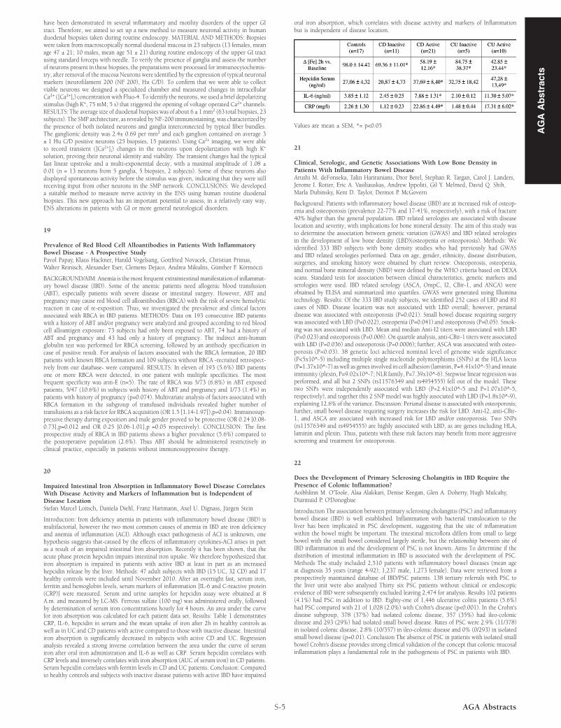
Introduction: Iron deficiency anemia in patients with inflammatory bowel disease (IBD) ismultifactorial, however the two most common causes of anemia in IBD are iron deficiencyand anemia of inflammation (ACI). Although exact pathogenesis of ACI is unknown, onehypothesis suggests that-caused by the effects of inflammatory cytokines-ACI arises in partas a result of an impaired intestinal Iron absorption. Recently it has been shown, that theacute phase protein hepcidin impairs intestinal iron uptake. We therefore hypothesized thatiron absorption is impaired in patients with active IBD at least in part as an increasedhepcidin release by the liver. Methods: 47 adult subjects with IBD (15 UC, 32 CD) and 17healthy controls were included until November 2010. After an overnight fast, serum iron,ferritin and hemoglobin levels, serum markers of inflammation [IL-6 and C-reactive protein(CRP)] were measured. Serum and urine samples for hepcidin assay were obtained at 8A.m. and measured by LC-MS. Ferrous sulfate (100 mg) was administered orally, followedby determination of serum iron concentrations hourly for 4 hours. An area under the curvefor iron absorption was calculated for each patient data set. Results: Table 1 demonstratesCRP, IL-6, hepcidin in serum and the mean uptake of iron after 2h in healthy controls aswell as in UC and CD patients with active compared to those with inactive disease. Intestinaliron absorption is significantly decreased in subjects with active CD and UC. Regressionanalysis revealed a strong inverse correlation between the area under the curve of serumiron after oral iron administration and IL-6 as well as CRP. Serum hepcidin correlates withCRP levels and inversely correlates with iron absorption (AUC of serum iron) in CD patients.Serum hepcidin correlates with ferritin levels in CD and UC patients. Conclusion: Comparedto healthy controls and subjects with inactive disease patients with active IBD have impaired

S-5 AGA Abstracts

oral iron absorption, which correlates with disease activity and markers of Inflammationbut is independent of disease location.

Values are mean ± SEM, *= p<0.05

21

Clinical, Serologic, and Genetic Associations With Low Bone Density inPatients With Inflammatory Bowel DiseaseArushi M. deFonseka, Talin Haritunians, Dror Berel, Stephan R. Targan, Carol J. Landers,Jerome I. Rotter, Eric A. Vasiliauskas, Andrew Ippoliti, Gil Y. Melmed, David Q. Shih,Marla Dubinsky, Kent D. Taylor, Dermot P. McGovern

Background: Patients with inflammatory bowel disease (IBD) are at increased risk of osteop-enia and osteoporosis (prevalence 22-77% and 17-41%, respectively), with a risk of fracture40% higher than the general population. IBD related serologies are associated with diseaselocation and severity, with implications for bone mineral density. The aim of this study wasto determine the association between genetic variation (GWAS) and IBD related serologiesin the development of low bone density (LBD)(osteopenia or osteoporosis). Methods: Weidentified 333 IBD subjects with bone density studies who had previously had GWASand IBD related serologies performed. Data on age, gender, ethnicity, disease distribution,surgeries, and smoking history were obtained by chart review. Osteoporosis, osteopenia,and normal bone mineral density (NBD) were defined by the WHO criteria based on DEXAscans. Standard tests for association between clinical characteristics, genetic markers andserologies were used. IBD related serology (ASCA, OmpC, I2, CBir-1, and ANCA) wereobtained by ELISA and summarized into quartiles. GWAS were generated using Illuminatechnology. Results: Of the 333 IBD study subjects, we identified 252 cases of LBD and 81cases of NBD. Disease location was not associated with LBD overall; however, perianaldisease was associated with osteoporosis (P=0.021). Small bowel disease requiring surgerywas associated with LBD (P=0.022), osteopenia (P=0.041) and osteoporosis (P=0.05). Smok-ing was not associated with LBD. Mean and median Anti-I2 titers were associated with LBD(P=0.023) and osteoporosis (P=0.006). On quartile analysis, anti-CBir-1 titers were associatedwith LBD (P=0.036) and osteoporosis (P=0.0006); further, ASCA was associated with osteo-porosis (P=0.03). 38 genetic loci achieved nominal level of genome wide significance(P<5x10^-5) including multiple single nucleotide polymorphisms (SNPs) at the HLA locus(P=1.37x10^-7) as well as genes involved in cell adhesion (laminin, P=4.41x10^-5) and innateimmunity (plexin, P=9.02x10^-7; NLR family, P=7.39x10^-6). Stepwise linear regression wasperformed, and all but 2 SNPs (rs11576349 and rs4954555) fell out of the model. Thesetwo SNPs were independently associated with LBD (P=2.41x10^-5 and P=1.07x10^-5,respectively), and together this 2 SNP model was highly associated with LBD (P=1.8x10^-9),explaining 12.6% of the variance. Discussion: Perianal disease is associated with osteoporosis;further, small bowel disease requiring surgery increases the risk for LBD. Anti-I2, anti-CBir-1, and ASCA are associated with increased risk for LBD and/or osteoporosis. Two SNPs(rs11576349 and rs4954555) are highly associated with LBD, as are genes including HLA,laminin and plexin. Thus, patients with these risk factors may benefit from more aggressivescreening and treatment for osteoporosis.

22

Does the Development of Primary Sclerosing Cholangitis in IBD Require thePresence of Colonic Inflammation?Aoibhlinn M. O'Toole, Alaa Alakkari, Denise Keegan, Glen A. Doherty, Hugh Mulcahy,Diarmuid P. O'Donoghue

Introduction The association between primary sclerosing cholangitis (PSC) and inflammatorybowel disease (IBD) is well established. Inflammation with bacterial translocation to theliver has been implicated in PSC development, suggesting that the site of inflammationwithin the bowel might be important. The intestinal microflora differs from small to largebowel with the small bowel considered largely sterile, but the relationship between site ofIBD inflammation in and the development of PSC is not known. Aims To determine if thedistribution of intestinal inflammation in IBD is associated with the development of PSC.Methods The study included 2,510 patients with inflammatory bowel diseases (mean ageat diagnosis 35 years (range 4-92); 1,237 male, 1,273 female). Data were retrieved from aprospectively maintained database of IBD/PSC patients. 138 tertiary referrals with PSC tothe liver unit were also analysed Thirty six PSC patients without clinical or endoscopicevidence of IBD were subsequently excluded leaving 2,474 for analysis. Results 102 patients(4.1%) had PSC in addition to IBD. Eighty-one of 1,446 ulcerative colitis patients (5.6%)had PSC compared with 21 of 1,028 (2.0%) with Crohn's disease (p<0.001). In the Crohn'sdisease subgroup, 378 (37%) had isolated colonic disease, 357 (35%) had ileo-colonicdisease and 293 (29%) had isolated small bowel disease. Rates of PSC were 2.9% (11/378)in isolated colonic disease, 2.8% (10/357) in ileo-colonic disease and 0% (0/293) in isolatedsmall bowel disease (p=0.01). Conclusion The absence of PSC in patients with isolated smallbowel Crohn's disease provides strong clinical validation of the concept that colonic mucosalinflammation plays a fundamental role in the pathogenesis of PSC in patients with IBD.

AG

AA

bst

ract

s