Embed Size (px)
Citation preview
PREVALENCE OF ANAEMIA, IRON DEFICIENCY, IRON DEFICIENCY
ANAEMIA AND RISK FACTORS FOR IRON DEFICIENCY
IN KELANTANESE CHILDREN
·By
DR. SITI NOOR BT. All SHIBRAMULISI
. .. f ( '·. '' i t ;·' L .: \ •
Dissertation Submitted In Partial Fulfilment Of The
Requirements For The Degree Of Master Of Medicine
(Paediatrics)
UNIVERSITI SAINS MALAYSIA 2001
ACKNOWLEDGEMENTS
First and foremost, I am greatly indebted to Associate Professor Dr. Quah
Ban Seng and Dr. Wan Maziah Wan Mohamad for their guidance and
assistance in every aspect of this dissertation and for making this
dissertation possible.
I am profoundly grateful to Professor Hamish Simpson, Dr. Narazah, Dr.
Sharifah Ainon Ismail Mokhtar and Dr. Bina Menon for their assistance,
advices and encouragement in the preparation of this dissertation.
My warmest appreciation to Kementerian Kesihatan and Pejabat Kesihatan
Kota Bharu for allowing me to conduct my study in their health clinics.
To my dearest husband, Dr. Nazri Mohd. Yusof and to my beloved parents I
am very grateful for their unconditional love, support and prayers which have
made my life possible throughout the preparation of this dissertation and the
whole post-graduate programme.
1 would like to express my sincere gratitude to all the nurses and doctors at
the health clinics and staff of Haematology laboratory HUSM for their kind
cooperation during the data collection and analysis of the blood samples.
Last but not least, my special thanks to the parents and children who
participated in this study in which without them this project would have never
been possible.
11

TABLE OF CONTENTS
CONTENTS
ACKNOWLEDGEMENT
TABLE OF CONTENTS
ABBREVIATIONS
LIST OF TABLES
LIST OF FIGURES
ABSTRACT
Malay version
English version
1. INTRODUCTION
2. REVIEW OF LITERATURE
3. OBJECTIVES
4. METHODOLOGY
4.1 Design of study
4.2 Questionnaire
4.3 Exclusion
4.4 Blood collection and measurements
4.5 Feedback and follow-up
4.6 Limitations
PAGE
ii
iii
v
vi
viii
ix
xii
1
4
39
39
43
44
44
45
46
111
IV
4.7 Ethics Approval 48
4.8 Statistical Analysis 48
4.9 Definition of terms 49
5. RESULTS
5.1 Sample collection 52
5.2 Demographic data 53
5.3 Prevalences of anaemia, ID and IDA 57
5.4 Clinical characteristics of all children 61 in the study
5.5 Characteristics of children in the iron 63 sufficient & iron deficient groups
5.6 Multivariate analysis 68
5.7 Clinical assessment of pallor 69
6. DISCUSSION 72
7. CONCLUSIONS 94
8. RECOMMENDATIONS 95
9. REFERENCES 97
APPENDIX 118
v
ABBREVIATIONS
dl Decilitre
FEP Free erythrocyte protoporphyrin
g Gram
Hb Haemoglobin
Hct Haematocrit
HUSM Hospital University Sains Malaysia
ID I ron deficiency
IDA Iron deficiency anaemia
IS I ron sufficient
kg Kilogram
MCHC Mean corpuscular haemoglobin concentration
MCV Mean corpuscular volume
OD Odds ratio
PPV Positive predictive value
RBC Red blood cell
RDW Red cell distribution width
RM Ringgit Malaysia
SF Serum ferritin
SPSS Statistical Package for the Social Sciences
TS Transferrin saturation
us United States
WHO World Health Organisation
LIST OF TABLES
TABLE 1.1 Prevalence of ID and IDA in various developing countries
TABLE 1.2 Prevalence of ID and IDA in various developing countries
{continuation)
TABLE 2.1 Prevalence of ID and IDA in various developed countries
TABLE 2.2 Prevalence of ID and IDA in various developed countries
{continuation)
TABLE 3 Recommended schedule of immunisation for Malaysian
children aged two years and below.
TABLE 4 Schedule for visits to Health Clinics in Kota Bharu.
TABLE 5. 1 Definiton of terms for risk factors of I D in this study.
TABLE 5.2 Definition of terms for risk factors of ID in this study
(continuation).
TABLE 6 Distribution of children based on different ethnic groups in each
health clinics.
TABLE 7 Prevalence of ID ba·sed on different definitions.
TABLE 8 Prevalence of IDA based on different definitions.
Vl
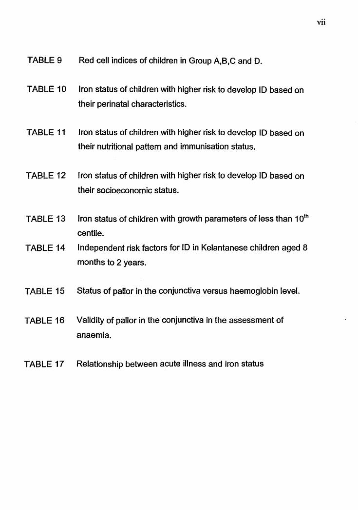
TABLE 9 Red cell indices of children in Group A,B,C and D.
TABLE 10 Iron status of children with higher risk to develop ID based on
their perinatal characteristics.
TABLE 11 Iron status of children with higher risk to develop ID based on
their nutritional pattern and immunisation status.
TABLE 12 Iron status of children with higher risk to develop ID based on
their socioeconomic status.
TABLE 13 Iron status of children with growth parameters of less than 10th
centile.
TABLE 14 Independent risk factors for ID in Kelantanese children aged 8
months to 2 years.
TABLE 15 Status of pallor in the conjunctiva versus haemoglobin level.
TABLE 16 Validity of pallor in the conjunctiva in the assessment of
anaemia.
TABLE 17 Relationship between acute illness and iron status
Vll
LIST OF FIGURES
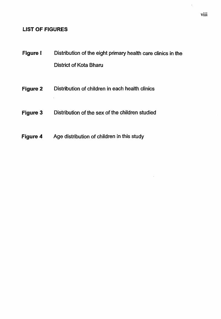
Figure I
Figure 2
Figure 3
Figure 4
Distribution of the eight primary health care clinics in the
District of Kota Bharu
Distribution of children in each health clinics
Distribution of the sex of the children studied
Age distribution of children in this study
Vlll
IX
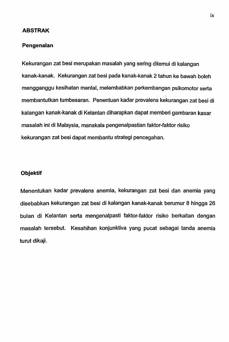
ABSTRAK
Pengenalan
Kekurangan zat besi merupakan masalah yang sering ditemui di kalangan
kanak-kanak. Kekurangan zat besi pada kanak-kanak 2 tahun ke bawah boleh
mengganggu kesihatan mental, melembabkan perkembangan psikomotor serta
membantutkan tumbesaran. Penentuan kadar prevalens kekurangan zat besi di
kalangan kanak-kanak di Kelantan diharapkan dapat memberi gambaran kasar
masalah ini di Malaysia, manakala pengenalpastian faktor-faktor risiko
kekurangan zat besi dapat membantu strategi pencegahan.
Objektif
Menentukan kadar prevalens anemia, kekurangan zat besi dan anemia yang
disebabkan kekurangan zat besi di kalangan kanak-kanak berumur 8 hingga 26
bulan di Kelantan serta mengenalpasti faktor-faktor risiko berkaitan dengan
masalah tersebut. Kesahihan konjunktiva yang pucat sebagai tanda anemia
turut dikaji.

X
Metodologi
Kajian hirisan lintang berdasarkan komuniti telah dijalankan dari bulan
September 1999 sehingga November 1999 di Iapan buah klinik kesihatan di
daerah Kota Bharu. Kanak-kanak berusia 8 hingga 26 bulan yang hadir di klinik
klinik kesihatan tersebut dijemput mengambil bahagian dalam kajian ini. Kanak
kanak yang mengalami penyakit akut pada waktu temu-ramah atau yang
berpenyakit kronik dikecualikan dari kajian. lbu-bapa ditemuramah
menggunakan pro forma piawai dan darah kanak-kanak diambil setelah
mendapat persetujuan lisan. Soalan-soalan yang dikemukakan meliputi data
demografi, faktor pemakanan, maklumat sosial dan faktor tumbesaran kanak
kanak.
Kepucatan pada konjunktiva dikenalpasti. Darah dianalisakan untuk
pemeriksaan gambaran darah dan penentuan kadar feritin. Faktor-faktor risiko
diantara kumpulan kanak-kanak yang kurang zat besi dengan kumpulan zat besi
mencukupi telah dibandingkan.
Keputusan
Hampir dua pertiga (65.1%) (95% Cl: 60.7-69.2%) daripada sejumlah 490 orang
kanak-kanak mengalami masalah anemia (Hb<11.0g/dl). Kadar prevalens
kekurangan zat besi (SF<12.0)Jg/L) ialah 38.9o/o (95o/o Cl: 34.7-43.5o/o) manakala
kadar prevalens anemia yang disebabkan kekurangan zat besi (Hb <11.0g/dL &

XI
SF <12.01-Jg/L) ialah 31.6% (95% Cl: 27.6-36.0%).
Faktor-faktor risiko independen bagi kekurangan zat besi adalah penyusuan
susu badan yang berpanjangan (OR 2.5; 95°kCI: 1.4-4.4%), kegagalan memberi
susu formula (OR 1.7; 95% Cl: 1.1-2.5%) dan kelewatan memberi makanan
pejal (OR 0.37; 95°k Cl 0.15-0.90% ). Faktor-faktor pemakanan lain, faktor
perinatal, faktor-faktor sosial dan ekonomi keluarga serta faktor tumbesaran
didapati tidak mencapai batas kemaknaan. Kepucatan konjunktiva mempunyai
kesensitifan 70.5°k dan kespesifikan 54.4o/o pada paras Hb <11.0g/dL.
Kesimpulan
Sebilangan besar kanak-kanak di Kelantan mengalami masalah anemia dan
kekurangan zat besi. _Kadar prevalens ini yang jauh lebih tinggi berbanding
dengan perangkaan di negara~negara maju amat membimbangkan.
Kepucatan konjunktiva bukanlah tanda yang sesuai untuk meramalkan anemia.
Pengubahsuaian faktor-faktor pemakanan berkaitan dengan masalah ini
(penyusuan susu badan berpanjangan dan kegagalan memberikan susu
formula) diharapkan dapat mengurangkan kadar insidens anemia serta
masalah kekurangan zat besi di negara ini.
xu
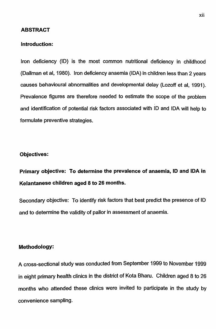
ABSTRACT
Introduction:
Iron deficiency (ID) is the most common nutritional deficiency in childhood
(Dallman et al, 1980). Iron deficiency anaemia (IDA) in children less than 2 years
causes behavioural abnormalities and developmental delay (Lozoff et al, 1991 ).
Prevalence figures are therefore needed to estimate the scope of the problem
and identification of potential risk factors associated with ID and IDA will help to
formulate preventive strategies.
Objectives:
Primary objective: To determine the prevalence of anaemia, ID and IDA in
Kelantanese children aged 8 to 26 months.
Secondary objective: To identify risk factors that best predict the presence of ID
and to determine the validity of pallor in assessment of anaemia.
Methodology:
A cross-sectional study was conducted from September 1999 to November 1999
in eight primary health clinics in the district of Kota Bharu. Children aged 8 to 26
months who attended these clinics were invited to participate in the study by
convenience sampling.
Xlll
Children with chronic diseases, thalassaemia or an acute infection at the time of
visit were excluded. Parents were interviewed using a standard pro forma
containing questions on demographic data, dietary history and socio-economic
influences. Pallor of conjunctiva was noted. Blood samples which were taken
after obtaining a verbal consent were analysed for full blood count and ferritin
level. The possible risk factors for ID were compared between the iron deficient
(SF<121Jg/L) and iron sufficient (SF>121Jg/L) groups using bivariate analysis and
multiple logistic regression.
Results
Among 490 children studied, 65.1% (95°/o Cl: 60.7-69.2o/o) were anaemic (Hb
<11.0g/dl). The prevalence of ID (SF<12.0J.Jg/L) was 38.9o/o (95°/o Cl: 34.7-
43.5%) and prevalence of IDA (Hb<11.0g/dl & SF <12.01Jg/L) was 31.6°/o (95o/o
Cl: 27.6-36.0%). Independent risk factors for ID were prolonged breast feeding,
(OR 2.5; 95% Cl: 1.4-4.4%), failure to give formula milk (OR 1.7; 95% Cl: 1.1-
2.5%) and delayed weaning (OR 0.37; 95% Cl: 0.15-0.90%).
Other dietary factors were not significantly associated with 10. None of the
perinatal factors, socio-economic factors or the growth parameters were
independently associated with 10. The sensitivity and specificity of pallor for
detecting a Hb < 11.0g/dl were 70.5o/o 54.4o/o respectively.

XlV
Conclusion
ID and IDA is a common problem among Kelantanese children. Prolonged
breast feeding and failure to give formula milk were significantly associated with
I D. Pallor of conjunctiva was not a sensitive or specific indicator for anaemia.
Improvements and appropriate interventions of these potentially modifiable risk
factors may reduce the incidence of anaemia and ID in Kelantanese children.
1
1. INTRODUCTION
1.1 Background of the study
Anaemia is a common problem in childhood especially in children aged 6 to 24
months old. It was estimated that 12o/o of children aged 0-4 years in developed
countries and 51% in developing countries were anaemic (DeMaeyer, 1985).
Iron deficiency anaemia (IDA) is the most commonly recognized form of
nutritional deficiency in developing countries (Dallman, 1980) as well as in
developed countries (Mills 1990). Worldwide, approximately 600 million
individuals had IDA (DeMaeyer, 1985). The deficiency is in most cases of
dietary origin and probably due to inadequate weaning (Mills, 1990). Other
causes of anaemia include infection, inflammatory diseases, malignancies and
hereditary disorders such as haemoglobinopathies.
IDA can no longer be considered a simple anaemia readily reversed by iron
therapy (Dallman, 1982). It has been associated with lowered scores on tests of
mental and motor development in infancy (Dallman, 1986), poorer psychomotor
development and behavioural changes of young children (Walter, 1989).
1
1. INTRODUCTION
1.1 Background of the study
Anaemia is a common problem in childhood especi~lly in children aged 6 to 24
months old. It was estimated that 12% of children aged 0-4 years in developed
countries and 51 o/o in developing countries were anaemic {DeMaeyer, 1985).
Iron deficiency anaemia (IDA) is the most commonly recognized form of
nutritional deficiency in developing countries {Dallman, 1980) as well as in
developed countries (Mills 1990). Worldwide, approximately 600 million
individuals had IDA (DeMaeyer, 1985). The deficiency is in most cases of
dietary origin and probably due to inadequate weaning (Mills, 1990). Other
causes of anaemia include infection, inflammatory diseases, malignancies and
hereditary disorders such as haemoglobinopathies.
IDA can no longer be considered a simple anaemia readily reversed by iron
therapy {Dallman, 1982). It has been associated with lowered scores on tests of
mental and motor development in infancy {Dallman, 1986), poorer psychomotor
development and behavioural changes of young children (Walter, 1989).
2
Iron deficiency (I D) has many other negative effects on health including changes
in immune function, cognitive development, temperature regulation, energy
metabolism and work performance (Lozoff et al., 1982). Iron is necessary for
maintaining normal structure and function of virtually all mammalian cells. It is
also involved in the immune and non-immune host defence. In vitro studies
have shown that iron and iron-binding proteins are important for lymphocyte
proliferation, for satisfactory functioning of natural killer cells, for B cells and
antibody production and for the activity of phagocytic cells (Farthing, 1 989).
Children who have IDA in infancy was found to be at risk for long lasting
developmental disadvantages as compared with their peers with better iron
status (Lozoff et al., 1986). Thus, it has been suggested that infants and
toddlers should be screened for I D.
Screening for anaemia was mostly carried out in Western countries where it was
found that children of Asian origin were at higher risk of being iron deficient
(Ehrhardt 1986, Grindulis et al., 1986). Similar studies of screening for IDA is
lacking in our population. The prevalence of ID and IDA in this country is
expected to be higher as compared to the figures in the developed countries.
However, to the best of our knowledge, only few studies had been carried out to
determine the prevalence for ID and IDA in children less than 2 years old in this
country.
3
This study was carried out to investigate the prevalence and the risk factors for
ID and IDA among the Kelantanese children aged 8 to 26 months. From this
study, we will be able to identify a group of children at risk of developing I D and
IDA in this part of country and it is hoped that in the future, children with these
particular risk factors will be looked into seriously to prevent iron deficency
anaemia thus preventing them from developing the complications that might
occur as a consequence of this disease.
4
2. REVIEW OF LITERATURE
2.1 IRON REQUIREMENTS
At birth, most term infants have about 75 mg of elemental iron per kilogram of
body weight, found primarily as haemoglobin (Hb) (75%) but also as storage
(15%) and tissue protein (10%) ( Oski, 1982). Infants of mothers with poorly
controlled diabetes and small-for-gestational-age infants have approximately
10% and 40% of normal storage iron, respectively, meaning that they may have
less of a buffer protection from postnatal ID (Petry et al., 1992, Georgieff et al.,
1995).
During the first 4 postnatal months, excess fetal red cells break down and the
infant retain the iron. This iron is used, along with dietary iron, to support the
expansion of the red blood cell mass as the infant grows.
The Nutrition Committee of the American Academy of Pediatrics , recommended
that term infants be given 1 mg iron per kg of elemental iron daily to a maximum
of 15 mg, starting no later than 4 months of age and continuing until 3 years of
age (American Academy of Pediatrics, 1976). Low birth weight infants however
require a higher amount at 2mg/kg per day to a maximum of 15 mg daily starting
no later than 2 months of age. Higher doses have been suggested for infants in
the lowest birth-weight categories {Dallman 1990). Infants with birth weights of
less than 1 000 g should receive 4 mg/kg per day, and infants with birth
5
weights between 1000 and 1500 g should receive 3 mg/kg per day.
For these infants, iron supplementation at the higher dose should continue
throughout the first year life (Oski, 1993).
The above recommendation are nutritional guidelines advocated to prevent IDA.
The final amount of iron being absorbed is dependent on the availability of iron
stores in the body as well as on the type and composition of food in the child's
diet.
2.2 IRON METABOLISM IN INFANCY
Iron deficiency is the most commonly recognized form of nutritional deficiencies
in developing countries as well as in affluent societies (Dallman et al., 1980). It
is particularly prevalent among infants and young children because their rapid
growth imposes large iron needs but on the other hand most infant diets contain
only a marginal supply of iron.
6
I ron containing compounds in the body may be divided into 2 categories:
I. Those that ser-Ve metabolic or enzymatic functions such as Hb. and
cytochromes. These compounds account for 25 to 55mg/kg body weight,
more than 80o/o of which is in Hb (Sjolin & Wranne 1968, Smith & Rios
1974).
II. Those that are associated with iron storage. These primarily consist of
ferritin and haemosiderin and account for 5 to 25 mg/kg body weight.
These compounds are involved in the maintenance of iron haemostasis
(Dallman et al., 1 980).
7
2.2.1 Iron Balance
The quantity of iron in the body is normally maintained within narrow limits at
each stage of growth and development. It is regulated mainly by the intake and
absorption of iron. Iron excretion occurs primarily through desquamation of cells
in the intestinal mucosa (Green et al. 1968, Finch et al. 1977).
The amount of iron absorbed from the duodenum depends on the adequacy of
iron stores, the form of iron in foods in the diet {Monsen et al., 1978). In adults,
about 95% of the iron required for the production of red blood cells is recycled
from the breakdown of senescent red cells and only 5% comes from dietary
sources (Hillman et al., 197 4 ). In contrast, the one year old infant, due to his
rapid growth, is estimated to derive less than 70% of red cell iron from senescent
red cells and requires about 30% from the diet (Fomon, 197 4 ).
2.2.2 Food Iron
Iron deficiency is more common than iron excess because most of the iron in
food and the environment is relatively insoluble and difficult to assimilate. As a
result, the form of iron in the diet is more important than the amount (Dallman,
1986). In infants, milk is the main form of diet. Breast and cow's milk each
contains less than 1.5mg of iron per 1000 calories (0.5 to 1.0 mg/L).
8
The bioavailability of iron is influenced by the type of foods in the diet. It is
therefore important to know how much iron is absorbed from each food item. It
has been found that a much higher percentage of iron was absorbed from breast
milk (about 50%) than from cow's milk or formula prepared cow's milk (Saarinen
et al. 1977, Oski et al. 1980).
The lower calcium and protein content of breast milk compared to cow's milk,
and the presence of high concentration of the iron-binding protein and lactoferrin,
have been postulated to play a role in the better iron absoption from breast milk
as compared to cow's milk or infant formula (Saarinen et al., 1977). However,
infants who were exclusively breast-fed for more than 6 months may eventually
become iron deficient (Siimes et al., 1984 ). This is because the amount of iron
in breast milk is limited even though the absorption of iron in breast milk is better
than cow's or formula milk.
Estimates of iron absorbtion from infant formula range from less than 5% in term
infants fed casein - predominant formula to to 40% in very low birth weight
infants fed whey-predominat formula (Stekel 1986, Ehrenkranz 1992, Saarinen
1977). About 7°/o to 12% of iron being absorbed in infants being fed with cow
milk formula. The percentage of iron absorbed from soy formula is lower than
from cow milk formula and ranges from less than 1 o/o to 7o/o. Nevertheless,
infants fed soy formula containing 12mg/L of iron remain comparably iron
9
sufficient to infants fed iron-fortified cow milk formula (Hertrampf et al., 1986).
Iron occurs in the food primarily as non-haem iron. A smaller amount is found in
the haem proteins, haemoglobin and myoglobin, which are present in meat.
Haem iron and non-haem iron differ markedly in their mechanism of absorption
and bioavailability (Hallberg 1981, Charlton et al., 1983). The non-haem iron in
food is primarily in the form of ferric complexes.
During digestion, this iron is reduced to the more readily absorbed ferrous form.
This is facilitated in the stomach by the hydrochloric acid- containing gastric juice
and continues in the small intestine. Achlorhydia will reduce the absorption of
ferric iron administered with food by about 50°k (Jacobs et al 1964 ). Assimilation
of ionic iron is enhanced by the formation of readily absorbed complexes with
other components of the diet, such as fructose, ascorbic acid, citrate and some
amino acids.
On the other hand, absorption is decreased by the formation of insoluble
compounds such as phosphates, tannates, polyphenols and oxalates. Bran in
cereals, polyphenols in many vegetables and tannin in tea can all play a major
role in inhibiting iron absorption (Charlton et al., 1983). Solid foods that are fed
near the time of a breast feeding can substantially inhibit the absorption of the
iron from that breast milk feeding (Oski et al., 1980, Saarinen et al., 1979).
10
Dietary iron in the form of haem protein is handled in a different manner. Haem
· is split from the globin portion of the molecule in the intestinal lumen. The haem
is then assimilated intact, and a haem-splitting enzyme within the mucosal cell
releases ionic iron. The absorption of haem iron is readily affected by other
dietary constituent, and a larger percentage of the total tends to be assimilated,
in comparison to non-haem iron {Bjom-Ramussen et al. 197 4, Layrisse et al.
1974).
2.2.3 Mechanism of Intestinal Absorption
The major portion of iron is absorbed in the duodenum and diminishing amounts
are absorbed as food advances toward the ileum. The mucosal cell of the
duodenum and jejunum is believed to take up iron from the intestinal lumen and
release it to the blood stream by a carrier system involving transferrin (Huebers
et al., 1983). The mucosal cell, by virtue of its brief 2- to 3- day life span,
constitues a temporary holding zone for the ferritin iron between the intestinal
lumen and the blood. In the iron-loaded individual, much of the iron that is taken
up by the mucosal cell is retained and later returned to the luminal contents by
desquamation (Dallman et al., 1980). By contrast, in 10 more iron crosses
through the mucosa into the circulation, and very little is retained within the cell
to be lost by desquamation.
11
Normally the diet contains 5 to 20 times the amount absorbed. Iron absorption
increases during the period of development that are characterized by a rapid rate
of growth (e.g. during infancy and adolescence) and consequently diminished
iron stores. At all ages, the more iron that is ingested, the less is the percentage
absorbed, but the greater is the absolute amount that is absorbed (Dallman et
al., 1980).
12
2.2.4 Iron losses
Iron losses from the body are small and relatively fixed, in contrast to wide
variations in iron intake and lesser fluctuations in absorption. In the normal
infant, the loss of iron is at least 20J.Jg per kg per day (Garby et al., 1964).
Cow milk feeding in early infancy is commonly associated with occult intestinal
blood loss detectable by the guaiac test (Forman et al., 1981 ). Over a one
month period of observation, about 40% of a group of 4 month old infants had at
least one guaiac-positive stools collection in contrast to 1 0°/o among formula fed
infants. At 6 months of age, there was no longer a significant difference between
the two groups. Severe anaemia and more substantial blood loss is occasionally
associated with ingestion of large volumes of fresh cow's milk (Woodruff et al.
1972, Wilson et al. 197 4 ).
13
2.3 CAUSES OF IRON DEFICIENCY
The most common factors that contribute to the development of ID in children
are rapid growth, blood loss and insufficient absorption of iron; many cases
result from combination of three (Dallman, 1980).
2.3.1 Rapid growth
Rapid growth and lack of dietary iron are usually of primary importance. At two
to three months of age the concentration of Hb decreases in response to its
lowest point and there will be increase in the rate of erythropoeisis. Iron stores
gradually decreases at 3 months of age and subsequently become depleted
unless replenished by an adequate exogenous supply of iron. In adult men only \
about 5% of the iron required for Hb production is derived from the diet; the
remaining 95% is recycled from the red blood cell (RBC) degradation. By
contrast, in the infant because of the rapid expansion of blood volume during
growth, 30% must come from the diet and only 70% is from recycled iron
(Dallman, 1987).
2.3.2 Blood loss
Blood loss as the sole cause of ID in children is less frequent than in adults. It is
a common primary cause of ID only when associated with ingestion of
14
unprocessed cow's milk in infancy (Forman et al., 1981) and in areas where
hookworm infestation and other parasitic infections are prevalent. Hookworm
infestation particularly Ancylostoma duodenale are likely to contribute to high
prevalence of I D and anaemia particularly in children and women (Hopkins et al.,
1997). Tasker (1958) found an inverse relationship of the Hb level with the
proportion of patients who had hookworms infestation, the lower the Hb level, the
higher the proportion of patients had hookworms. Using a radioactive tracer
technique, he estimated that daily blood loss increases from about 2 mi. with a
light infestation of about 20 hookworms to about 90 mi. in a heavy infestation of
greater than 1 ,500 hookworms.
There is evidence that blood and serum protein losses represent an intestinal
intolerance to large amounts of fresh cow's milk (Woodruff & Clark 1972, Wilson
& Lahey 197 4 ). It has been postulated that intestinal blood loss is to some
extent s~condary to the effects of ID on the mucosal lining (Kimber & Weintraub
1968), for example by a deficiency of iron containing enzymes in this tissue.
What distinguish this type of bleeding from that associated with gross anatomic
lesions is that it ceases shortly after initiation of treatment with iron (Woodruff &
Clark 1972, Wilson & Lahey 1974).
1 ndeed, providing processed formula milk even in the absence of iron treatment
has proved effective in stopping blood loss in some infants. In rare instances,
15
intestinal blood loss is associated with severe IDA and hypoproteinaemia
(Kimber & Weintraub, 1968). The abnormality appears to be reversed by iron
treatment alone, without a change in diet.
A common but often neglected form of intestinal blood loss is associated with the
use of medications that inhibit platelet aggregation and thereby prolong the
bleeding time. The most important of these is aspirin which even in relatively
small doses increases occult intestinal blood Joss to 5 ml per day, greater than 5
times the normal value of less than 1 ml per day (Person et al., 1961 ).
In children, blood loss due to anatomic lesions is easy to overlook because it is
rare. Occult intestinal blood loss should be suspected when there is no dietary
basis for anaemia, when anaemia persists or recurs despite iron treatment or
when severe anaemia is detected after infancy. Causes of bleeding in the
perinatal period include foetal-maternal haemorrhage, placental injury about the
time of delivery and twin-to-twin transfusion through placental communications.
An exchange transfusion also involves blood loss because blood with a high
haematocrit concentration is usually replaced by blood with a lower haematocrit
level (Dallman, 1987).
16
Beyond infancy, recurrent ID particularly in the absence of parasitic infestation or
symptomatology, should suggest a Meckel's diverticulum, often a cause of
intermittent painless intestinal blood loss in children (Brayton 1964, Spencer
1964, Shandling 1965). Other congenital anomalies, such as intestinal
duplications and intestinal haemorrhagic telangiectasia are less common but
may also result in IDA.
As in adults, bleeding ulcers and hiatus hernia are usually symptomatic but these
disorders are relatively rare in children. ID may also develop in patients with
haemophilia and other bleeding disorders not only as a result of external blood
loss but also because the iron lost through soft tissue bleeding may not be
completely resorbed and reutilized.
17
2.3.3 Insufficient absorption of iron
A reduced absorption of iron into the body can occur as the result either of low
levels of dietary iron or a poor biological availability of dietary iron or both.
These are important causes of IDA in developing countries where the economy
is restricted, so that the diets are commonly made up almost wholly of rice or
maize and very little foods of animal origin. Dietary inadequacy amongst
vegetarians would also be an important factor for I D.
Food iron exists primarily in the non-haem form of inorganic iron Ill (ferric)
complexes which are broken down during digestion and the iron being reduced
to the more readily absorbed iron II (ferrous) form (Dallman et al., 1980).
A lesser amount of food iron is present in the haem proteins, Hb and myoglobin,
which are present in foods of animal origin. Haem iron in the diet is easily
absorbed whereas non-haem iron is of a very much poorer bioavailability
(Dallman et al. 1980, Cook et al. 1972).
Dietary iron content and the bioavailability of iron in the diet will determine the
quantity of iron available to the intestine for absorption. The intestinal mucosa,

18
however does not necessarily take up all the available iron. Absorption will be
determined by the iron status of the body. Absorption normally decreases as
stores increase and increases as stores become depleted. (Heinrich 1970, Aisen
1982).
Mean iron absorption from foods of plants origin, vary from 1 % - 5% as
compared to 3% - 22% of iron absorption from foods of animal origin (Layrisse,
1975). However, foods of animal origin are frequently not present in the diets of
poorer communities so that only a small proportion of the iron in the diet is haem
iron.
Mild ID tends to be self-correcting, since iron absorption from food is increased
as iron stores in the body diminish. On the other hand, severely iron-deficient
children, as well as chronically deficient animals, may lose the normal adaptation
response of absorbing higher amounts of dietary iron. However, this abnormality
is promptly corrected after treatment with iron.
19
2.4 CONSEQUENCES OF IRON DEFICIENCY
Iron deficiency, even in the absence of anaemia, results in biochemical
alterations that impair behaviour in infants who are 9 to 12 months of age {Oski
et al., 1983).
Children who had severe, chronic ID in infancy scored lower on measures of
mental and motor functioning. After control for background factors, differences
remained statistically significant in arithmetic achievement and written
expression, motor functioning and some specific cognitive processes {spatial
memory, selective recall and tachistocopic threshold) {Lozoff, 2000).
More of the formerly iron-deficient children had repeated a grade and /or been
referred for special services or tutoring. Their parents and teachers rated their
behaviour as more problematic in several areas, agreeing in increased concerns
about anxiety or depression and attention problems (Lozoff, 2000). The
administration of iron produced a significant increase in the mental
developmental index scores in the infant with ID (Oski et al., 1983).
10 is significantly associated with retardation of physical and psychomotor
parameters {Walter et al 1989, Lozoff et al 1991 ), while repletion of the body's
iron stores rapidly reverses these developmental anomalies (Addy 1986, Aukett
et al., 1986). Studies in animals have shown that irrespective of anaemia, 10
produced metabolic and functional defects in muscle and impaired function of
20
white cells (Mackler et al., 1984 ). Functional abnormalities of both lymphocytes
and neutrophils have been shown in anaemic children (Srikantia et al., 1976).
A study in California found ID to be more common in 1-year-old infants who had
a history of recurrent mild infections (Reeves et al., 1984). Whether the
infections preceded or followed the ID is not clear. Most importantly, there is
now substantial evidence that ID has an adverse effect on brain function. In rats
ID leads to disturbed enzyme function in the brain affecting cerebral serotonin
metabolism (Mackler B et al., 1978) and learning ability (Massaro & Widmayer,
1981 ).
Pica in children is a symptom of ID and more than 50o/o of patients with ID have
pica (Crosby, 1976). Pica is quickly cured by therapy with iron. After a week or
two of therapy with iron, the pica invariably ceased, well before the anaemia had
been corrected (Cottman, 1969).
Oski and Honig (1985) studied 24 iron deficient anaemic children aged 9 to 26
months. Twelve were given intramuscular iron and 12 placebos, and tests of
mental development and of behaviour were administered before the injection
and five to eight days later. Improvement was found at the second testing in
those who had been given iron but not in the placebo group. In particular, the
treated children tended to become more responsive to their environment.
20
white cells (Mackler et al., 1984). Functional abnormalities of both lymphocytes
and neutrophils have been shown in anaemic children {Srikantia et al., 1976).
A study in California found ID to be more common in 1-year-old infants who had
a history of recurrent mild infections (Reeves et al., 1984). Whether the
infections preceded or followed the I D is not clear. Most importantly, there is
now substantial evidence that ID has an adverse effect on brain function. In rats
ID leads to disturbed enzyme function in the brain affecting cerebral serotonin
metabolism (Mackler 8 et al., 1978) and learning ability (Massaro & Widmayer,
1981 ).
Pica in children is a symptom of ID and more than 50o/o of patients with ID have
pica {Crosby, 1976). Pica is quickly cured by therapy with iron. After a week or
two of therapy with iron, the pica invariably ceased, well before the anaemia had
been corrected (Coltman, 1969).
Oski and Honig (1985) studied 24 iron deficient anaemic children aged 9 to 26
months. Twelve were given intramuscular iron and 12 placebos, and tests of
mental development and of behaviour were administered before the injection
and five to eight days later. Improvement was found at the second testing in
those who had been given iron but not in the placebo group. In particular, the
treated children tended to become more responsive to their environment.
21
In similar study in Guatemala, Lozoff (1982) and her colleagues showed that
children with IDA scored less well than other children in tests of mental
development and that the iron-deficient children were more tense and fearful but
otherwise less responsive. In their study no improvement in test scores was
seen after one week of treatment with oral iron.
Oski et al (1983) showed an improvement in mental development test scores
one week after intramuscular iron injection in babies aged 9 to 12 months who
were iron-deficient but not anaemic. Walter et al ( 1983) in Chile obtained similar
results in 15 months old children using treatment with oral iron and retesting after
11 days. The most noticeable behavioural characteristic of the anaemic Chilean
babies was that they were more unhappy than non-anaemic babies. Studies in
Java and Egypt have shown deficits in mental performance in schoolchildren
with ID, which was reversible with treatment (Pollitt et al., 1985).
The other consequences of IDA include abnormalities of immune function. ID
may at least contribute to impaired T lymphocyte function (defect in cell
mediated immunity and bacterial killing) and subsequently leads to an increased
risk of infections (Dallman, 1987), poor growth (Chwan et al., 1988) delayed
language and cognitive development (Walter, 1983), attention deficit disorder,
impaired school performance, impaired exercise capacity (Booth et al., 1997)
and clumsiness (Cantwell, 197 4 ).
22
Neurological sequelae such as irritability, lethargy, headache, papilloedema and
stroke have also been reported (Hartfield, 1997). Studie~ in young children and
adults indicate that both cell-mediated immunity and bactericidal activity of
neutrophils are impaired in patients with IDA (Chandra, 1973) although the
phagocytic function of the neutrophil may be normal, bacterial killing is
diminished. Neutrophils are defective in reducing the dye nitroblue tetrazolium,
suggesting the possibility that an iron containing enzyme required for this
reduction may be present in diminished amounts. After administration of
parenteral iron, the bactericidal abnormalities and the nitroblue tetrazolium test
were corrected within four to seven days, before the Hb concentration would
have increased appreciably.
Early identification and treatment of ID and IDA is therefore one of the highest
health care priorities. Treatment with iron has been shown to improve the
behaviour, cognitive skills and general learning ability in children with 10 (Oski,
1985). To avoid long term morbidity, ID can be prevented by dietary
modification, iron fortification of nutritional products and iron supplementation
(Hallberg, 1994 ). Children who received iron supplement had an increased rate
of weight gain and achieved the expected rate of development (Aukett, 1986).
23
2.5 DIAGNOSIS OF IRON DEFICIENCY
Iron deficiency anaemia develops as the end result of a series of steps that
begins with depletion of stored iron (Oski, 1993). First, iron disappears from the
bone marrow and the red cell distribution width becomes abnormal. Next, there
is a loss of transport iron, reflected by a reduce in serum iron level. Then
erythropoeisis becomes iron-deficient, as indicated by a reduced men
corpuscular volume and increased concentration of red-cell protoporphyrin. The
end result is overt anaemia (Oski, 1993).
The staging of iron status by Oski et al (1983) is a useful concept and various
measurements can be used to define the stages.
(A) Iron sufficiency:
1 ron stores and erythropoeisis normal
(B) Iron depleted: ·
Erythropoeisis normal but iron stores reduced.
In normal subjects, SF is directly proportional to body iron stores (Cook,
1982). During infancy and childhood, SF closely parallels the
developmental changes in iron status. The advantage of SF is its relative
stability with repeated measurements in the same subjects. SF value of
< 12ug/L indicates reduction of iron in the bone marrow, liver and other
parts of the reticuloendothelial system.
24
(C) Iron-deficient erythropoeisis.
When iron stores have been exhausted, the serum iron and transferrin saturation
{TS) will decline (Cook, 1982).
(a) Abnormal RBC biochemistry: Serum iron, total iron binding capacity &
free erythrocyte protoporphyrin:
1. decreased serum iron (<30J,Jg/dL in children aged 1-2 years)
n. Increased total iron-binding capacity {>480J,Jg/dL in children
aged 1-3 years)
nt. Elevated free erythrocyte protoporphyrin (FEP) level
(~90J,Jmol/mol of haem in children aged 1-5 years)
(b) Abnormal RBC morphology. Hypochromic microcytic red cells: MCV
<70fl, MCH less than 23pg , MCHC less than 30% and anisocytosis:
ROW >14.5% (Values vary with age)
(c) Reduced transport iron: Transferrin saturation< 8o/o in children 1-2 years.
There is a pronounced diurnal variation in serum iron and TS which can result in
a wide normal variation. Therefore, serum iron and TS are not recommended for
the routine confirmation of diagnosis of ID in favour of FEP and SF (Cook, 1982).
(D) Iron deficiency anaemia: The above plus Hb < 11.0g/dl