Embed Size (px)
Citation preview

4/11/11
1
Prematurity: Medica/on Impact on the Fetal Heart Rate
Rebecca Cypher, MSN, PNNP
Objec/ves
• Discuss normal physiologic characteris;cs of preterm fetal heart rate tracings
• Recognize common pharmacologic effects of medica;ons on the preterm fetal heart rate tracing
• Describe current assessment guidelines for the preterm fetus
Preterm birth US, 1998-2008
Preterm is less than 37 completed weeks gestation. Source: National Center for Health Statistics, final natality data. Retrieved April 9, 2011, from www.marchofdimes.com/peristats.
March of Dimes 2010 Prematurity Birth Report Card
Monitoring the Preterm Fetus
• Unique challenges • Limited research
• EFM implemented to establish fetal well being – Physiologic differences dependent on fetal development stage
– Response and/or tolerance to interrup;ons in oxygena;on pathway differ from those of the term fetus
Influence on FHR Regula/on • Autonomic nervous system
– Parasympathe;c • Vagus Nerve
– Medulla Oblongata
– Sympathe;c • Nerve fibers of myocardium
• Baroreceptors • Chemoreceptors
4/11/11
2
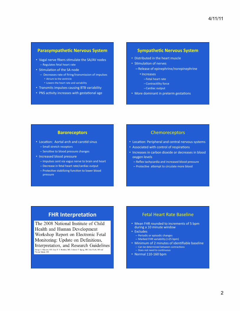
Parasympathe/c Nervous System
• Vagal nerve fibers s;mulate the SA/AV nodes – Regulates fetal heart rate
• S;mula;on of the SA node – Decreases rate of firing/transmission of impulses
• Atrium to the ventricle
• Lowers the heart rate and variability
• Transmits impulses causing BTB variability
• PNS ac;vity increases with gesta;onal age
Sympathe/c Nervous System • Distributed in the heart muscle • S;mula;on of nerves
– Release of epinephrine/norepinephrine • Increases
– Fetal heart rate – Contrac;lity force – Cardiac output
• More dominant in preterm gesta;ons
Baroreceptors
• Loca;on: Aortal arch and caro;d sinus – Small stretch receptors
– Sensi;ve to blood pressure changes • Increased blood pressure
– Impulses sent via vagus nerve to brain and heart – Decrease in fetal heart rate/cardiac output – Protec;ve stabilizing func;on to lower blood pressure
Chemoreceptors
• Loca;on: Peripheral and central nervous systems • Associated with control of respira;ons • Increases in carbon dioxide or decreases in blood oxygen levels – Reflex tachycardia and increased blood pressure – Protec;ve aUempt to circulate more blood
FHR Interpreta/on Fetal Heart Rate Baseline
• Mean FHR rounded to increments of 5 bpm during a 10 minute window
• Excludes – Periodic or episodic changes – Marked FHR variability (>25 bpm)
• Minimum of 2 minutes of iden;fiable baseline – Can be determined between contrac;ons – Does not need to con;nuous
• Normal 110-‐160 bpm

4/11/11
3
Is there an inverse rela;onship between EGA and FHR baseline?
Development of FHR Pa1erns During Normal Pregnancy
Pillai & James, 1990 • 43 low risk women in 2nd/3rd trimester
• Synchronized recordings in 4 week intervals – EFM for 90-‐100 minutes
• 9 am and 6 pm
– Ultrasound • Fetal eye movement, limb/body movement, fetal breathing, and mouthing movements
Results: Baseline Rate
• Nega;ve correla;on with gesta;onal age • Mean fall in baseline: 16 weeks to “term”
– 24 bpm
– 1 bpm per week of gesta;onal age
• Rate of fall greatest between 16-‐20 weeks – Less marked in last trimester
• Establishment of rest/ac;vity cycles
Variability
• Fluctua;ons in the baseline FHR that are irregular in amplitude and frequency
• Quan;fied as the amplitude of the peak and trough in bpm
• Excludes – Periodic or episodic changes
• Determined in 10-‐minute window
Minimal: ≤ 5 bpm
Amplitude range > undetectable
Moderate: 6-‐25 bpm
Marked: > 25 bpm
Baseline Variability
Is it all right to see minimal variability in the preterm fetus?

4/11/11
4
Fetal Heart Rate Variability Reveals DifferenCal Dynamics
Schneider et al, 2009
• 92 singletons • Two subgroups
– 24+1 to 32+0 weeks – 32+1 to 41+6 weeks
• Magnetocardiogram sessions – Measurement of magne;c fields produced by electrical ac;vity in the heart
Magnetocardiogram
Results
• Inverse rela;onship with gesta;onal age • FHR PaUern I
– FHR with “small oscilla;on bandwidth” <5 bpm
– 24+1 to 32 +0 gesta;ons • FHR PaUern II
– FHR with oscilla;ons >5 bpm
– >32+1 to 41+6 gesta;ons
Accelera/on
• Visually apparent abrupt increase in FHR • Peak ≥ 15 bpm from baseline and las;ng ≥ 15 seconds
• Preterm gesta/on (32 weeks) – Peak ≥ 10 bpm from baseline and las;ng ≥ 10 seconds
Decelera/ons
• Most frequent in 20-‐30 weeks • Absence of uterine contrac;ons • Fetal movement
• Reflec;on of developing cardioregulatory mechanisms and CNS maturity
AssociaCon Between FHR Pa1erns and Fetal Movements Between 20-‐30 Weeks GestaCon
Sorokin et al, 1982
• Low risk pa;ents – 20-‐22 weeks (10 pa;ents) – 28-‐30 weeks (10 pa;ents)
• Fetal monitoring – Day sessions las;ng 1-‐2 hours in quiet room – 1-‐2 hours ajer a meal
– Semi-‐fowlers with lateral ;lt
4/11/11
5
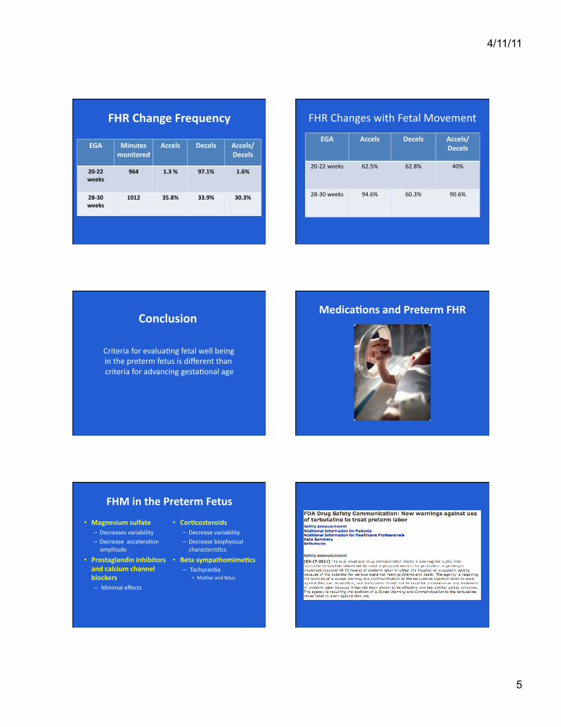
FHR Change Frequency
EGA Minutes monitored
Accels Decels Accels/Decels
20-‐22 weeks
964 1.3 % 97.1% 1.6%
28-‐30 weeks
1012 35.8% 33.9% 30.3%
FHR Changes with Fetal Movement
EGA Accels Decels Accels/Decels
20-‐22 weeks 62.5% 62.8% 40%
28-‐30 weeks 94.6% 60.3% 90.6%
Conclusion
Criteria for evalua;ng fetal well being in the preterm fetus is different than criteria for advancing gesta;onal age
Medica/ons and Preterm FHR
FHM in the Preterm Fetus
• Magnesium sulfate – Decreases variability – Decrease accelera;on amplitude
• Prostaglandin inhibitors and calcium channel blockers – Minimal effects
• Cor/costeroids – Decrease variability – Decrease biophysical characteris;cs
• Beta sympathomime/cs – Tachycardia
• Mother and fetus
29

4/11/11
6
Antenatal Cor;costeroid Therapy for Fetal Lung Matura;on
• 24-‐ 34 weeks gesta;on – At risk for preterm delivery within 7 days
• Single course • Betamethasone
– 12 mg IM 24 hours apart x2 doses
• Dexamethasone – 6 mg IM q12 hours x4 doses
ACOG, 2011
Does cor/costeroid administra/on suppress FHR characteris/cs and biophysical ac/vi/es?
Effects of Maternal Betamethasone AdministraCon on the Fetus
Derks et al, 1995
• 31 women who had received 2 doses for PTL – Gesta;onal age: 26-‐32 weeks
• EFM for 5 successive days (0-‐4)
• Ultrasound (Days 0, 2 and 4) – Fetal body movement – Fetal breathing – Eye Movements
Results • FHR variability below normal range for EGA
– 1/3 cases • Body movement
– Reduced by 50% on Day 2 • Breathing movement
– Absent on Day 2 • Eye movements: unchanged
• Day 4: return to normal state
Conclusion
• Considerable decrease in biophysical characteris;cs – Except eye movement
• Transient reduc;on in movements/ac;vity
• ? Glucocor;coid receptor mediated process
Effect of Dexamethasone and Betamethasone on FHR Variability
Senat et al, 1998
• 82 pa;ents with preterm labor – 25-‐33 weeks EG (median 29)
– Singletons, twins, triplets • Randomly assigned
– Betamethasone: 42 – Dexamethasone: 40
• FHR tracings reviewed (30 minutes) – Before, during and ajer treatment

4/11/11
7
Betamethasone Group
• Increase in FHR baseline – 2.6% (145 bpm to 147-‐148 bpm)
• Variability decreased by 28% – Returned to baseline or above baseline
• 96 hours
• Accelera;ons – Gradual increase ajer treatment stopped
• No change in decelera;ons or contrac;ons
Dexamethasone Group
• No “significant” changes – Baseline – Variability – Accelera;ons
Conclusion
Dexamethasone preferred steroid
Less altera;on in FHR variability
Effect of Steroids on Biophysical Profile And Doppler Indices
Deren et al, 2001
• 35 singleton pregnancies – 28-‐34 weeks – Betamethasone
• Biophysical profile and dopplers – Pre-‐steroid – 24, 48, 72, 96 and 120 hrs ajer administra;on of 1st dose
Results
• Reduced biophysical characteris;cs – Movement, fetal breathing and reac;vity
• Amnio;c fluid index unchanged
• Not volume
• Fetal tone unchanged
Specific Biophysical Scores
• Pre steroid: 10 • 24 hours: 8 • 48 hours: 6 (maximum peak of steroid)
• 72 hours: 8 • 96 hours: 10 • 120 hours: 10

4/11/11
8
Dopplers
• Umbilical artery & middle cerebral artery – Peak systolic velocity – Pulsa;lity index – Resistance index – End diastolic velocity – S/D ra;os
• Unchanged!
Conclusion
Doppler flow studies in conjunc;on with biophysical profiles may assist with
interpreta;on of post-‐steroid depression in the fetus and avoid an unwarranted delivery
Magnesium Sulfate • Tocoly/c (Mercer et al, 2009)
– 4-‐6 gram loading dose over 30 minutes
– Con;nuous infusion • 2 grams/hr up to 5 grams/hour
• Neuroprotec/on prior to preterm birth – ACOG, 2010 – Variety of protocols
• Crowther: 4 gram load, 1 gram/hour x24 hours • Rouse: 6 gram load, 2 grams/hr up to 12 hours
The Effect of Magnesium Sulfate On FHR Parameters
Hallack et al, 1999
• 34 pa;ents • > 30 weeks EGA (nonlaboring) • 800 kcal meal
• Randomized – Magnesium Sulfate (6 gram load and 2 gram/hr) – Placebo
• One hour monitoring sessions – Baseline, 1 hour and 3 hours of infusion
Results
FHR Group 0 hour 1 hour 3 hour
Baseline Placebo 134.4 ± 6.3 134.4 ±7.1 134.6 ±7.1
Mag Sulfate
136.6 ± 6.4 135.1 ± 6.6 132.3 ± 7.6
Variability Placebo 2.75 ± 0.33 2.81 ± 0.30 2.71 ± 0.52
Mag Sulfate
2.82 ± 0.29 2.84 ± 0.28 2.67 ± 0.36
Accels Placebo 10.2 ± 8.3 10.3 ± 8.2 10.4 ± 6.9
Mag Sulfate
11.1 ± 6.2 10.3 ± 8.2 7.4 ± 4.1
Conclusion
Administra;on of magnesium sulfate was associated with FHR changes but the clinical significance is ques;onable

4/11/11
9
Effects of Magnesium Sulfate On Cerebral Blood Flow
Twickler et al, 2010
• 38 pa;ents – Singletons/twins (24-‐31 weeks EGA) – 18 Magnesium Sulfate (Rouse protocol) – 10 Placebo
• Middle cerebral artery measurements – Before medica;on administra;on
– 1, 2, 3, and 4 hour intervals
Results
• Decrease in FHR baseline (doppler waveforms) – 8-‐10 bpm
– Confirmed results in Hallack trial
• No significant difference – Peak systolic velocity – Vessel diameter
– Volume flow
Conclusion
Magnesium sulfate has no significant effects on fetal cerebral blood flow
Assessment Guidelines
High Risk Antepartum Care • 1 nurse to 3 women if “stable condi;on”
– 1:1 if not stable • Magnesium sulfate administra;on
– 1 nurse in con/nuous bedside abendance • First hour of administra/on
– 1:1 ra;o con;nues un;l the pa;ent is no longer contrac;ng to “the degree that preterm birth is an imminent concern”
– 1 nurse to 2 women when no longer contrac/ng….
Fetal Assessments
• Rate • Variability • Periodic and Episodic Changes • Uterine Ac;vity • PaUern Evolu;on • Associated Clinical Findings • Communica;on

4/11/11
10
Frequency of Assessments
• Guidance on preterm labor pa;ents – AWHONN
– “Expert opinion” • Refer to unit policies
“When EFM is used to record FHR data permanently, periodic documentaAon can be used to summarize evaluaAon of fetal status at
the frequencies recommended by AAP/ACOG….A summary note including findings of the fetal status may be documented in the
medical record frequently.”
AWHONN, 2009


![Fetal membrane imaging and the prediction of preterm birth ... · fetal membranes and is located overlying the lower uterine pole and cervix in term fetal membranes [10, 13–15]](https://img.dokumen.tips/doc/110x75/5edac122434f4178104f9331/fetal-membrane-imaging-and-the-prediction-of-preterm-birth-fetal-membranes-and.jpg)