Embed Size (px)
Citation preview
Osimertinib in models of EGFR-mutant NSCLC brain metastases
1
Preclinical Comparison of Osimertinib with Other EGFR-TKIs in
EGFR-Mutant NSCLC Brain Metastases Models, and Early Evidence
of Clinical Brain Metastases Activity
Running title (52/limit of ~60 characters): Osimertinib in models of EGFR-mutant
NSCLC brain metastases
Peter Ballard1, James W.T. Yates2, Zhenfan Yang3, Dong-Wan Kim4, James Chih-Hsin
Yang5, Mireille Cantarini6, Kathryn Pickup1, Angela Jordan1, Mike Hickey7, Matthew
Grist1, Matthew Box1, Peter Johnström8,9, Katarina Varnäs9, Jonas Malmquist9,
Kenneth S. Thress10, Pasi A. Jänne11, and Darren Cross1
1iMED Oncology, AstraZeneca, Macclesfield, UK; 2iMED Oncology, AstraZeneca,
Cambridge, UK; 3Asia and Emerging Markets iMED, AstraZeneca, Shanghai, China;
4Department of Internal Medicine, Seoul National University Hospital, Seoul, Korea;
5National Taiwan University Hospital, Taipei City, Taiwan; 6Global Medicines Development,
AstraZeneca, Macclesfield, UK; 7AstraZeneca, Cambridge, UK; 8AstraZeneca Translational
Science Centre, Stockholm, Sweden; 9Karolinska Institutet, Department of Clinical
Neuroscience, Stockholm, Sweden; 10iMED Oncology, AstraZeneca, Gatehouse Park,
Waltham, MA, USA; 11Dana-Farber Cancer Institute, Boston, MA, USA
Corresponding author:
Peter Ballard
iMED Oncology, AstraZeneca
Alderley Park
Macclesfield
Cheshire
SK10 4TG
UK
Research. on April 10, 2018. © 2016 American Association for Cancerclincancerres.aacrjournals.org Downloaded from
Author manuscripts have been peer reviewed and accepted for publication but have not yet been edited. Author Manuscript Published OnlineFirst on July 19, 2016; DOI: 10.1158/1078-0432.CCR-16-0399
Osimertinib in models of EGFR-mutant NSCLC brain metastases
2
Email: [email protected]
Phone: +44 1625 510092
Fax: +44 1625 513253
Financial Support
This research was funded by AstraZeneca.
Disclosure of Potential Conflicts of Interest
Peter Ballard, James W.T. Yates, Zhenfan Yang, Mireille Cantarini, Kathryn Pickup, Angela
Jordan, Mike Hickey, Matthew Grist, Matthew Box, Peter Johnström, Kenneth S. Thress, and
Darren Cross are employees and shareholders of AstraZeneca. James Chi-Hsin Yang has
received grants and/or personal fees for advisory boards and speaking from Boehringer
Ingelheim, Eli Lilly, Pfizer, Clovis Oncology, Roche/Genentech/Chugai, Merck Sharp &
Dohme, Merck Serono, Astellas, Novartis, Bayer, and Celgene. Katarina Varnäs and Jonas
Malmquist have research collaboration agreements with AstraZeneca. Pasi A. Jänne has
received consulting fees from AstraZeneca, Boehringer Ingelheim, Pfizer, and Genentech,
has a sponsored research agreement with AstraZeneca, and is co-inventor on a Dana-
Farber Cancer Institute owned patent on EGFR mutations licensed to Lab Corp. Dong-Wan
Kim has declared no conflicts of interest.
Authors’ Contributions
Conception and design: Peter Ballard, James W.T. Yates, Zhenfan Yang, Mireille Cantarini,
Peter Johnström, Kenneth S. Thress, Darren Cross
Development and methodology: Peter Johnström, Jonas Malmquist
Acquisition of data: Zhenfan Yang, Dong-Wan Kim, James Chih-Hsin Yang, Mireille
Cantarini, Kathryn Pickup, Matthew Grist, Peter Johnström, Pasi A. Jänne
Research. on April 10, 2018. © 2016 American Association for Cancerclincancerres.aacrjournals.org Downloaded from
Author manuscripts have been peer reviewed and accepted for publication but have not yet been edited. Author Manuscript Published OnlineFirst on July 19, 2016; DOI: 10.1158/1078-0432.CCR-16-0399
Osimertinib in models of EGFR-mutant NSCLC brain metastases
3
Analysis and interpretation of data: Peter Ballard, James W.T. Yates, Zhenfan Yang, Dong-
Wan Kim, James Chih-Hsin Yang, Kathryn Pickup, Peter Johnström, Katarina Varnäs,
Kenneth S. Thress, Darren Cross
Writing, review, and/or revision of manuscript: Peter Ballard, James W.T. Yates, Zhenfan
Yang, Dong-Wan Kim, James Chih-Hsin Yang, Mireille Cantarini, Kathryn Pickup, Angela
Jordan, Mike Hickey, Matthew Grist, Matthew Box, Peter Johnström, Katarina Varnäs, Jonas
Malmquist, Kenneth S. Thress, Pasi A. Jänne, Darren Cross
Administrative, technical, or material support: Peter Ballard, James Chih-Hsin Yang, Angela
Jordan, Matthew Box, Jonas Malmquist
Study supervision: Peter Ballard, Mireille Cantarini
Other: Matthew Box, Matthew Grist (synthesized compounds for the trial)
Word count: 5499 (limit = 5500 [+10% as per correspondence with CCR Senior Editorial
Associate, and approved by Editorial Assistant])
Abstract: 158 (limit = 250)
References: 47 (limit = 50)
Tables and figures: 2 tables; 5 figures (additional figure approved by Editorial Assistant)
Key words
Lung cancer; animal models of cancer; brain metastases; tyrosine kinase inhibitors;
osimertinib
Research. on April 10, 2018. © 2016 American Association for Cancerclincancerres.aacrjournals.org Downloaded from
Author manuscripts have been peer reviewed and accepted for publication but have not yet been edited. Author Manuscript Published OnlineFirst on July 19, 2016; DOI: 10.1158/1078-0432.CCR-16-0399
Osimertinib in models of EGFR-mutant NSCLC brain metastases
4
Statement of Translational Relevance
There is a clinical need for novel epidermal growth factor receptor tyrosine kinase inhibitors
(EGFR-TKIs) with improved efficacy against brain lesions. Disease progression due to brain
metastases is common in patients with non-small cell lung cancer (NSCLC) harboring
tumors with EGFR-TKI-sensitizing mutations (EGFRm). As such, the exposure and activity in
the brain of osimertinib was analyzed. We present data which indicate that osimertinib had
greater exposure in the brain compared with some other EGFR-TKIs (gefitinib, afatinib, and
rociletinib), and demonstrated activity against EGFRm NSCLC brain metastases in
preclinical models and early clinical reports. Osimertinib has potential as a pharmacologic
treatment for patients with EGFRm NSCLC and brain metastases and may provide an
important step forward, given the limitations experienced with existing treatment options.
Research. on April 10, 2018. © 2016 American Association for Cancerclincancerres.aacrjournals.org Downloaded from
Author manuscripts have been peer reviewed and accepted for publication but have not yet been edited. Author Manuscript Published OnlineFirst on July 19, 2016; DOI: 10.1158/1078-0432.CCR-16-0399
Osimertinib in models of EGFR-mutant NSCLC brain metastases
5
Abstract
Purpose: Approximately one-third of patients with non-small cell lung cancer (NSCLC)
harboring tumors with epidermal growth factor receptor tyrosine kinase inhibitor (EGFR-TKI)-
sensitizing mutations (EGFRm) experience disease progression during treatment due to
brain metastases. Despite anecdotal reports of EGFR-TKIs providing benefit in some
patients with EGFRm NSCLC brain metastases, there is a clinical need for novel EGFR-TKIs
with improved efficacy against brain lesions.
Experimental design: We performed preclinical assessments of brain penetration and
activity of osimertinib (AZD9291), an oral, potent, irreversible EGFR-TKI selective for
EGFRm and T790M resistance mutations, and other EGFR-TKIs in various animal models of
EGFR-mutant NSCLC brain metastases. We also present case reports of previously treated
patients with EGFRm advanced NSCLC and brain metastases who received osimertinib in
the phase I/II AURA study (NCT01802632).
Results: Osimertinib demonstrated greater penetration of the mouse blood-brain barrier
than gefitinib, rociletinib (CO-1686), or afatinib, and at clinically relevant doses induced
sustained tumor regression in an EGFRm PC9 mouse brain metastases model; rociletinib
did not achieve tumor regression. Under positron emission tomography micro-dosing
conditions, [11C]osimertinib showed markedly greater exposure in the cynomolgus monkey
brain than [11C]rociletinib and [11C]gefitinib. Early clinical evidence of osimertinib activity in
previously treated patients with EGFRm advanced NSCLC and brain metastases is also
reported.
Conclusions: Osimertinib may represent a clinically significant treatment option for patients
with EGFRm NSCLC and brain metastases. Further investigation of osimertinib in this
patient population is ongoing.
Research. on April 10, 2018. © 2016 American Association for Cancerclincancerres.aacrjournals.org Downloaded from
Author manuscripts have been peer reviewed and accepted for publication but have not yet been edited. Author Manuscript Published OnlineFirst on July 19, 2016; DOI: 10.1158/1078-0432.CCR-16-0399
Osimertinib in models of EGFR-mutant NSCLC brain metastases
6
Introduction
A number of epidermal growth factor receptor (EGFR) tyrosine kinase inhibitors (TKIs) are
recommended for first-line treatment of patients with advanced non-small cell lung cancer
(NSCLC) harboring an EGFR-TKI-sensitizing mutation (EGFRm) (1, 2). However, more than
30% of patients with NSCLC experience disease progression during treatment with
established EGFR-TKIs due to growth of synchronous or metachronous brain metastases (3,
4).
For successful treatment of brain metastases, a drug must first be able to cross the blood-
brain barrier (BBB). BBB penetration is influenced by factors such as a drug’s affinity for the
ATP-binding cassette efflux transporters, permeability glycoprotein (P-gp) and breast cancer
resistance protein (BCRP), which are involved in the removal of toxins, drugs, and
chemotherapies from the central nervous system (CNS) (5-9). Chemotherapy agents and
large monoclonal antibodies are generally unable to cross the BBB (7, 10). The ability of a
molecule to cross the BBB is affected by multiple factors, including molecular weight (11).
Active or untreated brain metastases are often exclusion criteria in trials of EGFR-TKIs,
however there are reports documenting efficacy of EGFR-TKIs in treatment of, and/or
preventing development of, brain metastases in patients with EGFRm NSCLC (12-15). In a
retrospective analysis of 155 patients, initial gefitinib or erlotinib treatment was associated
with a lower cumulative risk of CNS progression (1%, 6%, and 21% at 6, 12, and 24 months)
compared with chemotherapy (7%, 19%, and 32%, respectively) (12). Additionally, pulsatile
administration of high-dose erlotinib can control CNS metastases from EGFR-mutant
NSCLC (16).
Despite reports of tumor responses, the TKIs gefitinib, erlotinib, and afatinib are considered
to have generally poor biopharmaceutical properties for penetrating the BBB, perhaps
attributable to interactions with P-gp and BCRP (17-19). However, penetration may be
increased in patients with more advanced brain metastases where BBB disruption has
Research. on April 10, 2018. © 2016 American Association for Cancerclincancerres.aacrjournals.org Downloaded from
Author manuscripts have been peer reviewed and accepted for publication but have not yet been edited. Author Manuscript Published OnlineFirst on July 19, 2016; DOI: 10.1158/1078-0432.CCR-16-0399
Osimertinib in models of EGFR-mutant NSCLC brain metastases
7
already occurred (20-22). Additionally, there is a cumulative increase in brain metastases
incidence in patients with EGFRm NSCLC over time (23). Although many patients die of
systemic progression, rather than brain lesion progression, quality of life is significantly
worsened, both directly and as a result of whole brain radiotherapy (WBRT), which degrades
cognitive function (24). Additionally, as systemic therapies improve for patients with EGFRm
NSCLC, the brain may increasingly become a sanctuary site where the BBB may offer
protection from pharmacological agents (22). Therefore there exists a clinical need for
EGFR-TKIs with improved BBB penetration, and it is important that new mutant-selective
agents, such as osimertinib (AZD9291), an oral, potent, irreversible EGFR-TKI selective for
sensitizing and T790M resistance mutations (25, 26), and rociletinib (CO-1686) (27), are
explored in this context.
Osimertinib was recently approved by the US Food and Drug Administration for treatment of
patients with NSCLC harboring a T790M mutation, and whose disease has progressed
following treatment with another EGFR-TKI (28). We examined the brain exposure and
distribution of osimertinib and the active metabolites AZ5104 and AZ7550 in the preclinical
setting. We compared brain distribution, pharmacokinetics (PK), and in vivo brain xenograft
efficacy of osimertinib with other EGFR-TKIs and simulated potential clinical efficacy based
on these data. Furthermore, brain penetration of radiolabeled osimertinib and other EGFR-
TKIs was examined in a non-human primate model. We also present early evidence of
clinical efficacy of osimertinib against brain metastases as part of the ongoing AURA trial
(NCT01802632).
Research. on April 10, 2018. © 2016 American Association for Cancerclincancerres.aacrjournals.org Downloaded from
Author manuscripts have been peer reviewed and accepted for publication but have not yet been edited. Author Manuscript Published OnlineFirst on July 19, 2016; DOI: 10.1158/1078-0432.CCR-16-0399
Osimertinib in models of EGFR-mutant NSCLC brain metastases
8
Materials and Methods
Test compounds and cell lines
Madin-Darby canine kidney (MDCK) epithelial cells were obtained from: US National
Institutes of Health, Bethesda, MD, USA and Netherlands Cancer Institute (multidrug
resistance protein 1 [MDR1]-MDCK); Absorption Systems, Exton, PA, USA and
AstraZeneca, Alderley Park, Macclesfield, UK (BCRP-MDCK); American Type Culture
Collection, Manassas, VA, USA (Caco2). Cells were authenticated during each experiment
by monitoring transepithelial electrical resistance, and with positive controls. H1975 cells
were obtained from American Type Culture Collection in 2004, and authenticated by short-
tandem repeat analysis in November 2012. PC9 cells (exon 19 deletion) were obtained in
November 2011 from Akiko Hiraide at Preclinical Sciences R&D, AstraZeneca, Japan, and
tested and authenticated by short-tandem repeat analysis in May 2013.
Details on cell line maintenance and test compounds are presented as supplementary
information.
Permeability glycoprotein and breast cancer resistance protein substrate assessment
P-gp (also known as MDR1) and BCRP substrate assessments of osimertinib, AZ7550,
AZ5104, rociletinib, afatinib, and erlotinib were performed using transfected MDCK cells
(MDR1-MDCK; BCRP-MDCK) and the Caco2 (colon carcinoma) cell line. Digoxin, cladribine,
and Ko143 10 mM, valspodar 1 mM, and atorvastatin 0.075 mM stock solutions in dimethyl
sulfoxide (DMSO) were prepared as positive controls or inhibitors.
Cell monolayers were grown onto collagen-coated, 12-well, polycarbonate membranes in
Costar Transwell plates (1.13 cm2 insert area, 0.4 µm pore size [Corning Life Sciences, New
York, NY, USA]). Test and control compound substrate assessments were carried out in
triplicate in each direction (apical [AP]-to-basolateral [BL], and BL-to-AP), co-dosed with
200 µM Lucifer yellow (LY). Aliquots were taken from the test compound receiver
compartment at pre-selected time points and replaced with an equal volume of fresh
Research. on April 10, 2018. © 2016 American Association for Cancerclincancerres.aacrjournals.org Downloaded from
Author manuscripts have been peer reviewed and accepted for publication but have not yet been edited. Author Manuscript Published OnlineFirst on July 19, 2016; DOI: 10.1158/1078-0432.CCR-16-0399
Osimertinib in models of EGFR-mutant NSCLC brain metastases
9
transport buffer. Positive control receiver samples were taken at 120 minutes. Liquid
chromatography (LC)-tandem mass spectrometry (MS/MS) was used for analysis of the test
compound, and LY concentration measured using a BMG microplate reader (excitation
485 nm; emission 540 nm). Donor sample aliquots were taken at selected time points
without replacement.
Relative efflux ratios of compounds between MDR1-MDCK or BCRP-MDCK cells, compared
with non-transfected MDCK cells, and co-dosing with 1 µM valspodar or 10 µM Ko143, were
utilized to verify whether test compounds were P-gp and BCRP substrates, respectively.
For Caco2 drug transport assays, atenolol (low permeability marker), minoxidil (high
permeability marker), and digoxin (control efflux marker) were prepared in DMSO to a test
concentration of 10 µM. To determine test and control compound transport rates in AP-to-BL
and BL-to-AP directions, LY 100 µM was run alongside all compounds. Donor samples were
taken at 60 and 120 minutes, and receiver samples taken additionally at 30 and 60 minutes
and replaced with an equal volume of fresh transport buffer.
Apparent permeability coefficients of test compounds across Caco2 monolayers was
estimated using high performance LC (HPLC)-MS/MS; LY concentrations were measured
using an Infinite 200 PRO microplate reader (485 nM excitation; 530 nM emission).
Rat quantitative whole body autoradiography
Tissue distribution of radioactivity in male partially pigmented (Lister-Hooded) rats was
determined following a single oral dose of [14C]osimertinib (4 mg/kg, 7.4 MBq/kg) as a
suspension in aqueous hydroxypropyl methylcellulose (HPMC) (0.5% w/v). Rats were
terminated under anesthesia by cold shock at 0.5, 1, 6, and 24 hours, and 2, 7, 21, and 60
days post-dose. Animals were immersed in a freezing mixture for ≥30 minutes and subjected
to sagittal plane whole body sectioning (nominal thickness 30 μm). Concentration of
radioactivity in tissues was determined using quantitative whole body autoradiography
Research. on April 10, 2018. © 2016 American Association for Cancerclincancerres.aacrjournals.org Downloaded from
Author manuscripts have been peer reviewed and accepted for publication but have not yet been edited. Author Manuscript Published OnlineFirst on July 19, 2016; DOI: 10.1158/1078-0432.CCR-16-0399
Osimertinib in models of EGFR-mutant NSCLC brain metastases
10
(QWBA) by Covance Laboratories Ltd (Harrogate, UK) using a validated image analysis
system.
In a separate experiment, the distribution of a single oral dose of [14C]gefitinib (5 mg/kg,
8.3 MBq/kg) in male Pievald Virol Glaxo pigmented rats was assessed. [14C]gefitinib was
administered as a suspension in 0.5% HPMC in 0.1% polysorbate 80. Rats were terminated
by CO2 narcosis at 1, 2, 6, 24, 48, and 96 hours post-dose and frozen and sectioned as
above. Radioactivity was quantified using a Phosphor Imager SF (Molecular Dynamics).
Brain binding in vitro
Brain tissue from treatment-naïve nude mice was homogenized with phosphate-buffered
saline (PBS, 1:3 w/w), and [3H]osimertinib spiked into the blank brain homogenate at a 5 µM
final concentration. Radioactive content of spiked brain homogenate was assessed by liquid
scintillation counting (LSC) using Ultima Gold Scintillation cocktail. Samples were counted
for 10 minutes, or until 105 counts had accumulated; test compound concentrations were
calculated using the radioactive content of each sample in conjunction with the specific
activity of the material used. Spiked brain homogenate was dialyzed in triplicate against PBS
for 4 hours at 37°C using Spectra/Por2 equilibrium dialysis membrane discs (molecular
weight cut-off of 12–14 kDa; Spectrum Laboratories Inc., Rancho Dominguez, CA, USA).
Aliquots of brain homogenate and PBS were assessed by LSC; the free fraction of test
compounds in brain homogenate was calculated and overall recovery of radioactivity from
cells determined. The free fraction in undiluted brain was determined from these data.
The in vitro brain binding assays of unlabeled osimertinib was carried out on a HT-Dialysis
plate (HTD 96 b, Cat#1006). Blank brain homogenate (1:3 with Dulbecco’s PBS pH 7.4) was
spiked with 5 µM test compound (in triplicate) and dialyzed (molecular weight cut-off 12–
14 kDa) against 100 mM PBS buffer (pH 7.4), slowly rotated at 37°C for 4 hours. Receiver
and donor aliquots were taken following incubation, and donor samples further diluted.
Paired samples were matrix-matched with buffer or blank brain homogenate, and
Research. on April 10, 2018. © 2016 American Association for Cancerclincancerres.aacrjournals.org Downloaded from
Author manuscripts have been peer reviewed and accepted for publication but have not yet been edited. Author Manuscript Published OnlineFirst on July 19, 2016; DOI: 10.1158/1078-0432.CCR-16-0399
Osimertinib in models of EGFR-mutant NSCLC brain metastases
11
precipitated with cold acetonitrile with internal standard. After centrifuging at 4000 rpm for 20
minutes, supernatant was diluted with 0.1% formic acid aqueous solution and analyzed by
LC-MS/MS (API 4000, Applied Biosystems, Foster City, CA, USA).
The in vitro brain binding assay of unlabeled gefitinib was carried out on a RED device
system (Thermo Fisher Scientific, Grand Island, NY, USA) with a semi-permeable
membrane. Gefitinib 10 mM was prepared in DMSO, and a 1 mM working solution spiked in
triplicate into blank brain homogenate (1:3 with PBS pH 7.4) to a 5 µM final concentration;
the final percentage volume of the organic solvent was 0.5%. Spiked brain homogenate was
dialyzed against PBS pH 7.4 for 4 hours at 37°C with 5% CO2 for 4 hours on a slowly
rotating orbital shaker. Following incubation, a 50 µL receiver aliquot, and a 5 µL donor
aliquot were taken; the donor sample was diluted with 45 µL of blank brain homogenate.
Paired samples were matrix-matched with buffer or blank brain homogenate, mixed for 2
minutes, and precipitated with 150 µL cold acetonitrile containing internal standard. Samples
were centrifuged at 4000 g for 20 minutes and supernatant diluted 1:2 with distilled water for
LC/MS/MS (Waters Xevo TQ).
Mouse pharmacokinetic studies
Osimertinib was administered orally to female CB17 Sever combined immunodeficient
(SCID) mice bearing H1975 tumor xenografts at Oncodesign Biotechnology (Dijon, France).
Three mice per dose group and time point received osimertinib 5 mg/kg or 25 mg/kg (as
suspension in 0.5% HPMC). Mice were terminated (isoflurane overdose) and blood, brain
and tumor samples collected at 0.5, 1, 2, 4, 6, and 24 hours post-dose.
Osimertinib 25 mg/kg, gefitinib 6.25 mg/kg, rociletinib 100 mg/kg, or afatinib 7.5 mg/kg were
dosed to naïve female nude mice (Alderley Park, Macclesfield, UK), with animals terminated
(rising CO2 dose) and blood and brain samples collected at 1, 2, and 6 hours post-dose.
Plasma was prepared from blood in lithium heparin chilled tubes immediately after collection.
Test compounds were extracted by protein precipitation using acetonitrile, containing internal
Research. on April 10, 2018. © 2016 American Association for Cancerclincancerres.aacrjournals.org Downloaded from
Author manuscripts have been peer reviewed and accepted for publication but have not yet been edited. Author Manuscript Published OnlineFirst on July 19, 2016; DOI: 10.1158/1078-0432.CCR-16-0399
Osimertinib in models of EGFR-mutant NSCLC brain metastases
12
standard (AZ10024306) in a 4:1 solvent:sample ratio. Samples were centrifuged for 10
minutes at 3000 rpm; 50 µL of supernatant was combined with 300 µL of water before
HPLC-MS/MS.
Brain and tumor samples were cut into two portions prior to snap freezing in liquid nitrogen,
stored at -80°C, and transferred to Oncology DMPK, AstraZeneca R&D. Following thawing,
samples were transferred to FastPrep™ Lysing Matrix A tubes (MP Biomedicals LLC, Santa
Ana, CA, USA). Water was added (1:3 tissue:water w/v) and samples homogenized;
homogenate was treated in the same way as plasma.
Exposure was expressed as area under the plasma or tissue-concentration time curve
(AUC) from 0–6 or 0–24 hours according to the last concentration measured. Maximum
observed concentration (Cmax) and time to Cmax (tmax) were direct observations of
concentration versus time data. PK parameters were obtained by means of non-
compartmental analysis using Phoenix32 version 6.4 (Pharsight Corporation, Mountain
View, CA, USA).
Cynomolgus monkey positron emission tomography micro-dosing
A PET micro-dosing study was performed in four cynomolgus monkeys (Supplementary
Table S2). Monkeys were anesthetized using ketamine, and anesthesia maintained by a
mixture of O2, air, and sevoflurane (2–8%). Anesthesia, heart rate, blood pressure, and body
temperature were continuously monitored. Radiolabeled compounds were injected as a
bolus into a sural vein. Injected radioactivity was 141 ± 25 MBq corresponding to <3 µg of
radiolabeled osimertinib, AZ5104, gefitinib, and rociletinib. The distribution of radioactivity
following intravenous (IV) micro-dosing was assessed using PET imaging over 125-minutes
with a Siemens Molecular Imaging high resolution research tomograph system (29). Arterial
blood samples were collected and analyzed for radioactivity in blood. Separate experiments
were conducted to determine radioactivity distribution to the brain and abdomen.
Research. on April 10, 2018. © 2016 American Association for Cancerclincancerres.aacrjournals.org Downloaded from
Author manuscripts have been peer reviewed and accepted for publication but have not yet been edited. Author Manuscript Published OnlineFirst on July 19, 2016; DOI: 10.1158/1078-0432.CCR-16-0399
Osimertinib in models of EGFR-mutant NSCLC brain metastases
13
Radioactivity concentration in brain was calculated and normalized to arterial blood
radioactivity concentration as described in the supplementary material.
Mouse brain metastases xenograft
Effects of chronic once daily (QD) dosing of osimertinib, gefitinib, and rociletinib was
assessed in a mouse brain metastases model. The human NSCLC cell line PC9 (exon 19
deletion) was transfected with the GL4.50[luc2/CMV/Hygro] vector containing the luciferase
gene (PC9_Luc) using lipofectamine LTX in order to monitor brain tumor growth by
measuring bioluminescence signals in tumor cells (30, 31). Signal intensity was measured
using the Bright-Glo™ Luciferase Assay System (Promega, Madison, WI, USA). The EGFR
exon 19 deletion mutation was further confirmed in the PC9_Luc cell line.
For the brain metastases model, PC9_Luc cells were implanted into 6–8-week-old specific-
pathogen-free immunodeficient nude mice purchased from Vital River (Beijing, China) by
intra-internal carotid artery injection (32, 33). Bioluminescence signals were measured with
an IVIS® Xenogen imaging system weekly to monitor tumor growth. When signals reached
the range of 107 photons/second, mice were randomized to be treated orally with osimertinib
5 mg/kg or 25 mg/kg QD, gefitinib 6.25 mg/kg QD, rociletinib 100 mg/kg QD, or vehicle for 8
weeks. Bioluminescence signals (anti-tumor efficacy) and body weight (tolerability) were
measured weekly; tumor growth inhibition was assessed by comparing mean
bioluminescence intensity change for control and treated groups.
Pharmacokinetic-pharmacodynamic modeling
A series of modeling studies was undertaken to predict active doses of osimertinib in
humans by incorporating human PK data into a previously developed PK-pharmacodynamic
(PKPD) PC9 subcutaneous xenograft efficacy model that linked phosphorylated EGFR
(pEGFR) inhibition to reductions in tumour volume (34). The studies also accounted for
reduced free concentrations in the brain due to higher levels of binding than observed in
plasma.
Research. on April 10, 2018. © 2016 American Association for Cancerclincancerres.aacrjournals.org Downloaded from
Author manuscripts have been peer reviewed and accepted for publication but have not yet been edited. Author Manuscript Published OnlineFirst on July 19, 2016; DOI: 10.1158/1078-0432.CCR-16-0399
Osimertinib in models of EGFR-mutant NSCLC brain metastases
14
The pharmacodynamic model represents pEGFR, relative to control, as a pool
homogenously distributed across the cell and cells in the tumour. The reversible interaction
(docking) of the molecule with the receptor is assumed to be more rapid than the covalent
binding step and subsequent deactivation of the receptor. The system is simplified with the
reversible interaction characterised by the binding affinity, and the irreversible interaction
characterised by a binding constant for unbound parent and metabolite. Further information
on the model is presented as supplementary material.
Clinical case studies
We present two clinical case studies from the dose escalation/expansion component of the
ongoing phase I/II AURA study (ClinicalTrials.gov: NCT01802632) (35). In these cases,
presence of brain metastases was documented at study entry; patients underwent magnetic
resonance imaging of the brain at baseline and every 6 weeks thereafter as part of the
systemic evaluation of disease status according to RECIST version 1.1 until disease
progression.
Research. on April 10, 2018. © 2016 American Association for Cancerclincancerres.aacrjournals.org Downloaded from
Author manuscripts have been peer reviewed and accepted for publication but have not yet been edited. Author Manuscript Published OnlineFirst on July 19, 2016; DOI: 10.1158/1078-0432.CCR-16-0399
Osimertinib in models of EGFR-mutant NSCLC brain metastases
15
Results
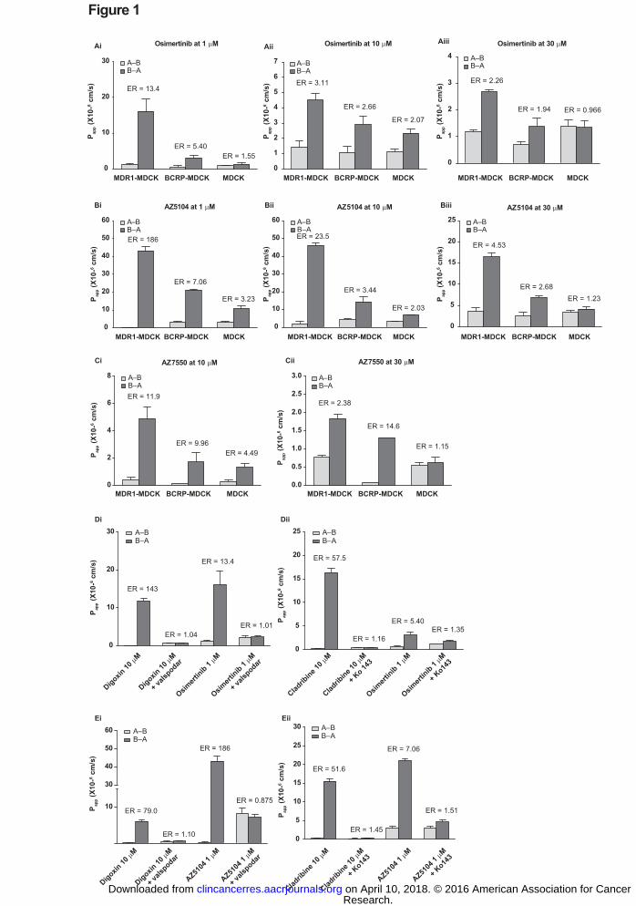
Osimertinib is a substrate of P-gp and BCRP transporters
Two efflux transporters, P-gp and BCRP, prevent molecules from crossing the BBB, so it
was important to determine the level of osimertinib substrate activity against these
transporters. Assessments were carried out in transwell plates in AP-to-BL and BL-to-AP
directions (Fig. 1). Relative efflux ratios of osimertinib or its metabolite AZ5104 between
MDR1-MDCK and normal MDCK cells and between BCRP-MDCK and MDCK cells suggest
that these agents are P-gp and BCRP transporter substrates. When dosed in the presence
of transporter inhibitors valspodar or Ko143, efflux ratios were similar to that observed in
non-transfected MDCK or BCRP-MDCK cells, respectively. Collectively, data confirm that
osimertinib and AZ5104 are substrates of P-gp and BCRP. The apparent permeability of
AZ7550 in the BL-to-AP direction of MDR1-MDCK cells in the presence of valspodar or
BCRP-MDCK cells in the presence of Ko143 suggests that AZ7550 is also a substrate of
P-gp and BCRP.
In MDCK-MDR1 cells, efflux ratios of test compounds dosed at 1 µM were 13.4 for
osimertinib, 5.38 for rociletinib, 4.62 for afatinib, and 4.63 for erlotinib. Efflux ratios in MDCK-
BCRP cells were 5.4 for osimertinib, 54.6 for afatinib, and 6.39 for erlotinib.
Osimertinib demonstrated a passive permeability profile across Caco2 cell monolayers, with
no concentration dependence when tested at 1, 10, and 50 µM, and an efflux ratio <2.0. The
high binding propensity of osimertinib to cells and plasticware led to low recovery values in
this in vitro study (data not shown). The present osimertinib data indicate an efflux ratio of
0.372 for osimertinib 50 µM (Supplementary Table S1). In this cell line, the efflux ratios were
4.61 for rociletinib 10 µM and 11.5 for afatinib 10 µM.
Rat quantitative whole body autoradiography indicates that osimertinib achieves
brain exposure
Research. on April 10, 2018. © 2016 American Association for Cancerclincancerres.aacrjournals.org Downloaded from
Author manuscripts have been peer reviewed and accepted for publication but have not yet been edited. Author Manuscript Published OnlineFirst on July 19, 2016; DOI: 10.1158/1078-0432.CCR-16-0399
Osimertinib in models of EGFR-mutant NSCLC brain metastases
16
To begin to directly explore the extent that osimertinib can achieve brain exposure following
oral dosing (4 mg/kg), we utilized QWBA in rat with radiolabeled osimertinib. The tissue
exposure pattern in male pigmented animals indicated that radioactivity associated with
[14C]osimertinib was rapidly absorbed and distributed into the brain, suggesting that drug-
related radioactivity may have crossed the BBB. Maximum concentrations of total
radioactivity were recorded in the blood and various tissues, including the brain at 6 hours
post-dose; the concentration of total radioactivity in blood and brain was 0.552 nmol
equivalents/g and 1.02 nmol equivalents/g, respectively. The maximum brain:blood ratio was
approximately 2.2, achieved by 60 minutes post-dose (Supplementary Fig. S2). Distribution
was maintained in the brain up to 21 days after a single dose, although the brain:blood ratio
(0.2) had decreased, and was below the lower limit of quantification in blood and brain at 60
days.
In comparison, gefitinib achieved little brain exposure following oral dosing (5 mg/kg), with
0.36 nmol equivalents/g detected in brain and a brain:blood ratio of approximately 0.69,
achieved by 6 hours post-dose. Gefitinib levels in the brain were below the limit of
quantification by 24 hours post-dose.
Osimertinib exhibits high brain binding in vitro and high distribution to mouse brain
Osimertinib was highly bound in mouse brain tissue in vitro. Dilution-corrected fractions of
unbound osimertinib and [3H]osimertinib in brain homogenate were 0.0009 and 0.0012
(standard error for both = 0.0002), with no statistically significant difference between
datasets (P = 0.3059). The fraction of unbound gefitinib was 0.0052 (standard error =
0.0002).
Although protein binding and efflux activity are important parameters contributing to BBB
penetrance, they may not predict overall brain distribution. The rat QWBA study indicated
that osimertinib-related material could penetrate the brain, so we were interested to discover
if this radioactivity could be attributed to osimertinib and/or its metabolites. As a
Research. on April 10, 2018. © 2016 American Association for Cancerclincancerres.aacrjournals.org Downloaded from
Author manuscripts have been peer reviewed and accepted for publication but have not yet been edited. Author Manuscript Published OnlineFirst on July 19, 2016; DOI: 10.1158/1078-0432.CCR-16-0399
Osimertinib in models of EGFR-mutant NSCLC brain metastases
17
consequence, we measured osimertinib brain distribution directly in vivo in mouse following
oral dosing at 5 and 25 mg/kg. Osimertinib was highly distributed in brain, to a similar extent
as in tumor, resulting in AUC tissue:plasma ratios of 1.7–2.8 (Table 1). Furthermore,
osimertinib was more highly distributed to mouse brain than gefitinib, rociletinib, or afatinib
(Table 2). The brain:plasma Cmax ratio for osimertinib was 3.41. In comparison, ratios for
gefitinib, rociletinib and afatinib were only 0.21, <0.08 and <0.36, respectively, with brain
concentrations of rociletinib and afatinib below the assay lower limit of quantification. The
unbound brain-to-plasma partition ratio (Kpuu,brain) was 0.39 and 0.02, for osimertinib and
gefitinib, respectively, but could not be determined for rociletinib and afatinib.
Of interest, based on AUC from time 0 to time t (AUC0–t), the osimertinib metabolites AZ5104
and AZ7550 were observed at 21–44% and 24–34% of osimertinib at 5 and 25 mg/kg,
respectively. AZ5104 exposure in tumor was at somewhat lower levels than observed in
plasma; tumor:plasma ratios ranged from 0.26–0.86 (Table 1). However, in contrast to
parent, no AZ5104 concentrations above the assay limit of detection (0.08 μM) were
measurable in brain (Table 1). Similarly, AZ7550 exposure in tumor was at similar levels to
that observed in plasma; tumor:plasma ratios ranged from 0.63–2.0 (Table 1). AZ7550
showed minimal brain distribution with a brain:plasma ratio of 0.1 determined at the 25
mg/kg dose; distribution in brain was below the assay lower limit of detection (0.02 μM) at 5
mg/kg (Table 1).
Osimertinib displays brain exposure in a cynomolgus monkey positron emission
tomography micro-dosing model
To further investigate osimertinib brain exposure using a quantitative imaging of radiolabeled
drug approach, we used positron emission tomography (PET) micro-dosing in cynomolgus
monkeys. Under micro-dosing conditions (total dose <3 µg), [11C]osimertinib showed marked
exposure in the cynomolgus monkey brain in contrast to its active metabolite [11C]AZ5104,
and other EGFR-TKIs (Fig. 2). Following IV administration of [11C]osimertinib, distribution to
Research. on April 10, 2018. © 2016 American Association for Cancerclincancerres.aacrjournals.org Downloaded from
Author manuscripts have been peer reviewed and accepted for publication but have not yet been edited. Author Manuscript Published OnlineFirst on July 19, 2016; DOI: 10.1158/1078-0432.CCR-16-0399
Osimertinib in models of EGFR-mutant NSCLC brain metastases
18
the brain was fast, plateauing at 1.29% ± 0.42% (N = 3) of injected radioactivity within 10
minutes. In contrast, administration of [11C]AZ5104 (0.17%; N = 2), [11C]gefitinib (0.11%;
N = 2) and [11C]rociletinib (0.023%; N = 2) resulted in very low brain exposure, with no
regional differences in brain radioactivity observed. Ratios of the area under the brain
radioactivity concentration-time curve from 0 to 90 minutes to that for blood radioactivity
were calculated for [11C]osimertinib (2.62 ± 1.42), [11C]AZ5104 (0.35), [11C]gefitinib (0.28),
and [11C]rociletinib (0.025).
Assessment of whole body distribution up to 120 minutes post-dose revealed extensive
hepatobiliary excretion of [11C]rociletinib; hepatobiliary excretion of [11C]osimertinib and
[11C]gefitinib occurred to a lower degree at a slower rate (Fig. 2).
No adverse effects or significant changes in physiological or blood parameters related to
administration of radioactive test compounds were observed.
Osimertinib causes regression in a mouse EGFRm brain metastases model
As the PK and PET studies supported brain penetration of osimertinib, but not its
metabolites, we explored how this translated into anti-tumor activity in a mouse PC9 (exon
19 deletion) xenograft brain metastases model. For this aggressively growing tumor model,
there was only one control animal from a cohort of six still on study after day 50, as the
tumor in this mouse grew slower than in other controls. Consequently, the control growth
curve is observed to decrease after day 50 (Fig. 3A). A dose-dependent tumor regression
was achieved with osimertinib (Fig. 3A), which correlated with overall survival (Fig. 3B). The
dose of osimertinib 25 mg/kg QD, roughly equating to the 80 mg QD clinical dose of
osimertinib in terms of exposure, was well tolerated and induced sustained tumor regression
until study end at day 60, with a little weight loss at the initial time point and no subsequent
decrease throughout the dosing period (Fig. 3C). Although the lower 5 mg/kg QD dose of
osimertinib also caused tumor regression, it was more transient, only occurring in the first 3
weeks (Fig. 3A). In contrast, no tumor regression was achieved with rociletinib 100 mg/kg,
Research. on April 10, 2018. © 2016 American Association for Cancerclincancerres.aacrjournals.org Downloaded from
Author manuscripts have been peer reviewed and accepted for publication but have not yet been edited. Author Manuscript Published OnlineFirst on July 19, 2016; DOI: 10.1158/1078-0432.CCR-16-0399
Osimertinib in models of EGFR-mutant NSCLC brain metastases
19
approximately equivalent to a 500 mg twice daily human dose, and no survival benefit was
observed (Fig. 3). A clinically relevant dose of gefitinib 6.25 mg/kg, approximating to the
exposure of a 250 mg QD human clinical dose, also demonstrated only transient tumor
regression, for up to 20 days (Supplementary Fig. S3).
Predicting osimertinib clinical brain metastasis activity using pharmacokinetic-
pharmacodynamic modeling
Overall, preclinical data indicated that osimertinib could potentially achieve efficacy in mutant
EGFRm brain metastases. We therefore wanted to translate this into a clinical context using
a PKPD modeling approach. The plasma PKPD model for osimertinib was adjusted
according to mouse BBB penetration and binding data. After adjusting for control growth,
simulated efficacy suggested that the model adequately predicted efficacy (Fig. 4), validating
the assumption of adjusting for free exposure in the brain metastases model. Simulation
based on subcutaneous xenograft models and free brain exposure to osimertinib revealed
that higher doses were needed to achieve the same percentage tumor growth inhibition of
brain metastases as seen in the primary tumor (Fig. 4), potentially due to lower free
exposure of osimertinib and AZ5104 in the brain than systemically. Tumor growth
simulations using human exposure of osimertinib predicted that a human dose of at least
osimertinib 80 mg QD could be sufficient to target EGFRm NSCLC brain metastases.
Proof of principle clinical brain metastases activity of osimertinib in clinical case
studies
As clinical proof of principle to support the preclinical findings, we report two clinical case
studies (Fig 5.) demonstrating evidence of clinical activity of osimertinib in brain metastases
observed in the AURA phase I/II study (NCT01802632) in patients with acquired resistance
to current EGFR-TKIs (35).
Case study 1 is of a 62-year-old Asian female diagnosed with EGFRm (exon 19 deletion)
advanced NSCLC. This patient had been previously treated with gemcitabine/cisplatin for
Research. on April 10, 2018. © 2016 American Association for Cancerclincancerres.aacrjournals.org Downloaded from
Author manuscripts have been peer reviewed and accepted for publication but have not yet been edited. Author Manuscript Published OnlineFirst on July 19, 2016; DOI: 10.1158/1078-0432.CCR-16-0399
Osimertinib in models of EGFR-mutant NSCLC brain metastases
20
four cycles (outcome: stable disease [SD]), gefitinib between June 2011 and October 2012
(partial response [PR]), and pemetrexed for 10 cycles between November 2012 and June
2013 (SD), with WBRT between December 2012 and January 2013. Biopsy in July 2013
identified T790M mutation. The patient started osimertinib 40 mg QD in a T790M positive
expansion cohort of the AURA clinical study on August 7, 2013. PR was achieved from the
12-week Response Evaluation Criteria In Solid Tumors (RECIST) 1.1 scan in October 2013,
with non-complete response-non-progressive disease (in effect, SD) reported in the non-
target lesions (including brain metastases). The patient was ongoing with systemic PR as of
May 1, 2015, 632 days after starting osimertinib.
Case study 2 is of a 59-year-old Asian female diagnosed with EGFRm (L858R) advanced
NSCLC. This patient was previously treated with erlotinib between October 2011 and
October 2012 (outcome: PR), pemetrexed/cisplatin/carboplatin for five cycles between
October 2012 and January 2013 (SD), erlotinib between January 2013 and March 2013
(non-evaluable [NE]), docetaxel for three cycles between April 2013 and June 2013 (SD),
and gemcitabine for two cycles between June 2013 and July 2013 (NE). Biopsy in August
2013 identified a T790M mutation. The patient started osimertinib 80 mg QD in a T790M
positive expansion cohort of the AURA clinical study on September 2, 2013 and PR was
achieved from the 6-week RECIST 1.1 scan in October 2013, with non-complete response-
non-progressive disease (in effect, SD) reported in the non-target lesions (including brain
metastases). PR remained for the 12- and 18-week systemic assessments. Disease
progression was observed in the brain as non-target lesions at the 24-week scan in February
2014, although extracranial target lesions remained in response. The patient discontinued
osimertinib on March 10, 2014 after 189 days on study treatment, and commenced
vinorelbine on March 10, 2014 and whole brain irradiation on March 17, 2014.
Research. on April 10, 2018. © 2016 American Association for Cancerclincancerres.aacrjournals.org Downloaded from
Author manuscripts have been peer reviewed and accepted for publication but have not yet been edited. Author Manuscript Published OnlineFirst on July 19, 2016; DOI: 10.1158/1078-0432.CCR-16-0399
Osimertinib in models of EGFR-mutant NSCLC brain metastases
21
Discussion
Identification of brain metastases in patients with NSCLC has increased over recent
decades; contributing factors include improved survival as a result of more effective systemic
therapies, and improved imaging quality and accessibility allowing detection of asymptomatic
lesions (36). Although approximately one-third of patients with NSCLC progress during
treatment by the development of brain metastases (3), there are few effective treatment
options available.
Traditionally, WBRT has been regarded as the cornerstone of treatment; however, there are
concerns regarding its long-term neurotoxicity profile (37). In recent years, brain metastases
management has been refined, and now includes local therapies such as surgical resection
for single brain lesions and stereotactic radiosurgery for oligometastatic lesions (22).
Incomplete BBB penetration is widely viewed as the reason for the high prevalence of brain
metastases in patients with NSCLC who have achieved good systemic control with
chemotherapy regimens (20). As currently available EGFR-TKIs have a limited ability to
penetrate the BBB, there remains an unmet need for EGFR-TKIs with improved clinical
efficacy against brain lesions. This is particularly important as patients with EGFRm
advanced NSCLC are living longer, and managing long-term neurotoxicity related to WBRT
is challenging. We therefore describe preclinical and clinical evidence supporting that
osimertinib may be an EGFR-TKI with improved brain exposure for treatment of brain
metastases in the EGFRm NSCLC setting.
Kpuu,brain is well established as a good predictor of BBB permeability, with values greater than
0.3 indicative of good diffusion across the BBB (38). Osimertinib was shown to have a good
Kpuu,brain value (0.39) compared with other currently available TKIs and rociletinib, suggesting
it has the potential to achieve good brain exposure. This was despite evidence that
osimertinib is a substrate of P-gp and BCRP efflux transporters, which are involved in the
removal of toxins, drugs, and chemotherapies from the CNS, ultimately leading to drug
Research. on April 10, 2018. © 2016 American Association for Cancerclincancerres.aacrjournals.org Downloaded from
Author manuscripts have been peer reviewed and accepted for publication but have not yet been edited. Author Manuscript Published OnlineFirst on July 19, 2016; DOI: 10.1158/1078-0432.CCR-16-0399

Osimertinib in models of EGFR-mutant NSCLC brain metastases
22
resistance in the brain, when measured in cell lines (MDCK-MDR1, MDCK-BCRP)
overexpressing these transporters (6–9). In the Caco2 cell line, which expresses P-gp/BCRP
at physiological levels, osimertinib was the only agent without an efflux ratio, suggesting that
permeability of osimertinib is sufficient to overcome the efflux in this non-transfected human
cell line. In contrast, the other EGFR-TKIs assessed were restricted by efflux. By analogy,
this same phenomenon could be happening at the BBB, resulting in the superior brain
penetration of osimertinib, compared with the other TKI agents; however, these data need to
be confirmed before any firm conclusions can be drawn. Kpuu data presented here
consistently showed that osimertinib could achieve significant exposure in the brain.
Moreover, preclinical data showed that osimertinib caused durable shrinkage in an in vivo
EGFRm brain metastases model at clinically relevant doses, consistent with its efficacy in
extracranial preclinical models (25).
Importantly, the improved brain exposure of osimertinib indicated by these preclinical studies
may result in improved clinical activity compared with currently available EGFR-TKIs, and
also rociletinib. In these studies, osimertinib was more highly distributed to the mouse brain
than gefitinib, afatinib, and rociletinib, and penetration of the rat brain was greater than
previously described for gefitinib (39). Interestingly, low uptake into brain was also observed
for erlotinib in xenografted mice (40). Osimertinib also demonstrated markedly more
penetration of the non-human primate brain than rociletinib and gefitinib at micro-dosing
levels. In the PC9 EGFRm mouse brain metastases model, osimertinib 25 mg/kg QD
induced sustained tumor regression, with the anti-tumor activity correlating with overall
survival. Although a dose of gefitinib 6.25 mg/kg, which roughly equates to a 250 mg QD
human clinical dose, also demonstrated tumor regression, this was only for up to 20 days.
Interestingly, consistent with distribution studies, no tumor regression was achieved with
rociletinib at a dose of 100 mg/kg, and no survival benefit observed. It should be noted that
at a dose of 25 mg/kg, plasma exposure of the active metabolites AZ5104 and AZ7550 was
~24–34% that of osimertinib, whereas human exposure of the metabolites has been
Research. on April 10, 2018. © 2016 American Association for Cancerclincancerres.aacrjournals.org Downloaded from
Author manuscripts have been peer reviewed and accepted for publication but have not yet been edited. Author Manuscript Published OnlineFirst on July 19, 2016; DOI: 10.1158/1078-0432.CCR-16-0399
Osimertinib in models of EGFR-mutant NSCLC brain metastases
23
reported as ~10% (41). In addition, the plasma terminal half-life of osimertinib was ~3 hours
in mouse models, and reported as at least 50 hours in healthy volunteers (25).
An EGFR-TKI designed specifically to penetrate the BBB (AZD3759) is currently being
investigated for treatment of patients with NSCLC with brain metastases. AZD3759 is active
against EGFR-TKI sensitizing mutations, and preclinical evidence indicates that this
compound shows good penetration of the BBB and induces profound tumor regression in
animal models. In addition, in an ongoing phase I study in patients with EGFRm NSCLC
(BLOOM; NCT02228369), AZD3759 was well tolerated and was associated with intracranial
tumor shrinkage (42). While data are encouraging, it is important to note that, unlike
osimertinib, this compound is not selective for T790M resistance mutations.
PET micro-dosing has been shown to be a robust method of predicting brain exposure
compared with pharmacological dosing (43) and is comparable to micro-dialysis for
confirming adequate brain exposure of CNS drug candidates (44). The low extent of brain
exposure for gefitinib in the PET studies is consistent with human clinical experience,
lending support to this approach being predictive. Indeed, PET micro-dosing in non-human
primates has been used to confirm adequate brain exposure with AZD3241, a drug targeting
the CNS to support to the conduct of phase IIa studies in patients (45, 46).
The greater distribution of osimertinib in the brain has the potential to translate into clinical
benefit versus other EGFR-TKIs. Based on PKPD modeling, doses of up to 240 mg QD were
simulated for brain metastases. These tumor growth simulations predicted that osimertinib at
the current clinically recommended dose of 80 mg QD could be sufficient to target human
EGFRm NSCLC brain metastases, although 160 mg QD may be more effective. This
potency modelling is based on the observed lack of metabolite exposure in pre-clinical brain
models, which contrasts to systemic plasma levels; it will be important to determine whether
metabolites have similar lack of exposure in the clinical setting. Indeed in strong support of
these preclinical predictions, we also present early evidence of clinical activity of osimertinib
Research. on April 10, 2018. © 2016 American Association for Cancerclincancerres.aacrjournals.org Downloaded from
Author manuscripts have been peer reviewed and accepted for publication but have not yet been edited. Author Manuscript Published OnlineFirst on July 19, 2016; DOI: 10.1158/1078-0432.CCR-16-0399
Osimertinib in models of EGFR-mutant NSCLC brain metastases
24
in brain metastases, observed in two case studies of patients enrolled in the phase I AURA
study. Both of these patients’ cases achieved benefit from osimertinib treatment for
controlling brain lesion growth.
The collective preclinical results reported here are promising, and suggest that osimertinib
could offer a new clinically significant treatment option for patients with EGFRm brain
metastases. Nonetheless, further investigation of osimertinib in patients with EGFRm
NSCLC and brain metastases is warranted. An analysis of osimertinib PK in cerebrospinal
fluid is an exploratory objective in the ongoing AURA3 (NCT02151981) trial, in which
patients with EGFRm advanced NSCLC and stable brain metastases have been enrolled. As
patients with brain metastases frequently present with concurrent leptomeningeal
metastases (LM) (47), osimertinib and AZD3759 are also being investigated in patients with
LM in a phase I study (NCT02228369).
Research. on April 10, 2018. © 2016 American Association for Cancerclincancerres.aacrjournals.org Downloaded from
Author manuscripts have been peer reviewed and accepted for publication but have not yet been edited. Author Manuscript Published OnlineFirst on July 19, 2016; DOI: 10.1158/1078-0432.CCR-16-0399
Osimertinib in models of EGFR-mutant NSCLC brain metastases
25
Acknowledgments
This research was funded by AstraZeneca. We thank Jon Moran, PhD, from iMed Comms,
an Ashfield Company, part of UDG healthcare plc, who provided medical writing support
funded by AstraZeneca. We also thank Sue Ashton and Martine Mellor (plasma and brain in
vivo), Ryan Bragg (radiolabels), and M. Raymond V. Finlay (coordination of chemistry) from
AstraZeneca, members of the Karolinska Institutet PET group (PET micro-dosing), and Zack
Cheng and Kan Chen (ICC; gefitinib in vitro brain binding data) for their contributions to
these studies.
Research. on April 10, 2018. © 2016 American Association for Cancerclincancerres.aacrjournals.org Downloaded from
Author manuscripts have been peer reviewed and accepted for publication but have not yet been edited. Author Manuscript Published OnlineFirst on July 19, 2016; DOI: 10.1158/1078-0432.CCR-16-0399
Osimertinib in models of EGFR-mutant NSCLC brain metastases
26
References
1. NCCN. NCCN Clinical Practice Guidelines in Oncology NSCLC (version 7.2015), 2015. Available at http://www.nccn.org/professionals/physician_gls/pdf/nscl.pdf, accessed on 16 July 2015.
2. Masters GA, Temin S, Azzoli CG, Giaccone G, Baker S, Jr., Brahmer JR, et al. Systemic therapy for Stage IV non-small-cell lung cancer: American Society of Clinical Oncology Clinical Practice Guideline update. J Clin Oncol 2015;epub ahead of print.
3. Mujoomdar A, Austin JH, Malhotra R, Powell CA, Pearson GD, Shiau MC, et al. Clinical predictors of metastatic disease to the brain from non-small cell lung carcinoma: primary tumor size, cell type, and lymph node metastases. Radiology 2007;242:882-8.
4. Heon S, Yeap BY, Britt GJ, Costa DB, Rabin MS, Jackman DM, et al. Development of central nervous system metastases in patients with advanced non-small cell lung cancer and somatic EGFR mutations treated with gefitinib or erlotinib. Clin Cancer Res 2010;16:5873-82.
5. Garg P, Dhakne R, Belekar V. Role of breast cancer resistance protein (BCRP) as active efflux transporter on blood-brain barrier (BBB) permeability. Mol Divers 2015;19:163-72.
6. Togashi Y, Masago K, Masuda S, Mizuno T, Fukudo M, Ikemi Y, et al. Cerebrospinal fluid concentration of gefitinib and erlotinib in patients with non-small cell lung cancer. Cancer Chemother Pharmacol 2012;70:399-405.
7. Bartolotti M, Franceschi E, Brandes AA. EGF receptor tyrosine kinase inhibitors in the treatment of brain metastases from non-small-cell lung cancer. Expert Rev Anticancer Ther 2012;12:1429-35.
8. Ding YL, Shih YH, Tsai FY, Leong MK. In silico prediction of inhibition of promiscuous breast cancer resistance protein (BCRP/ABCG2). PLoS One 2014;9:e90689.
9. Elmeliegy MA, Carcaboso AM, Tagen M, Bai F, Stewart CF. Role of ATP-binding cassette and solute carrier transporters in erlotinib CNS penetration and intracellular accumulation. Clin Cancer Res 2011;17:89-99.
10. Lampson LA. Monoclonal antibodies in neuro-oncology: Getting past the blood-brain barrier. MAbs 2011;3:153-60.
11. Pardridge WM. Drug transport across the blood-brain barrier. J Cereb Blood Flow Metab 2012;32:1959-72.
12. Heon S, Yeap BY, Lindeman NI, Joshi VA, Butaney M, Britt GJ, et al. The impact of initial gefitinib or erlotinib versus chemotherapy on central nervous system progression in advanced non-small cell lung cancer with EGFR mutations. Clin Cancer Res 2012;18:4406-14.
13. Hoffknecht P, Tufman A, Wehler T, Pelzer T, Wiewrodt R, Schutz M, et al. Efficacy of the irreversible ErbB family blocker afatinib in epidermal growth factor receptor (EGFR) tyrosine kinase inhibitor (TKI)-pretreated non-small-cell lung cancer patients with brain metastases or leptomeningeal disease. J Thorac Oncol 2015;10:156-63.
Research. on April 10, 2018. © 2016 American Association for Cancerclincancerres.aacrjournals.org Downloaded from
Author manuscripts have been peer reviewed and accepted for publication but have not yet been edited. Author Manuscript Published OnlineFirst on July 19, 2016; DOI: 10.1158/1078-0432.CCR-16-0399

Osimertinib in models of EGFR-mutant NSCLC brain metastases
27
14. Iuchi T, Shingyoji M, Sakaida T, Hatano K, Nagano O, Itakura M, et al. Phase II trial of gefitinib alone without radiation therapy for Japanese patients with brain metastases from EGFR-mutant lung adenocarcinoma. Lung Cancer 2013;82:282-7.
15. Park SJ, Kim HT, Lee DH, Kim KP, Kim SW, Suh C, et al. Efficacy of epidermal growth factor receptor tyrosine kinase inhibitors for brain metastasis in non-small cell lung cancer patients harboring either exon 19 or 21 mutation. Lung Cancer 2012;77:556-60.
16. Grommes C, Oxnard GR, Kris MG, Miller VA, Pao W, Holodny AI, et al. "Pulsatile" high-dose weekly erlotinib for CNS metastases from EGFR mutant non-small cell lung cancer. Neuro Oncol 2011;13:1364-9.
17. CHMP. Committee for Medicinal Products for Human Use (CHMP) assessment report for Giotrif (afatinib). European Medicines Agency 2013.
18. de Vries NA, Buckle T, Zhao J, Beijnen JH, Schellens JH, van Tellingen O. Restricted brain penetration of the tyrosine kinase inhibitor erlotinib due to the drug transporters P-gp and BCRP. Invest New Drugs 2012;30:443-9.
19. EMA. Iressa Summary of Product Characteristics, 2009. Available accessed on 22 October 2015.
20. Omuro AM, Kris MG, Miller VA, Franceschi E, Shah N, Milton DT, et al. High incidence of disease recurrence in the brain and leptomeninges in patients with nonsmall cell lung carcinoma after response to gefitinib. Cancer 2005;103:2344-8.
21. Fidler IJ, Yano S, Zhang RD, Fujimaki T, Bucana CD. The seed and soil hypothesis: vascularisation and brain metastases. Lancet Oncol 2002;3:53-7.
22. Zimmermann S, Dziadziuszko R, Peters S. Indications and limitations of chemotherapy and targeted agents in non-small cell lung cancer brain metastases. Cancer Treat Rev 2014;40:716-22.
23. Rangachari D, Yamaguchi N, VanderLaan PA, Folch E, Mahadevan A, Floyd SR, et al. Brain metastases in patients with EGFR-mutated or ALK-rearranged non-small-cell lung cancers. Lung Cancer 2015;88:108-11.
24. Li J, Bentzen SM, Li J, Renschler M, Mehta MP. Relationship between neurocognitive function and quality of life after whole-brain radiotherapy in patients with brain metastasis. Int J Radiat Oncol Biol Phys 2008;71:64-70.
25. Cross DA, Ashton SE, Ghiorghiu S, Eberlein C, Nebhan CA, Spitzler PJ, et al. AZD9291, an irreversible EGFR TKI, overcomes T790M-mediated resistance to EGFR inhibitors in lung cancer. Cancer Discov 2014;4:1046-61.
26. Finlay MR, Anderton M, Ashton S, Ballard P, Bethel PA, Box MR, et al. Discovery of a potent and selective EGFR inhibitor (AZD9291) of both sensitizing and T790M resistance mutations that spares the wild type form of the receptor. J Med Chem 2014;57:8249-67.
27. Sequist LV, Soria JC, Goldman JW, Wakelee HA, Gadgeel SM, Varga A, et al. Rociletinib in EGFR-mutated non-small-cell lung cancer. N Engl J Med 2015;372:1700-9.
28. FDA. FDA approves new pill to treat certain patients with non-small cell lung cancer, 2015. Available at
Research. on April 10, 2018. © 2016 American Association for Cancerclincancerres.aacrjournals.org Downloaded from
Author manuscripts have been peer reviewed and accepted for publication but have not yet been edited. Author Manuscript Published OnlineFirst on July 19, 2016; DOI: 10.1158/1078-0432.CCR-16-0399

Osimertinib in models of EGFR-mutant NSCLC brain metastases
28
http://www.fda.gov/NewsEvents/Newsroom/PressAnnouncements/ucm472525.htm, accessed on 13 November 2015.
29. Varrone A, Sjöholm N, Eriksson L, Gulyas B, Halldin C, Farde L. Advancement in PET quantification using 3D-OP-OSEM point spread function reconstruction with the HRRT. Eur J Nucl Med Mol Imaging 2009;36:1639-50.
30. Hochgrafe K, Mandelkow EM. Making the brain glow: in vivo bioluminescence imaging to study neurodegeneration. Mol Neurobiol 2013;47:868-82.
31. Kemper EM, Leenders W, Kusters B, Lyons S, Buckle T, Heerschap A, et al. Development of luciferase tagged brain tumour models in mice for chemotherapy intervention studies. Eur J Cancer 2006;42:3294-303.
32. Schackert G, Fan D, Nayar R, Fidler IJ. Arrest and retention of multilamellar liposomes in the brain of normal mice or mice bearing experimental brain metastases. Sel Cancer Ther 1989;5:73-9.
33. Schackert G, Fidler IJ. Site-specific metastasis of mouse melanomas and a fibrosarcoma in the brain or meninges of syngeneic animals. Cancer Res 1988;48:3478-84.
34. Ballard P, Ashton S, Cross D, Dimelow R, Yates J. Integrating the pre-clinical pharmacokinetic, pharmacodynamics, and efficacy data for AZD9291, an oral, irreversible inhibitor of EGFR activating (EGFRm+) and resistant (EGFRm+/T790M) mutations and an active metabolite to predict the human pharmacokinetics and potential efficacious dose in patients. Mol Cancer Ther 2013;12(11 Suppl):B212.
35. Jänne PA, Yang JC, Kim DW, Planchard D, Ohe Y, Ramalingam SS, et al. AZD9291 in EGFR inhibitor-resistant non-small-cell lung cancer. N Engl J Med 2015;372:1689-99.
36. Norden AD, Wen PY, Kesari S. Brain metastases. Curr Opin Neurol 2005;18:654-61.
37. McTyre E, Scott J, Chinnaiyan P. Whole brain radiotherapy for brain metastasis. Surg Neurol Int 2013;4:S236-44.
38. Varadharajan S, Winiwarter S, Carlsson L, Engkvist O, Anantha A, Kogej T, et al. Exploring in silico prediction of the unbound brain-to-plasma drug concentration ratio: model validation, renewal, and interpretation. J Pharm Sci 2015;104:1197-206.
39. McKillop D, Hutchison M, Partridge EA, Bushby N, Cooper CM, Clarkson-Jones JA, et al. Metabolic disposition of gefitinib, an epidermal growth factor receptor tyrosine kinase inhibitor, in rat, dog and man. Xenobiotica 2004;34:917-34.
40. Memon AA, Jakobsen S, Dagnaes-Hansen F, Sorensen BS, Keiding S, Nexo E. Positron emission tomography (PET) imaging with [11C]-labeled erlotinib: a micro-PET study on mice with lung tumor xenografts. Cancer Res 2009;69:873-8.
41. Planchard D, Dickinson PA, Brown KH, Kim D, Kim S, Ohe Y, et al. Preliminary AZD9291 Western and Asian clinical pharmacokinetics in patients and healthy volunteers: implications for formulation, dose and dosing frequency in pivotal clinical studies. Ann Oncol 2014;25(Suppl 4):Abstract 464P.
42. Kim D, Yang JC, Chen K, Z. C, Yin L, Martin PD, et al. AZD3759, an EGFR inhibitor with blood brain barrier (BBB) penetration for the treatment of non-small cell lung cancer (NSCLC) with brain metastasis (BM): preclinical evidence and clinical cases. J Clin Oncol 2015;33(Suppl):Abstract 8016.
Research. on April 10, 2018. © 2016 American Association for Cancerclincancerres.aacrjournals.org Downloaded from
Author manuscripts have been peer reviewed and accepted for publication but have not yet been edited. Author Manuscript Published OnlineFirst on July 19, 2016; DOI: 10.1158/1078-0432.CCR-16-0399
Osimertinib in models of EGFR-mutant NSCLC brain metastases
29
43. Schou M, Varnäs K, Lundquist S, Nakao R, Amini N, Takano A, et al. Large variation in brain exposure of reference CNS drugs: a PET study in nonhuman primates. Int J Neuropsychopharmacol 2015;18:pii: pyv036. doi: 10.1093/ijnp/pyv036.
44. Johnström P, Varnäs K, Bergman L, Malmquist J, Halldin C, Farde L. Estimation of the unbound brain to plasma ratio for CNS drug candidates – comparing results obtained with PET microdosing and microdialysis in non-human primates. J Labelled Compd Radiopharm 2015;58:S314.
45. Johnström P, Bergman L, Varnäs K, Malmquist J, Halldin C, Farde L. Development of rapid multistep carbon-11 radiosynthesis of the myeloperoxidase inhibitor AZD3241 to assess brain exposure by PET microdosing. Nucl Med Biol 2015;42:555-60.
46. Jucaite A, Svenningsson P, Rinne JO, Cselenyi Z, Varnäs K, Johnström P, et al. Effect of the myeloperoxidase inhibitor AZD3241 on microglia: a PET study in Parkinson's disease. Brain 2015;138:2687-700.
47. Herrlinger U, Forschler H, Kuker W, Meyermann R, Bamberg M, Dichgans J, et al. Leptomeningeal metastasis: survival and prognostic factors in 155 patients. J Neurol Sci 2004;223:167-78.
Research. on April 10, 2018. © 2016 American Association for Cancerclincancerres.aacrjournals.org Downloaded from
Author manuscripts have been peer reviewed and accepted for publication but have not yet been edited. Author Manuscript Published OnlineFirst on July 19, 2016; DOI: 10.1158/1078-0432.CCR-16-0399
Osimertinib in models of EGFR-mutant NSCLC brain metastases
30
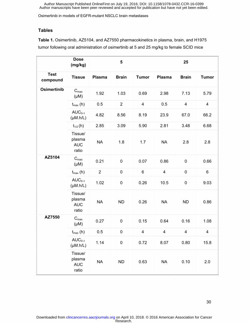
Tables
Table 1. Osimertinib, AZ5104, and AZ7550 pharmacokinetics in plasma, brain, and H1975
tumor following oral administration of osimertinib at 5 and 25 mg/kg to female SCID mice
Dose (mg/kg) 5 25
Test compound Tissue Plasma Brain Tumor Plasma Brain Tumor
Osimertinib Cmax (μM) 1.92 1.03 0.69 2.98 7.13 5.79
tmax (h) 0.5 2 4 0.5 4 4
AUC0–t (μM.h/L) 4.82 8.56 8.19 23.9 67.0 66.2
t1/2 (h) 2.85 3.09 5.90 2.81 3.48 6.68
Tissue/ plasma
AUC ratio
NA 1.8 1.7 NA 2.8 2.8
AZ5104 Cmax (μM) 0.21 0 0.07 0.86 0 0.66
tmax (h) 2 0 6 4 0 6
AUC0–t
(μM.h/L) 1.02 0 0.26 10.5 0 9.03
Tissue/ plasma
AUC ratio
NA ND 0.26 NA ND 0.86
AZ7550 Cmax (μM) 0.27 0 0.15 0.64 0.16 1.08
tmax (h) 0.5 0 4 4 4 4
AUC0–t
(μM.h/L) 1.14 0 0.72 8.07 0.80 15.8
Tissue/ plasma
AUC ratio
NA ND 0.63 NA 0.10 2.0
Research. on April 10, 2018. © 2016 American Association for Cancerclincancerres.aacrjournals.org Downloaded from
Author manuscripts have been peer reviewed and accepted for publication but have not yet been edited. Author Manuscript Published OnlineFirst on July 19, 2016; DOI: 10.1158/1078-0432.CCR-16-0399
Osimertinib in models of EGFR-mutant NSCLC brain metastases
31
AUC, area under the plasma or tissue concentration-time curve; AUC0–t, area under the plasma or
tissue concentration-time curve from time 0 to time t; Cmax, maximum plasma concentration; NA, not
applicable; ND, not determined; SCID, Sever combined immunodeficient; t1/2, terminal half-life; tmax,
time to Cmax.
Research. on April 10, 2018. © 2016 American Association for Cancerclincancerres.aacrjournals.org Downloaded from
Author manuscripts have been peer reviewed and accepted for publication but have not yet been edited. Author Manuscript Published OnlineFirst on July 19, 2016; DOI: 10.1158/1078-0432.CCR-16-0399

Osimertinib in models of EGFR-mutant NSCLC brain metastases
32
Table 2. Distribution to mouse brain of osimertinib, gefitinib, rociletinib, and afatinib following
oral administration
Osimertinib Gefitinib Rociletinib Afatinib
Dose (mg/kg) 25 6.25 100 7.5
Plasma Cmax (µM) 0.82 0.82 3.32 0.14
Brain Cmax (µM) 2.78 0.17 BLQ BLQ
Brain/plasma Cmax ratio
3.41 0.21 <0.08 <0.36
Doses equivalent to clinical doses or reported previously.
BLQ, below limit of quantification (rociletinib 0.25 µM, afatinib 0.05 µM); Cmax, maximum plasma
concentration.
Research. on April 10, 2018. © 2016 American Association for Cancerclincancerres.aacrjournals.org Downloaded from
Author manuscripts have been peer reviewed and accepted for publication but have not yet been edited. Author Manuscript Published OnlineFirst on July 19, 2016; DOI: 10.1158/1078-0432.CCR-16-0399
Osimertinib in models of EGFR-mutant NSCLC brain metastases
33
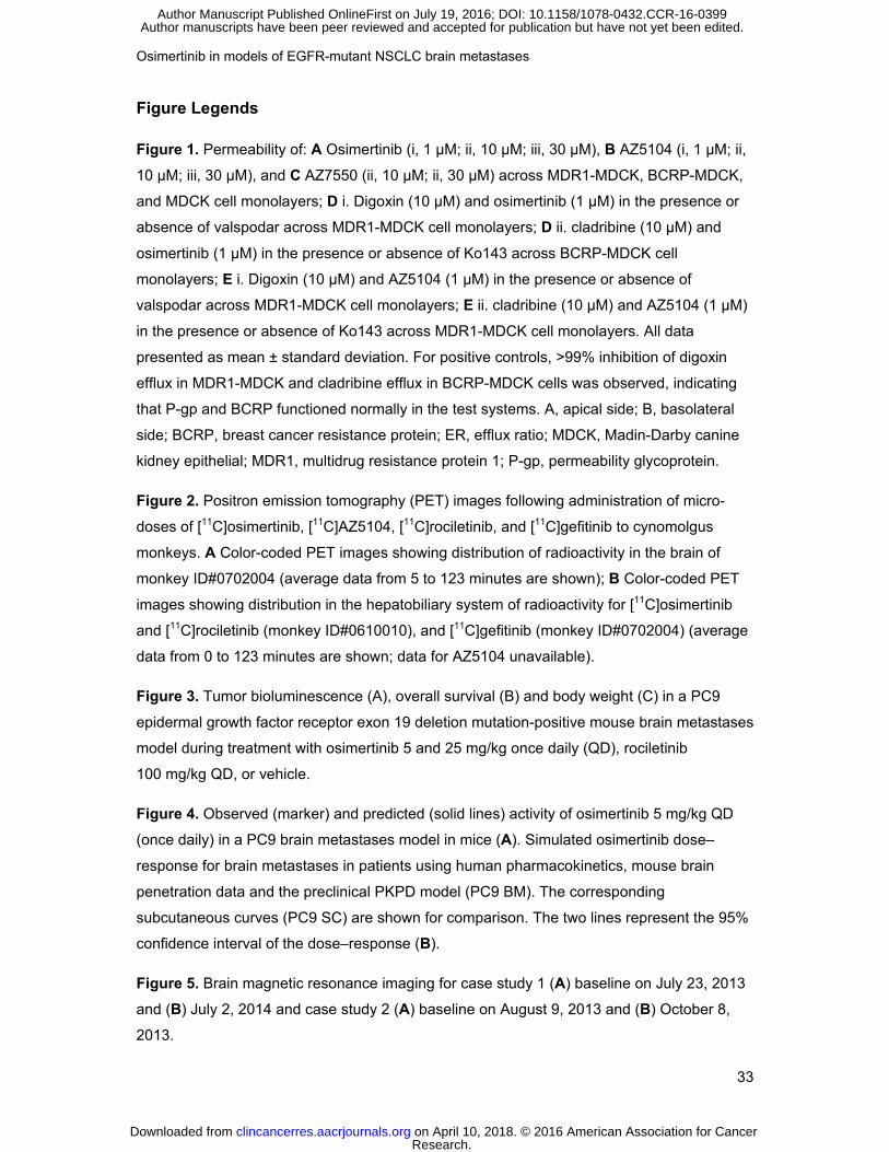
Figure Legends
Figure 1. Permeability of: A Osimertinib (i, 1 μM; ii, 10 μM; iii, 30 μM), B AZ5104 (i, 1 μM; ii,
10 μM; iii, 30 μM), and C AZ7550 (ii, 10 μM; ii, 30 μM) across MDR1-MDCK, BCRP-MDCK,
and MDCK cell monolayers; D i. Digoxin (10 μM) and osimertinib (1 μM) in the presence or
absence of valspodar across MDR1-MDCK cell monolayers; D ii. cladribine (10 μM) and
osimertinib (1 μM) in the presence or absence of Ko143 across BCRP-MDCK cell
monolayers; E i. Digoxin (10 μM) and AZ5104 (1 μM) in the presence or absence of
valspodar across MDR1-MDCK cell monolayers; E ii. cladribine (10 μM) and AZ5104 (1 μM)
in the presence or absence of Ko143 across MDR1-MDCK cell monolayers. All data
presented as mean ± standard deviation. For positive controls, >99% inhibition of digoxin
efflux in MDR1-MDCK and cladribine efflux in BCRP-MDCK cells was observed, indicating
that P-gp and BCRP functioned normally in the test systems. A, apical side; B, basolateral
side; BCRP, breast cancer resistance protein; ER, efflux ratio; MDCK, Madin-Darby canine
kidney epithelial; MDR1, multidrug resistance protein 1; P-gp, permeability glycoprotein.
Figure 2. Positron emission tomography (PET) images following administration of micro-
doses of [11C]osimertinib, [11C]AZ5104, [11C]rociletinib, and [11C]gefitinib to cynomolgus
monkeys. A Color-coded PET images showing distribution of radioactivity in the brain of
monkey ID#0702004 (average data from 5 to 123 minutes are shown); B Color-coded PET
images showing distribution in the hepatobiliary system of radioactivity for [11C]osimertinib
and [11C]rociletinib (monkey ID#0610010), and [11C]gefitinib (monkey ID#0702004) (average
data from 0 to 123 minutes are shown; data for AZ5104 unavailable).
Figure 3. Tumor bioluminescence (A), overall survival (B) and body weight (C) in a PC9
epidermal growth factor receptor exon 19 deletion mutation-positive mouse brain metastases
model during treatment with osimertinib 5 and 25 mg/kg once daily (QD), rociletinib
100 mg/kg QD, or vehicle.
Figure 4. Observed (marker) and predicted (solid lines) activity of osimertinib 5 mg/kg QD
(once daily) in a PC9 brain metastases model in mice (A). Simulated osimertinib dose–
response for brain metastases in patients using human pharmacokinetics, mouse brain
penetration data and the preclinical PKPD model (PC9 BM). The corresponding
subcutaneous curves (PC9 SC) are shown for comparison. The two lines represent the 95%
confidence interval of the dose–response (B).
Figure 5. Brain magnetic resonance imaging for case study 1 (A) baseline on July 23, 2013
and (B) July 2, 2014 and case study 2 (A) baseline on August 9, 2013 and (B) October 8,
2013.
Research. on April 10, 2018. © 2016 American Association for Cancerclincancerres.aacrjournals.org Downloaded from
Author manuscripts have been peer reviewed and accepted for publication but have not yet been edited. Author Manuscript Published OnlineFirst on July 19, 2016; DOI: 10.1158/1078-0432.CCR-16-0399
Pap
p (
X10
-6 c
m/s
)
Pap
p (
X10
-6 c
m/s
)
Pap
p (
X10
-6 c
m/s
)
0
10
20
30
40
50
60 A–B
B–A
Bi
ER = 186
ER = 7.06
ER = 3.23
0
10
20
30
40
50
60 A–B
B–A
Bii
ER = 23.5
ER = 3.44
ER = 2.03
0
5
10
15
20
25 A–B
B–A
Biii
ER = 4.53
ER = 2.68
ER = 1.23
MDR1-MDCK BCRP-MDCK MDCK MDR1-MDCK BCRP-MDCK MDCK MDR1-MDCK BCRP-MDCK MDCK
AZ5104 at 1 µM AZ5104 at 10 µM AZ5104 at 30 µM
Pap
p (
X10
-6 c
m/s
)
Osimertinib at 30 µMOsimertinib at 10 µMOsimertinib at 1 µM
MDR1-MDCK
0
10
20
30 A–B
B–A
Ai
ER = 13.4
ER = 5.40
ER = 1.55
0
1
2
3
4
5
6
7 A–B
B–A
Aii
ER = 3.11
ER = 2.66
ER = 2.07
0
1
2
3
4 A–B
B–A
Aiii
ER = 2.26
ER = 1.94 ER = 0.966
BCRP-MDCK MDCK MDR1-MDCK BCRP-MDCK MDCK MDR1-MDCK BCRP-MDCK MDCK
Pap
p (
X10
-6 c
m/s
)
Pap
p (
X10
-6 c
m/s
)
0
2
4
6
8 A–B
B–A
Ci
ER = 11.9
ER = 9.96
ER = 4.49
0.0
0.5
1.0
1.5
2.0
2.5
3.0 A–B
B–A
Cii
ER = 2.38
ER = 14.6
ER = 1.15
MDR1-MDCK BCRP-MDCK MDCK MDR1-MDCK BCRP-MDCK MDCK
AZ7550 at 10 µM AZ7550 at 30 µM
Pap
p (
X10
-6 c
m/s
)
Pap
p (
X10
-6 c
m/s
)
0
10
20
30 A–B
B–A
ER = 143
ER = 1.04
ER = 13.4
ER = 1.01
0
5
10
15
20
25 A–B
B–A
ER = 57.5
ER = 1.16
ER = 5.40
ER = 1.35
Pap
p (
X10
-6 c
m/s
)
Pap
p (
X10
-6 c
m/s
)
Dig
oxin 1
0 µM
Dig
oxin 1
0 µM
+ va
lspodar
Osi
mer
tinib
1 µ
M
Osi
mer
tinib
1 µ
M
+ va
lspodar
Cla
dribin
e 10
µM
Cla
dribin
e 10
µM
+ Ko 1
43
Osi
mer
tinib
1 µ
M
Osi
mer
tinib
1 µ
M
+ Ko14
3
Di Dii
10
30
40
50
60 A–B
B–A
ER = 79.0
ER = 1.10
ER = 186
ER = 0.875
0
5
10
15
20
25
30 A–B
B–A
ER = 51.6
ER = 1.45
ER = 7.06
ER = 1.51Pap
p (
X10
-6 c
m/s
)
Pap
p (
X10
-6 c
m/s
)
Dig
oxin 1
0 µM
Dig
oxin 1
0 µM
+ va
lspodar
AZ51
04 1
µM
AZ51
04 1
µM
+ va
lspodar
Cla
dribin
e 10
µM
Cla
dribin
e 10
µM
+ Ko14
3
AZ51
04 1
µM
AZ51
04 1
µM
+ Ko14
3
Ei Eii
Figure 1
Research. on April 10, 2018. © 2016 American Association for Cancerclincancerres.aacrjournals.org Downloaded from
Author manuscripts have been peer reviewed and accepted for publication but have not yet been edited. Author Manuscript Published OnlineFirst on July 19, 2016; DOI: 10.1158/1078-0432.CCR-16-0399
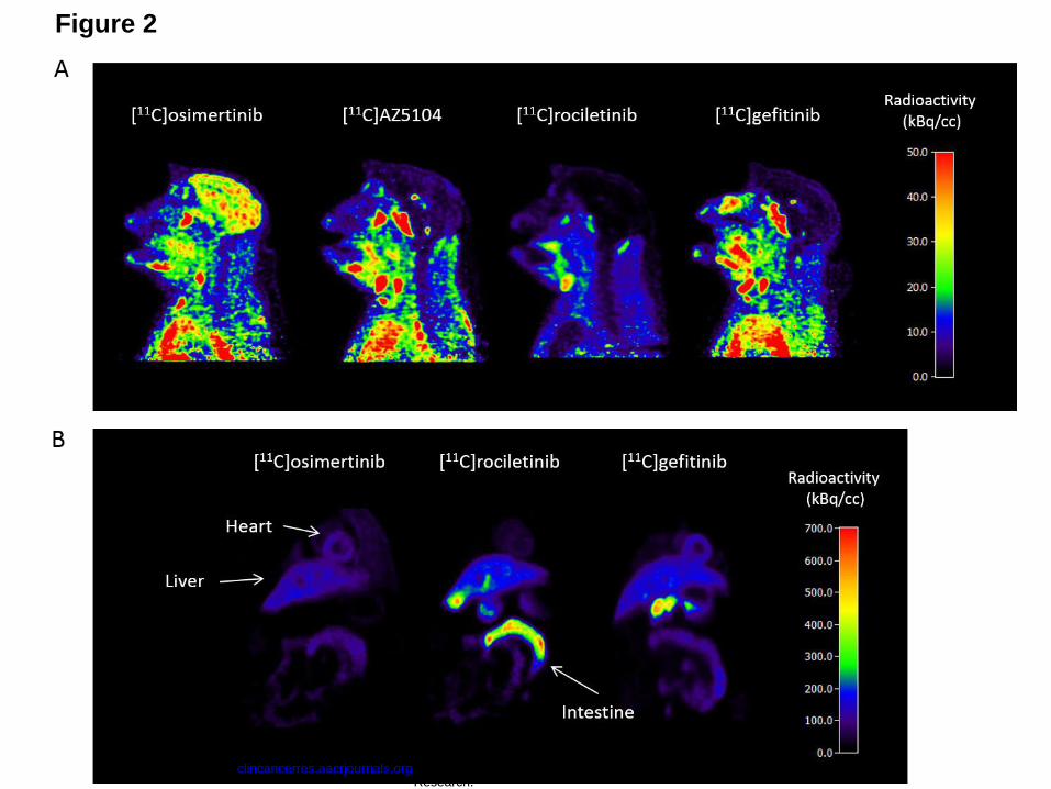
Figure 2
Research. on April 10, 2018. © 2016 American Association for Cancerclincancerres.aacrjournals.org Downloaded from
Author manuscripts have been peer reviewed and accepted for publication but have not yet been edited. Author Manuscript Published OnlineFirst on July 19, 2016; DOI: 10.1158/1078-0432.CCR-16-0399

-3
-2
-1
0
1
2
3 vehicle control
osimertinib 5 mg/kg QD
osimertinib 25 mg/kg QD
rociletinib 100 mg/kg QD
Lo
g r
ela
tive
bio
lum
ine
scence (
p/s
)
Time (days)
706040 503020100
A
B
-0
20
40
60
80
100
Su
rviv
al (%
)
Time (days)
8040 60200
-20
-15
-10
-5
0
5
10
15
20
Bo
dy w
eig
ht
ch
an
ge
(%
)
C
vehicle control
osimertinib 5 mg/kg QD
osimertinib 25 mg/kg QD
rociletinib 100 mg/kg QD
vehicle control
osimertinib 5 mg/kg QD
osimertinib 25 mg/kg QD
rociletinib 100 mg/kg QD
Time (days)
706040 503020100
Figure 3
Research. on April 10, 2018. © 2016 American Association for Cancerclincancerres.aacrjournals.org Downloaded from
Author manuscripts have been peer reviewed and accepted for publication but have not yet been edited. Author Manuscript Published OnlineFirst on July 19, 2016; DOI: 10.1158/1078-0432.CCR-16-0399
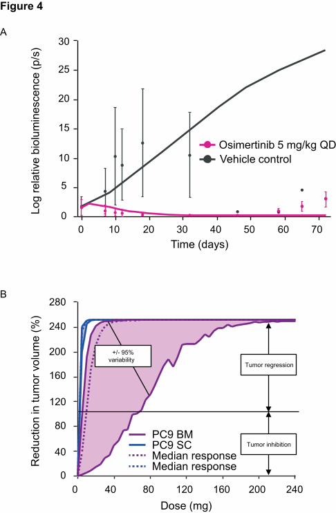
706040 503020100
0
5
10
15
20
25
30
Vehicle control
Osimertinib 5 mg/kg QD
Lo
g r
ela
tive
bio
lum
ine
scence (
p/s
)
Time (days)
A
240160 20012080400
0
80
120
160
200
240
280
40
PC9 BM
Re
du
ctio
n in
tu
mo
r vo
lum
e (
%)
Dose (mg)
B
PC9 SCMedian responseMedian response
Figure 4
Tumor inhibition
Tumor regression
+/- 95%
variability
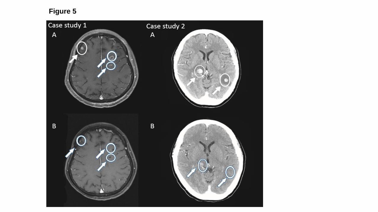
Figure 5

Published OnlineFirst July 19, 2016.Clin Cancer Res Peter Ballard, James WT Yates, Zhenfan Yang, et al. Evidence of Clinical Brain Metastases ActivityEGFR-Mutant NSCLC Brain Metastases Models, and Early Preclinical Comparison of Osimertinib with Other EGFR-TKIs in
Updated version
10.1158/1078-0432.CCR-16-0399doi:
Access the most recent version of this article at:
Material
Supplementary
http://clincancerres.aacrjournals.org/content/suppl/2016/07/19/1078-0432.CCR-16-0399.DC1
Access the most recent supplemental material at:
Manuscript
Authoredited. Author manuscripts have been peer reviewed and accepted for publication but have not yet been
E-mail alerts related to this article or journal.Sign up to receive free email-alerts
Subscriptions
Reprints and
To order reprints of this article or to subscribe to the journal, contact the AACR Publications
Permissions
Rightslink site. Click on "Request Permissions" which will take you to the Copyright Clearance Center's (CCC)
.http://clincancerres.aacrjournals.org/content/early/2016/07/19/1078-0432.CCR-16-0399To request permission to re-use all or part of this article, use this link
Research. on April 10, 2018. © 2016 American Association for Cancerclincancerres.aacrjournals.org Downloaded from
Author manuscripts have been peer reviewed and accepted for publication but have not yet been edited. Author Manuscript Published OnlineFirst on July 19, 2016; DOI: 10.1158/1078-0432.CCR-16-0399